Embed Size (px)
Citation preview


Ease of UseSetting up the Single Balloon Enteroscope System is
a snap so getting ready for an examination is never a
bother. All you have to do is moisten the lining of the
splinting tube with sterile water, connect it to the balloon
control unit and pass the scope through.
The “Next” Evolution in Enteroscopy: The Single Balloon Enteroscope System from OLYMPUS.
2 3
Clinical EfficiencySince the Single Balloon Enteroscope System has
only a single balloon, complex operation is reduced.
Just press a single button on the compact remote
control as required to manipulate the inflation and
deflation of the balloon.
Hypoallergenic, latex-free designTo achieve a patient-friendly, latex-free design, all
components that comprise the splinting tube of the Single
Balloon Enteroscope System — from the tube shaft to the
balloon and tube tip — are made of silicone. In addition, a
hydrophilic lubricant coating has been applied to the lining
of the splinting tube. This provides excellent lubrication
between the scope and splinting tube, effectively
supporting insertion into the deep small intestine.
Advanced Imaging and ConnectivitySupports Narrow Band Imaging (NBI) observation,
which enables more detailed observation of mucosal
morphology, and compatible with a wide range of
systems, from the EVIS 240 to the EVIS LUCERA to
the EVIS LUCERA SPECTRUM.
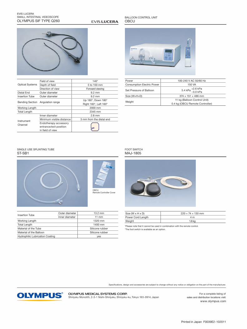
Single Balloon Enteroscope System
Despite the rapid technological advances of the 21st century, enteroscopy continues to prove more difficult
than upper gastrointestinal endoscopy or colonoscopy. Now, thanks to our groundbreaking Single Balloon
Enteroscope System, OLYMPUS has created a simple yet efficient system that radically redefines the nature
of enteroscopy. The new Single Balloon Enteroscope maintains OLYMPUS' signature high quality image,
while offering breakthrough capabilities in terms of operability and functionality that shed a new light on a
region once considered the “Last Frontier” of the human body.
Simple operation at every step of the way from setup to observation and treatment
Efficient hand or foot controls and automatic pressure control, eliminate complex operations and reduce procedure time
Effective high quality image and improved treatment performance achieved through the use of OLYMPUS' latest technology
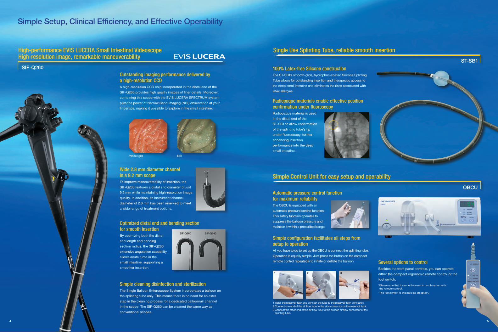
Optimized distal end and bending section for smooth insertionBy optimizing both the distal
end length and bending
section radius, the SIF-Q260
extensive angulation capability
allows acute turns in the
small intestine, supporting a
smoother insertion.
Automatic pressure control function for maximum reliabilityThe OBCU is equipped with an
automatic pressure control function.
This safety function operates to
suppress the balloon pressure and
maintain it within a prescribed range.
4 5
Simple Setup, Clinical Efficiency, and Effective Operability
High-performance EVIS LUCERA Small Intestinal Videoscope High-resolution image, remarkable maneuverability
100% Latex-free Silicone constructionThe ST-SB1’s smooth-glide, hydrophilic-coated Silicone Splinting
Tube allows for outstanding insertion and therapeutic access to
the deep small intestine and eliminates the risks associated with
latex allergies.
Radiopaque materials enable effective position confirmation under fluoroscopyRadiopaque material is used
in the distal end of the
ST-SB1 to allow confirmation
of the splinting tube’s tip
under fluoroscopy, further
enhancing insertion
performance into the deep
small intestine.
Simple configuration facilitates all steps from setup to operationAll you have to do to set up the OBCU is connect the splinting tube.
Operation is equally simple. Just press the button on the compact
remote control repeatedly to inflate or deflate the balloon.
Simple Control Unit for easy setup and operability
White light NBI
Wide 2.8 mm diameter channel in a 9.2 mm scopeTo improve maneuverability of insertion, the
SIF-Q260 features a distal end diameter of just
9.2 mm while maintaining high-resolution image
quality. In addition, an instrument channel
diameter of 2.8 mm has been reserved to meet
a wide range of treatment options.
Simple cleaning disinfection and sterilizationThe Single Balloon Enteroscope System incorporates a balloon on
the splinting tube only. This means there is no need for an extra
step in the cleaning process for a dedicated balloon/air channel
in the scope. The SIF-Q260 can be cleaned the same way as
conventional scopes.
Single Use Splinting Tube, reliable smooth insertion
SIF-Q260ST-SB1
1 Install the reservoir tank and connect the tube to the reservoir tank connector.2 Connect one end of the air flow tube to the side connector on the reservoir tank.3 Connect the other end of the air flow tube to the balloon air flow connector of the splinting tube.
Several options to controlBesides the front panel controls, you can operate
either the compact ergonomic remote control or the
foot switch.
*Please note that it cannot be used in combination with the remote control.
*The foot switch is available as an option.
Outstanding imaging performance delivered by a high-resolution CCDA high-resolution CCD chip incorporated in the distal end of the
SIF-Q260 provides high quality images of finer details. Moreover,
combining this scope with the EVIS LUCERA SPECTRUM system
puts the power of Narrow Band Imaging (NBI) observation at your
fingertips, making it possible to explore in the small intestine.
1 2 3
SIF-Q260 SIF-Q240
OBCU

6 7
Groundbreaking mechanism to plicate the intestinal tract
The single balloon scope can be inserted into the deep small
intestine by manipulating the balloon on the distal end of the
splinting tube and the angulation mechanism of the scope.
First, insert the scope deeply into the gastrointestinal tract.
Second, advance the splinting tube and inflate the balloon.
Next, withdraw both the scope and splinting tube to plicate the
intestinal tract.
By repeating these steps, you can pleat and reduce the small
intestine for deep small bowel intubation.
Antegrade approach
Retrograde approach
White light
NBI observation
Antegrade approach under fluoroscopy
Retrograde approach under fluoroscopy
: Scope motion : Splinting tube motion
❶ Insert the scope as deep as possible into the gastrointestinal tract.
❷ Advance the splinting tube.
❸ Inflate the balloon.
❹Withdraw both the splinting tube and scope in tandem to reduce the intestinal tract.
Principles of insertion Clinical Case Images
The SIF-Q260’s wide connectivity means that it is
compatible with the EVIS 240 to the EVIS LUCERA
systems you already use. When it is combined with the
EVIS LUCERA SPECTRUM system, NBI observation
is possible, facilitating more advanced observation of
mucosal morphology.
NBI observation is possible when the SIF-Q260 is combined with the EVIS LUCERA SPECTRUM system
A list of SIF-Q260 compatible systems
Jejunum
Ileum
NBI
SIF-Q260
CV-240
CV-260ACV-260B
CV-260SL


![Towards Systematic Privacy and Operability (PRIOP) Studies · Hazard and Operability Studies The international standard IEC 61882 [2] de nes what a Hazard and Operability (HAZOP)](https://img.pdfslide.us/doc/110x75/5e9fb7d8f9d766473e1e43a5/towards-systematic-privacy-and-operability-priop-studies-hazard-and-operability.jpg)