Embed Size (px)
Citation preview
University of Groningen
The neuropsychological rehabilitation of visual agnosia and Balint’s syndromeHeutink, Joost; Indorf, Dana L.; Cordes, Christina
Published in:Neuropsychological Rehabilitation
DOI:10.1080/09602011.2017.1422272
IMPORTANT NOTE: You are advised to consult the publisher's version (publisher's PDF) if you wish to cite fromit. Please check the document version below.
Document VersionPublisher's PDF, also known as Version of record
Publication date:2019
Link to publication in University of Groningen/UMCG research database
Citation for published version (APA):Heutink, J., Indorf, D. L., & Cordes, C. (2019). The neuropsychological rehabilitation of visual agnosia andBalint’s syndrome. Neuropsychological Rehabilitation, 29(10), 1489-1508 .https://doi.org/10.1080/09602011.2017.1422272
CopyrightOther than for strictly personal use, it is not permitted to download or to forward/distribute the text or part of it without the consent of theauthor(s) and/or copyright holder(s), unless the work is under an open content license (like Creative Commons).
Take-down policyIf you believe that this document breaches copyright please contact us providing details, and we will remove access to the work immediatelyand investigate your claim.
Downloaded from the University of Groningen/UMCG research database (Pure): http://www.rug.nl/research/portal. For technical reasons thenumber of authors shown on this cover page is limited to 10 maximum.
Download date: 13-03-2020
Full Terms & Conditions of access and use can be found athttps://www.tandfonline.com/action/journalInformation?journalCode=pnrh20
Neuropsychological RehabilitationAn International Journal
ISSN: 0960-2011 (Print) 1464-0694 (Online) Journal homepage: https://www.tandfonline.com/loi/pnrh20
The neuropsychological rehabilitation of visualagnosia and Balint’s syndrome
Joost Heutink, Dana L. Indorf & Christina Cordes
To cite this article: Joost Heutink, Dana L. Indorf & Christina Cordes (2019) Theneuropsychological rehabilitation of visual agnosia and Balint’s syndrome, NeuropsychologicalRehabilitation, 29:10, 1489-1508, DOI: 10.1080/09602011.2017.1422272
To link to this article: https://doi.org/10.1080/09602011.2017.1422272
© 2018 The Author(s). Published by InformaUK Limited, trading as Taylor & FrancisGroup
Published online: 24 Jan 2018.
Submit your article to this journal
Article views: 3425
View related articles
View Crossmark data
Citing articles: 1 View citing articles
The neuropsychological rehabilitation of visual agnosiaand Balint’s syndromeJoost Heutink a,b, Dana L. Indorfa and Christina Cordesa,b
aDepartment of Clinical and Developmental Neuropsychology, University of Groningen, Groningen,The Netherlands; bRoyal Dutch Visio, Centre of Expertise for Visually Impaired and Blind People,Department of Knowledge, Expertise & Innovation, Huizen, The Netherlands
ABSTRACTVisual agnosia and Balint’s syndrome are complex neurological disorders of the highervisual system that can have a remarkable impact on individuals’ lives. Rehabilitation ofthese individuals is important to enable participation in everyday activities despite theimpairment. However, the literature about the rehabilitation of these disorders isvirtually silent. Therefore, the aim of this systematic review is to give an overview ofavailable literature describing treatment approaches and their effectiveness withregard to these disorders. The search engines Psychinfo, Amed, and Medline wereused, resulting in 22 articles meeting the criteria for inclusion. Only articlesdescribing acquired disorders were considered. These articles revealed that there issome information available on the major subtypes of visual agnosia as well as onBalint’s syndrome which practising clinicians can consult for guidance. With regardto the type of rehabilitation, compensatory strategies have proven to be beneficialin most of the cases. Restorative training on the other hand has produced mixedresults. Concluding, although still scarce, a scientific foundation about therehabilitation of visual agnosia and Balint’s syndrome is evolving. The availableapproaches give valuable information that can be built upon in the future.
ARTICLE HISTORY Received 22 August 2017; Accepted 21 December 2017
KEYWORDS Visual perception; Visual agnosia; Balint’s syndrome; Neuropsychological rehabilitation;Rehabilitation
Introduction
Due to the complexity of neurological disorders and the individual nature of impair-ments, neuropsychological rehabilitation is a difficult endeavour. Especially the rehabi-litation of visual (perceptual) disorders is multifaceted, since visual disturbances maytake place at lower and/or at higher function levels. Vision is often affected in patientswith neurological disorders. For example, about 30% of the patients with acquired braininjury show deficits in vision (Zihl, 2003) and 20–40% of the patients with cerebrovas-cular acquired brain injury or traumatic brain injury (TBI) have higher visual disorders
© 2018 The Author(s). Published by Informa UK Limited, trading as Taylor & Francis GroupThis is an Open Access article distributed under the terms of the Creative Commons Attribution-NonCommercial-NoDerivativesLicense (http://creativecommons.org/licenses/by-nc-nd/4.0/), which permits non-commercial re-use, distribution, and reproduc-tion in any medium, provided the original work is properly cited, and is not altered, transformed, or built upon in any way.
CONTACT Joost Heutink [email protected] Department of Clinical and Developmental Neuropsy-chology, University of Groningen, Grote Kruisstraat 2/1, 9712 TS Groningen, The Netherlands
NEUROPSYCHOLOGICAL REHABILITATION2019, VOL. 29, NO. 10, 1489–1508https://doi.org/10.1080/09602011.2017.1422272
(Zihl & Kennard, 2003). Visual perceptual disorders can have a tremendous impact on anindividual’s spatial orientation, learning, and motor activities (Zihl, 2003), which in turncan affect a person’s independence, social participation, and vocational life. Therefore,rehabilitation of visual perceptual disorders is a central topic in clinical practice.
Two approaches can be distinguished in neuropsychological rehabilitation(Spikman & Fasotti, 2017). The first type, restoration, aims to improve a particular func-tion by training the impaired function and thereby the damaged brain structuredirectly and repetitively. The second type, compensation, refers to using an intactfunction to compensate for the loss of the other one. Thus, the impaired functionitself is not targeted, but functioning on an activity and participation level is ratherattempted to be improved. Depending on the level of impairment of the patient,environmental adaptation, for example signposting a route, could also beimplemented if the patient’s ability to learn compensatory strategies is limited.Often, a combination of these approaches may be necessary in a rehabilitationregime. The type of treatment chosen should also consider the type of brain injurythe patient suffers from.
Visual (perceptual) disorders such as visual field defects, neglect, or difficulties withcontrasts and colours may arise from either acquired brain injury (ABI; e.g., stroke) orneurodegenerative disorders (e.g., dementia, Parkinson’s disease, and multiple scler-osis). Although there are several treatment options available for the more frequent dis-orders like hemianopia and neglect (Bowen, Hazelton, Pollock, & Lincoln, 2013; De Haan,Heutink, Melis-Dankers, Tucha, & Brouwer, 2014; Pollock et al., 2011), some visual dis-orders have received only little attention regarding their rehabilitation. Two disordersfor which the latter is true are visual agnosia and Balint’s syndrome, which can manifestin patients following ABI as well as in neurodegenerative diseases.
According to Zihl (2003), visual agnosia is a difficulty or inability to identify familiarstimuli via the visual modality, although the patient possesses sufficient visual percep-tual, cognitive, and verbal functioning, and is able to recognise the stimulus using othermodalities. Pure visual agnosia is a relatively rare condition, with an estimated preva-lence of about 1–3% (Zihl & Kennard, 2003). At least four types of visual agnosia canbe differentiated: prosopagnosia (the inability to recognise familiar faces and to learnnew faces); object agnosia (the inability to identify stimuli in the same class or differen-tiate between classes of stimuli); topographical agnosia (difficulties with geographicalorientation); and letter agnosia or pure alexia (inability to recognise individual lettersand their combinations; Damasio, Tranel, & Rizzo, 2000; Roberts, 1992; Zihl, 2003).However, also finer distinctions can be made and very specific agnosias can be foundin the literature as well (e.g., colour agnosia; Nijboer & Heutink, 2017).
A remarkable syndrome arising from disturbances to the higher visual system isBalint’s syndrome (Balint, 1909). Balint’s syndrome usually occurs after damage to bilat-eral posterior-parietal brain areas, and can occur after ABI or due to neurodegenerativediseases, usually dementia (Kerkhoff & Heldmann, 1999). Damasio et al. (2000), defineBalint’s syndrome as
An acquired disturbance of the ability to perceive the visual field as a whole, resulting in theunpredictable perception and recognition of only parts of it (simultanagnosia); which isaccompanied by an impairment of target pointing under visual guidance (optic ataxia) and aninability to shift gaze at will toward new visual stimuli (ocular apraxia). (p. 353)
1490 J. HEUTINK ET AL.
Of these three symptoms, simultanagnosia is described as the essence of the syndrome.The impact of Balint’s syndrome is often so grave, that patients might appear as if theywere blind (Kerkhoff & Heldmann, 1999).
Individuals with acquired visual agnosia or Balint’s syndrome require intensive reha-bilitation to help them to overcome their deficits. In addition to that, spontaneousrecovery in both visual agnosia and Balint’s syndrome is assumed to be low (Zihl &Kennard, 2003). This emphasises the importance of rehabilitation as an invaluabletool for affected individuals. However, due to the much lower prevalence of thesevisual-perceptual disorders compared to hemianopia or neglect, there are not manystudies on the rehabilitation of visual agnosia and Balint’s syndrome available. Thepurpose of this article is therefore to conduct a systematic literature review on the reha-bilitation of acquired visual agnosia and Balint’s syndrome to explore the available treat-ment approaches and their effectiveness, and to give an indication of which sources topursue for those interested in further detail. Thereby, we aim to provide a status updateon the rehabilitation of visual agnosia and Balint’s syndrome and give directions forfuture research.
Method
A comprehensive systematic literature search was conducted to retrieve literatureaddressing cognitive rehabilitation of acquired visual agnosia and Balint’s syndrome.
Search strategy
This study utilised the academic databases Psychinfo, Medline, and Amed to search forpeer-reviewed publications published in either English or German. Keywords used were“treatment” or “rehabilitation” in combination with each of the terms “visual agnosia,”“simultanagnosia,” “object agnosia,” “prosopagnosia,” “associative agnosia,” “appercep-tive agnosia,” “colour agnosia,” “form agnosia,” “semantic agnosia,” “topographicalagnosia,” and “Balint’s syndrome” in either English or German translation. We did notdefine a certain time period for the inclusion of the listed results and did not includeunpublished data. Abstracts of the listed results were reviewed to identify those articlesaddressing the topic at hand.
Study selection
The time period of the listed results ranged from 1948 until 2017. Figure 1 shows thesteps followed during the literature search (PRISMA flow diagram; Moher, Liberati,Tetzlaff, Altman & The PRISMA Group 2009). Exclusion criteria were: (a) if thedeficit was caused by a congenital condition, meaning the visual system had notdeveloped normally before the occurrence of the impairment; (b) if the rehabilitationwas on pure alexia as there have been publications reviewing the current status ofrehabilitation (e.g., Starrfelt, Ólafsdóttir, & Arendt, 2013); and (c) if the sectionsdealing with the rehabilitation of visual agnosia and Balint’s syndrome only did soby describing other researchers’ work. We included studies: (a) providing evidenceabout the effectiveness of a rehabilitation by empirically evaluating it; (b) giving rec-ommendations for rehabilitation based on scientific foundations or clinical experi-ence as long as the recommendations were specific enough to give guidance to
NEUROPSYCHOLOGICAL REHABILITATION 1491
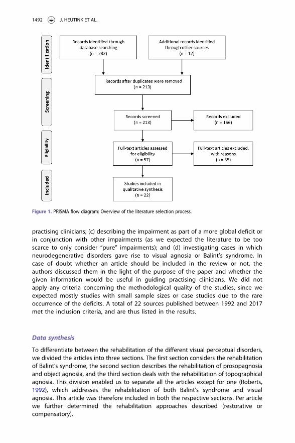
practising clinicians; (c) describing the impairment as part of a more global deficit orin conjunction with other impairments (as we expected the literature to be tooscarce to only consider “pure” impairments); and (d) investigating cases in whichneurodegenerative disorders gave rise to visual agnosia or Balint’s syndrome. Incase of doubt whether an article should be included in the review or not, theauthors discussed them in the light of the purpose of the paper and whether thegiven information would be useful in guiding practising clinicians. We did notapply any criteria concerning the methodological quality of the studies, since weexpected mostly studies with small sample sizes or case studies due to the rareoccurrence of the deficits. A total of 22 sources published between 1992 and 2017met the inclusion criteria, and are thus listed in the results.
Data synthesis
To differentiate between the rehabilitation of the different visual perceptual disorders,we divided the articles into three sections. The first section considers the rehabilitationof Balint’s syndrome, the second section describes the rehabilitation of prosopagnosiaand object agnosia, and the third section deals with the rehabilitation of topographicalagnosia. This division enabled us to separate all the articles except for one (Roberts,1992), which addresses the rehabilitation of both Balint’s syndrome and visualagnosia. This article was therefore included in both the respective sections. Per articlewe further determined the rehabilitation approaches described (restorative orcompensatory).
Figure 1. PRISMA flow diagram: Overview of the literature selection process.
1492 J. HEUTINK ET AL.
Results
Balint’s syndrome
A total of 10 articles addressing the rehabilitation of Balint’s syndrome met our criteria.Seven of these articles presented a case study including empirical validation of a treat-ment, while three articles gave suggestions and recommendations for treatment(Table 1). Generally, it can be observed that most rehabilitation approaches were multi-faceted, and led to variable improvements in the treated patients. Compensatory strat-egies seemed to be widely applied and usually relatively successful. A small number ofstudies attempted to train the specific deficits arising from Balint’s syndrome (Perez,Tunkel, Lachmann, & Nagler, 1996; Rosselli, Ardila, & Beltran, 2001; Zgaljardic, Yancy,Levinson, Morales, & Masel, 2011), with for example eye movement exercises or conver-gence exercises, which proved to be successful in some patients (Rosselli et al., 2001).Yet, some attempts remained less successful (Zgaljardic et al., 2011), allowing for littleconclusiveness about the effectiveness of neuropsychological training or eye move-ment exercises. Several authors emphasised the importance of psychoeducation, strat-egies to promote the transfer from training situations to real-life situations, functionalabilities, as well as building on strengths. Importantly, the choice for a rehabilitationapproach and the expectations for an outcome depended heavily on the aetiology ofBalint’s syndrome. Perez et al. (1996) proposed to focus on learning and using strategiesfor neurodegenerative disorders that increase participation in everyday activities toincrease coping and confidence in patients.
Based on the results of this search, we can conclude that a certain degree of rehabi-litation of Balint’s syndrome is possible for most patients, in so far as to regain functionalcapabilities and quality of life. A necessary condition might be that the treatment is indi-vidually tailored to the patient and the therapists maintain flexibility throughout theprocess.
Prosopagnosia and object agnosia
Much research on prosopagnosia has focused on the mechanisms by which we recog-nise faces and how these might be damaged. With regard to rehabilitation, two litera-ture reviews could be found. The 12 case studies already reviewed by these two articleswere not included in the present study. Instead, we added eight studies on the rehabi-litation of prosopagnosia that were not included in these literature reviews (Table 2).
Both literature reviews concluded that restorative training has not yet proven to bevery successful and that compensatory strategies appeared to be a more effectiveapproach for the rehabilitation of acquired prosopagnosia (Bate & Bennetts, 2014;DeGutis, Chiu, Grosso, & Cohan, 2014). The additional eight articles appeared to be inline with the outcome of the literature reviews, especially with regard to transferringthe training to real-life face recognition (Bate et al., 2015). The study by Davies-Thomp-son et al. (2017) describes a promising restorative approach, however training is veryintense and does not improve real-life facial recognition in all participants involved.Concluding, compensation strategies seemed to be most promising for the rehabilita-tion of prosopagnosia.
With regard to object agnosia, the literature search revealed seven articles, describ-ing the rehabilitation of object agnosia either in the context of other impairments (fourstudies) or as suggestions for rehabilitation strategies without clinical data (three
NEUROPSYCHOLOGICAL REHABILITATION 1493
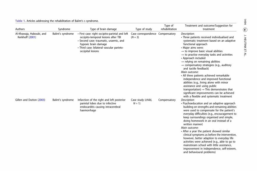
Table 1. Articles addressing the rehabilitation of Balint’s s syndrome.
Authors Syndrome Type of brain damage Type of studyType of
rehabilitationTreatment and outcome/Suggestion for
treatment
Al-Khawaja, Haboubi, andKerkhoff (2001)
Balint’s syndrome • First case: right occipito-parietal and leftoccipito-temporal lesions after TBI
• Second case: traumatic, uraemic, andhypoxic brain damage
• Third case: bilateral vascular parieto-occipital lesions
Case correspondence(N = 3)
Compensatory Description:• Three patients received individualised andsystematic treatment based on an adaptivefunctional approach
• Major aims were:→ to improve basic visual abilities→ to practise everyday tasks and activities• Approach included→ relying on remaining abilities→ compensatory strategies (e.g., auditoryand tactile feedback)
Main outcome:• All three patients achieved remarkableindependence and improved functionalabilities (e.g., living alone with minorassistance and using publictransportation)→ This demonstrates thatsignificant improvements can be achievedwith a flexible and systematic treatment
Gillen and Dutton (2003) Balint’s syndrome Infarction of the right and left posteriorparietal lobes due to infectiveendocarditis causing intracerebralhaemorrhage
Case study (child,N = 1)
Compensatory Description:• Psychoeducation and an adaptive approachbuilding on strengths and remaining abilitieswere used to compensate for the patient’severyday difficulties (e.g., encouragement tokeep surroundings organised and simple,doing homework in an oral instead of awritten manner)
Main outcome:• After a year the patient showed similarclinical symptoms as before the intervention,however, better adaption to everyday lifeactivities were achieved (e.g., able to go tomainstream school with little assistance,improvement in independence, self-esteem,and behavioural problems)
1494J.H
EUTIN
KET
AL.
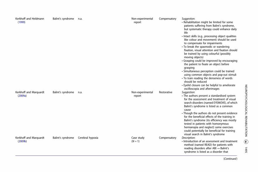
Kerkhoff and Heldmann(1999)
Balint’s syndrome n.a. Non-experimentalreport
Compensatory Suggestion:• Rehabilitation might be limited for somepatients suffering from Balint’s syndrome,but systematic therapy could enhance dailylife
• Intact skills (e.g., processing object qualitieslike colour and movement) should be usedto compensate for impairments
• To break the spasmodic or wanderingfixation, visual attention and fixation shouldbe trained by using colourful (possiblymoving objects)
• Grasping could be improved by encouragingthe patient to fixate an object beforegrasping
• Simultaneous perception could be trainedusing common objects and pop-out stimuli
• To train reading the denseness of wordsshould be reduced
• Eyelid closure can be helpful to ameliorateoscilloscopia and afterimages
Kerkhoff and Marquardt(2009a)
Balint’s syndrome n.a. Non-experimentalreport
Restorative Suggestion:• The authors present a standardised systemfor the assessment and treatment of visualsearch disorders (named EYEMOVE), of whichBalint’s syndrome is listed as a commoncause
• Though the authors do not present evidencefor the beneficial effects of the training inBalint’s syndrome (its efficiency was mostlytested in patients with homonymoushemianopia and neglect) some exercisescould potentially be beneficial for trainingvisual search in Balint’s syndrome
Kerkhoff and Marquardt(2009b)
Balint’s syndrome Cerebral hypoxia Case study(N = 1)
Compensatory Description:• Introduction of an assessment and treatmentmethod (named READ) for patients withreading disorders after ABI→ Balint’ssyndrome is listed as a disorder that
(Continued )
NEU
ROPSYC
HOLO
GICALREH
ABILITA
TION
1495
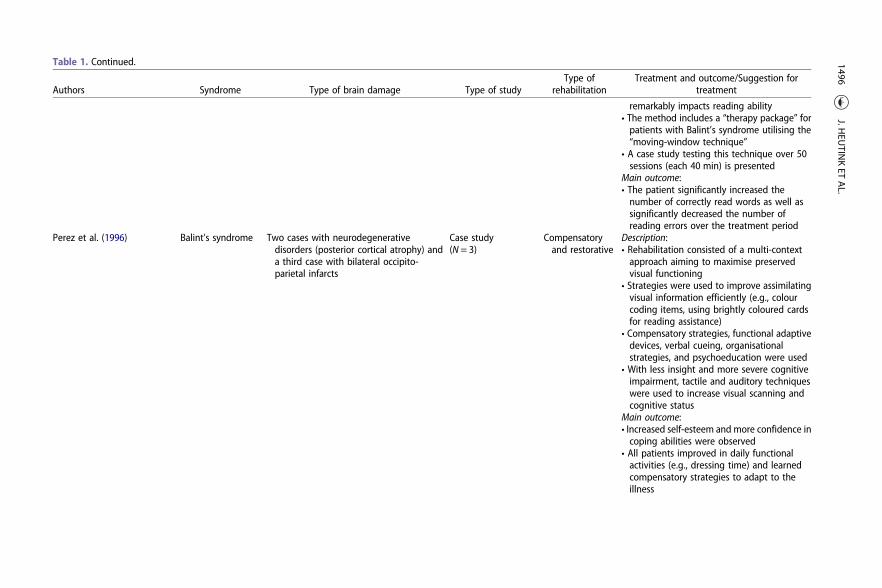
Table 1. Continued.
Authors Syndrome Type of brain damage Type of studyType of
rehabilitationTreatment and outcome/Suggestion for
treatment
remarkably impacts reading ability• The method includes a “therapy package” forpatients with Balint’s syndrome utilising the“moving-window technique”
• A case study testing this technique over 50sessions (each 40 min) is presented
Main outcome:• The patient significantly increased thenumber of correctly read words as well assignificantly decreased the number ofreading errors over the treatment period
Perez et al. (1996) Balint’s syndrome Two cases with neurodegenerativedisorders (posterior cortical atrophy) anda third case with bilateral occipito-parietal infarcts
Case study(N = 3)
Compensatoryand restorative
Description:• Rehabilitation consisted of a multi-contextapproach aiming to maximise preservedvisual functioning
• Strategies were used to improve assimilatingvisual information efficiently (e.g., colourcoding items, using brightly coloured cardsfor reading assistance)
• Compensatory strategies, functional adaptivedevices, verbal cueing, organisationalstrategies, and psychoeducation were used
• With less insight and more severe cognitiveimpairment, tactile and auditory techniqueswere used to increase visual scanning andcognitive status
Main outcome:• Increased self-esteem and more confidence incoping abilities were observed
• All patients improved in daily functionalactivities (e.g., dressing time) and learnedcompensatory strategies to adapt to theillness
1496J.H
EUTIN
KET
AL.
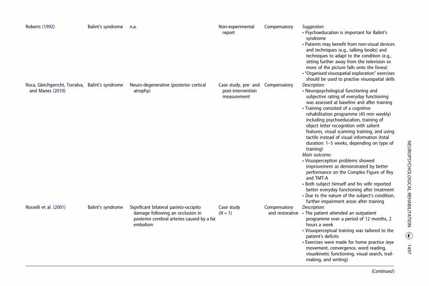
Roberts (1992) Balint’s syndrome n.a. Non-experimentalreport
Compensatory Suggestion:• Psychoeducation is important for Balint’ssyndrome
• Patients may benefit from non-visual devicesand techniques (e.g., talking books) andtechniques to adapt to the condition (e.g.,sitting further away from the television somore of the picture falls onto the fovea)
• “Organised visuospatial exploration” exercisesshould be used to practise visuospatial skills
Roca, Gleichgerrcht, Torralva,and Manes (2010)
Balint’s syndrome Neuro-degenerative (posterior corticalatrophy)
Case study, pre- andpost-interventionmeasurement
Compensatory Description:• Neuropsychological functioning andsubjective rating of everyday functioningwas assessed at baseline and after training
• Training consisted of a cognitiverehabilitation programme (45 min weekly)including psychoeducation, training ofobject letter recognition with salientfeatures, visual scanning training, and usingtactile instead of visual information (totalduration: 1–5 weeks, depending on type oftraining)
Main outcome:• Visuoperception problems showedimprovement as demonstrated by betterperformance on the Complex Figure of Reyand TMT-A
• Both subject himself and his wife reportedbetter everyday functioning after treatment
• Due to the nature of the subject’s condition,further impairment arose after training
Rosselli et al. (2001) Balint’s syndrome Significant bilateral parieto-occipitodamage following an occlusion inposterior cerebral arteries caused by a fatembolism
Case study(N = 1)
Compensatoryand restorative
Description:• The patient attended an outpatientprogramme over a period of 12 months, 2hours a week
• Visuoperceptual training was tailored to thepatient’s deficits
• Exercises were made for home practice (eyemovement, convergence, word reading,visuokinetic functioning, visual search, trail-making, and writing)
(Continued )
NEU
ROPSYC
HOLO
GICALREH
ABILITA
TION
1497
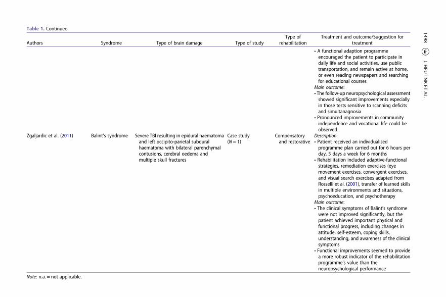
Table 1. Continued.
Authors Syndrome Type of brain damage Type of studyType of
rehabilitationTreatment and outcome/Suggestion for
treatment
• A functional adaption programmeencouraged the patient to participate indaily life and social activities, use publictransportation, and remain active at home,or even reading newspapers and searchingfor educational courses
Main outcome:• The follow-up neuropsychological assessmentshowed significant improvements especiallyin those tests sensitive to scanning deficitsand simultanagnosia
• Pronounced improvements in communityindependence and vocational life could beobserved
Zgaljardic et al. (2011) Balint’s syndrome Severe TBI resulting in epidural haematomaand left occipito-parietal subduralhaematoma with bilateral parenchymalcontusions, cerebral oedema andmultiple skull fractures
Case study(N = 1)
Compensatoryand restorative
Description:• Patient received an individualisedprogramme plan carried out for 6 hours perday, 5 days a week for 6 months
• Rehabilitation included adaptive-functionalstrategies, remediation exercises (eyemovement exercises, convergent exercises,and visual search exercises adapted fromRosselli et al. (2001), transfer of learned skillsin multiple environments and situations,psychoeducation, and psychotherapy
Main outcome:• The clinical symptoms of Balint’s syndromewere not improved significantly, but thepatient achieved important physical andfunctional progress, including changes inattitude, self-esteem, coping skills,understanding, and awareness of the clinicalsymptoms
• Functional improvements seemed to providea more robust indicator of the rehabilitationprogramme’s value than theneuropsychological performance
Note: n.a. = not applicable.
1498J.H
EUTIN
KET
AL.
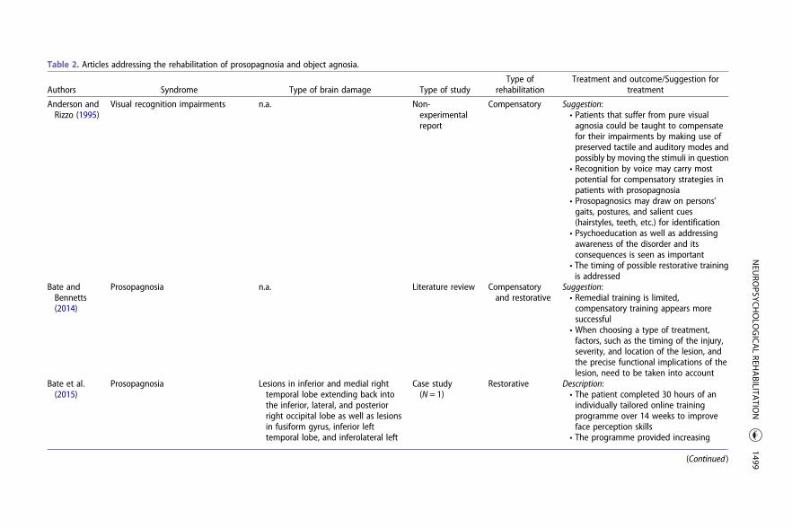
Table 2. Articles addressing the rehabilitation of prosopagnosia and object agnosia.
Authors Syndrome Type of brain damage Type of studyType of
rehabilitationTreatment and outcome/Suggestion for
treatment
Anderson andRizzo (1995)
Visual recognition impairments n.a. Non-experimentalreport
Compensatory Suggestion:• Patients that suffer from pure visualagnosia could be taught to compensatefor their impairments by making use ofpreserved tactile and auditory modes andpossibly by moving the stimuli in question
• Recognition by voice may carry mostpotential for compensatory strategies inpatients with prosopagnosia
• Prosopagnosics may draw on persons’gaits, postures, and salient cues(hairstyles, teeth, etc.) for identification
• Psychoeducation as well as addressingawareness of the disorder and itsconsequences is seen as important
• The timing of possible restorative trainingis addressed
Bate andBennetts(2014)
Prosopagnosia n.a. Literature review Compensatoryand restorative
Suggestion:• Remedial training is limited,compensatory training appears moresuccessful
• When choosing a type of treatment,factors, such as the timing of the injury,severity, and location of the lesion, andthe precise functional implications of thelesion, need to be taken into account
Bate et al.(2015)
Prosopagnosia Lesions in inferior and medial righttemporal lobe extending back intothe inferior, lateral, and posteriorright occipital lobe as well as lesionsin fusiform gyrus, inferior lefttemporal lobe, and inferolateral left
Case study(N = 1)
Restorative Description:• The patient completed 30 hours of anindividually tailored online trainingprogramme over 14 weeks to improveface perception skills
• The programme provided increasing
(Continued )
NEU
ROPSYC
HOLO
GICALREH
ABILITA
TION
1499
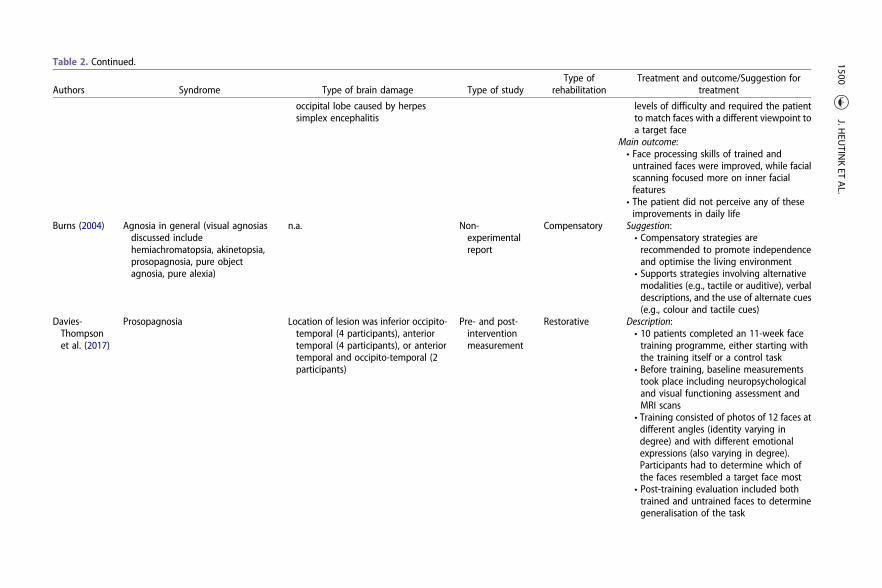
Table 2. Continued.
Authors Syndrome Type of brain damage Type of studyType of
rehabilitationTreatment and outcome/Suggestion for
treatment
occipital lobe caused by herpessimplex encephalitis
levels of difficulty and required the patientto match faces with a different viewpoint toa target face
Main outcome:• Face processing skills of trained anduntrained faces were improved, while facialscanning focused more on inner facialfeatures
• The patient did not perceive any of theseimprovements in daily life
Burns (2004) Agnosia in general (visual agnosiasdiscussed includehemiachromatopsia, akinetopsia,prosopagnosia, pure objectagnosia, pure alexia)
n.a. Non-experimentalreport
Compensatory Suggestion:• Compensatory strategies arerecommended to promote independenceand optimise the living environment
• Supports strategies involving alternativemodalities (e.g., tactile or auditive), verbaldescriptions, and the use of alternate cues(e.g., colour and tactile cues)
Davies-Thompsonet al. (2017)
Prosopagnosia Location of lesion was inferior occipito-temporal (4 participants), anteriortemporal (4 participants), or anteriortemporal and occipito-temporal (2participants)
Pre- and post-interventionmeasurement
Restorative Description:• 10 patients completed an 11-week facetraining programme, either starting withthe training itself or a control task
• Before training, baseline measurementstook place including neuropsychologicaland visual functioning assessment andMRI scans
• Training consisted of photos of 12 faces atdifferent angles (identity varying indegree) and with different emotionalexpressions (also varying in degree).Participants had to determine which ofthe faces resembled a target face most
• Post-training evaluation included bothtrained and untrained faces to determinegeneralisation of the task
1500J.H
EUTIN
KET
AL.
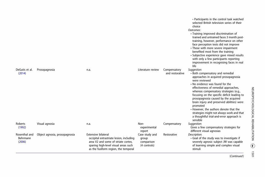
• Participants in the control task watchedselected British television series of theirchoice
Outcomes:• Training improved discrimination oftrained and untrained faces 3 month post-training, however, performance on otherface perception tests did not improve
• Those with more severe impairmentbenefited most from the training
• Subjective experience gave mixed resultswith only a few participants reportingimprovement in recognising faces in reallife
DeGutis et al.(2014)
Prosopagnosia n.a. Literature review Compensatoryand restorative
Suggestion:• Both compensatory and remedialapproaches in acquired prosopagnosiawere reviewed
• No evidence was found for theeffectiveness of remedial approaches,whereas compensatory strategies (e.g.,focusing on the specific deficit leading toprosopagnosia caused by the acquiredbrain injury and preserved abilities) werepromoted
• However, the authors denote that thestrategies might not always work and thata thoughtful trial-and-error approach issensible
Roberts(1992)
Visual agnosia n.a. Non-experimentalreport
Compensatory Suggestion:Gives a few compensatory strategies fordifferent visual agnosias
Rosenthal andBehrmann(2006)
Object agnosia, prosopagnosia Extensive bilateraloccipital extrastriate lesion, includingarea V2 and some of striate cortex,sparing high-level visual areas suchas the fusiform region, the temporal
Case study andgroupcomparison(4 controls)
Restorative Description:• Goal of the study was to investigate ifseverely agnosic subject JW was capableof learning simple and complex visualstimuli
(Continued )
NEU
ROPSYC
HOLO
GICALREH
ABILITA
TION
1501
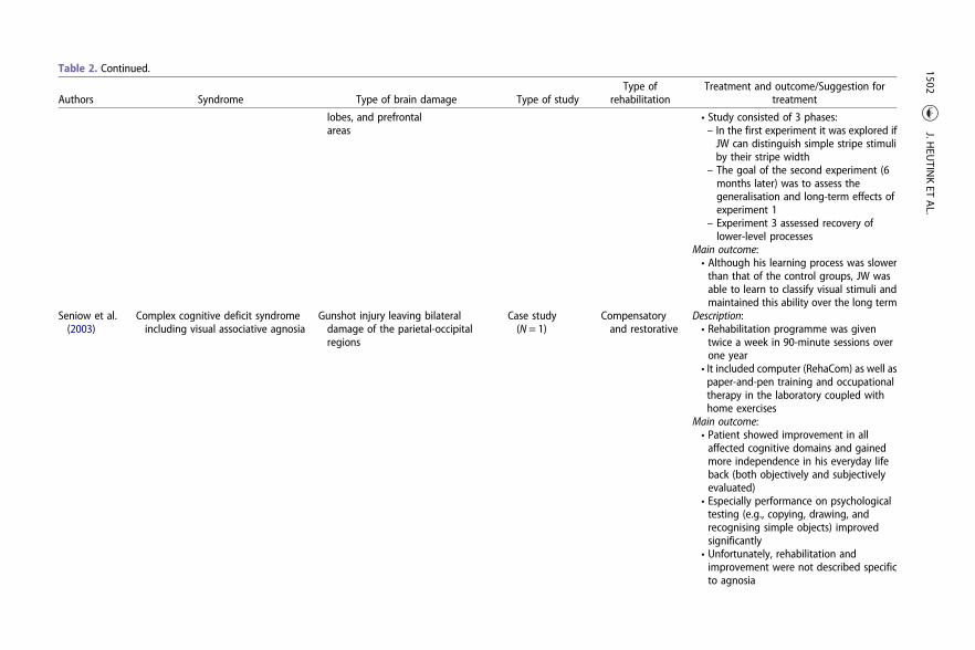
Table 2. Continued.
Authors Syndrome Type of brain damage Type of studyType of
rehabilitationTreatment and outcome/Suggestion for
treatment
lobes, and prefrontalareas
• Study consisted of 3 phases:– In the first experiment it was explored ifJW can distinguish simple stripe stimuliby their stripe width
– The goal of the second experiment (6months later) was to assess thegeneralisation and long-term effects ofexperiment 1
– Experiment 3 assessed recovery oflower-level processes
Main outcome:• Although his learning process was slowerthan that of the control groups, JW wasable to learn to classify visual stimuli andmaintained this ability over the long term
Seniow et al.(2003)
Complex cognitive deficit syndromeincluding visual associative agnosia
Gunshot injury leaving bilateraldamage of the parietal-occipitalregions
Case study(N = 1)
Compensatoryand restorative
Description:• Rehabilitation programme was giventwice a week in 90-minute sessions overone year
• It included computer (RehaCom) as well aspaper-and-pen training and occupationaltherapy in the laboratory coupled withhome exercises
Main outcome:• Patient showed improvement in allaffected cognitive domains and gainedmore independence in his everyday lifeback (both objectively and subjectivelyevaluated)
• Especially performance on psychologicaltesting (e.g., copying, drawing, andrecognising simple objects) improvedsignificantly
• Unfortunately, rehabilitation andimprovement were not described specificto agnosia
1502J.H
EUTIN
KET
AL.
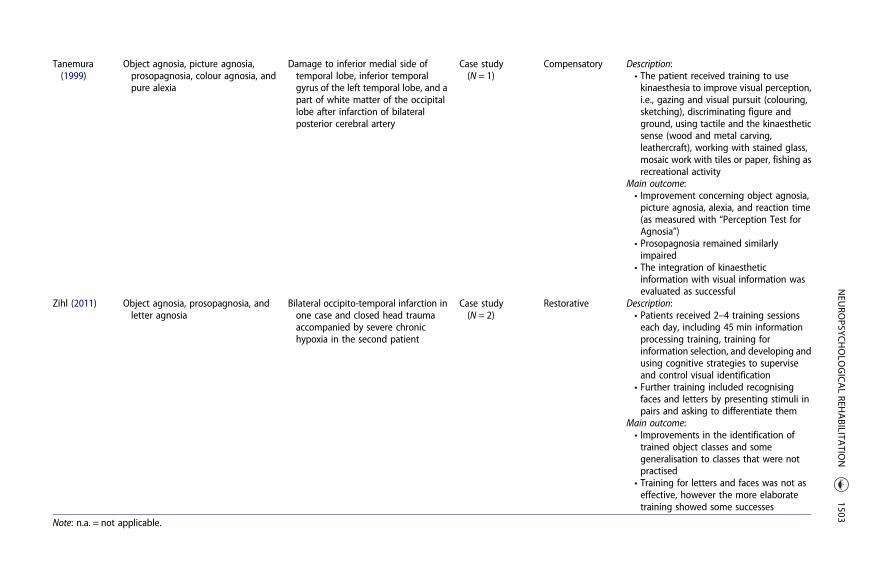
Tanemura(1999)
Object agnosia, picture agnosia,prosopagnosia, colour agnosia, andpure alexia
Damage to inferior medial side oftemporal lobe, inferior temporalgyrus of the left temporal lobe, and apart of white matter of the occipitallobe after infarction of bilateralposterior cerebral artery
Case study(N = 1)
Compensatory Description:• The patient received training to usekinaesthesia to improve visual perception,i.e., gazing and visual pursuit (colouring,sketching), discriminating figure andground, using tactile and the kinaestheticsense (wood and metal carving,leathercraft), working with stained glass,mosaic work with tiles or paper, fishing asrecreational activity
Main outcome:• Improvement concerning object agnosia,picture agnosia, alexia, and reaction time(as measured with “Perception Test forAgnosia”)
• Prosopagnosia remained similarlyimpaired
• The integration of kinaestheticinformation with visual information wasevaluated as successful
Zihl (2011) Object agnosia, prosopagnosia, andletter agnosia
Bilateral occipito-temporal infarction inone case and closed head traumaaccompanied by severe chronichypoxia in the second patient
Case study(N = 2)
Restorative Description:• Patients received 2–4 training sessionseach day, including 45 min informationprocessing training, training forinformation selection, and developing andusing cognitive strategies to superviseand control visual identification
• Further training included recognisingfaces and letters by presenting stimuli inpairs and asking to differentiate them
Main outcome:• Improvements in the identification oftrained object classes and somegeneralisation to classes that were notpractised
• Training for letters and faces was not aseffective, however the more elaboratetraining showed some successes
Note: n.a. = not applicable.
NEU
ROPSYC
HOLO
GICALREH
ABILITA
TION
1503
studies; Table 2). Generally, compensating for the deficit via tactile and auditory modes,as well as via verbal description and kinaesthetic information was frequently rec-ommended (Tanemura, 1999). Zihl (2011) trained object recognition in two patientsusing a restorative approach, which led to improvements in both patients with somegeneralisation to non-trained objects. Seniow, Polanowska, Mandat, and Laudanski(2003) achieved improvements using both restorative and compensatory training.Therefore, it is hard to draw conclusions about which the effective component was.To summarise, there is only limited data available on effective rehabilitation of objectagnosia, but evidence is beginning to emerge for the usefulness of both restorativeand compensatory strategies.
Topographical agnosia
Literature on the rehabilitation of topographical agnosia is scarce; only two studiesgiving information on treatment approaches could be included (Table 3). In the firststudy, Bouwmeester, Van de Wege, Haaxma, and Snoek (2015) described a patientwith a relatively severe clinical presentation who benefited from an individualised train-ing programme. The patient did not improve much in location recognition, but never-theless learned to reach desired destinations. This outcome provides evidence thattraining can significantly increase independence and quality of life even in patientswith severe deficits. The second study described a generally more highly functioningindividual, who suffered severely from topographical disorientation (Davis & Coltheart,1999). By focusing on the patient’s relevant environment and common routes, thepatient was taught to use mnemonic techniques and a map to reach desired desti-nations independently and more securely. To conclude, both studies used compensa-tory strategies for the rehabilitation and offered some specific ideas about what mayhave been effective in these two cases.
Discussion
The aim of the present literature review was to systematically collect information orrecommendations for the rehabilitation of visual agnosia and Balint’s syndrome.The results show that especially compensatory strategies have proven to be beneficialin most cases, whereas restorative training has produced mixed results. Evidence forthe effectiveness of restorative training in acquired prosopagnosia is currently notvery persuasive. For the other subtypes of agnosia, it is not possible to draw con-clusions about the effectiveness of restorative training as there is too little and contra-dictory research available. In Balint’s syndrome, restorative training has been shown tobe more successful, but there is also not enough research available to give a clear indi-cation. Altogether, though still very scarce, a scientific foundation about the rehabili-tation of these disorders is evolving and there are approaches giving valuableinformation that can be built upon in the future. Yet, a few issues require somefurther consideration.
First, for the treatment of agnosia, Groh-Bordin and Kerkhoff (2010) suggested that,based on their clinical experience, it might be more effective to treat deficits that co-occur with agnosia (e.g., hemianopia) rather than focusing on the agnosia itself. Thisapproach is based on the view that, if other visual deficits are enhanced, recognitionwill likely be improved as well. However, Zihl (2011) failed to prove this approach. He
1504 J. HEUTINK ET AL.
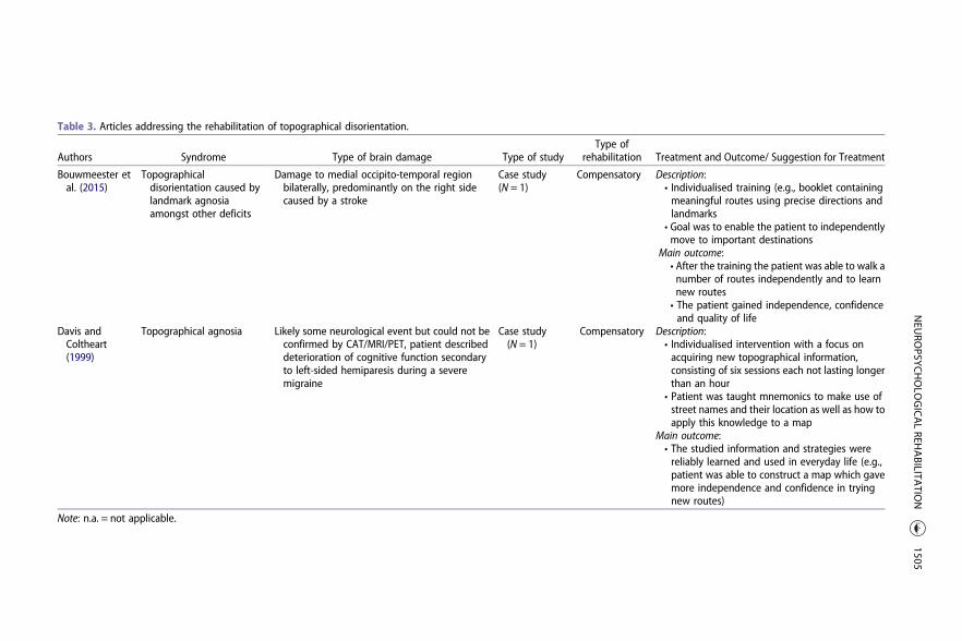
Table 3. Articles addressing the rehabilitation of topographical disorientation.
Authors Syndrome Type of brain damage Type of studyType of
rehabilitation Treatment and Outcome/ Suggestion for Treatment
Bouwmeester etal. (2015)
Topographicaldisorientation caused bylandmark agnosiaamongst other deficits
Damage to medial occipito-temporal regionbilaterally, predominantly on the right sidecaused by a stroke
Case study(N = 1)
Compensatory Description:• Individualised training (e.g., booklet containingmeaningful routes using precise directions andlandmarks
• Goal was to enable the patient to independentlymove to important destinations
Main outcome:• After the training the patient was able to walk anumber of routes independently and to learnnew routes• The patient gained independence, confidenceand quality of life
Davis andColtheart(1999)
Topographical agnosia Likely some neurological event but could not beconfirmed by CAT/MRI/PET, patient describeddeterioration of cognitive function secondaryto left-sided hemiparesis during a severemigraine
Case study(N = 1)
Compensatory Description:• Individualised intervention with a focus onacquiring new topographical information,consisting of six sessions each not lasting longerthan an hour
• Patient was taught mnemonics to make use ofstreet names and their location as well as how toapply this knowledge to a map
Main outcome:• The studied information and strategies werereliably learned and used in everyday life (e.g.,patient was able to construct a map which gavemore independence and confidence in tryingnew routes)
Note: n.a. = not applicable.
NEU
ROPSYC
HOLO
GICALREH
ABILITA
TION
1505
attempted to improve identification and recognition by training oculomotor scanningin patients with homonymous visual field loss and disorientation, which resulted inimprovement of the trained skill, yet none for recognition. Nevertheless, it seems plaus-ible that improved lower visual functions will increase the chances to rehabilitate recog-nition as well. We would argue that clinicians need to consider how much the patientsare – subjectively and objectively – impaired by the agnosia. If the agnosia dominatesimpairment caused by other deficient visual functions, rehabilitation should target thespecific agnostic deficit at least by training compensatory strategies.
Another factor to consider in clinical rehabilitation is the cost-effectiveness of acertain approach. As mentioned before, the effectiveness of restorative training invisual agnosia and Balint’s syndrome has not been fully proven. In addition tothat, successful rehabilitation using restorative training usually follows an extensiveperiod of training, sometimes up to hundreds of hours of practice with often onlysmall improvements in daily life (e.g., Zihl, 2011). This might lead to potential frustra-tion in patients. Compensatory strategies on the other hand have been shown toachieve meaningful progress in the patients’ daily functioning and might thereforebe more cost-effective. Yet, one pitfall may be that patients may not be ready to“accept” the impairment in early stages of the rehabilitation, decreasing the moti-vation to attempt compensation rather than training the deficit itself (Finauer,2009). Most important, decisions about rehabilitation approaches need to be indivi-dualised, taking the circumstances and unique impairments of the patients intoconsideration.
Future research and clinical practice
Based on the current literature available, an evidence-based rehabilitation programmecannot be established. We therefore encourage clinicians to share any cases of visualagnosia and Balint’s syndrome with the scientific community, and to make detailedrehabilitation plans available to benefit other practitioners. Especially the addition ofmore details about the rehabilitation approach, including pre- and post-measurements,would be helpful to establish an evidence-based practice. Future publications may takethis literature review as a basis for further research to confirm the effectiveness of par-ticular rehabilitation approaches and fill in further knowledge gaps. For example, exist-ing cognitive rehabilitation software that was created to train people with visual agnosiaand Balint’s syndrome (for example, visual exploration exercises), could be incorporatedinto future rehabilitation programmes to be systematically evaluated. Furthermore, theliterature described in the present manuscript rarely explored how well trained skillstransferred into the relevant context for the patient. It is important for future researchto not overlook this crucial step in determining the effectiveness of a certain rehabilita-tion approach.
Conclusion
The current article provides a basis and starting point for more systematic progresstowards an evidence-based treatment approach for patients with visual agnosia andBalint’s syndrome. Researchers and clinicians can use the information this articlerevealed to guide their research and fill in the knowledge gaps identified in thepresent literature review.
1506 J. HEUTINK ET AL.
Disclosure statement
No potential conflict of interest was reported by the authors.
ORCID
Joost Heutink http://orcid.org/0000-0002-4811-968X
References
Al-Khawaja, I., Haboubi, N. H. J., & Kerkhoff, G. (2001). Neurovisual rehabilitation in Balint’s syndrome.Journal of Neurology, Neurosurgery & Psychiatry, 70, 416–416. doi:10.1136/jnnp.70.3.416
Anderson, S. W., & Rizzo, M. (1995). Recovery and rehabilitation of visual cortical dysfunction.NeuroRehabilitation, 5(2), 129–140. doi:10.3233/NRE-1995-5204
Balint, R. (1909). Seelenlähmung des “Schauens”: Optische Ataxie, räumliche Störung der Aufmerksamkeit[Paralysis of “seeing”: Optical ataxia, spatial disorder of attention]. Monatszeitschift Für Psychiatrie UndNeurologie, 25(1), 51–66. doi:10.1159/000210464
Bate, S., & Bennetts, R. J. (2014). The rehabilitation of face recognition impairments: A critical review andfuture directions. Frontiers in Human Neuroscience, 8, 491–491. doi:10.3389/fnhum.2014.00491
Bate, S., Bennetts, R., Mole, J. A., Ainge, J. A., Gregory, N. J., Bobak, A. K., & Bussunt, A. (2015). Rehabilitationof face-processing skills in an adolescent with prosopagnosia: Evaluation of an online perceptual train-ing programme. Neuropsychological Rehabilitation, 25(5), 733–762. doi:10.1080/09602011.2014.973886
Bouwmeester, L., Van de Wege, A., Haaxma, R., & Snoek, J. W. (2015). Rehabilitation in a complex case oftopographical disorientation. Neuropsychological Rehabilitation, 25(1), 1–14. doi:10.1080/09602011.2014.923318
Bowen, A., Hazelton, C., Pollock, A., & Lincoln, N. B. (2013). Cognitive rehabilitation for spatial neglect fol-lowing stroke. Cochrane Database of Systematic Reviews, 7, CD003586. doi:10.1002/14651858.CD003586.pub3
Burns, M. S. (2004). Clinical management of agnosia. Topics in Stroke Rehabilitation, 11(1), 1–9.Damasio, A. R., Tranel, D., & Rizzo, M. (2000). Disorders of complex visual processing. In M. M. Mesualm
(Ed.), Principles of behavioral and cognitive neurology (pp. 332–364). New York: Oxford University Press.Davies-Thompson, J., Fletcher, K., Hills, C., Pancaroglu, R., Corrow, S. L., & Barton, J. J. S. (2017). Perceptual
learning of faces: A rehabilitative study of acquired prosopagnosia. Journal of Cognitive Neuroscience,29, 573–591. doi:10.1162/jocn_a_01063
Davis, S. J. C., & Coltheart, M. (1999). Rehabilitation of topographical disorientation: An experimentalsingle case study. Neuropsychological Rehabilitation, 9(1), 1–30. doi:10.1080/713755586
DeGutis, J. M., Chiu, C., Grosso, M. E., & Cohan, S. (2014). Face processing improvements in prosopagnosia:Successes and failures over the last 50 years. Frontiers in Human Neuroscience, 8, 561–561. doi:10.3389/fnhum.2014.00561
De Haan, G. A., Heutink, J., Melis-Dankers, B. J. M., Tucha, O., & Brouwer, W. H. (2014). Spontaneous recov-ery and treatment effects in patients with homonymous visual field defects: A meta-analysis of exist-ing literature in terms of the ICF framework. Survey of Ophthalmology, 59(1), 77–96. doi:10.1016/j.survophthal.2013.02.006
Finauer, G. (2009). Therapiemanuale für die neuropsychologische Rehabilitation: Kognitive und kompeten-zorientierte Therapie für die Gruppen- und Einzelbehandlung [Manual for neuropsychological rehabilita-tion: Cognitive and competence-oriented therapy for group- and indivudual treatment]. Berlin:Springer.
Gillen, J. A., & Dutton, G. N. (2003). Balint’s syndrome in a 10-year-old male. Developmental Medicine &Child Neurology, 45(5), 349–352.
Groh-Bordin, C., & Kerkhoff, G. (2010). Recovery and treatment of sensory perceptual disorders. In J. Gurd,U. Kischka, & J. Marshall (Eds.), Handbook of clinical neuropsychology (pp. 139–158). New York: OxfordUniversity Press.
Kerkhoff, G., & Groh-Bordin, C. (2010). Höhere visuelle Funktionen: Neglect, Raumorientierung, Balint-Holmes Syndrom und Agnosien [Higher visual functions: Neglect, spatial orientation, balint-holmes
NEUROPSYCHOLOGICAL REHABILITATION 1507
syndrome and agnosias]. In P. Frommelt & H. Lösslein (Eds.), NeuroRehabilitation (pp. 207–221). Berlin:Springer.
Kerkhoff, G., & Heldmann, B. (1999). Balint syndrome and associated disorders. Anamnesis–diagnosis–approaches to treatment. Der Nervenarzt, 70(10), 859–869.
Kerkhoff, G., & Marquardt, C. (2009a). EYEMOVE. Standardized assessment and treatment of visual searchdisorders. Der Nervenarzt, 80(10), 1190. doi:10.1007/s00115-009-2811-4
Kerkhoff, G., & Marquardt, C. (2009b). Erworbene, visuell bedingte Lesestörungen: StandardisierteDiagnostik und Therapie mit READ [Acquired, visually based reading disorders: Standardised assess-ment and treatment with READ]. Der Nervenarzt, 80(12), 1424–1439. doi:10.1007/s00115-009-2723-3
Moher, D., Liberati, A., Tetzlaff, J., & Altman, D. G. (2009). Preferred reporting items for systematic reviewsand meta-analyses: The PRISMA statement. PLoS Medicine, 6(7), e1000097. doi:10.1371/journal.pmed.1000097
Nijboer, T., & Heutink, J. (2017). Visual perception. In R. Kessels, P. Eling, R. Ponds, J. Spikman, & Van M.Zandvoort (Eds.), Clinical neuropsychology (pp. 137–157). Amsterdam: Boom.
Perez, F. M., Tunkel, R. S., Lachmann, E. A., & Nagler, W. (1996). Balint’s syndrome arising from bilateralposterior cortical atrophy or infarction: Rehabilitation strategies and their limitation. Disability andRehabilitation, 18(6), 300–304.
Pollock, A., Hazelton, C., Henderson, C. A., Angilley, J., Dhillon, B., Langhorne, P.,… Shahani, U. (2011).Interventions for visual field defects in patients with stroke. Cochrane Database of SystematicReviews, (10), CD008388. doi:10.1002/14651858.CD008388.pub2
Roberts, S. P. (1992). Visual disorders of higher cortical function. Journal of the American OptometricAssociation, 63(10), 723–732.
Roca, M., Gleichgerrcht, E., Torralva, T., & Manes, F. (2010). Cognitive rehabilitation in posterior corticalatrophy. Neuropsychological Rehabilitation, 20(4), 528–540. doi:10.1080/09602011003597408
Rosenthal, O., & Behrmann, M. (2006). Acquiring long-term representations of visual classes followingextensive extrastriate damage. Neuropsychologia, 44(5), 799–815. doi:10.1016/j.neuropsychologia.2005.07.010
Rosselli, M., Ardila, A., & Beltran, C. (2001). Rehabilitation of balint’s syndrome: A single case report. AppliedNeuropsychology, 8(4), 242–247. doi:10.1207/09084280152829093
Seniow, J., Polanowska, K., Mandat, T., & Laudanski, K. (2003). The cognitive impairments due to the occi-pito-parietal brain injury after gunshot. A successful neurorehabilitation case study. Brain Injury, 17(8),701–713. doi:10.1080/0269905031000088621
Spikman, J., & Fasotti, L. (2017). Recovery and treatment. In R. Kessels, P. Eling, R. Ponds, J. Spikman, & M.Van Zandvoort (Eds.), Clinical neuropsychology (pp. 113–133). Amsterdam: Boom.
Starrfelt, R., Ólafsdóttir, R. R., & Arendt, I.-M. (2013). Rehabilitation of pure alexia: A review.Neuropsychological Rehabilitation, 23(5), 755–779. doi:10.1080/09602011.2013.809661
Tanemura, R. (1999). Awareness in apraxia and agnosia. Topics in Stroke Rehabilitation, 6(1), 33–42. doi:10.1310/U9KU-M4X8-DMRQ-9TR7
Zgaljardic, D. J., Yancy, S., Levinson, J., Morales, G., & Masel, B. E. (2011). Balint’s syndrome and post-acutebrain injury rehabilitation: A case report. Brain Injury, 25(9), 909–917. doi:10.3109/02699052.2011.585506
Zihl, J. (2003). Recovery and rehabilitation of cerebral visual disorders. In M. Fahle & M. Greenlee (Eds.),The neuropsychology of vision (pp. 319–338). New York: Oxford University Press.
Zihl, J. (2011). Rehabilitation of visual disorders after brain injury. New York: Psychology Press.Zihl, J., & Kennard, C. (2003). Disorders of higher visual function. In T. Brandt (Ed.), Neurological disorders:
Course and treatment (pp. 255–261). Amsterdam: Academic Press.
1508 J. HEUTINK ET AL.