Embed Size (px)
Citation preview
Brain (1999),122,593–624
I N V I T E D R E V I E W
The neuropathology of schizophreniaA critical review of the data and their interpretation
Paul J. Harrison
University Department of Psychiatry, Warneford Hospital, Correspondence to: Dr P. J. Harrison, NeurosciencesOxford, UK Building, University Department of Psychiatry,
Warneford Hospital, Oxford OX3 7JX, UKE-mail: [email protected]
SummaryDespite a hundred years’ research, the neuropathology ofschizophrenia remains obscure. However, neither can thenull hypothesis be sustained—that it is a ‘functional’psychosis, a disorder with no structural basis. A numberof abnormalities have been identified and confirmedby meta-analysis, including ventricular enlargement anddecreased cerebral (cortical and hippocampal) volume.These are characteristic of schizophrenia as a whole,rather than being restricted to a subtype, and are presentin first-episode, unmedicated patients. There isconsiderable evidence for preferential involvement of thetemporal lobe and moderate evidence for an alterationin normal cerebral asymmetries. There are severalcandidates for the histological and molecular correlatesof the macroscopic features. The probable proximalexplanation for decreased cortical volume is reducedneuropil and neuronal size, rather than a loss of neurons.These morphometric changes are in turn suggestive ofalterations in synaptic, dendritic and axonal organization,a view supported by immunocytochemical and ultra-structural findings. Pathology in subcortical structures isnot well established, apart from dorsal thalamic nuclei,which are smaller and contain fewer neurons. Othercytoarchitectural features of schizophrenia which areoften discussed, notably entorhinal cortex heterotopiasand hippocampal neuronal disarray, remain to be
Keywords: Alzheimer’s disease; cytoarchitecture; morphometry; synapse; psychosis
Abbreviations: DLPFC 5 dorsolateral prefrontal cortex; 5-HT5 5-hydroxytryptamine; VBR5 ventricle : brain ratio
IntroductionA hundred years ago, Kraeplin described the syndrome nowcalled schizophrenia. He was convinced that it was an organicbrain disease, and it was his colleague Alzheimer who beganthe neuropathological investigation before moving to a more
© Oxford University Press 1999
confirmed. The phenotype of the affected neuronal andsynaptic populations is uncertain. A case can be made forimpairment of hippocampal and corticocortical excitatorypathways, but in general the relationship betweenneurochemical findings (which centre upon dopamine,5-hydroxytryptamine, glutamate and GABA systems) andthe neuropathology of schizophrenia is unclear. Gliosis isnot an intrinsic feature; its absence supports, but doesnot prove, the prevailing hypothesis that schizophrenia isa disorder of prenatal neurodevelopment. The cognitiveimpairment which frequently accompanies schizophreniais not due to Alzheimer’s disease or any other recognizedneurodegenerative disorder. Its basis is unknown.Functional imaging data indicate that the pathophysiologyof schizophrenia reflects aberrant activity in, andintegration of, the components of distributed circuitsinvolving the prefrontal cortex, hippocampus and certainsubcortical structures. It is hypothesized that theneuropathological features represent the anatomicalsubstrate of these functional abnormalities in neuralconnectivity. Investigation of this proposal is a goal ofcurrent neuropathological studies, which must also seek(i) to establish which of the recent histological findingsare robust and cardinal, and (ii) to define the relationshipof the pathological phenotype with the clinical syndrome,its neurochemistry and its pathogenesis.
fruitful research area. Subsequently the subject has continuedto fascinate and exasperate researchers in equal measure,generating more heat than light and being notable formemorable quotes rather than durable data. The most
at UC
HS
C D
enison Mem
orial Library Box A
-003 on March 30, 2010
http://brain.oxfordjournals.orgD
ownloaded from
594 P. J. Harrison
infamous, that schizophrenia is the ‘graveyard ofneuropathologists’ (Plum, 1972), was a statement which,together with critical reviews of the work up to that time(Corsellis, 1976), marked the nadir of the field.
Over the past 20 years, signs of life have appeared in thegraveyard, reflected in the return of schizophrenia to thelatest edition ofGreenfield’s Neuropathology(Robertset al.,1997), having been omitted from the previous two. Thesignificant progress which has been made began with CTfindings, followed by MRI and by post-mortem studies usingimproved methodologies and new techniques. The progressallowed Ron and Harvey (1990) to charge that ‘[to] haveforgotten that schizophrenia is a brain disease will go downas one of the great aberrations of twentieth century medicine’.In a similar vein, Weinberger (1995) stated ‘20 years ago,the principal challenge for schizophrenia research was togather objective scientific evidence that would implicate thebrain. That challenge no longer exists.’ On the other hand,it is undoubtedly an overstatement to claim that there is ‘anavalanche of consistent . . . evidence of microscopicpathology’ (Bloom, 1993); the current challenge is to establishthe characteristics of the pathological changes (Shapiro, 1993;Chua and McKenna, 1995). This review summarizes thepresent state of knowledge, including the issues ofhemispheric asymmetry, dementia in schizophrenia, neuro-development and neurochemistry. An integration of structurewith function is attempted, with elaboration of the proposalthat the neuropathology of schizophrenia represents theanatomical substrate of aberrant functional connectivity.
Review coverage and methodologyThe review focuses on the key points of agreement and ofcontroversy affecting the robustness of the data and theirinterpretation. It comprises a comprehensive survey ofcontemporary (post-1980) neurohistopathological research,with restricted coverage of earlier work and of related fieldssuch as neuroimaging and neurochemistry.
The sources for the review consisted of: (i) papers identifiedusing a range of keywords for on-line searches of Medline,PsycLIT andBiological Abstracts(last search, October 1998),(ii) weekly scanning ofReference Update(deluxe edition,customized to 350 journals) from 1989 to October 1998using a similar range of keywords, and (iii) an extensivereprint collection and perusal of each article’s reference list.Only data published in full papers in peer-reviewed English-language journals were considered for inclusion.
Clinical features of schizophreniaSchizophrenia remains a clinical diagnosis, based upon thepresence of certain types of delusions, hallucinations andthought disorder (McKenna, 1994; Andreasen, 1995). These‘positive’ symptoms are often complemented by the‘negative’ symptoms of avolition, alogia and affectiveflattening. The criteria of the Diagnostic and Statistical
Manual of Mental Disorders (American PsychiatricAssociation, 1994), used for most research studies, requiresymptoms to have been present for at least 6 months; theremust also be impaired personal functioning, and the symptomsmust not be secondary to another disorder (e.g. depression,substance abuse). The peak age of onset is in the thirddecade, occurring a few years earlier in males than in females(Hafneret al., 1998). The course and outcome are remarkablyvariable, but better than sometimes believed; only a minorityof patients have a chronic, deteriorating course, thoughmany others have enduring symptoms or functional deficits(Davidson and McGlashan, 1997; Huber, 1997). There is asignificant excess of mortality from suicide and natural causes(Brown, 1997). The lifetime risk of schizophrenia is justunder 1% (Cannon and Jones, 1996). It has a predominantlygenetic aetiology, but no chromosomal loci or genes havebeen unequivocally demonstrated (McGuffinet al., 1995).
The diagnosis of schizophrenia is reliable, but as with anyother syndromal diagnosis there are problems establishingits validity and debate as to where its external and internalboundaries should be drawn (Jablensky, 1995). These issueshave implications for research into its pathological basis justas they do for the search for the causative genes (Kennedy,1996). For example, is schizophrenia a categorical ordimensional construct? What is the relationship of schizo-affective and schizotypal disorders to schizophrenia? Arethere separate pathological counterparts of schizophrenicsubsyndromes or specific symptoms, given that each hasits own pathophysiological correlates (Liddleet al., 1992;Silbersweiget al., 1995; Sabriet al., 1997)? The delineationof type I and type II schizophrenia was an important, if nowrather outmoded, attempt to address this issue (Crow, 1980).As an analogy, is schizophrenia—neuropathologicallyspeaking—comparable to dementia, to a specific dementingdisorder or to a domain of memory impairment? Comparisonswith epilepsy are also pertinent (Brutonet al., 1994; Stevens,1997). Clearly, the prospects for success in finding theneuropathology of schizophrenia depend on which of theseparallels proves closest. These issues are touched upon laterin the review but for the most part, predicated on the designof the studies being discussed, schizophrenia is consideredas a single entity.
Structural imaging in schizophreniaThe cardinal findingsContemporary research into the structural basis ofschizophrenia can be traced to the landmark report ofJohnstoneet al. (1976) describing dilatation of the lateralventricles in a small group of patients with chronicschizophrenia. This CT finding, which was consistent withearlier pneumoencephalographic data (Haug, 1982), has beenfollowed by a large number of CT and MRI studies withever-improving resolution and sophistication of analysis. Thekey findings are as follows.
at UC
HS
C D
enison Mem
orial Library Box A
-003 on March 30, 2010
http://brain.oxfordjournals.orgD
ownloaded from
Neuropathology of schizophrenia 595
There is enlargement of the lateral and third ventricles inschizophrenia. The magnitude has been estimated in severalways. Comprehensive reviews of lateral ventricle : brainratio (VBR) indicate an increase of 20–75% (Danielet al.,1991; van Horn and McManus, 1992), whilst a meta-analysisof CT studies up to 1989 showed a VBR effect size (d) of0.70, corresponding to a 43% non-overlap between cases andcontrols (Raz and Raz, 1990). A median 40% increase inventricular size was reported in a recent systematic reviewof volumetric MRI studies (Lawrie and Abukmeil, 1998).Of note, VBR in schizophrenia follows a single normaldistribution, indicating that structural pathology, at least interms of this parameter, is not restricted to an ‘organic’subgroup but is present to a degree in all cases (Danielet al.,1991). Conversely, despite the group differences, there is asignificant overlap between subjects with schizophrenia andcontrols for every imaging (and neuropathological) parameterto be discussed. For this reason, as well as the fact thatchanges such as increased VBR and decreased brain sizelack diagnostic specificity, it is worth emphasizing thatschizophrenia cannot be diagnosed using either a brain scanor a microscope. It remains a moot point whether thissituation will change.
The ventricular enlargement is accompanied by a loss ofbrain tissue averaging 3% (Lawrie and Abukmeil, 1998) withd 5 –0.26 (Ward et al., 1996). However, no consistentcorrelation has been observed between the degree ofventricular enlargement and that of the decreased brainvolume. This may reflect the relative sizes of the ventriclesand cerebral cortex, such that a given percentage change inventricular volumes corresponds to a much smaller percentagechange in cortical substance (and hence one which is difficultto measure accurately). Or it may suggest that the ventricularenlargement is due to disproportionate reductions inunidentified, localized periventricular structures, or even thatindependent pathological processes are at work.
Evidence for regional pathology has emerged fromvolumetric MRI studies which indicate larger reductions inthe temporal lobe overall (~8%) and in medial temporalstructures (hippocampus, parahippocampal gyrus andamygdala, 4–12%; Lawrie and Abukmeil, 1998) present aftercorrection for total brain volume (Nelsonet al., 1998).In support of this conclusion, the brain size reduction issignificantly greater in the axial (d 5 –0.60) than the sagittal(d 5 –0.09) plane (Wardet al., 1996), suggesting a relativedecrease in mediolateral breadth and a greater involvementof regions typically included in axial slices, such as thetemporal lobes. Grey matter appears to be reduced morethan white matter (Lawrie and Abukmeil, 1998; Zipurskyet al., 1998).
Valuable information has come from imaging studies ofmonozygotic twins discordant for schizophrenia. In virtuallyall pairs the affected twin has the larger ventricles (Reveleyet al., 1982; Suddathet al., 1990) and smaller cortical andhippocampal size (Nogaet al., 1996). In the MRI study ofSuddathet al. (1990), the affected twin was distinguishable
even more clearly by the smaller size of his or her temporallobes and hippocampi. The discordant monozygotic twinstudy design allows two conclusions to be drawn. First,that structural abnormalities are a consistent finding inschizophrenia, their identification being aided by controllingfor genetic influences on neuroanatomy (Bartleyet al., 1997)and, to a large degree, for variation due to environmentalfactors. Secondly, that the alterations are associated withexpression of the schizophrenia phenotype rather than merelywith the underlying, shared genotype. Family studies supportthis interpretation, in that schizophrenics have biggerventricles and smaller brains than do their unaffected relatives(Honer et al., 1994; Sharmaet al., 1998; Silvermanet al.,1998). However, the relatives who are obligate carriers [i.e.unaffected by schizophrenia but transmitting the gene(s)]have larger ventricles than relatives who are not; moreover,both groups of relatives have larger ventricles and smallerbrain structures than equivalent control subjects from familieswithout schizophrenia (Lawrieet al., 1999; Sharmaet al.,1998). These data indicate that a proportion of the structuralpathology of schizophrenia may be a marker of geneticliability to the disorder. (By inference, the same applies tothe accompanying histological features, though there havebeen no post-mortem studies of relatives.)
Imaging of subcortical structures in schizophrenia hasproduced few clear findings. One firm conclusion is that thestriatal enlargement reported in some studies is, unlike theother changes, due to antipsychotic medication (Chakoset al.,1994; Keshavanet al., 1994b). Indeed, in unmedicated andfirst-episode patients, caudate volumes are probably reduced(Keshavanet al., 1998; Shihabuddinet al., 1998). Two MRIstudies suggest that the thalamus is smaller in schizophrenia(Andreasenet al., 1994; Buchsbaumet al., 1996); thoughthis evidence is weak (Portaset al., 1998), it is complementedby relatively strong neuropathological data (see below).Finally, reports of structural abnormalities in the cerebellumin schizophrenia (Katsetoset al., 1997) merit furtherinvestigation, given accumulating evidence for its patho-physiological involvement in the disorder (Andreasenet al., 1996).
Progression, heterogeneity andclinicopathological correlationsKnowledge of the timing of the brain changes is essentialfor understanding their aetiological significance. Ventricularenlargement and cortical volume reduction are both presentin first-episode cases (Degreefet al., 1992; Limet al., 1996;Gur et al., 1998; Whitworthet al., 1998; Zipurskyet al.,1998), excluding the possibility that they are a consequenceof chronic illness or its treatment. Moreover, adolescents andyoung adults who are at high risk of developing schizophreniaby virtue of their family history show enlarged ventricles(Cannonet al., 1993) and smaller medial temporal lobes(Lawrie et al., 1999), suggesting that the brain pathology
at UC
HS
C D
enison Mem
orial Library Box A
-003 on March 30, 2010
http://brain.oxfordjournals.orgD
ownloaded from
596 P. J. Harrison
precedes the onset of symptoms (Harrison, 1999a) andsupporting a neurodevelopmental model of schizophrenia(discussed below).
It is less clear what happens to the structural pathologyafter symptoms emerge. Neither VBR nor cortical volumereduction, nor the smaller size of the medial temporallobe (Marshet al., 1994), correlate with disease duration,suggesting that the alterations are largely static. However,longitudinal studies, which now span 4–8 years, are equivocal.Some support the view that there is no progression (Jaskiwet al., 1994; Vitaet al., 1997) whilst others find continuingdivergence from controls (DeLisiet al., 1997a; Nair et al.,1997; Gur et al., 1998). This may reflect a subgroup ofsubjects with a deteriorating course (Daviset al., 1998) orwho receive high doses of antipsychotics (Madsenet al.,1998), but other studies have not shown such correlations.Overall, the question whether brain pathology inschizophrenia is progressive or static, or even fluctuating,remains controversial, and has an uncertain relationship withthe clinical heterogeneity of the syndrome.
It is uncertain whether sex is a confounder. Greaterstructural abnormalities in men than women with schizo-phrenia have been reported (Flaumet al., 1990; Nopouloset al., 1997), perhaps related to sex differences in clinicaland aetiological factors (Tamminga, 1997). However, sexdifferences have not been found consistently (Laurielloet al.,1997) and they were not apparent in the meta-analysis ofLawrie and Abukmeil (1998).
Numerous correlations have been reported between brainstructure and the individual subtypes and symptoms ofschizophrenia, but they are less well established than thoseinvolving cerebral metabolism (e.g. Buchananet al., 1993;Gur et al., 1994). One of the few reasonably robustcorrelations is that between decreased superior temporalgyrus size and the severity of thought disorder and auditoryhallucinations (Bartaet al., 1990; Shentonet al., 1992; Marshet al., 1997).
In the rare childhood-onset schizophrenia, similar brainand ventricular abnormalities are observed as in adults(Frazieret al., 1996), with progression of the changes duringthe early phase of the illness (Rapaportet al., 1997; Jacobsenet al., 1998).
Neuropathological findings in schizophreniaBy 1980, the growing evidence for structural brain changesin schizophrenia provided by CT studies had spurred a returnto post-mortem investigations. These have focused on threeoverlapping areas, which I consider in turn. First, attemptshave been made to confirm whether the alterations werereplicable in direct measurements of the brain. Secondly,research has sought to clarify the frequency and nature ofneurodegenerative abnormalities in schizophrenia, especiallyto ascertain whether gliosis is present and whetherAlzheimer’s disease occurs at an increased frequency, asearlier authors had suggested. As will be seen, the results
indicate strongly that neurodegenerative processes do notrepresent the neuropathology of schizophrenia and theycannot explain the smaller brain volume. In the context ofthese negative findings, the third, and largest, area of researchhas been to investigate the cytoarchitecture of the cerebralcortex.
Contemporary neuropathological investigations ofschizophrenia have, unlike their predecessors, been by andlarge well designed and appropriately analysed. Theirrenaissance has coincided with the advent of moleculartechniques and computerized image analysis, allowing morepowerful and quantitative experimental approaches (Harrison,1996). Nevertheless, it is worth mentioning three limitationswhich continue to apply, to varying degrees, to most studies.First, few have been carried out according to stereologicalprinciples (Howard and Reed, 1998) and hence are subjectto errors and biases which may be particularly important inthis instance, given the subtlety of the alterations beingsought. Secondly, research groups have tended to use differingmethods, measuring different parameters, and have studieddifferent regions of the brain. It is therefore difficult to knowwhether inconsistent results reflect genuine pathological oranatomical heterogeneity or methodological factors, or aresimply contradictory. Thirdly, sample sizes have continuedto be small, leading inevitably to both false-positive andfalse-negative results and meaning that potential complexities,such as diagnosis3 gender interactions and discreteclinicopathological correlations, have barely been addressed.
Macroscopic featuresThe CT and MRI findings in schizophrenia are partly butnot unequivocally corroborated by measurements of the brainpost-mortem. The key positive autopsy studies report adecrease in brain weight (Brownet al., 1986; Pakkenberg,1987; Brutonet al., 1990), brain length (Brutonet al., 1990)and volume of the cerebral hemispheres (Pakkenberg, 1987).Concerning regional alterations, there are several post-mortemreplications of the imaging findings, especially enlargementof the lateral ventricles (Brownet al., 1986; Pakkenberg,1987; Crow et al., 1989), reduced size of temporal lobestructures (Bogertset al., 1985, 1990b; Brown et al., 1986;Falkai and Bogerts, 1986; Falkaiet al., 1988; Jeste and Lohr,1989; Altshuleret al., 1990; Vogeleyet al., 1998), decreasedthalamic volume (Pakkenberg, 1990, 1992; Danoset al.,1998) and enlarged basal ganglia (Heckerset al., 1991a).Whilst this convergence of autopsy andin vivo results isencouraging, there are negative post-mortem reports for eachparameter (Rosenthal and Bigelow, 1972; Bogertset al.,1990b; Heckerset al., 1990; Pakkenberg, 1990; Arnoldet al.,1995a; for further details, see Arnold and Trojanowski, 1996;Dwork, 1997).
As a meta-analysis of the post-mortem studies is notfeasible, the robustness of the positive findings and the sourceof the discrepancies remain unclear. In any event, the relianceupon such measurements has been diminished by MRI, which
at UC
HS
C D
enison Mem
orial Library Box A
-003 on March 30, 2010
http://brain.oxfordjournals.orgD
ownloaded from
Neuropathology of schizophrenia 597
allows most of the indices to be measured accurately in life.The real value of neuropathological studies, and hence theprimary focus here, is now in elucidating the microscopicand molecular features of schizophrenia which remain beyondthe reach of neuroimaging.
Coincidental pathological abnormalitiesA high proportion (~50%) of brains from patients withschizophrenia contain non-specific focal degenerativeabnormalities, such as small infarcts and white matter changes(Stevens, 1982; Jellinger, 1985; Brutonet al., 1990; Riedereret al., 1995). These are presumably coincidental, in that theyare variable in distribution and nature, do not affect theclinical picture (Johnstoneet al., 1994) and in some instancesare documented as having occurred long after the onset ofsymptoms. The issue is whether the frequency of lesions isa sign that the brain in schizophrenia is vulnerable toneurodegenerative and vascular impairment, perhaps inconjunction with chronic antipsychotic treatment, or whetherthe finding is merely a collection artefact (see below). Arelated point is that ~3–5% of cases diagnosed asschizophrenia turn out to be due to an atypical presentationof a neurological disorder, such as temporal lobe epilepsy,syphilis, Wilson’s disease and metachromatic leucodystrophy(Davison, 1983; Johnstoneet al., 1987). One school ofthought argues that cases in both these categories should beincluded in neuropathological studies of schizophrenia sincethere are no grounds a priori for exclusion, and these ‘outliers’may provide crucial and unexpected clues—and if not willat least help establish the pathological heterogeneity of thesyndrome (Heckers, 1997; Stevens, 1997). On the other hand,the omission of subjects with coincidental pathologies andthose with a neurological schizophrenia-like disorder allows‘true’ schizophrenia to be examined (Brutonet al., 1990;Dwork, 1997); an argument in favour of the latter strategyis that the excess of miscellaneous lesions in schizophreniamay be an artefact of how tissue is acquired: researchers canafford to pick and choose control brains, but cases withschizophrenia are scarce and hence more likely to be includedeven if there is a complex or incomplete medical history.Note that the cytoarchitectural findings to be discussedlater all come from brain series which were ‘purified’ tovarying extents.
GliosisStevens (1982), in keeping with observations going back asfar as Alzheimer (Nieto and Escobar, 1972; Fisman, 1975),found fibrillary gliosis (reactive astrocytosis) in ~70% of hercases of schizophrenia. The gliosis was usually located inperiventricular and subependymal regions of the diencephalonor in adjacent basal forebrain structures. As gliosis is a signof past inflammation (Kreutzberget al., 1997), this findingsupported a number of aetiopathogenic scenarios for
schizophrenia involving infective, ischaemic, autoimmune orneurodegenerative processes.
Because of these implications for the nature of the diseaseand its position as the first major neuropathological study ofschizophrenia in the modern era, Stevens’ paper has beenimportant and influential. However, many subsequentinvestigations of schizophrenia have not found gliosis(Robertset al., 1986, 1987; Stevenset al., 1988b; Casanovaet al., 1990; Arnoldet al., 1996). The illuminating study ofBruton et al. (1990) found that, when gliosis was present,it was in the cases exhibiting separate neuropathologicalabnormalities mentioned above. These findings togethersuggest strongly that gliosis is not a feature of the diseasebut is a sign of coincidental or superimposed pathologicalchanges (Harrison, 1997b). Though this view is now widelyaccepted, it is subject to several caveats. First, the recognitionand definition of gliosis is not straightforward (Miyakeet al.,1988; da Cunha, 1993; Hallidayet al., 1996). Secondly,several of the key studies have determined gliosis by GFAP(glial fibrillary acidic protein) immunoreactivity (Robertset al., 1986, 1987; Arnoldet al., 1996), but the sensitivityof this method for detection of chronic gliosis relative to thetraditional Holzer technique has been questioned (Stevenset al., 1988a, 1992). An alternative method sometimes used,that of counting or sizing glia in Nissl-stained material (Beneset al., 1986; Pakkenberg, 1990; Rajkowskaet al., 1998),though reassuringly reaching the same negative conclusionin schizophrenia, has the problem of distinguishing astrocytesfrom small neurons and other cell types. Thirdly, recentstudies have focused on the cerebral cortex rather than onthe diencephalic regions where the gliosis of Stevens (1982)were concentrated. Since lesions do not always producegliosis in distant areas, even those heavily interconnected, itcannot be assumed that a lack of gliosis in the cortexprecludes it in other structures (Anezakiet al., 1992; Jones,1997a). Finally, the subgroup of schizophrenics who aredemented (see below) do have an increased number of GFAP-positive astrocytes (Arnoldet al., 1996). Inclusion of suchcases in post-mortem studies, where the cognitive status ofindividuals is usually unknown, may therefore contribute tothe uncertainty concerning gliosis in schizophrenia.
The gliosis debate has been fuelled by the implications ithas for the nature of schizophrenia. The gliotic response issaid not to occur until the end of the second trimesterinutero (Friede, 1989). Hence an absence of gliosis is taken asprima facie evidence for an early neurodevelopmental originof schizophrenia (discussed below), whereas the presence ofgliosis would imply that the disease process occurred afterthat time and raise the possibility that it is a progressive anddegenerative disorder. In this respect the lack of gliosis is animportant issue. Unfortunately, there are problems with thisdichotomous view of the meaning of gliosis. Despite thewidely cited time point at which the glial response is said tobegin, it has not been well investigated (Roessmann andGambetti, 1986; Aquinoet al., 1996) and may be regionallyvariable (Ajtaiet al., 1997). Hence it is prudent not to time
at UC
HS
C D
enison Mem
orial Library Box A
-003 on March 30, 2010
http://brain.oxfordjournals.orgD
ownloaded from
598 P. J. Harrison
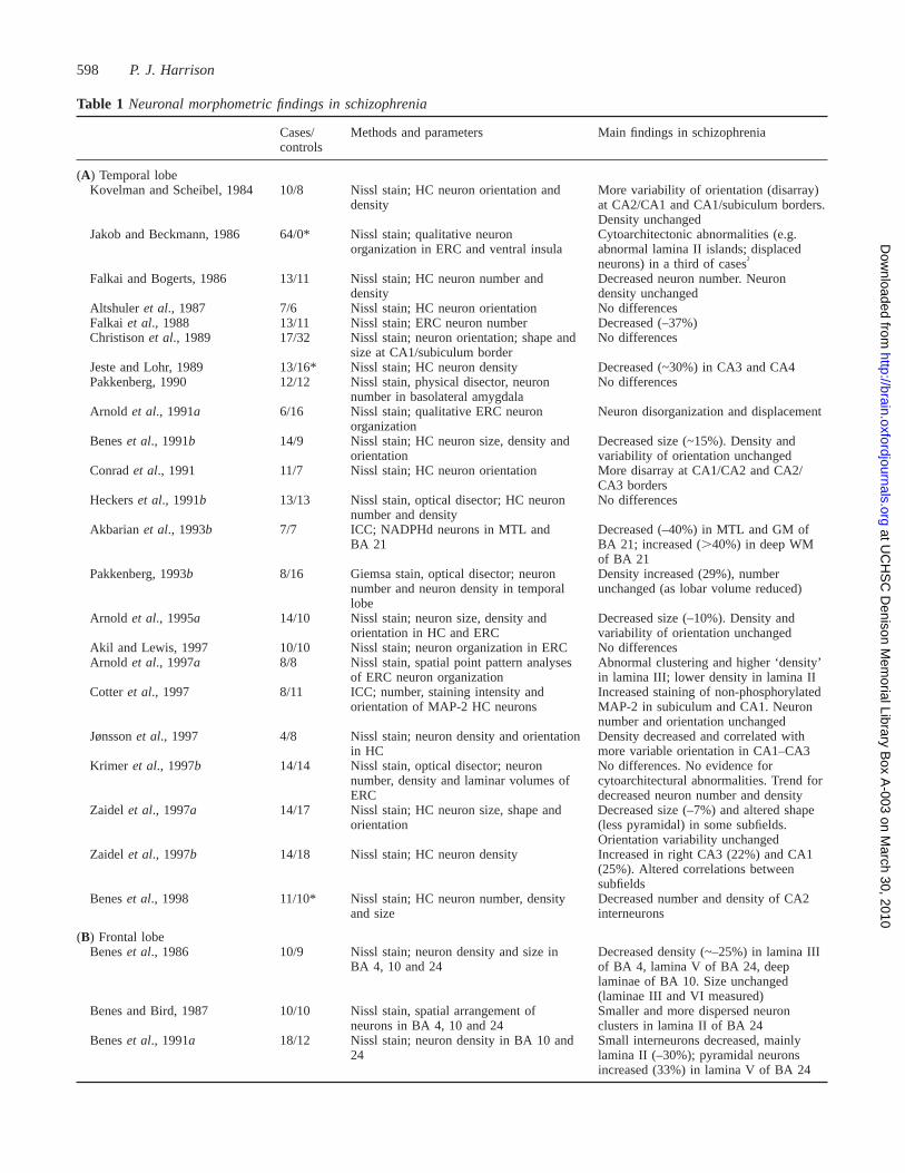
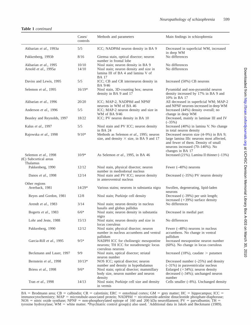
Table 1 Neuronal morphometric findings in schizophrenia
Cases/ Methods and parameters Main findings in schizophreniacontrols
(A) Temporal lobeKovelman and Scheibel, 1984 10/8 Nissl stain; HC neuron orientation and More variability of orientation (disarray)
density at CA2/CA1 and CA1/subiculum borders.Density unchanged
Jakob and Beckmann, 1986 64/0* Nissl stain; qualitative neuron Cytoarchitectonic abnormalities (e.g.organization in ERC and ventral insula abnormal lamina II islands; displaced
neurons) in a third of cases†
Falkai and Bogerts, 1986 13/11 Nissl stain; HC neuron number and Decreased neuron number. Neurondensity density unchanged
Altshuler et al., 1987 7/6 Nissl stain; HC neuron orientation No differencesFalkai et al., 1988 13/11 Nissl stain; ERC neuron number Decreased (–37%)Christisonet al., 1989 17/32 Nissl stain; neuron orientation; shape and No differences
size at CA1/subiculum borderJeste and Lohr, 1989 13/16* Nissl stain; HC neuron density Decreased (~30%) in CA3 and CA4Pakkenberg, 1990 12/12 Nissl stain, physical disector, neuron No differences
number in basolateral amygdalaArnold et al., 1991a 6/16 Nissl stain; qualitative ERC neuron Neuron disorganization and displacement
organizationBeneset al., 1991b 14/9 Nissl stain; HC neuron size, density and Decreased size (~15%). Density and
orientation variability of orientation unchangedConradet al., 1991 11/7 Nissl stain; HC neuron orientation More disarray at CA1/CA2 and CA2/
CA3 bordersHeckerset al., 1991b 13/13 Nissl stain, optical disector; HC neuron No differences
number and densityAkbarianet al., 1993b 7/7 ICC; NADPHd neurons in MTL and Decreased (–40%) in MTL and GM of
BA 21 BA 21; increased (.40%) in deep WMof BA 21
Pakkenberg, 1993b 8/16 Giemsa stain, optical disector; neuron Density increased (29%), numbernumber and neuron density in temporal unchanged (as lobar volume reduced)lobe
Arnold et al., 1995a 14/10 Nissl stain; neuron size, density and Decreased size (–10%). Density andorientation in HC and ERC variability of orientation unchanged
Akil and Lewis, 1997 10/10 Nissl stain; neuron organization in ERC No differencesArnold et al., 1997a 8/8 Nissl stain, spatial point pattern analyses Abnormal clustering and higher ‘density’
of ERC neuron organization in lamina III; lower density in lamina IICotteret al., 1997 8/11 ICC; number, staining intensity and Increased staining of non-phosphorylated
orientation of MAP-2 HC neurons MAP-2 in subiculum and CA1. Neuronnumber and orientation unchanged
Jønssonet al., 1997 4/8 Nissl stain; neuron density and orientation Density decreased and correlated within HC more variable orientation in CA1–CA3
Krimer et al., 1997b 14/14 Nissl stain, optical disector; neuron No differences. No evidence fornumber, density and laminar volumes of cytoarchitectural abnormalities. Trend forERC decreased neuron number and density
Zaidel et al., 1997a 14/17 Nissl stain; HC neuron size, shape and Decreased size (–7%) and altered shapeorientation (less pyramidal) in some subfields.
Orientation variability unchangedZaidel et al., 1997b 14/18 Nissl stain; HC neuron density Increased in right CA3 (22%) and CA1
(25%). Altered correlations betweensubfields
Beneset al., 1998 11/10* Nissl stain; HC neuron number, density Decreased number and density of CA2and size interneurons
(B) Frontal lobeBeneset al., 1986 10/9 Nissl stain; neuron density and size in Decreased density (~–25%) in lamina III
BA 4, 10 and 24 of BA 4, lamina V of BA 24, deeplaminae of BA 10. Size unchanged(laminae III and VI measured)
Benes and Bird, 1987 10/10 Nissl stain, spatial arrangement of Smaller and more dispersed neuronneurons in BA 4, 10 and 24 clusters in lamina II of BA 24
Beneset al., 1991a 18/12 Nissl stain; neuron density in BA 10 and Small interneurons decreased, mainly24 lamina II (–30%); pyramidal neurons
increased (33%) in lamina V of BA 24
at UC
HS
C D
enison Mem
orial Library Box A
-003 on March 30, 2010
http://brain.oxfordjournals.orgD
ownloaded from
Neuropathology of schizophrenia 599
Table 1 continued
Cases/ Methods and parameters Main findings in schizophreniacontrols
Akbarianet al., 1993a 5/5 ICC; NADPHd neuron density in BA 9 Decreased in superficial WM, increasedin deep WM
Pakkenberg, 1993b 8/16 Giemsa stain, optical disector; neuron No differencesnumber in frontal lobe
Akbarianet al., 1995 10/10 Nissl stain; neuron density in BA 9 No differencesArnold et al., 1995a 14/10 Nissl stain; neuron density and size in No differences
lamina III of BA 4 and lamina V ofBA 17
Daviss and Lewis, 1995 5/5 ICC; CB and CR interneuron density in Increased (50%) CB neuronsBA 9/46
Selemonet al., 1995 16/19* Nissl stain, 3D-counting box; neuron Pyramidal and non-pyramidal neurondensity in BA 9 and 17 density increased by 17% in BA 9 and
10% in BA 17Akbarianet al., 1996 20/20 ICC; MAP-2, NADPHd and NPNF All decreased in superficial WM; MAP-2
neurons in WM of BA 46 and NPNF neurons increased in deep WMAndersonet al., 1996 5/5 ICC; MAP-2 neuron density and size in Increased (44%) density overall; no
WM of BA 9/46 change in deep WMBeasley and Reynolds, 1997 18/22 ICC; PV neuron density in BA 10 Decreased, mainly in laminae III and IV
(–35%)Kalus et al., 1997 5/5 Nissl stain and PV ICC; neuron density Increased (40%) in lamina V. No change
in BA 24 in total neuron densityRajowskaet al., 1998 9/10* Methods as Selemonet al., 1995; neuron Decreased neuron size (4–9%) in BA 9;
size, and density3 size, in BA 9 and 17 large lamina IIIc neurons most affected,and fewer of them. Density of smallneurons increased (70–140%). Nochanges in BA 17
Selemonet al., 1998 10/9* As Selemonet al., 1995, in BA 46 Increased (21%). Lamina II thinner (–13%)(C) Subcortical areas
ThalamusPakkenberg, 1990 12/12 Nissl stain, physical disector; neuron Fewer (–40%) neurons
number in mediodorsal nucleusDanoset al., 1998 12/14 Nissl stain and PV ICC; neuron density Decreased (–35%) PV neuron density
in anteroventral nucleusOther regions
Averback, 1981 14/29* Various stains; neurons in substantia nigra Swollen, degenerating, lipid-ladenneurons
Reyes and Gordon, 1981 12/8 Nissl stain; Purkinje cell density Decreased (–39%) per unit length;increased (139%) surface density
Arendt et al., 1983 3/14 Nissl stain; neuron density in nucleus No differencesbasalis and globus pallidus
Bogertset al., 1983 6/6* Nissl stain; neuron density in substantia Decreased in medial partnigra
Lohr and Jeste, 1988 15/13 Nissl stain; neuron density and size in No differenceslocus coeruleus
Pakkenberg, 1990 12/12 Nissl stain; physical disector; neuron Fewer (–48%) neurons in nucleusnumber in nucleus accumbens and ventral accumbens. No change in ventralpallidum pallidum
Garcia-Rill et al., 1995 9/5* NADPH ICC for cholinergic mesopontine Increased mesopontine neuron numberneurons; TH ICC for noradrenergic locus (60%). No change in locus coeruleuscoeruleus neurons
Beckmann and Lauer, 1997 9/9 Nissl stain, optical disector; striatal Increased (18%), caudate. putamenneuron number
Bernsteinet al., 1998 10/13* NOS ICC; optical disector; neuron Decreased number (–25%) and densitynumber and density in hypothalamus (–31%) in paraventricular nucleus
Briesset al., 1998 9/6* Nissl stain; optical disector; mammillary Enlarged (134%); neuron densitybody size, neuron number and neuron decreased (–34%); unchanged neurondensity number
Tran et al., 1998 14/13 Nissl stain; Purkinje cell size and density Cells smaller (–8%). Unchanged densityin vermis
BA 5 Brodmann area; CB5 calbindin; CR5 calretinin; ERC5 entorhinal cortex; GM5 grey matter; HC5 hippocampus; ICC5immunocytochemistry; MAP5 microtubule-associated protein; NADPHd5 nicotinamide-adenine dinucleotide phosphate-diaphorase;NOS 5 nitric oxide synthase; NPNF5 non-phosphorylated epitope of 160 and 200 kDa neurofilament; PV5 parvalbumin; TH5tyrosine hydroxylase; WM5 white matter. *Psychiatric control group(s) also used.†Additional data in Jakob and Beckmann (1989).
at UC
HS
C D
enison Mem
orial Library Box A
-003 on March 30, 2010
http://brain.oxfordjournals.orgD
ownloaded from
600 P. J. Harrison
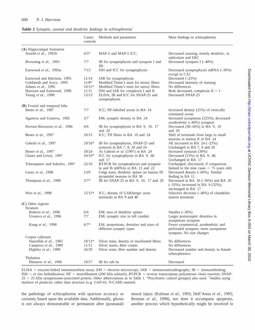
Table 2 Synaptic, axonal and dendritic findings in schizophrenia†
Cases/ Methods and parameters Main findings in schizophreniacontrols
(A) Hippocampal formationArnold et al., 1991b 6/5* MAP-2 and MAP-5 ICC. Decreased staining, mainly dendritic, in
subiculum and ERCBrowning et al., 1993 7/7 IB for synaptophysin and synapsin I and Decreased synapsin I (–40%)
IIbEastwoodet al., 1995a 7/13 ISH and ICC for synaptophysin Decreased synaptophysin mRNA (–30%)
except in CA1Eastwood and Harrison, 1995 11/14 IAR for synaptophysin Decreased (–25%)Goldsmith and Joyce, 1995 11/8* Modified Timm’s stain for mossy fibres Decreased intensity of stainingAdamset al., 1995 10/11* Modified Timm’s stain for mossy fibres No differencesHarrison and Eastwood, 1998 11/11 ISH and IAR for complexin I and II Both decreased, complexin II. IYoung et al., 1998 13/13 ELISA, IB and ICC for SNAP-25 and Decreased SNAP-25
synaptophysin
(B) Frontal and temporal lobeBeneset al., 1987 7/7 ICC; NF-labelled axons in BA 24 Increased density (25%) of vertically
orientated axonsAganova and Uranova, 1992 5/7 EM; synaptic density in BA 24 Increased axospinous (225%), decreased
axodendritic (–40%) synapsesPerrone-Bizzozeroet al., 1996 6/6 IB for synaptophysin in BA 9, 10, 17 Decreased (30–50%) in BA 9, 10
and 20 and 20Beneset al., 1997 10/15 ICC; TH fibres in BA 10 and 24 Shift of terminals from large to small
neurons in lamina II of BA 24Gabrielet al., 1997 19/16* IB for synaptophysin, SNAP-25 and All increased in BA 24 (~25%).
syntaxin in BA 7, 8, 20 and 24 Unchanged in BA 7, 8 and 20Honeret al., 1997 18/24 As Gabrielet al. (1997) in BA 24 Increased syntaxin (30%)Glantz and Lewis, 1997 10/10* ICC for synaptophysin in BA 9, 46 Decreased (15%) in BA 9, 46.
and 17 Unchanged in BA 17Tcherepanov and Sokolov, 1997 22/10 RT-PCR for synaptophysin and synapsin Unchanged. (Increased, if comparison
Ia and Ib mRNAs in BA 21 and 22 limited to the nine cases, 75 years old)Gareyet al., 1998 13/9 Golgi stain; dendritic spines on lamina III Decreased density (–60%). Similar
pyramidal neurons in BA 38 finding in BA 11Thompsonet al., 1998 5/7* IB for SNAP-25 in BA 9, 10, 17 and 20 Decreased in BA 10 (–56%) and BA 20
(–33%); increased in BA 9 (32%);unchanged in BA 17
Woo et al., 1998 15/15* ICC; density of GABAergic axon Selective decrease (–40%) of chandelierterminals in BA 9 and 46 neuron terminals
(C) Other regionsStriatum
Robertset al., 1996 6/6 EM; area of dendritic spines Smaller (–30%)Uranovaet al., 1996 7/7 EM; synaptic size in left caudate Larger postsynaptic densities in
axospinous synapsesKung et al., 1998 6/7* EM; proportions, densities and sizes of Fewer symmetrical, axodendritic and
different synaptic types perforated synapses; more axospinoussynapses. No size changes
Corpus callosumNasrallahet al., 1983 18/11* Silver stain; density of myelinated fibres No differencesCasanovaet al., 1989 11/13 Silver stains; fibre counts No differencesHighley et al., 1999 26/29 Silver stain; fibre number and density Decreased number and density in female
schizophrenicsThalamus
Blennowet al., 1996 19/27 IB for rab-3a Decreased
ELISA 5 enzyme-linked immunosorbent assay; EM5 electron microscopy; IAR5 immunoautoradiography; IB5 immunoblotting;ISH 5 in situ hybridization; NF5 neurofilament (200 kDa subunit); RT-PCR5 reverse transcriptase–polymerase chain reaction; SNAP-25 5 25 kDa synaptosome-associated protein. Other abbreviations as in Table 1. *Psychiatric control group(s) also used.†Studies usingmarkers of plasticity rather than structure (e.g. GAP-43, N-CAM) omitted.
the pathology of schizophrenia with spurious accuracy orcertainty based upon the available data. Additionally, gliosisis not always demonstrable or permanent after (postnatal)
neural injury (Kalmanet al., 1993; Dell’Annaet al., 1995;Berman et al., 1998), nor does it accompany apoptosis,another process which hypothetically might be involved in
at UC
HS
C D
enison Mem
orial Library Box A
-003 on March 30, 2010
http://brain.oxfordjournals.orgD
ownloaded from
Neuropathology of schizophrenia 601
schizophrenia. Furthermore, it is a moot point whether thesubtle kinds of morphometric disturbance to be described inschizophrenia, whenever and however they occurred, wouldbe sufficient to trigger gliosis or other signs of ongoingneurodegeneration (Hortonet al., 1993). Thus the lack ofgliosis does not mean, in isolation, that schizophrenia mustbe a neurodevelopmental disorder of prenatal origin; it ismerely one argument in favour of that conclusion.
Schizophrenia, its dementia and Alzheimer’sdiseaseCognitive impairment has been a neglected feature ofschizophrenia. Its importance is now being appreciatedclinically as a major factor contributing to the failure torehabilitate some patients despite relief of their psychoticsymptoms (Green, 1996), and as being a putative therapeutictarget (Davidson and Keefe, 1995). Neuropsychologicalabnormalities are demonstrable in first-episode patients (Hoffet al., 1992; Saykinet al., 1994; Kennyet al., 1997) andpremorbidly (Jones, 1997b; Russellet al., 1997), and thoughtheir progression remains unclear (Bilderet al., 1992;Goldberget al., 1993; Waddington and Youssef, 1996), in asizeable minority of chronic schizophrenics their severitywarrants the label of dementia (Davidsonet al., 1996).There is particular involvement of memory and executivefunctioning (McKennaet al., 1990; Saykinet al., 1991)against a background of a generalized deficit (Blanchard andNeale, 1994; for review, see David and Cutting, 1994). (Aswith the neuropathological abnormalities, it is worth pointingout that the mean size of these differences is small. Manyindividuals with schizophrenia score within the normal range,and some are well above average. On the other hand, thereis no evidence that cognitive impairment is limited to asubgroup, and it may be that even in high-functioning subjectsthere has been a decline from, or failure to attain, theirfull neuropsychological potential.) The final controversiesregarding neurodegenerative processes in schizophreniaconcern the neuropathological explanation for the cognitivedeficits, and the alleged increased prevalence of Alzheimer’sdisease in schizophrenia (e.g. Plum, 1972).
The belief that Alzheimer’s disease is commoner inschizophrenia, regardless of cognitive status, seems to haveoriginated from two German papers in the 1930s (Corsellis,1962). It was supported by three retrospective, uncontrolledstudies (Buhl and Bojsen-Møller, 1988; Soustek, 1989;Prohovniket al., 1993) and the suggestion that antipsychoticdrugs promote Alzheimer-type changes (Wisniewskiet al.,1994). However, corroborating Corsellis’ opinion (Corsellis,1962),ameta-analysis (Baldessarinietal.,1997)andadditionalmethodologically sound studies show conclusively thatAlzheimer’s disease is not commoner than expected inschizophrenia (Arnoldetal., 1998;Murphyetal.,1998;Niizatoet al., 1998; Purohitet al., 1998). Even amongst elderlyschizophrenics with unequivocal, prospectively assessed
dementia (mean Mini-Mental State score5 12), detailedimmunocytochemical analyses find no evidence forAlzheimer’s disease or any other neurodegenerative disorder(Arnold et al., 1996, 1998). In keeping with this negativeconclusion, apolipoprotein E4 allele frequencies areunchanged (Arnoldet al., 1997b; Powchiket al., 1997; Thibautet al., 1998) and cholinergic markers are preserved (Arendtet al., 1983; Haroutunianet al., 1994) in schizophrenia.Moreover, the evidence as a whole does not support the viewthat antipsychotic drugs predispose to neurofibrillarypathology (Baldessariniet al., 1997; Harrisonet al., 1997b).
How, therefore, is the cognitive impairment of schizophreniaexplained? One possibility is that it is a more severemanifestation of whatever substrate underlies schizophreniaitself rather than resulting from the superimposition of aseparate process. Or it may be that the brain in schizophreniais rendered more vulnerable to cognitive impairment inresponse to a normal age-related amount of neurodegeneration,or even that the pathological findings so far discovered actuallyrelate to the cognitive impairment rather than to the psychoticfeatures by which the disorder is defined. A final, speculativesuggestion is that the gliosis observed in dementedschizophrenics (Arnoldet al., 1996) is a sign of an as yetunrecognized novel neurodegenerative disorder. Thesepossibilities cannot be distinguished at present since fewneuropsychologically evaluated patients have been studiedneuropathologically; inclusion of subjects with comorbidschizophrenia and mental retardation may be valuable whenaddressing the issue (Doodyet al., 1998).
The cytoarchitecture of schizophreniaSince neurodegenerative abnormalities are uncommon in,and probably epiphenomenal to, schizophrenia, the questionis raised as to what the pathology of the disorder is, and howthe macroscopic findings are explained at the microscopiclevel. This brings us to the heart of recent schizophrenianeuropathology research, which has been the increasinglysophisticated measurement of the cortical cytoarchitecture.The focus has been mainly on the extended limbic system[hippocampus, dorsolateral prefrontal cortex (DLPFC) andcingulate gyrus], encouraged by suggestions that psychoticsymptoms originate in these regions (Stevens, 1973; Torreyand Peterson, 1974).
Table 1 summarizes the morphometric investigations inwhich neuronal parameters such as density, number, size,shape, orientation, location and clustering have beendetermined. Table 2 summarizes the studies of synapses,dendrites and axons, evaluated either ultrastructurally orindirectly using immunological and molecular markers. Bothtables are subdivided by brain region. Only the major findingsare listed; details such as laterality effects are omitted. Inthe following sections the main themes of this literature arediscussed, although even the choice of what to highlight isproblematic given that controversy surrounds nearly everypoint.
at UC
HS
C D
enison Mem
orial Library Box A
-003 on March 30, 2010
http://brain.oxfordjournals.orgD
ownloaded from
602 P. J. Harrison
Studies of neurons
Cytoarchitectural abnormalities in entorhinalcortex. An influential paper reported the presence of variousabnormalities in the cytoarchitecture and lamination of theentorhinal cortex (anterior parahippocampal gyrus) inschizophrenia (Jakob and Beckmann, 1986). The changeswere prominent in lamina II, with a loss of the normalclustering of the constituent pre-α cells, which appearedshrunken, misshapen and heterotopic. Despite extensions(Jakob and Beckmann, 1989) and partial replications (Arnoldet al., 1991a), the findings remain questionable for severalreasons, which are elaborated because of the importanceattributed to them in the neurodevelopmental model ofschizophrenia. First, no normal control group was used; thecomparisons were made with brains from 10 patients withother psychiatric or neurological disorders. Whilst thiscriticism does not apply to the study of Arnoldet al. (1991a),both are limited by the lack of objective criteria for thecytoarchitectural disturbance. The later work of Arnoldet al.(1995a, 1997a) overcomes this deficiency in different waysand provides some further evidence for a disturbance in thelocation, clustering and/or size of entorhinal cortex neurons,though of much lesser magnitude and frequency than reportedby Jakob and Beckmann. The most serious problem is thatnone of these studies have fully allowed for the heterogeneouscytoarchitecture of the entorhinal cortex (Beall and Lewis,1992; Insausti et al., 1995) and its variation betweenindividuals (Heinsenet al., 1996; Krimeret al., 1997a; Westand Slomianka, 1998). For example, as Akil and Lewis(1997) point out, Jakob and Beckmann (1986) sampled theirmaterial based on external landmarks which may shift relativeto the entorhinal cortex in schizophrenia, resulting indifferences in the rostrocaudal location of the sections, whichcould account for their findings. Notably, the two studies thatmost closely attend to the issue of anatomical complexitywithin the entorhinal cortex found no differences in itscytoarchitecture in schizophrenia (Akil and Lewis, 1997;Krimer et al., 1997a).
Disarray of hippocampal pyramidal neurons. Asecond parameter of cytoarchitectural disturbance inschizophrenia, a disarray of hippocampal pyramidal neurons,has also been given prominence disproportionate to thestrength of the data. Normally, pyramidal neurons in Ammon’shorn are aligned, as in a palisade, with the apical dendriteorientated towards the stratum radiatum. Kovelman andScheibel (1984) reported that this orientation was morevariable and even reversed in schizophrenia, hence theterm ‘neuronal disarray’. The disarray was present at theboundaries of CA1 with CA2 and subiculum. The basicfinding of greater variability of hippocampal neuronalorientation was extended in subsequent studies from the samegroup (Altshuler et al., 1987; Conradet al., 1991) andindependently (Jønssonet al., 1997; Zaidelet al., 1997a).However, none of these studies constitutes true replication.
Conradet al. (1991) came closest, but located the disarrayat the boundaries of CA2 rather than CA1; Altshuleret al.(1987) found no differences between cases and controls—merely a correlation between the degree of disarray and theseverity of psychosis within the schizophrenic group; thedisarray in the small study of Jønssonet al. (1997) was inthe central part of each CA field, and Zaidelet al. (1997a)found no overall difference in orientation but, in apost hocanalysis, found an asymmetrical variability limited to a partof CA3. Furthermore, there are three entirely negative studies(Christisonet al., 1989; Beneset al., 1991b; Arnold et al.,1995a). Thus, even a charitable overview of the data wouldaccept that the site and frequency of hippocampal neuronaldisarray in schizophrenia remains uncertain, while a scepticalview would be that the phenomenon has not beenunequivocally demonstrated. Certainly, as with the entorhinalcortex abnormalities, it seems inappropriate to place too muchinterpretative weight on such insecure empirical foundations.
Location of cortical subplate neurons.The subplateis a key structure in the formation of the cortex and theorderly ingrowth of thalamic axons (Allendoerfer and Shatz,1994). Some of the subplate neurons persist as interstitialneurons in the subcortical white matter and contribute tocortical and corticothalamic circuits. Stimulated by theentorhinal and hippocampal cytoarchitectural findingssuggestive of aberrant neuronal migration, subplate neuronshave been studied in schizophrenia, since changes in thedensity and distribution of these neurons would probably bea correlate of such a disturbance. Using nicotinamide-adeninedinucleotide phosphate-diaphorase histochemistry as amarker, these neurons were found to be distributed moredeeply in the frontal and temporal cortex white matter inschizophrenics than in controls (Akbarianet al., 1993a, b).A subsequent survey using additional markers and a largersample confirmed the observation of fewer interstitial neuronsin superficial white matter compartments of DLPFC inschizophrenia (Akbarianet al., 1996).
These data are more convincing than the reports ofentorhinal cortex dysplasias and hippocampal neuron disarray,and the studies are noteworthy for being embedded in theknown cellular biology of cortical development. Nevertheless,it would be premature to consider maldistribution of survivingsubplate neurons, and by inference aberrant neuronalmigration, to be an established feature of schizophrenia. First,Dwork (1997) has drawn attention to the doubtful statisticalsignificance of the original results (Akbarianet al., 1993a, b).Secondly, in the follow-up study (Akbarianet al., 1996) theabnormalities were milder and less prevalent, and theirstatistical significance was enhanced by the apparent retentionof the original cases. Thirdly, considerable variation in theabundance of interstitial neurons has been found betweenindividuals and between frontal and temporal white matter(Rojiani et al., 1996), suggesting that sample sizes largerthan those employed to date may be necessary to identifyclearly any alterations associated with schizophrenia. Finally,
at UC
HS
C D
enison Mem
orial Library Box A
-003 on March 30, 2010
http://brain.oxfordjournals.orgD
ownloaded from
Neuropathology of schizophrenia 603
as shown in Table 1B, Andersonet al.(1996) found essentiallythe opposite result from that of Akbarianet al. (1996).Further investigations are therefore essential to corroboratethe potentially key observations of Akbarian and colleagues.
Hippocampal and cortical neuron density andnumber.A loss of hippocampal neurons is another oft-stated feature of schizophrenia. In fact only two studies havefound reductions in neuron density (Jeste and Lohr, 1989;Jønssonet al., 1997) and one reported a lower number ofpyramidal neurons (Falkai and Bogerts, 1986). In contrast,several have found no change in density (Kovelman andScheibel, 1984; Falkai and Bogerts, 1986; Beneset al.,1991b; Arnold et al., 1995a) and one found a localizedincrease (Zaidelet al., 1997b). Since none of these studieswere stereological, their value is limited by the inherentweaknesses of neuron counts when measured in this way(Mayhew and Gundersen, 1996)—although not to the extentthat they should be discounted (Guillery and Herrup, 1997).Nevertheless, the fact that the single stereological study thathas been carried out found no difference in neuronal numberor density in any subfield (Heckerset al., 1991a) supportsthe view that there is no overall change in the neuron contentof the hippocampus in schizophrenia. In this context, singlereports of altered neuronal density restricted to a specificneuronal type or subfield (Zaidelet al., 1997a; Beneset al.,1998) must be replicated before discussion is warranted.
The prefrontal cortex has also been examined. A carefulstereologically based study found an increased neuronaldensity in DLPFC (Selemonet al., 1995, 1998), and a similartrend was seen for the whole frontal lobe by Pakkenberg(1993b). The higher packing density identified by Selemonand colleagues affected small and medium-sized neuronsmore than large pyramidal ones. Other neuronal densitystudies in the prefrontal cortex have not produced consistentfindings (Table 1B). For example, Beneset al. (1986, 1991a)identified a variety of lamina-, area- and cell type-specificdifferences, whilst unaltered neuronal density has beenreported in the motor cortex (Arnoldet al., 1995a) andDLPFC (Akbarianet al., 1995). These discrepancies may bedue to anatomical heterogeneity or may be the consequenceof differences in the stereological purity of the studies. Thetotal number of neurons in the frontal cortex is not alteredin schizophrenia (Pakkenberg, 1993b), which probablyreflects the net effect of anatomical variation in the neuronaldensity changes within the frontal lobe and/or the trend forcortical grey matter to be thinner in schizophrenia, whichcompensates for the increased packing density of neuronstherein (Pakkenberg, 1987; Selemonet al., 1998; Wooet al., 1998).
Hippocampal and cortical neuronal size.With theadvent of user-friendly image analysis it has become relativelystraightforward to measure the size of the cell body ofneurons, either by tracing around the perikaryal outline orby measuring the smallest circle within which the soma fits.
Three studies, each counting large numbers of neurons, havenow identified a smaller mean size of hippocampal pyramidalneurons in schizophrenia (Beneset al., 1991a; Arnold et al.,1995a; Zaidel et al., 1997a). Although different individualsubfields reached significance in the latter two studies, thesame downward trend was present in all CA fields and inthe subiculum. The non-replications comprise Christisonet al. (1989) and Beneset al. (1998), perhaps becausemeasurements were limited to a restricted subset of neurons.Smaller neuronal size has also been reported in DLPFC,especially affecting large lamina IIIc neurons (Rajkowskaet al., 1998). A degree of anatomical specificity to the sizereductions is apparent, since this study found no differencesin the visual cortex of the same cases, in agreement with theunchanged cell size found in that region as well as in themotor cortex by Arnoldet al.(1995a) and Beneset al.(1986).
Neuronal morphometric changes in otherregions.Outside the cerebral cortex, consistent cyto-architectural data are limited to the thalamus (Table 1C).Pakkenberg (1990) found markedly lower numbers of neuronsin the dorsomedial nucleus, which projects mainly to theprefrontal cortex. A similar finding was observed in theanteroventral nucleus, which also has primarily prefrontalconnections, the significant deficit affecting parvalbumin-immunoreactive cells, a marker for thalamocortical neurons(Danos et al., 1998). Whether similar changes occur inthalamic nuclei not intimately related to cortical regionsimplicated in schizophrenia remains to be determined.
In summary, a range of differences in neuronal parametershave been reported to occur in schizophrenia. Theabnormalities most often taken to be characteristic of thedisorder—disarray, displacement and paucity of hippocampaland cortical neurons—are in fact features which have notbeen clearly demonstrated. This undermines attempts to datethe pathology of schizophrenia to the second trimesterin uterobased on their presence (see below). In contrast, decreasedneuron size, especially affecting neurons in the hippocampusand DLPFC, has been shown fairly convincingly; somestudies suggest that the size reduction is accompaniedby increased neuron density. The other relatively robustcytoarchitectural abnormality in schizophrenia is in the dorsalthalamus, which is smaller and contains fewer neurons.
Studies of synapses and dendrites.Synapticabnormalities represent a potential site for significantpathology in schizophrenia which would be undetectableusing standard histological approaches. The term ‘synapticpathology’ is used here to denote abnormalities in axons anddendrites in addition to those affecting the synaptic terminalsthemselves.
Practical issues.Qualitative studies identified a range ofultrastructural abnormalities of neuronal and synapticelements in schizophrenia (Tatetsuet al., 1964; Miyakawaet al., 1972; Averback, 1981; Soustek, 1989; Ong and Garey,
at UC
HS
C D
enison Mem
orial Library Box A
-003 on March 30, 2010
http://brain.oxfordjournals.orgD
ownloaded from
604 P. J. Harrison
1993). However, because of the difficulties and limitationsof electron microscopy in post-mortem human brain tissue,especially for quantitative analysis, much contemporaryresearch into synaptic pathology in schizophrenia has adopteda complementary approach whereby the expression andabundance of proteins concentrated in presynaptic terminals,such as synaptophysin, are used as proxies for synapses. Thisapproach has been validated in several experimental anddisease states (Masliah and Terry, 1993; Eastwoodet al.,1994a). For example, in Alzheimer’s disease, synaptophysinmRNA and protein levels correlate inversely with the clinicaland pathological severity of dementia (Terryet al., 1991;Heffernanet al., 1998). Note, however, that although synapticprotein measurements are widely interpreted as reflectingsynaptic density, an assumption almost certainly true inneurodegenerative disorders, in principle changes in synapticprotein expression could instead be due to alterations insynaptic size or number of vesicles per terminal, or toa structural abnormality of the presynaptic region. Suchpossibilities should not be ignored in schizophrenia, giventhat ultrastructural features of this kind were suggested bysome of the electron microscopy studies mentioned above.
Hippocampal formation.Synaptic protein determina-tions in the hippocampal formation (hippocampus andparahippocampal gyrus) in schizophrenia have fairlyconsistently found levels to be reduced (Table 2A), althoughnot all reach statistical significance for reasons other than justinadequate sample size. First, subfields may be differentiallyaffected (Eastwood and Harrison, 1995; Eastwoodet al.,1995a), and localized changes may be masked if homogenizedtissue is used. Secondly, the synaptic proteins studied changeto varying degrees, probably reflecting their concentration indifferentially affected synaptic populations. For example,synaptophysin, which is present in all synapses, shows onlyslight reductions (Browninget al., 1993; Eastwood andHarrison, 1995; Eastwoodet al., 1995a), whereas SNAP-25(Young et al., 1998) and complexin II (Harrison andEastwood, 1998), which are both concentrated in subsets ofsynapses, show greater decrements. Furthermore,complexin II is primarily expressed by excitatory neurons,unlike complexin I, which is mainly present in inhibitoryneurons and is less affected in schizophrenia (Harrison andEastwood, 1998). Thus, these data suggest a particularinvolvement of excitatory pathways in this region, aconclusion in keeping with neurochemical studies of theglutamatergic system (see below). A final example of currentattempts to dissect out the nature of hippocampal synapticinvolvement in schizophrenia is provided by a study ofthe expression of the neuronal growth-associated protein-43(GAP-43), a marker of synaptic plasticity (Benowitz andRouttenberg, 1997). A loss of hippocampal GAP-43 mRNAwas found, suggesting that hippocampal synapses may beremodelled less actively in schizophrenia (Eastwood andHarrison, 1998).
Less attention has been paid to postsynaptic elements of
the hippocampal circuitry. However, dendritic abnormalitieshave been reported, with decreased and aberrant expressionof the dendritic microtubule-associated protein MAP-2 insome subfields (Arnoldet al., 1991b; Cotteret al., 1997).
Neocortex.Two studies have found synaptophysin to bereduced in DLPFC in schizophrenia (Perrone-Bizzozeroet al.,1996; Glantz and Lewis, 1997). The inferred decrease ofpresynaptic terminals is complemented by a lower densityof dendritic spines (to which many of the synapses areapposed) on layer III pyramidal neurons (Gareyet al.,1998). The pattern of synaptophysin alteration is not uniformthroughout the cortex, since levels are unchanged in thevisual cortex (Perrone-Bizzozeroet al., 1996; Glantz andLewis, 1997) and increased in the cingulate gyrus (Gabrielet al., 1997). The suggestion that there is a discrete profileof synaptic pathology in the cingulate gyrus is noteworthygiven the other cytoarchitectural and ultrastructural findingsin that region (Tables 1B and 2B), such as increasedglutamatergic axons (Beneset al., 1987, 1992a) andaxospinous synapses (Aganova and Uranova, 1992), anddeficits in inhibitory interneurons (Beneset al., 1991a) whichhave not been reported elsewhere. However, further directcomparisons are needed before it can be concluded that thecingulate exhibits a different pattern of pathology.
Thalamus.A marked reduction of the synaptic proteinrab3a from the thalamus was found in a large group ofschizophrenics compared with controls (Blennowet al.,1996). These data, in concert with the morphometric andimaging findings (Table 1C), highlight the thalamus asmeriting active investigation in schizophrenia (Jones, 1997a),a somewhat belated return to the one brain region for whichthe earlier generation of studies had produced potentiallymeaningful findings (David, 1957).
Striatum. In the striatum, electron microscopy rather thanimmunocytochemical measurements has continued to be usedto investigate synaptic pathology in schizophrenia. Alteredsizes and proportions of synapses in the caudate nucleushave been found compared with controls (Table 2C). It isdifficult to interpret these findings and integrate them withthose in other regions because of the methodologicaldifferences and the greater concern about confounding effectsof antipsychotic medication in basal ganglia (see below).Nevertheless, they broadly support the view that synapticorganization is altered in schizophrenia.
In summary, synaptic studies in the hippocampus andDLPFC in schizophrenia show decrements in presynapticmarkers and, though less extensively studied, in postsynapticmarkers too. The simplest interpretation is that these changesreflect a reduction in the number of synaptic contacts formedand received in these areas, bearing in mind the caveat aboutalternative possibilities such as abnormal synaptic vesiclecomposition or even dysregulation of synaptic protein genetranscription. In pathogenic terms, the direction of the
at UC
HS
C D
enison Mem
orial Library Box A
-003 on March 30, 2010
http://brain.oxfordjournals.orgD
ownloaded from
Neuropathology of schizophrenia 605
synaptic alterations in the hippocampus and DLPFC supportshypotheses of excessive (Keshavanet al., 1994a) rather thaninadequate (Feinberg, 1982) synaptic pruning inschizophrenia. Since the reductions are not uniform inmagnitude or location, it is likely that certain synapticpopulations are more affected than others; preliminaryevidence suggests glutamatergic synapses may be especiallyvulnerable in the hippocampus and perhaps the DLPFC, withpredominantly GABAergic involvement in the cingulategyrus. There is a need not only to extend the work (e.g.to include confocal microscopy and to measure additionalsynaptic proteins) but to integrate it with further Golgi stainingand electron microscope investigations directly visualizingsynapses and dendrites.
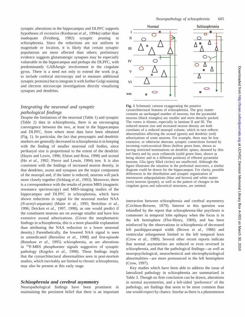
Integrating the neuronal and synapticpathological findingsDespite the limitations of the neuronal (Table 1) and synaptic(Table 2) data in schizophrenia, there is an encouragingconvergence between the two, at least in the hippocampusand DLPFC, from where most data have been obtained(Fig. 1). In particular, the fact that presynaptic and dendriticmarkers are generally decreased in schizophrenia is in keepingwith the finding of smaller neuronal cell bodies, sinceperikaryal size is proportional to the extent of the dendritic(Hayes and Lewis, 1996; Elston and Rosa, 1998) and axonal(Ho et al., 1992; Pierce and Lewin, 1994) tree. It is alsoconsistent with the findings of increased neuron density, inthat dendrites, axons and synapses are the major componentof the neuropil and, if the latter is reduced, neurons will packmore closely together (Schlauget al., 1993). Moreover, thereis a correspondence with the results of proton MRS (magneticresonance spectroscopy) and MRS-imaging studies of thehippocampus and DLPFC in schizophrenia, which haveshown reductions in signal for the neuronal marker NAA(N-acetyl-aspartate) (Maieret al., 1995; Bertolinoet al.,1996; Deickenet al., 1997, 1998), as one would predict ifthe constituent neurons are on average smaller and have lessextensive axonal arborizations. (Given the morphometricfindings in schizophrenia, this is a more plausible explanationthan attributing the NAA reduction to a lower neuronaldensity.) Parenthetically, the lowered NAA signal is seenin unmedicated (Bertolinoet al., 1998) and first-episode(Renshawet al., 1995) schizophrenia, as are alterationsin 31P-MRS phosphoester signals suggestive of synapticpathology (Kegeleset al., 1998). These findings implythat the cytoarchitectural abnormalities seen in post-mortemstudies, which inevitably are limited to chronic schizophrenia,may also be present at this early stage.
Schizophrenia and cerebral asymmetryNeuropathological findings have been prominent inmaintaining the persistent belief that there is an important
Fig. 1 Schematic cartoon exaggerating the putativecytoarchitectural features of schizophrenia. The grey mattercontains an unchanged number of neurons, but the pyramidalneurons (black triangles) are smaller and more densely packed.The cortex is thinner, especially in laminae II and III. Thereduced neuron size and increased neuron density are bothcorrelates of a reduced neuropil volume, which in turn reflectsabnormalities affecting the axonal (green) and dendritic (red)arborizations of some neurons. For example, there may be lessextensive, or otherwise aberrant, synaptic connections formed byincoming corticocortical fibres (hollow green lines, shown ashaving restricted terminations on dendritic spines, denoted by thinred lines) and by axon collaterals (solid green lines, shown asbeing shorter and in a different position) of efferent pyramidalneurons. Glia (grey filled circles) are unaffected. Although thefigure illustrates the situation in the prefrontal neocortex, a similardiagram could be drawn for the hippocampus. For clarity, possibledifferences in the distribution and synaptic organization ofinterneuron subpopulations (blue and brown) and white matter(wm) neurons (purple), as well as the pattern of changes in thecingulate gyrus and subcortical structures, are omitted.
interaction between schizophrenia and cerebral asymmetry(Crichton-Browne, 1879). Interest in this question wasrekindled by the report that schizophrenia-like psychosis iscommoner in temporal lobe epilepsy when the focus is inthe left hemisphere (Flor-Henry, 1969), and has beenreinforced by the observations in schizophrenia of decreasedleft parahippocampal width (Brownet al., 1986) andventricular enlargement limited to the left temporal horn(Crow et al., 1989). Several other recent reports indicatethat normal asymmetries are reduced or even reversed inschizophrenia, and that the pathological findings—as well asneuropsychological, neurochemical and electrophysiologicalabnormalities—are more pronounced in the left hemisphere(Crow, 1997).
Key studies which have been able to address the issue oflateralized pathology in schizophrenia are summarized inTable 3. Though no firm conclusion can be drawn, alterationsin normal asymmetries, and a left-sided ‘preference’ of thepathology, are findings that seem to be more common thanone would expect by chance. Insofar as there is a phenomenon
at UC
HS
C D
enison Mem
orial Library Box A
-003 on March 30, 2010
http://brain.oxfordjournals.orgD
ownloaded from
606 P. J. Harrison
Table 3 Cerebral asymmetry and the neuropathology of schizophrenia
Key positive reports* Relevant negative reports†
Macroscopic featuresDecreased fronto-occipital torque Bilderet al., 1994; DeLisiet al., 1997b DeLisi et al., 1997bDecreased size of left superior temporal gyrus Shentonet al., 1992 Flaumet al., 1995Reversal of left. right planum temporale size Falkaiet al., 1995; Bartaet al., 1997‡ Kulynych et al., 1996asymmetryLoss of left. right sylvian fissure length Falkaiet al., 1992Left parahippocampal thinning Brownet al., 1986§
Left temporal horn enlargement Crowet al., 1989Left medial temporal lobe reductions Bogertset al., 1990a; Pearlsonet al., 1997 Altshuleret al., 1990; Bogerts
et al., 1990b; Nelsonet al, 1998Progressive left ventricular enlargement in severe Daviset al., 1998cases
Cytoarchitectural asymmetriesAsymmetrical size and shape changes in Zaidelet al., 1997ahippocampal neuronsIncreased right hippocampal neuron density Zaidelet al., 1997bLoss of synaptic protein from left thalamus Blennowet al., 1996
For additional references see Falkai and Bogerts (1993) and Crow (1997). *With clear evidence for lateralised change (e.g.diagnosis3 side interaction on ANOVA).†Studies where the change was found bilaterally.‡Planum temporale area reduced unilaterally,but volume reduced bilaterally.§Relative to affective disorder controls.
to be explained, two hypotheses exist. Crow’s evolving andevolutionary theory is that schizophrenia, cerebral dominance,handedness and language are inextricably and causally linkedto each other and to a single gene (Crow, 1990, 1997).Alternatively, altered asymmetry in schizophrenia is viewedas an epiphenomenon of itsin uteroorigins, a process whichinterferes with subsequent brain lateralization (Bracha, 1991;Roberts, 1991). Clarifying how the neuropathology interactswith cerebral asymmetry thus requires not only additional,appropriately designed studies of schizophrenia and otherneurodevelopmental disorders, but also a better understandingof the causes and consequences of histological asymmetriesper se(Galaburda, 1994; Hayes and Lewis, 1996; Andersonand Rutledge, 1996). Interactions between asymmetry, genderand schizophrenia introduce further complexity to the issue(Highley et al., 1998, 1999; Vogeleyet al., 1998).
Interpretation of the neuropathology ofschizophreniaNeuropathology and the neurodevelopmentalmodelThe concept of developmental insanity was proposed byClouston in 1891 (Murray and Woodruff, 1995) andelaborated in neuropathological terms early this century(Southard, 1915). However, it is only in the past decade thata neurodevelopmental origin for schizophrenia has becomethe prevailing pathogenic hypothesis for the disorder; indeedthe principle is now largely unchallenged (Murray and Lewis,1987; Weinberger, 1987, 1995). The model receives supportfrom various sources, the neuropathological data forming animportant component of the evidence (Table 4) (Harrison,1997a; Raedleret al., 1998).
The most influential and specific form of the theory is thatthe pathology of schizophrenia originates in the middle stageof intrauterine life (Roberts, 1991; Bloom, 1993; Robertset al., 1997). An earlier timing is excluded since overtabnormalities in the structure and cellular content of thecerebral cortex would be expected if neurogenesis wereaffected, whilst the absence of gliosis is taken to mean thatthe changes must have occurred prior to the third trimester.The conclusion that, by default, the pathological processoriginates in the second trimester is bolstered by referenceto certain of the cytoarchitectural abnormalities ofschizophrenia. However, this ‘strong’ form of the neuro-developmental model is weak on two grounds. First, becauseof the limitations of the absence-of-gliosis argumentmentioned earlier. Secondly, the types of cytoarchitecturaldisturbance adduced in favour are those of neuronal disarray,heterotopias and malpositioning suggestive of aberrantmigration (Kovelman and Scheibel, 1984; Jakob andBeckmann, 1986; Arnoldet al., 1991a; Akbarian et al.,1993a, b), processes which occur at the appropriate gestationalperiod; yet, as described above, none of these cyto-architectural abnormalities has been unequivocallyestablished to be a feature of schizophrenia. By comparison,the other cytoarchitectural findings, such as alterations inneuronal size and synaptic and dendritic organization, couldwell originate much later, being susceptible to ongoingenvironmental influences (Jones and Schallert, 1994; Moseret al., 1994; Saitoet al., 1994; Andradeet al., 1996; Kolband Whishaw, 1998), ageing (Huttenlocher, 1979; Braak andBraak, 1986; Masliahet al., 1993; de Brabanderet al., 1998)and perhaps also to genetic factors (Vaughnet al., 1977;Williams et al., 1998).
Other versions of the neurodevelopmental theory of
at UC
HS
C D
enison Mem
orial Library Box A
-003 on March 30, 2010
http://brain.oxfordjournals.orgD
ownloaded from
Neuropathology of schizophrenia 607
Table 4 Key points of evidence for a neurodevelopmental origin of schizophrenia
Neuropathological evidenceVentricular enlargement and decreased cortical volume present at onset of symptoms, if not earlierPresence and nature of cytoarchitectural abnormalitiesAbsence of gliosis and other neurodegenerative featuresIncreased prevalence of abnormal septum pellucidum (Shioiriet al., 1996; Kwonet al., 1998)
Other evidenceThe environmental risk factors are mostly obstetric complications (Geddes and Lawrie, 1995)Children destined to develop schizophrenia in adulthood show neuromotor, behavioural and intellectual impairment (Jones, 1997b)Increased prevalence of abnormal dermatoglyphics and minor physical anomalies (Buckley, 1998)Experimental neonatal lesions have delayed effects on relevant behavioural and neurochemical indices
For additional references see text.
schizophrenia postulate additional or alternative abnormalitiesin processes such as cell adhesion, myelination and synapticpruning (e.g. Keshavanet al., 1994a; Beneset al., 1994;Akbarianet al., 1996; Arnold and Trojanowski, 1996; Lewis,1997) or allow for a mixture of maturational and degenerativeprocesses (e.g. Murrayet al., 1992; Garver, 1997). Overall,a parsimonious view is that the extant cytoarchitecturalabnormalities and lack of gliosis are indicative merely of anessentially neurodevelopmental as opposed to neuro-degenerative disease process, rather than as pointing directlyto a particular mechanism or timing. It is only by considerationof the pathological features in conjunction with the otherevidence (Table 4) that a strong case for a significant earlychildhood, including foetal, component to schizophrenia canbe made. Even then, it is unknown whether neuro-developmental deviance is either necessary or sufficient.Moreover, any such model has a problem explaining theonset and outcome of the disorder: how is an abnormality inthe cortical cytoarchitecture, present since early in life andpresumably persistent, reconciled with the onset of symptomsin adulthood and a typically relapsing and remitting coursethereafter? Regarding the explanation for the timing ofpsychosis, one can take refuge in the similar difficultiesin explaining some epilepsies, and point to the fact thatpathological and behavioural effects can clearly be longdelayed after relevant neonatal lesions (Beauregardet al.,1995; Lipska and Weinberger, 1995; Saunderset al., 1998).It can also be argued that the expression of psychoticsymptoms requires a brain which has reached a certain stageof biochemical and anatomical maturation. Explaining thecourse of the disorder is more difficult and entirelyspeculative. It may be hypothesized that the aberrant circuitryis rendered ‘unstable’ (e.g. is more susceptible to neuro-chemical fluctuations which precipitate recurrence) or isunable to undergo normal plasticity in response to age-relatedand environmental factors (Stevens, 1992; DeLisi, 1997;Liebermanet al., 1997).
Neuropathology and neurochemistryThe neurochemical pathology of schizophrenia is outside thescope of this review, but it is pertinent to summarize some
key recent findings (Table 5) and their relationship to theneuropathology. For more extensive coverage see Owen andSimpson (1995) and Reynolds (1995).
DopamineThe dopamine hypothesis proposes that the symptoms ofschizophrenia are due to dopaminergic overactivity. Thismight arise due to excess dopamine itself or to an elevatedsensitivity to it, e.g. because of increased numbers ofdopamine receptors. The hypothesis originated with thediscovery that all effective antipsychotic drugs are dopamine(D2) receptor antagonists, and that dopamine-releasing agentssuch as amphetamine can produce a paranoid psychosis. Itreceived support from findings of increased dopamine contentand higher densities of D2 receptors in schizophrenia(summarized in Robertset al., 1997). However, despite thelongevity of this hypothesis there is still no consensus as tothe nature of the supposed abnormality or any evidence thatdopamine has a causal role in the disorder (Daviset al., 1991;Joyce and Meador-Woodruff, 1997). There are two maindifficulties. First, antipsychotics have marked effects onthe dopamine system, seriously confounding all studies ofmedicated subjects. Secondly, the molecular characterizationof the dopamine receptor family has greatly increased thenumber of potential sites of dysfunction and the mechanismsby which it might occur in schizophrenia.
There is no doubt that D2 receptor densities are increasedin schizophrenia, but considerable doubt as to what proportionis not attributable to antipsychotic treatment (Zakzanis andHansen, 1998), especially given that PET studies of D2
receptors in drug-naive, first-episode cases are largelynegative (Nordstro¨m et al., 1995). There are reports of alteredD1 (Okubo et al., 1997) and D3 (Gurevich et al., 1997)receptors in schizophrenia, but these are either unconfirmedor contradicted by other studies (see Harrison, 1999b). TheD4 receptor has proved particularly controversial followinga report that its density was increased several-fold inschizophrenia, seemingly independently of medication(Seemanet al., 1993). However, it appears that the resultwas due to a ‘D4-like site’ rather than the true D4 receptor(Reynolds, 1996; Seemanet al., 1997). In summary, the status
at UC
HS
C D
enison Mem
orial Library Box A
-003 on March 30, 2010
http://brain.oxfordjournals.orgD
ownloaded from
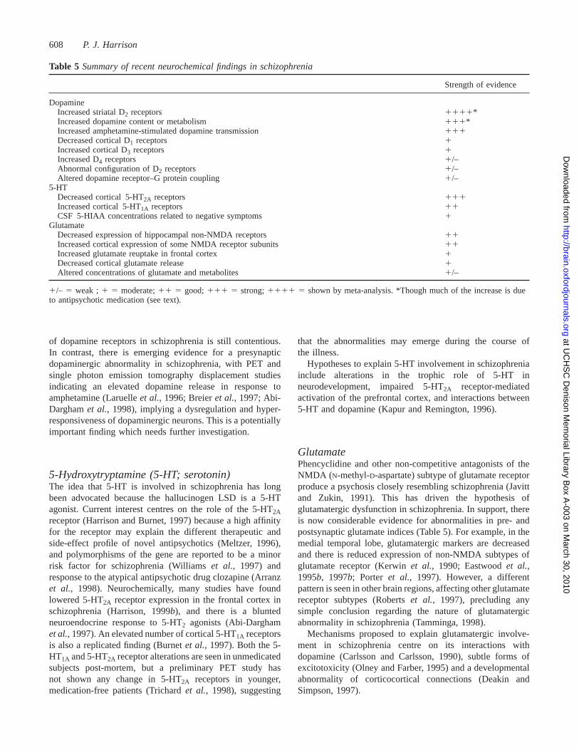
608 P. J. Harrison
Table 5 Summary of recent neurochemical findings in schizophrenia
Strength of evidence
DopamineIncreased striatal D2 receptors 1111*Increased dopamine content or metabolism 111*Increased amphetamine-stimulated dopamine transmission 111Decreased cortical D1 receptors 1Increased cortical D3 receptors 1Increased D4 receptors 1/–Abnormal configuration of D2 receptors 1/–Altered dopamine receptor–G protein coupling 1/–
5-HTDecreased cortical 5-HT2A receptors 111Increased cortical 5-HT1A receptors 11CSF 5-HIAA concentrations related to negative symptoms 1
GlutamateDecreased expression of hippocampal non-NMDA receptors 11Increased cortical expression of some NMDA receptor subunits 11Increased glutamate reuptake in frontal cortex 1Decreased cortical glutamate release 1Altered concentrations of glutamate and metabolites 1/–
1/– 5 weak ;1 5 moderate;11 5 good;111 5 strong;1111 5 shown by meta-analysis. *Though much of the increase is dueto antipsychotic medication (see text).
of dopamine receptors in schizophrenia is still contentious.In contrast, there is emerging evidence for a presynapticdopaminergic abnormality in schizophrenia, with PET andsingle photon emission tomography displacement studiesindicating an elevated dopamine release in response toamphetamine (Laruelleet al., 1996; Breieret al., 1997; Abi-Darghamet al., 1998), implying a dysregulation and hyper-responsiveness of dopaminergic neurons. This is a potentiallyimportant finding which needs further investigation.
5-Hydroxytryptamine (5-HT; serotonin)The idea that 5-HT is involved in schizophrenia has longbeen advocated because the hallucinogen LSD is a 5-HTagonist. Current interest centres on the role of the 5-HT2A
receptor (Harrison and Burnet, 1997) because a high affinityfor the receptor may explain the different therapeutic andside-effect profile of novel antipsychotics (Meltzer, 1996),and polymorphisms of the gene are reported to be a minorrisk factor for schizophrenia (Williamset al., 1997) andresponse to the atypical antipsychotic drug clozapine (Arranzet al., 1998). Neurochemically, many studies have foundlowered 5-HT2A receptor expression in the frontal cortex inschizophrenia (Harrison, 1999b), and there is a bluntedneuroendocrine response to 5-HT2 agonists (Abi-Darghamet al., 1997). An elevated number of cortical 5-HT1A receptorsis also a replicated finding (Burnetet al., 1997). Both the 5-HT1A and 5-HT2A receptor alterations are seen in unmedicatedsubjects post-mortem, but a preliminary PET study hasnot shown any change in 5-HT2A receptors in younger,medication-free patients (Trichardet al., 1998), suggesting
that the abnormalities may emerge during the course ofthe illness.
Hypotheses to explain 5-HT involvement in schizophreniainclude alterations in the trophic role of 5-HT inneurodevelopment, impaired 5-HT2A receptor-mediatedactivation of the prefrontal cortex, and interactions between5-HT and dopamine (Kapur and Remington, 1996).
GlutamatePhencyclidine and other non-competitive antagonists of theNMDA (N-methyl-D-aspartate) subtype of glutamate receptorproduce a psychosis closely resembling schizophrenia (Javittand Zukin, 1991). This has driven the hypothesis ofglutamatergic dysfunction in schizophrenia. In support, thereis now considerable evidence for abnormalities in pre- andpostsynaptic glutamate indices (Table 5). For example, in themedial temporal lobe, glutamatergic markers are decreasedand there is reduced expression of non-NMDA subtypes ofglutamate receptor (Kerwinet al., 1990; Eastwoodet al.,1995b, 1997b; Porter et al., 1997). However, a differentpattern is seen in other brain regions, affecting other glutamatereceptor subtypes (Robertset al., 1997), precluding anysimple conclusion regarding the nature of glutamatergicabnormality in schizophrenia (Tamminga, 1998).
Mechanisms proposed to explain glutamatergic involve-ment in schizophrenia centre on its interactions withdopamine (Carlsson and Carlsson, 1990), subtle forms ofexcitotoxicity (Olney and Farber, 1995) and a developmentalabnormality of corticocortical connections (Deakin andSimpson, 1997).
at UC
HS
C D
enison Mem
orial Library Box A
-003 on March 30, 2010
http://brain.oxfordjournals.orgD
ownloaded from
Neuropathology of schizophrenia 609
Relationship between neurochemical andneuropathological findingsEven from this brief, selective review it is apparent that thereis still no clear picture as to the cardinal neurochemicalfeatures of schizophrenia or their position in the pathogenesisof the disorder. The main point,vis a vis neuropathology, isthat the presence of structural abnormalities, however slight,must be taken into account. That is, a change in the level ofa neurotransmitter, receptor or any other molecule may bedue to dysfunction in the cells producing it or a change inthe cellular constituents of the tissue being evaluated, ratherthan being indicative of a molecularly specific abnormality.This applies to in vivo functional imaging as well toneurochemistry, and affects discussions as to the putativecauses and consequences of any alteration. Whilst an obviouspoint to make, it has not always been appreciated inschizophrenia research, perhaps because of the belief that thebrain is entirely normal, structurally speaking. As an example,consider the case of 5-HT receptors. In the prefrontal cortexin schizophrenia, we and others have found a loss of 5-HT2A
receptors and an accompanying increase of 5-HT1A receptors(Burnetet al., 1996, 1997). In this region, the 5-HT2A receptoris expressed by pyramidal neurons, interneurons and perhapsglia (Burnetet al., 1995; Jakab and Goldman-Rakic, 1998),whilst the 5-HT1A receptor is expressed exclusively, orvirtually so, by pyramidal neurons (Burnetet al., 1995). Thusit is unclear whether the 5-HT receptor changes seen inschizophrenia arise from differential involvement of the celltypes concerned or from opposing abnormalities in theregulation of expression of each receptor subtype. Similarly,it is difficult to interpret unambiguously the variousglutamatergic and dopaminergic abnormalities reported inschizophrenia, or the deficits in GABA which are alsoapparent (Simpsonet al., 1989; Beneset al., 1992b; Akbarianet al., 1995), independently of the morphometric alterationsin excitatory and inhibitory neurons and their synapsesdiscussed above. More sophisticated analyses are needed inorder to establish when a neurochemical finding inschizophrenia is really that, and when it is a reflection ofneuropathology.
Neuropathology and aberrant functionalconnectivityBleuler, who coined the term schizophrenia, stated ‘thethousands of associations guiding our thought are interruptedby this disease . . . The thought processes, as a result, becomestrange and illogical, and the associations find new paths’(Bleuler, 1950). His view that the key symptoms ofschizophrenia were those of ‘psychic splitting’ now have theircounterparts in neuropsychological models and in imagingstudies which have implicated aberrant functionalconnectivity between different brain regions as the patho-physiological mechanism of psychosis (Friston and Frith,1995; McGuire and Frith, 1996; Andreasenet al., 1996;
Bullmore et al., 1998). Although the evidence in favour ofaltered connectivity in schizophrenia remains circumstantialand its details poorly specified, the concept has been widelypromulgated. Examples are shown in Table 6.
Here the specific hypothesis is elaborated that aberrantconnectivity in schizophrenia has neuroanatomical roots: theneuropathology of the disorder is that of a miswiring of theneural circuitry within and between certain brain regions.This ‘hard-wiring’ theme (Mesulam, 1990), which can betraced back to Wernicke, is apparent in a number ofexperimental and theoretical perspectives (Table 6). However,aberrant functional connectivity does not presuppose orrequire an anatomical substrate, and neither is it synonymouswith regional brain dysfunction (Friston, 1998). Hence thepathological evidence in schizophrenia must be consideredon its own merits before attempts are made to integratestructure with function. Certainly, schizophrenia is not adisconnectivity syndrome akin to Alzheimer’s disease, inwhich there is frank loss of connections due to neuronal andsynaptic degeneration (Pearson and Powell, 1989; De Lacosteand White, 1993), but it is argued here that schizophrenia isa dysconnectivity or misconnectivity syndrome which affectsthe precise organization of the neural circuitry, and perhapsits plasticity characteristics (Randall, 1983; Haracz, 1985;Goodman, 1989; Walker and Diforio, 1997). It is in suchterms that the cytoarchitectural abnormalities reported inschizophrenia can reasonably be interpreted as being aputative basis for at least a proportion of the aberrantconnectivity. That is, the types of neuronal, synaptic anddendritic findings reviewed above are entirely consistent withaberrant functional connectivity. For example (Fig. 1), inDLPFC, lamina III pyramidal neurons are smaller (Rajkowskaet al., 1998), have fewer dendritic spines (Gareyet al., 1998)and receive fewer inhibitory inputs (Wooet al., 1998);moreover there is a reduction in synaptophysin in this region(Glantz and Lewis, 1997). Since the layer III pyramidalneurons are the origin of corticocortical projections, it isreasonable to propose that this combination of abnormalitieswill result in dysfunction of pathways to and from theDLPFC, in keeping with the many neuropsychological andfunctional imaging data attesting to the involvement of thisregion in the pathophysiology of schizophrenia (Weinberger,1987; Andreasenet al., 1996; Panteliset al., 1997).
Much remains to be done before the hypothesizedassociation between structure and function can be confirmedand shown to be causal. It will not be easy to falsify or toclarify its detail, and the overriding need at present isstill to improve the robustness of the contributory data.Nevertheless, the goal should be kept firmly in mind whendesigning neuropathological investigations into schizo-phrenia.
Methodological issuesIt is all too evident that schizophrenia pushes neuropathologyto its technical and conceptual limit. As well as sharing the
at UC
HS
C D
enison Mem
orial Library Box A
-003 on March 30, 2010
http://brain.oxfordjournals.orgD
ownloaded from
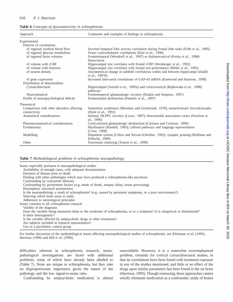
610 P. J. Harrison
Table 6 Concepts of dysconnectivity in schizophrenia
Approach Comments and examples of findings in schizophrenia
ExperimentalPatterns of correlations
of regional cerebral blood flow Inverted temporal lobe activity correlation during frontal lobe tasks (Frithet al., 1995)of regional glucose metabolism Fewer corticothalamic correlations (Katzet al., 1996)of regional brain volumes Frontotemporal (Woodruffet al., 1997) or thalamocortical (Portaset al., 1998)
dissociationof volume with rCBF Hippocampal size correlates with frontal rCBF (Weinbergeret al., 1992)of volume with function Hippocampal size correlates with frontal test performance (Bilderet al., 1995)of neuron density Asymmetrical change in subfield correlations within and between hippocampi (Zaidel
et al., 1997b)of gene expression Increased inter-areal correlations of GAP-43 mRNA (Eastwood and Harrison, 1998)
Distribution of abnormalitiesCytoarchitectural Hippocampal (Arnoldet al., 1995a) and corticocortical (Rajkowskaet al., 1998)
pathwaysNeurochemical Frontotemporal glutamatergic circuitry (Deakin and Simpson, 1997)
Profile of neuropsychological deficits Frontostriatal dysfunction (Panteliset al., 1997)
TheoreticalComparison with other disorders affecting Inattention syndromes (Mesulam and Geschwind, 1978); metachromatic leucodystrophyconnectivity (Hydeet al., 1992)Anatomical considerations Intrinsic DLPFC circuitry (Lewis, 1997); heteromodal association cortex (Pearlsonet
al., 1996)Pharmacoanatomical considerations Corticostriatal glutamatergic dysfunction (Carlsson and Carlsson, 1990)Evolutionary Myelination (Randall, 1983); callosal pathways and language representation
(Crow, 1998)Modelling Dopamine system (Cohen and Servan-Schreiber, 1992); synaptic pruning (Hoffman and
Dobscha, 1989)Other Functional clustering (Tononiet al., 1998)
Table 7 Methodological problems in schizophrenia neuropathology
Issues especially pertinent to neuropathological studiesAvailability of enough cases, with adequate documentationDuration of disease prior to deathDealing with other pathologies which may have produced a schizophrenia-like psychosisConfounding by concurrent illnessesConfounding by perimortem factors (e.g. mode of death, autopsy delay, tissue processing)Hemispheric structural asymmetriesIs the neuropathology a result of schizophrenia? (e.g. caused by persistent symptoms, or a poor environment?)Selecting which brain areas to studyAdherence to stereological principles
Issues common to all schizophrenia researchValidity of the diagnosisDoes the variable being measured relate to the syndrome of schizophrenia, or to a symptom? Is it categorical or dimensional?Is there heterogeneity?Is the variable affected by antipsychotic drugs or other treatments?Are subjects included in research representative?Use of a psychiatric control group
For further discussion of the methodological issues affecting neuropathological studies of schizophrenia, see Kleinmanet al. (1995),Harrison (1996) and Hillet al. (1996).
difficulties inherent in schizophrenia research, neuro-pathological investigations are faced with additionalproblems, some of which have already been alluded to(Table 7). None are unique to schizophrenia, but they takeon disproportionate importance given the nature of thepathology and the low signal-to-noise ratio.
Confounding by antipsychotic medication is almost
unavoidable. However, it is a somewhat overemphasizedproblem, certainly for cortical cytoarchitectural studies, inthat no correlations have been found with treatment exposurein any of the studies mentioned, and little or no effect of thedrugs upon similar parameters has been found in the rat brain(Harrison, 1993). Though reassuring, these approaches cannotwholly eliminate medication as a confounder; study of brains
at UC
HS
C D
enison Mem
orial Library Box A
-003 on March 30, 2010
http://brain.oxfordjournals.orgD
ownloaded from
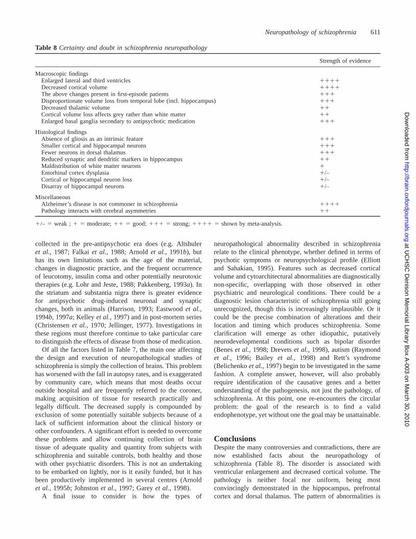
Neuropathology of schizophrenia 611
Table 8 Certainty and doubt in schizophrenia neuropathology
Strength of evidence
Macroscopic findingsEnlarged lateral and third ventricles 1111Decreased cortical volume 1111The above changes present in first-episode patients 111Disproportionate volume loss from temporal lobe (incl. hippocampus) 111Decreased thalamic volume 11Cortical volume loss affects grey rather than white matter 11Enlarged basal ganglia secondary to antipsychotic medication 111
Histological findingsAbsence of gliosis as an intrinsic feature 111Smaller cortical and hippocampal neurons 111Fewer neurons in dorsal thalamus 111Reduced synaptic and dendritic markers in hippocampus 11Maldistribution of white matter neurons 1Entorhinal cortex dysplasia 1/–Cortical or hippocampal neuron loss 1/–Disarray of hippocampal neurons 1/–
MiscellaneousAlzheimer’s disease is not commoner in schizophrenia 1111Pathology interacts with cerebral asymmetries 11
1/– 5 weak ;1 5 moderate;11 5 good;111 5 strong;1111 5 shown by meta-analysis.
collected in the pre-antipsychotic era does (e.g. Altshuleret al., 1987; Falkaiet al., 1988; Arnoldet al., 1991b), buthas its own limitations such as the age of the material,changes in diagnostic practice, and the frequent occurrenceof leucotomy, insulin coma and other potentially neurotoxictherapies (e.g. Lohr and Jeste, 1988; Pakkenberg, 1993a). Inthe striatum and substantia nigra there is greater evidencefor antipsychotic drug-induced neuronal and synapticchanges, both in animals (Harrison, 1993; Eastwoodet al.,1994b, 1997a; Kelley et al., 1997) and in post-mortem series(Christensenet al., 1970; Jellinger, 1977). Investigations inthese regions must therefore continue to take particular careto distinguish the effects of disease from those of medication.
Of all the factors listed in Table 7, the main one affectingthe design and execution of neuropathological studies ofschizophrenia is simply the collection of brains. This problemhas worsened with the fall in autopsy rates, and is exaggeratedby community care, which means that most deaths occuroutside hospital and are frequently referred to the coroner,making acquisition of tissue for research practically andlegally difficult. The decreased supply is compounded byexclusion of some potentially suitable subjects because of alack of sufficient information about the clinical history orother confounders. A significant effort is needed to overcomethese problems and allow continuing collection of braintissue of adequate quality and quantity from subjects withschizophrenia and suitable controls, both healthy and thosewith other psychiatric disorders. This is not an undertakingto be embarked on lightly, nor is it easily funded, but it hasbeen productively implemented in several centres (Arnoldet al., 1995b; Johnstonet al., 1997; Gareyet al., 1998).
A final issue to consider is how the types of
neuropathological abnormality described in schizophreniarelate to the clinical phenotype, whether defined in terms ofpsychotic symptoms or neuropsychological profile (Elliottand Sahakian, 1995). Features such as decreased corticalvolume and cytoarchitectural abnormalities are diagnosticallynon-specific, overlapping with those observed in otherpsychiatric and neurological conditions. There could be adiagnostic lesion characteristic of schizophrenia still goingunrecognized, though this is increasingly implausible. Or itcould be the precise combination of alterations and theirlocation and timing which produces schizophrenia. Someclarification will emerge as other idiopathic, putativelyneurodevelopmental conditions such as bipolar disorder(Beneset al., 1998; Drevetset al., 1998), autism (Raymondet al., 1996; Bailey et al., 1998) and Rett’s syndrome(Belichenkoet al., 1997) begin to be investigated in the samefashion. A complete answer, however, will also probablyrequire identification of the causative genes and a betterunderstanding of the pathogenesis, not just the pathology, ofschizophrenia. At this point, one re-encounters the circularproblem: the goal of the research is to find a validendophenotype, yet without one the goal may be unattainable.
ConclusionsDespite the many controversies and contradictions, there arenow established facts about the neuropathology ofschizophrenia (Table 8). The disorder is associated withventricular enlargement and decreased cortical volume. Thepathology is neither focal nor uniform, being mostconvincingly demonstrated in the hippocampus, prefrontalcortex and dorsal thalamus. The pattern of abnormalities is
at UC
HS
C D
enison Mem
orial Library Box A
-003 on March 30, 2010
http://brain.oxfordjournals.orgD
ownloaded from
612 P. J. Harrison
suggestive of a disturbance of connectivity within andbetween these regions, most likely originating during braindevelopment. At the cellular level, the changes are manifestedas abnormalities, mainly decrements, in perikaryal,presynaptic and dendritic parameters (Tables 1 and 2; Fig. 1).The phenotype of the affected neurons and synapses isunclear, though there is some evidence for preferentialglutamatergic involvement in the hippocampus. The sortsof cytoarchitectural changes being proposed, whilst stillspeculative and perhaps better viewed as aberrantneuroanatomy rather than neuropathologyper se, are inkeeping with the clinical complexity of the syndrome whichthey are being invoked to explain, as well as with theresults of various functional imaging, neurochemical andneuropsychological findings. Progress is now constrainedmore by the subtlety of the target pathology than bymethodological limitations of the work; indeed, the bestcontemporary studies are prime examples of innovative,quantitative neuropathology research.
In 1915 the neuropathologist Southard boldly stated that‘structural (visible or invisible) changes of a mal-developmental nature lie at the bottom of the [schizophrenia]disease process . . . Aside from the left-sidedness of thelesions and internal hydrocephalus, very striking is thepreference of these changes to occupy the association centresof Fleschig’. A positive view of the field today is thatempirical data are belatedly proving the correctness of theseassertions. Conversely, a cynic might suggest that it is theinvisibility which has proved the most robustly demonstrable,or that the current histological findings remain sufficientlyvague and numerous to cover all possibilities, ensuring thatone will turn out, with hindsight, to be similarly prescient.Certainly it is time to implement still better research, drivenby specific hypotheses (e.g. Benes, 1998; Weickert andWeinberger, 1998). In this way the small but increasinglysteady steps which are finally being taken can proceed toa clear identification of the pathological substrate(s) ofschizophrenia.
AcknowledgementsI am grateful to the Wellcome Trust and the StanleyFoundation for research support. I wish to thank Tim Crow,Sharon Eastwood, Margaret Esiri, Gareth Roberts and themany other colleagues upon whose findings and ideas thereview is based. Margaret Cousin kindly providedsecretarial support.
ReferencesAbi-Dargham A, Laruelle M, Aghajanian GK, Charney D, KrystalJ. The role of serotonin in the pathophysiology and treatment ofschizophrenia. [Review]. J Neuropsychiatry Clin Neurosci 1997; 9:1–17.
Abi-Dargham A, Gil R, Krystal J, Baldwin RM, Seibyl JP, BowersM, et al. Increased striatal dopamine transmission in schizophrenia:confirmation in a second cohort. Am J Psychiatry 1998; 155: 761–7.
Adams CE, DeMasters BK, Freedman R. Regional zinc staining inpostmortem hippocampus from schizophrenic patients. SchizophrRes 1995; 18: 71–7.
Aganova EA, Uranova NA. Morphometric analysis of synapticcontacts in the anterior limbic cortex in the endogenous psychoses.Neurosci Behav Physiol 1992; 22: 59–65.
Ajtai BM, Kallai L, Kalman M. Capability for reactive gliosisdevelops prenatally in the diencephalon but not in the cortex ofrats. Exp Neurol 1997; 146: 151–8.
Akbarian S, Bunney WE Jr, Potkin SG, Wigal SB, Hagman JO,Sandman CA, et al. Altered distribution of nicotinamide-adeninedinucleotide phosphate-diaphorase cells in frontal lobe ofschizophrenics implies disturbances of cortical development. ArchGen Psychiatry 1993a; 50: 169–77.
Akbarian S, Vinuela A, Kim JJ, Potkin SG, Bunney WE Jr, JonesEG. Distorted distribution of nicotinamide-adenine dinucleotidephosphate-diaphorase neurons in temporal lobe of schizophrenicsimplies anomalous cortical development. Arch Gen Psychiatry1993b; 50: 178–87.
Akbarian S, Kim JJ, Potkin SG, Hagman JO, Tafazzoli A, BunneyWE Jr, et al. Gene expression for glutamic acid decarboxylase isreduced without loss of neurons in prefrontal cortex ofschizophrenics [see comments]. Arch Gen Psychiatry 1995; 52:258–66.
Akbarian S, Kim JJ, Potkin SG, Hetrick WP, Bunney WE Jr, JonesEG. Maldistribution of interstitial neurons in prefrontal white matterof the brains of schizophrenic patients. Arch Gen Psychiatry 1996;53: 425–36.
Akil M, Lewis DA. Cytoarchitecture of the entorhinal cortex inschizophrenia. Am J Psychiatry 1997; 154: 1010–2.
Allendoerfer KL, Shatz CJ. The subplate, a transient neocorticalstructure: its role in the development of connections betweenthalamus and cortex. [Review]. Annu Rev Neurosci 1994; 17:185–218.
Altshuler LL, Conrad A, Kovelman JA, Scheibel A. Hippocampalpyramidal cell orientation in schizophrenia. A controlledneurohistologic study of the Yakovlev collection. Arch GenPsychiatry 1987; 44: 1094–8.
Altshuler LL, Casanova MF, Goldberg TE, Kleinman JE. Thehippocampus and parahippocampus in schizophrenia, suicide, andcontrol brains [published erratum appears in Arch Gen Psychiatry1991; 48; 442]. Arch Gen Psychiatry 1990; 47: 1029–34.
American Psychiatric Association. Diagnostic and statistical manualof mental disorders. DSM-IV. 4th ed. Washington (DC): AmericanPsychiatric Association; 1994.
Anderson B, Rutledge V. Age and hemisphere effects on dendriticstructure. Brain 1996; 119: 1983–90.
Anderson SA, Volk DW, Lewis DA. Increased density of microtubuleassociated protein 2-immunoreactive neurons in the prefrontal whitematter of schizophrenic subjects. Schizophr Res 1996; 19: 111–9.
at UC
HS
C D
enison Mem
orial Library Box A
-003 on March 30, 2010
http://brain.oxfordjournals.orgD
ownloaded from
Neuropathology of schizophrenia 613
Andrade JP, Castanheira-Vale AJ, Paz-Dias PG, Madeira MD, Paula-Barbosa MM. The dendritic trees of neurons from the hippocampalformation of protein-deprived adult rats. A quantitative Golgi study.Exp Brain Res 1996; 109: 419–33.
Andreasen NC. Symptoms, signs, and diagnosis of schizophrenia.[Review]. Lancet 1995; 346: 477–81.
Andreasen NC, Arndt S, Swayze V 2nd, Cizadlo T, Flaum M,O’Leary D, et al. Thalamic abnormalities in schizophrenia visualizedthrough magnetic resonance image averaging [see comments].Science 1994; 266: 294–8. Comment in: Science 1994; 266: 221.
Andreasen NC, O’Leary DS, Cizadlo T, Arndt S, Rezai K, PontoLL, et al. Schizophrenia and cognitive dysmetria: a positron-emission tomography study of dysfunctional prefrontal-thalamic-cerebellar circuitry. Proc Natl Acad Sci USA 1996; 93: 9985–90.
Anezaki T, Yanagisawa K, Takahashi H, Nakajima T, Miyashita K,Ishikawa A, et al. Remote astrocytic response of prefrontal cortexis caused by the lesions in the nucleus basalis of Meynert, but notin the ventral tegmental area. Brain Res 1992; 574: 63–9.
Aquino DA, Padin C, Perez JM, Peng D, Lyman WD, Chiu FC.Analysis of glial fibrillary acidic protein, neurofilament protein,actin and heat shock proteins in human fetal brain during the secondtrimester. Brain Res Dev Brain Res 1996; 91: 1–10.
Arendt T, Bigl V, Arendt A, Tennstedt A. Loss of neurons in thenucleus basalis of Meynert in Alzheimer’s disease, paralysis agitansand Korsakoff’s disease. Acta Neuropathol (Berl) 1983; 61: 101–8.
Arnold SE, Trojanowski JQ. Recent advances in defining theneuropathology of schizophrenia. [Review]. Acta Neuropathol (Berl)1996; 92: 217–31.
Arnold SE, Hyman BT, Van Hoesen GW, Damasio AR. Somecytoarchitectural abnormalities of the entorhinal cortex inschizophrenia. Arch Gen Psychiatry 1991a; 48: 625–32.
Arnold SE, Lee VM-Y, Gur RE, Trojanowski JQ. Abnormalexpression of two microtubule-associated proteins (MAP2 andMAP5) in specific subfields of the hippocampal formation inschizophrenia. Proc Natl Acad Sci USA 1991b; 88: 10850–4.
Arnold SE, Franz BR, Gur RC, Gur RE, Shapiro RM, Moberg PJ,et al. Smaller neuron size in schizophrenia in hippocampal subfieldsthat mediate cortical-hippocampal interactions. Am J Psychiatry1995a; 152: 738–48.
Arnold SE, Gur RE, Shapiro RM, Fisher KR, Moberg PJ, GibneyMR, et al. Prospective clinicopathologic studies of schizophrenia:accrual and assessment of patients. Am J Psychiatry 1995b; 152:731–7.
Arnold SE, Franz BR, Trojanowski JQ, Moberg PJ, Gur RE.Glial fibrillary acidic protein-immunoreactive astrocytosis in elderlypatients with schizophrenia and dementia. Acta Neuropathol (Berl)1996; 91: 269–77.
Arnold SE, Ruscheinsky DD, Han LY. Further evidence of abnormalcytoarchitecture of the entorhinal cortex in schizophrenia usingspatial point pattern analyses. Biol Psychiatry 1997a; 42: 639–47.
Arnold SE, Joo E, Martinoli MG, Roy N, Trojanowski JQ, Gur RE,et al. Apolipoprotein E genotype in schizophrenia: frequency, ageof onset, and neuropathologic features [see comments]. Neuroreport1997b; 8: 1523–6. Comment in: Neuroreport 1997; 8: i–ii.
Arnold SE, Trojanowski JQ, Gur RE, Blackwell P, Han LY, ChoiC. Absence of neurodegeneration and neural injury in the cerebralcortex in a sample of elderly patients with schizophrenia. Arch GenPsychiatry 1998; 55: 225–32.
Arranz MJ, Munro J, Sham P, Kirov G, Murray RM, Collier DA,et al. Meta-analysis of studies on genetic variation in 5-HT2Areceptors and clozapine response. Schizophr Res 1998; 32: 93–9.
Averback P. Lesions of the nucleus ansae peduncularis inneuropsychiatric disease. Arch Neurol 1981; 38: 230–5.
Bailey A, Luthert P, Dean A, Harding B, Janota I, Montgomery M,et al. A clinicopathological study of autism. Brain 1998; 121:889–905.
Baldessarini RJ, Hegarty JD, Bird ED, Benes FM. Meta-analysisof postmortem studies of Alzheimer’s disease-like neuropathologyin schizophrenia [published erratum appears in Neuroreport 1997;154: 1180]. Neuroreport 1997; 154: 861–3.
Barta PE, Pearlson GD, Powers RE, Richards SS, Tune LE. Auditoryhallucinations and smaller superior temporal gyral volume inschizophrenia. Am J Psychiatry 1990; 147: 1457–62.
Barta PE, Pearlson GD, Brill LB 2nd, Royall R, McGilchristIK, Pulver AE, et al. Planum temporale asymmetry reversal inschizophrenia: replication and relationship to gray matterabnormalities. Am J Psychiatry 1997; 154: 661–7.
Bartley AJ, Jones DW, Weinberger DR. Genetic variability of humanbrain size and cortical gyral patterns. Brain 1997; 120: 257–69.
Beall MJ, Lewis DA. Heterogeneity of layer II neurons in humanentorhinal cortex. J Comp Neurol 1992; 321: 241–66.
Beasley CL, Reynolds GP. Parvalbumin-immunoreactive neuronsare reduced in the prefrontal cortex of schizophrenics. SchizophrRes 1997; 24: 349–55.
Beauregard M, Malkova L, Bachevalier J. Stereotypies and lossof social affiliation after early hippocampectomy in primates.Neuroreport 1995; 6: 2521–6.
Beckmann H, Lauer M. The human striatum in schizophrenia. II.Increased number of striatal neurons in schizophrenics. PsychiatryRes 1997; 68: 99–109.
Belichenko PV, Hagberg B, Dahlstro¨m A. Morphological study ofneocortical areas in Rett syndrome. Acta Neuropathol (Berl) 1997;93: 50–61.
Benes FM. Model generation and testing to probe neural circuitryin the cingulate cortex of postmortem schizophrenic brain. [Review].Schizophr Bull 1998; 24: 219–30.
Benes FM, Bird ED. An analysis of the arrangement of neurons inthe cingulate cortex of schizophrenic patients. Arch Gen Psychiatry1987; 44: 608–16.
Benes FM, Davidson J, Bird ED. Quantitative cytoarchitecturalstudies of the cerebral cortex of schizophrenics. Arch Gen Psychiatry1986; 43: 31–5.
Benes FM, Majocha RE, Bird ED, Marotta CA. Increased verticalaxon numbers in cingulate cortex of schizophrenics. Arch GenPsychiatry 1987; 44: 1017–21.
at UC
HS
C D
enison Mem
orial Library Box A
-003 on March 30, 2010
http://brain.oxfordjournals.orgD
ownloaded from
614 P. J. Harrison
Benes FM, McSparren J, Bird ED, SanGiovanni JP, Vincent SL.Deficits in small interneurons in prefrontal and cingulate corticesof schizophrenic and schizoaffective patients. Arch Gen Psychiatry1991a; 48: 996–1001.
Benes FM, Sorensen I, Bird ED. Reduced neuronal size in posteriorhippocampus of schizophrenic patients. Schizophr Bull 1991b; 17:597–608.
Benes FM, Sorensen I, Vincent SL, Bird ED, Sathi M. Increaseddensity of glutamate-immunoreactive vertical processes insuperficial laminae in cingulate cortex of schizophrenic brain. CerebCortex 1992a; 2: 503–12.
Benes FM, Vincent SL, Alsterberg G, Bird ED, SanGiovanni JP.Increased GABAA receptor binding in superficial layers of cingulatecortex in schizophrenics. J Neurosci 1992b; 12: 924–9.
Benes FM, Turtle M, Khan Y, Farol P. Myelination of a key relayzone in the hippocampal formation occurs in the human brain duringchildhood, adolescence, and adulthood. Arch Gen Psychiatry 1994;51: 477–84.
Benes FM, Todtenkopf MS, Taylor JB. Differential distribution oftyrosine hydroxylase fibers on small and large neurons in layer IIof anterior cingulate cortex of schizophrenic brain. Synapse 1997;25: 80–92.
Benes FM, Kwok EW, Vincent SL, Todtenkopf MS. A reductionof nonpyramidal cells in sector CA2 of schizophrenics and manicdepressives. Biol Psychiatry 1998; 44: 88–97.
Benowitz LI, Routtenberg A. GAP-43: an intrinsic determinant ofneuronal development and plasticity. [Review]. Trends Neurosci1997; 20: 84–91.
Berman NE, Raymond LA, Warren KA, Raghavan R, Joag SV,Narayan O, et al. Fractionator analysis shows loss of neurons in thelateral geniculate nucleus of macaques infected with neurovirulentsimian immunodeficiency virus. Neuropathol Appl Neurobiol 1998;24: 44–52.
Bernstein HG, Stanarius A, Baumann B, Henning H, Krell D, DanosP, et al. Nitric oxide synthase-containing neurons in the humanhypothalamus: reduced number of immunoreactive cells in theparaventricular nucleus of depressive patients and schizophrenics.Neuroscience 1998; 83: 867–75.
Bertolino A, Nawroz S, Mattay VS, Barnett AS, Duyn JH, MoonenCT, et al. Regionally specific pattern of neurochemical pathologyin schizophrenia as assessed by multislice proton magnetic resonancespectroscopic imaging. Am J Psychiatry 1996; 153: 1554–63.
Bertolino A, Callicott JH, Elman I, Mattay VS, Tedeschi G, FrankJA, et al. Regionally specific neuronal pathology in untreatedpatients with schizophrenia: a proton magnetic resonancespectroscopic imaging study. Biol Psychiatry 1998; 43: 641–8.
Bilder RM, Lipschutz-Broch L, Reiter G, Geisler SH, MayerhoffDI, Lieberman JA. Intellectual deficits in first-episode schizophrenia:evidence for progressive deterioration. Schizophr Bull 1992; 18:437–48.
Bilder RM, Wu H, Bogerts B, Degreef G, Ashtari M, Alvir JM,et al. Absence of regional hemispheric volume asymmetries in first-episode schizophrenia. Am J Psychiatry 1994; 151: 1437–47.
Bilder RM, Bogerts B, Ashtari M, Wu H, Alvir JM, Jody D,et al. Anterior hippocampal volume reductions predict frontal lobedysfunction in first episode schizophrenia. Schizophr Res 1995; 17:47–58.
Blanchard JJ, Neale JM. The neuropsychological signature ofschizophrenia: generalized or differential deficit? Am J Psychiatry1994; 151: 40–8.
Blennow K, Davidsson P, Gottfries C-G, Ekman R, Heilig M.Synaptic degeneration in thalamus in schizophrenia [letter]. Lancet1996; 348: 692–3.
Bleuler E. Dementia praecox or the group of schizophrenias 1911.Transl. J. Zinkin. New York: International Universities Press; 1950.
Bloom FE. Advancing a neurodevelopmental origin forschizophrenia. [Review]. Arch Gen Psychiatry 1993; 50: 224–7.
Bogerts B, Ha¨ntsch J, Herzer M. A morphometric study of thedopamine-containing cell groups in the mesencephalon of normals,Parkinson patients, and schizophrenics. Biol Psychiatry 1983; 18:951–69.
Bogerts B, Meertz E, Schonfeldt-Bausch R. Basal ganglia andlimbic system pathology in schizophrenia. Arch Gen Psychiatry1985; 42: 784–91.
Bogerts B, Ashtari M, Degreef G, Alvir JM, Bilder RM, LiebermanJA. Reduced temporal limbic structure volumes on magneticresonance images in first episode schizophrenia. Psychiatry Res1990a; 35: 1–13.
Bogerts B, Falkai P, Haupts M, Greve B, Ernst S, Tapernon-FranzU, et al. Post-mortem volume measurements of limbic system andbasal ganglia structures in chronic schizophrenics. Initial resultsfrom a new brain collection. Schizophr Res 1990b; 3: 295–301.
Braak H, Braak E. Ratio of pyramidal cells versus non-pyramidalcells in the human frontal isocortex and changes in ratio with ageingand Alzheimer’s disease. Prog Brain Res 1986; 70: 185–212.
Bracha HS. Etiology of structural asymmetry in schizophrenia: analternative hypothesis [letter]. Schizophr Bull 1991; 17: 551–3.
Breier A, Su TP, Saunders R, Carson RE, Kolachana BS, deBartolomeis A, et al. Schizophrenia is associated with elevatedamphetamine-induced synaptic dopamine concentrations: evidencefrom a novel positron emission tomography method. Proc NatlAcad Sci USA 1997; 94: 2569–74.
Briess D, Cotter D, Doshi R, Everall I. Mammillary bodyabnormalities in schizophrenia [letter]. Lancet 1998; 352: 789–90.
Brown S. Excess mortality of schizophrenia. A meta-analysis. Br JPsychiatry 1997; 171: 502–8.
Brown R, Colter N, Corsellis JA, Crow TJ, Frith CD, JagoeR, et al. Postmortem evidence of structural brain changes inschizophrenia. Differences in brain weight, temporal horn area, andparahippocampal gyrus compared with affective disorder. Arch GenPsychiatry 1986; 43: 36–42.
Browning MD, Dudek EM, Rapier JL, Leonard S, Freedman R.Significant reductions in synapsin but not synaptophysin specificactivity in the brains of some schizophrenics. Biol Psychiatry 1993;34: 529–35.
at UC
HS
C D
enison Mem
orial Library Box A
-003 on March 30, 2010
http://brain.oxfordjournals.orgD
ownloaded from
Neuropathology of schizophrenia 615
Bruton CJ, Crow TJ, Frith CD, Johnstone EC, Owens DG, RobertsGW. Schizophrenia and the brain: a prospective clinico-neuropathological study. Psychol Med 1990; 20: 285–304.
Bruton CJ, Stevens JR, Frith CD. Epilepsy, psychosis, andschizophrenia: clinical and neuropathologic correlations. Neurology1994; 44: 34–42.
Buchanan RW, Breier A, Kirkpatrick B, Elkashef A, Munson RC,Gellad F, et al. Structural abnormalities in deficit and nondeficitschizophrenia. Am J Psychiatry 1993; 150: 59–65.
Buchsbaum MS, Someya T, Teng CY, Abel L, Chin S, Najafi A,et al. PET and MRI of the thalamus in never-medicated patientswith schizophrenia. Am J Psychiatry 1996; 153: 191–9.
Buckley PF. The clinical stigmata of aberrant neurodevelopment inschizophrenia. [Review]. J Nerv Ment Dis 1998; 186: 79–86.
Buhl L, Bojsen-Møller M. Frequency of Alzheimer’s disease in apostmortem study of psychiatric patients. Dan Med Bull 1988; 35:288–90.
Bullmore ET, Woodruff PW, Wright IC, Rabe-Hesketh S, HowardRJ, Shuriquie N, et al. Does dysplasia cause anatomicaldysconnectivity in schizophrenia? Schizophr Res 1998; 30: 127–35.
Burnet PW, Eastwood SL, Lacey K, Harrison PJ. The distributionof 5-HT1A and 5-HT2A receptor mRNA in human brain. Brain Res1995; 676: 157–68.
Burnet PW, Eastwood SL, Harrison PJ. 5-HT1A and 5-HT2A receptormRNAs and binding site densities are differentially altered inschizophrenia. Neuropsychopharmacology 1996; 15: 442–55.
Burnet PW, Eastwood SL, Harrison PJ. [3H]WAY-100635 for 5-HT1A receptor autoradiography in human brain: a comparison with[3H]8-OH-DPAT and demonstration of increased binding in thefrontal cortex in schizophrenia. Neurochem Int 1997; 30: 565–74.
Cannon M, Jones P. Schizophrenia. [Review]. J Neurol NeurosurgPsychiatry 1996; 60: 604–13.
Cannon TD, Mednick SA, Parnas J, Schulsinger F, Praestholm J,Vestergaard A. Developmental brain abnormalities in the offspringof schizophrenic mothers. I. Contributions of genetic and perinatalfactors [see comments]. Arch Gen Psychiatry 1993; 50: 551–64.Comment in: Arch Gen Psychiatry 1995; 52: 157–9.
Carlsson M, Carlsson A. Schizophrenia: a subcorticalneurotransmitter imbalance syndrome? [Review]. Schizophr Bull1990; 16: 425–32.
Casanova MF, Zito M, Bigelow LB, Berthot B, Sanders RD,Kleinman JE. Axonal counts of the corpus callosum of schizophrenicpatients. J Neuropsychiatry Clin Neurosci 1989; 1: 391–3.
Casanova MF, Stevens JR, Kleinman JE. Astrocytosis in themolecular layer of the dentate gyrus: a study in Alzheimer’s diseaseand schizophrenia. Psychiatry Res 1990; 35: 149–66.
Chakos MH, Lieberman JA, Bilder RM, Borenstein M, Lerner G,Bogerts B, et al. Increase in caudate nuclei volumes of first-episodeschizophrenic patients taking antipsychotic drugs. Am J Psychiatry1994; 151: 1430–6.
Crichton-Browne J. On the weight of the brain and its componentparts in the insane. Brain 1879; 2: 42–67.
Christensen E, Moller JE, Faurbye A. Neuropathologicalinvestigation of 28 brains from patients with dyskinesia. ActaPsychiatr Scand 1970; 46: 14–23.
Christison GW, Casanova MF, Weinberger DR, Rawlings R,Kleinman JE. A quantitative investigation of hippocampal pyramidalcell size, shape, and variability of orientation in schizophrenia. ArchGen Psychiatry 1989; 46: 1027–32.
Chua SE, McKenna PJ. Schizophrenia—a brain disease? A criticalreview of structural and functional cerebral abnormality in thedisorder. [Review]. Br J Psychiatry 1995; 166: 563–82.
Cohen JD, Servan-Schreiber D. Context, cortex, and dopamine: aconnectionist approach to behavior and biology in schizophrenia.[Review]. Psychol Rev 1992; 99: 45–77.
Conrad AJ, Abebe T, Austin R, Forsythe S, Scheibel AB.Hippocampal pyramidal cell disarray in schizophrenia as a bilateralphenomenon. Arch Gen Psychiatry 1991; 48: 413–7.
Corsellis JAN. Mental illness and the ageing brain. Institute ofPsychiatry Maudsley Monographs No. 9. London: Oxford UniversityPress; 1962.
Corsellis JAN. Psychoses of obscure pathology. In: Blackwood W,Corsellis JAN, editors. Greenfield’s neuropathology. 3rd ed. London:Edward Arnold; 1976. p. 903–15.
Cotter D, Kerwin R, Doshi B, Martin CS, Everall IP. Alterationsin hippocampal non-phosphorylated MAP2 protein expression inschizophrenia. Brain Res 1997; 765: 238–46.
Crow TJ. Molecular pathology of schizophrenia: more than onedisease process? [Review]. Br Med J 1980; 280: 66–8.
Crow TJ. Temporal lobe asymmetries as the key to the etiology ofschizophrenia. [Review]. Schizophr Bull 1990; 16: 433–43.
Crow TJ. Schizophrenia as failure of hemispheric dominance forlanguage [see comments]. [Review]. Trends Neurosci 1997; 20:339–43. Comment in: Trends Neurosci 1998; 21: 145–7.
Crow TJ. Schizophrenia as a transcallosal misconnection syndrome.Schizophr Res 1998; 30: 111–4.
Crow TJ, Ball J, Bloom SR, Brown R, Bruton CJ, Colter N, et al.Schizophrenia as an anomaly of development of cerebral asymmetry.Arch Gen Psychiatry 1989; 46: 1145–50.
da Cunha A, Jefferson JJ, Tyor WR, Glass JD, Jannotta FS, VitkovicL. Gliosis in human brain: relationship to size but not otherproperties of astrocytes. Brain Res 1993; 600: 161–5.
Daniel DG, Goldberg TE, Gibbons RD, Weinberger DR. Lack of abimodal distribution of ventricular size in schizophrenia: a Gaussianmixture analysis of 1056 cases and controls. Biol Psychiatry 1991;30: 887–903.
Danos P, Baumann B, Bernstein H-G, Franz M, Stauch R, NorthoffG, et al. Schizophrenia and anteroventral thalamic nucleus: selectivedecrease of parvalbumin-immunoreactive thalamocortical projectionneurons. Psychiatry Res 1998; 82: 1–10.
David GB. The pathological anatomy of the schizophrenias. In:Richter D, editor. Schizophrenia: somatic aspects. London:Pergamon Press; 1957. p. 93–130.
at UC
HS
C D
enison Mem
orial Library Box A
-003 on March 30, 2010
http://brain.oxfordjournals.orgD
ownloaded from
616 P. J. Harrison
David AS, Cutting JC. The neuropsychology of schizophrenia. Hove(UK): Lawrence Erlbaum; 1994.
Davidson M, Keefe RS. Cognitive impairment as a target forpharmacological treatment in schizophrenia. [Review]. SchizophrRes 1995; 17: 123–9.
Davidson L, McGlashan TH. The varied outcomes of schizophrenia[review]. Can J Psychiatry 1997; 42: 34–43.
Davidson M, Harvey P, Welsh KA, Powchik P, Putnam KM, MohsRC. Cognitive functioning in late-life schizophrenia: a comparisonof elderly schizophrenic patients and patients with Alzheimer’sdisease. Am J Psychiatry 1996; 153: 1274–9.
Davis KL, Kahn RS, Ko G, Davidson M. Dopamine in schizophrenia:a review and reconceptualization [see comments]. Am J Psychiatry1991; 148: 1474–86. Comment in: Am J Psychiatry 1992; 149:1284–5. Comment in: Am J Psychiatry 1992; 149: 1620–1.
Davis KL, Buchsbaum MS, Shihabuddin L, Spiegel-Cohen J,Metzger M, Frecska E, et al. Ventricular enlargement in poor-outcome schizophrenia [see comments]. Biol Psychiatry 1998; 43:783–93. Comment in: Biol Psychiatry 1998; 43: 781–2.
Davison K. Schizophrenia-like psychoses associated with organiccerebral disorders: a review. [Review]. Psychiatr Dev 1983; 1: 1–33.
Daviss SR, Lewis DA. Local circuit neurons of the prefrontal cortexin schizophrenia: selective increase in the density of calbindin-immunoreactive neurons. Psychiatry Res 1995; 59: 81–96.
de Brabander JM, Kramers RJK, Uylings HBM. Layer-specificdendritic regression of pyramidal cells with ageing in the humanprefrontal cortex. Eur J Neurosci 1998; 10: 1261–9.
De Lacoste M-C, White CL, 3d. The role of cortical connectivityin Alzheimer’s disease pathogenesis: a review and model system[see comments]. [Review]. Neurobiol Aging 1993; 14: 1–16.Comment in: Neurobiol Aging 1993; 14: 49–50, Comment in:Neurobiol Aging 1993; 14: 51–4; disc 55–6.
Deakin JF, Simpson MD. A two-process theory of schizophrenia:evidence from studies in post-mortem brain. [Review]. J PsychiatRes 1997; 31: 277–95.
Degreef G, Ashtari M, Bogerts B, Bilder RM, Jody DN, Alvir JM,et al. Volumes of ventricular system subdivisions measured frommagnetic resonance images in first-episode schizophrenic patients.Arch Gen Psychiatry 1992; 49: 531–7.
Deicken RF, Zhou L, Corwin F, Vinogradov S, Weiner MW.Decreased left frontal lobe N-acetylaspartate in schizophrenia. AmJ Psychiatry 1997; 154: 688–90.
Deicken RF, Zhou L, Schuff N, Fein G, Weiner MW. Hippocampalneuronal dysfunction in schizophrenia as measured by protonmagnetic resonance spectroscopy. Biol Psychiatry 1998; 43: 483–8.
DeLisi LE. Is schizophrenia a lifetime disorder of brain plasticity,growth and aging? [Review]. Schizophr Res 1997; 23: 119–29.
DeLisi LE, Sakuma M, Tew W, Kushner M, Hoff AL, Grimson R.Schizophrenia as a chronic active brain process: a study ofprogressive brain structural change subsequent to the onset ofschizophrenia. Psychiatry Res 1997a; 74: 129–40.
DeLisi LE, Sakuma M, Kushner M, Finer DL, Hoff AL, CrowTJ. Anomalous cerebral asymmetry and language processing in
schizophrenia [published erratum appears in Schizophr Bull 1997;23: 536]. Schizophr Bull 1997b; 23: 255–71.
Dell’Anna ME, Geloso MC, Draisci G, Luthman J. Transientchanges in fos and GFAP immunoreactivity precede neuronal lossin the rat hippocampus following neonatal anoxia. Exp Neurol1995; 131: 144–56.
Doody GA, Johnstone EC, Sanderson TL, Owens DGC, Muir WJ.‘Pfropfschizophrenie’ revisited: schizophrenia in people with mildlearning disability. Br J Psychiatry 1998; 173: 145–53.
Drevets WC, Ongur D, Price JL. Neuroimaging abnormalities inthe subgenual prefrontal cortex: implications for the pathophysiologyof familial mood disorders. Mol Psychiatry 1998; 3: 220–6.
Dwork AJ. Postmortem studies of the hippocampal formation inschizophrenia. [Review]. Schizophr Bull 1997; 23: 385–402.
Eastwood SL, Harrison PJ. Decreased synaptophysin in the medialtemporal lobe in schizophrenia demonstrated using immuno-autoradiography. Neuroscience 1995; 69: 339–43.
Eastwood SL, Harrison PJ. Hippocampal and cortical growth-associated protein-43 messenger RNA in schizophrenia.Neuroscience 1998; 86: 437–48.
Eastwood SL, Burnet PWJ, McDonald B, Clinton J, Harrison PJ.Synaptophysin gene expression in human brain: a quantitative insitu hybridization and immunocytochemical study. Neuroscience1994a; 59: 881–92.
Eastwood SL, Burnet PW, Harrison PJ. Striatal synaptophysinexpression and haloperidol-induced synaptic plasticity. Neuroreport1994b; 5: 677–80.
Eastwood SL, Burnet PW, Harrison PJ. Altered synaptophysinexpression as a marker of synaptic pathology in schizophrenia.Neuroscience 1995a; 66: 309–19.
Eastwood SL, McDonald B, Burnet PW, Beckwith JP, Kerwin RW,Harrison PJ. Decreased expression of mRNAs encoding non-NMDAglutamate receptors GluR1 and GluR2 in medial temporal lobeneurons in schizophrenia. Brain Res Mol Brain Res 1995b; 29:211–23.
Eastwood SL, Heffernan J, Harrison PJ. Chronic haloperidoltreatment differentially affects the expression of synaptic andneuronal plasticity-associated genes. Mol Psychiatry 1997a; 2:322–9.
Eastwood SL, Kerwin RW, Harrison PJ. Immunoautoradiographicevidence for a loss of alpha-amino-3-hydroxy-5-methyl-4-isoxazolepropionate-preferring non-N-methyl-D-aspartate glutamatereceptors within the medial temporal lobe in schizophrenia. BiolPsychiatry 1997b; 41: 636–43.
Elliott R, Sahakian, BJ. The neuropsychology of schizo-phrenia: relations with clinical and neurobiological dimen-sions. [Review]. Psychol Med 1995; 25: 581–94.
Elston GN, Rosa MG. Morphological variation of layer III pyramidalneurones in the occipitotemporal pathway of the macaque monkeyvisual cortex. Cereb Cortex 1998; 8: 278–94.
Falkai P, Bogerts B. Cell loss in the hippocampus of schizophrenics.Eur Arch Psychiatry Neurol Sci 1986; 236: 154–61.
at UC
HS
C D
enison Mem
orial Library Box A
-003 on March 30, 2010
http://brain.oxfordjournals.orgD
ownloaded from
Neuropathology of schizophrenia 617
Falkai P, Bogerts B. Cytoarchitectonic and developmental studiesin schizophrenia. In: Kerwin RW, editor. Neurobiology andpsychiatry, Vol. 2. Cambridge: Cambridge University Press; 1993.p. 43–70.
Falkai P, Bogerts B, Rozumek M. Limbic pathology in schizophrenia:the entorhinal region—a morphometric study. Biol Psychiatry 1988;24: 515–21.
Falkai P, Bogerts B, Greve B, Pfeiffer U, Machus B, Fo¨lsch-ReetzB, et al. Loss of sylvian fissure asymmetry in schizophrenia. Aquantitative post-mortem study. Schizophr Res 1992; 7: 23–32.
Falkai P, Bogerts B, Schneider T, Greve B, Pfeiffer U, Pilz K,et al. Disturbed planum temporale asymmetry in schizophrenia. Aquantitative post-mortem study. Schizophr Res 1995; 14: 161–76.
Feinberg I. Schizophrenia: caused by a fault in programmed synapticelimination during adolescence? J Psychiatr Res 1982; 17: 319–34.
Fisman M. The brain stem in psychosis. Br J Psychiatry 1975; 126:414–22.
Flaum M, Arndt S, Andreasen NC. The role of gender in studiesof ventricle enlargement in schizophrenia: a predominantly maleeffect [see comments]. Am J Psychiatry 1990; 147: 1327–32.Comment in: Am J Psychiatry 1991; 148; 1754–6.
Flaum M, Swayze VW 2nd, O’Leary DS, Yuh WT, Ehrhardt JC,Arndt SV, et al. Effects of diagnosis, laterality, and gender on brainmorphology in schizophrenia. Am J Psychiatry 1995; 152: 704–14.
Flor-Henry P. Psychosis and temporal lobe epilepsy. A controlledinvestigation. Epilepsia 1969; 10: 363–95.
Frazier JA, Giedd JN, Hamburger SD, Albus KE, Kaysen D, VaituzisAC, et al. Brain anatomic magnetic resonance imaging in childhood-onset schizophrenia. Arch Gen Psychiatry 1996; 53: 617–24.
Friede RL. Developmental neuropathology. Berlin: SpringerVerlag; 1989.
Friston KJ. The disconnection hypothesis. Schizophr Res 1998; 30:115–25.
Friston KJ, Frith CD. Schizophrenia: a disconnection syndrome?[Review]. Clin Neurosci 1995; 3: 89–97.
Frith CD, Friston KJ, Herold S, Silbersweig D, Fletcher P, CahillC, et al. Regional brain activity in chronic schizophrenic patientsduring the performance of a verbal fluency task. Br J Psychiatry1995; 167: 343–9.
Gabriel SM, Haroutunian V, Powchik P, Honer WG, Davidson M,Davies P, et al. Increased concentrations of presynaptic proteins inthe cingulate cortex of subjects with schizophrenia [publishederratum appears in Arch Gen Psychiatry 1997; 54: 912]. 559–66.
Galaburda AM. Anatomic basis for cerebral dominance. In:Richardson RE, Hugdahl K, editors. Cerebral asymmetry. Boston:MIT Press; 1994. p. 51–73.
Garcia-Rill E, Biedermann JA, Chambers T, Skinner RD, MrakRE, Husain M, et al. Mesopontine neurons in schizophrenia.Neuroscience 1995; 66: 321–35.
Garey LJ, Ong WY, Patel TS, Kanani M, Davis A, Mortimer AM,et al. Reduced dendritic spine density on cerebral cortical pyramidal
neurons in schizophrenia. J Neurol Neurosurg Psychiatry 1998; 65:446–53.
Garver DL. The etiologic heterogeneity of schizophrenia. HarvardRev Psychiatry 1997; 4: 317–27.
Geddes JR, Lawrie SM. Obstetric complications and schizophrenia:a meta-analysis. Br J Psychiatry 1995; 167: 786–93.
Glantz LA, Lewis DA. Reduction of synaptophysinimmunoreactivity in the prefrontal cortex of subjects withschizophrenia. Regional and diagnostic specificity [corrected andrepublished article originally appeared in Arch Gen Psychiatry1997; 54: 660–9]. Arch Gen Psychiatry 1997; 54: 943–52.
Goldberg TE, Hyde TM, Kleinman JE, Weinberger DR. Course ofschizophrenia: neuropsychological evidence for a staticencephalopathy. [Review]. Schizophr Bull 1993; 19: 797–804.
Goldsmith SK, Joyce JN. Alterations in hippocampal mossy fiberpathway in schizophrenia and Alzheimer’s disease. Biol Psychiatry1995; 37: 122–6.
Goodman R. Neuronal misconnections and psychiatric disorder. Isthere a link? [see comments]. [Review]. Br J Psychiatry 1989; 154:292–9. Comment in: Br J Psychiatry 1989; 155: 129–30.
Green MF. What are the functional consequences of neurocognitivedeficits in schizophrenia? [see comments]. [Review]. Am JPsychiatry 1996; 153: 321–30. Comment in: Am J Psychiatry 1997;154: 443–4.
Guillery RW, Herrup K. Quantification without pontification:choosing a method for counting objects in sectioned tissues[comment]. J Comp Neurol 1997; 386: 2–7. Comment on: J CompNeurol 1996; 364: 6–15.
Gur RE, Mozley PD, Shtasel DL, Cannon TD, Gallacher F, TuretskyB, et al. Clinical subtypes of schizophrenia: differences in brainand CSF volume [see comments]. Am J Psychiatry 1994; 151: 343–50. Comment in: Am J Psychiatry 1995; 152: 817–8.
Gur RE, Cowell P, Turetsky BI, Gallacher F, Cannon T, Bilker W,et al. A follow-up magnetic resonance imaging study ofschizophrenia. Arch Gen Psychiatry 1998; 55: 145–52.
Gurevich EV, Bordelon Y, Shapiro RM, Arnold SE, Gur RE, JoyceJN. Mesolimbic dopamine D3 receptors and use of antipsychoticsin patients with schizophrenia. A postmortem study. Arch GenPsychiatry 1997; 54: 225–32.
Hafner H, an der Heiden W, Behrens S, Gattaz WF, Hambrecht M,Loffler W, et al. Causes and consequences of the gender differencein age at onset of schizophrenia. [Review]. Schizophr Bull 1998;24: 99–113.
Halliday GM, Cullen KM, Kril JJ, Harding AJ, Harasty J. Glialfibrillary acidic protein (GFAP) immunohistochemistry in humancortex: a quantitative study using different antisera. Neurosci Lett1996; 209: 29–32.
Haracz JL. Neural plasticity in schizophrenia. Schizophr Bull 1985;11: 191–229.
Haroutunian V, Davidson M, Kanof PD, Perl DP, Powchik P,Losonczy M, et al. Cortical cholinergic markers in schizophrenia.Schizophr Res 1994; 12: 137–44.
at UC
HS
C D
enison Mem
orial Library Box A
-003 on March 30, 2010
http://brain.oxfordjournals.orgD
ownloaded from
618 P. J. Harrison
Harrison PJ. Effects of neuroleptics on neuronal and synapticstructure. In: Barnes TRE, editor. Antipsychotic drugs and theirside-effects. London: Academic Press; 1993. p. 99–110.
Harrison PJ. Advances in post-mortem molecular neurochemistryand neuropathology: examples from schizophrenia research.[Review]. Br Med Bull 1996; 52: 527–38.
Harrison PJ. Schizophrenia: a disorder of neurodevelopment?[Review]. Curr Opin Neurobiol 1997a; 7: 285–9.
Harrison PJ. Schizophrenia and its dementia. In: Esiri MM, MorrisJH, editors. The neuropathology of dementia. Cambridge: CambridgeUniversity Press; 1997b. p. 385–97.
Harrison PJ. Brains at risk of schizophrenia [Editorial]. Lancet1999a; 353: 3–4.
Harrison PJ. Neurochemical alterations in schizophrenia affectingthe putative targets of atypical antipsychotics: focus on dopamine(D1, D3, D4) and 5-HT2A receptors. Br J Psychiatry 1999b; 174Suppl 38: 41–51.
Harrison PJ, Burnet PW. The 5-HT2A (serotonin2A) receptor genein the aetiology, pathophysiology and pharmacotherapy ofschizophrenia. J Psychopharmacol (Oxf) 1997; 11: 18–20.
Harrison PJ, Eastwood SL. Preferential involvement of excitatoryneurons in the medial temporal lobe in schizophrenia. Lancet 1998;352: 1669–73.
Haug JO. Pneumoencephalographic evidence of brain atrophy inacute and chronic schizophrenic patients. Acta Psychiatr Scand1982; 66: 374–83.
Hayes TL, Lewis DA. Magnopyramidal neurons in the anterior motorspeech region. Dendritic features and interhemispheric comparisons.Arch Neurol 1996; 53: 1277–83.
Heckers S. Neuropathology of schizophrenia: cortex, thalamus,basal ganglia, and neurotransmitter-specific projection systems.[Review]. Schizophr Bull 1997; 23: 403–21.
Heckers S, Heinsen H, Heinsen YC, Beckmann H. Limbic structuresand lateral ventricle in schizophrenia: a quantitative postmortemstudy [see comments]. Arch Gen Psychiatry 1990; 47: 1016–22.Comment in: Arch Gen Psychiatry 1991; 48: 956–8.
Heckers S, Heinsen H, Heinsen Y, Beckmann H. Cortex, whitematter, and basal ganglia in schizophrenia: a volumetric postmortemstudy. Biol Psychiatry 1991a; 29: 556–66.
Heckers S, Heinsen H, Geiger B, Beckmann H. Hippocampal neuronnumber in schizophrenia: a stereological study. Arch Gen Psychiatry1991b; 48: 1002–8.
Heffernan JM, Eastwood SL, Nagy Z, Sanders MW, McDonald B,Harrison PJ. Temporal cortex synaptophysin mRNA is reduced inAlzheimer’s disease and is negatively correlated with the severityof dementia. Exp Neurol 1998; 150: 235–9.
Heinsen H, Go¨ssmann E, Ru¨b U, Eisenmenger W, Bauer M, UlmarG, et al. Variability in the human entorhinal region may confoundneuropsychiatric diagnoses. Acta Anat (Basel) 1996; 157: 226–37.
Highley JR, Esiri MM, McDonald B, Cortina-Borja M, Cooper SJ,Herron BM et al. Anomalies of cerebral asymmetry in schizophreniainteract with gender and age of onset: a post-mortem study.Schizophr Res 1998; 34: 13–25.
Highley JR, Esiri MM, McDonald B, Cortina-Borja M, Herron BM,Crow TJ. The size and fibre composition of the corpus callosumwith respect to gender and schizophrenia: a post-mortem study.Brain 1999; 122: 99–110.
Hill C, Keks N, Roberts S, Opeskin K, Dean B, MacKinnonA, et al. Problem of diagnosis in postmortem brain studies ofschizophrenia. Am J Psychiatry 1996; 153: 533–7.
Ho K-C, Gwozdz JT, Hause LL, Antuono PG. Correlation ofneuronal cell body size in motor cortex and hippocampus with bodyheight, body weight, and axonal length. Int J Neurosci 1992; 65:147–53.
Hoff AL, Riordan H, O’Donnell DW, Morris L, DeLisi LE.Neuropsychological functioning of first-episode schizophreniformpatients. Am J Psychiatry 1992; 149: 898–903.
Hoffman RE, Dobscha SK. Cortical pruning and the developmentof schizophrenia: a computer model [see comments]. SchizophrBull 1989; 15: 477–90. Comment in: Schizophr Bull 1990; 16:567–70.
Honer WG, Bassett AS, Smith GN, Lapointe JS, Falkai P. Temporallobe abnormalities in multigenerational families with schizophrenia.Biol Psychiatry 1994; 36: 737–43.
Honer WG, Falkai P, Young C, Wang T, Xie J, Bonner J, et al.Cingulate cortex synaptic terminal proteins and neural cell adhesionmolecule in schizophrenia. Neuroscience 1997; 78: 99–110.
Horton K, Forsyth CS, Sibtain N, Ball S, Bruton CJ, Royston MC,et al. Ubiquitination as a probe for neurodegeneration in the brainin schizophrenia: the prefrontal cortex. Psychiatry Res 1993; 48:145–52.
Howard CV, Reed MG. Unbiased stereology. Oxford: Bios; 1998.
Huber G. The heterogeneous course of schizophrenia. SchizophrRes 1997; 28: 177–85.
Huttenlocher PR. Synaptic density in human frontal cortex:developmental changes and effects of aging. Brain Res 1979; 163:195–205.
Hyde TM, Ziegler JC, Weinberger DR. Psychiatric disturbances inmetachromatic leukodystrophy: insights into the neurobiology ofpsychosis [see comments]. Arch Neurol 1992; 49: 401–6. Commentin: Arch Neurol 1993; 50: 131.
Insausti R, Tun˜on T, Sobreviela T, Insausti AM, Gonzalo LM. Thehuman entorhinal cortex: a cytoarchitectonic analysis. J CompNeurol 1995; 355: 171–98.
Jablensky A. Schizophrenia: recent epidemiologic issues. [Review].Epidemiol Rev 1995; 17: 10–20.
Jacobsen LK, Giedd JN, Castellanos FX, Vaituzis AC, HamburgerSD, Kumra S, et al. Progressive reduction of temporal lobe structuresin childhood-onset schizophrenia. Am J Psychiatry 1998; 155:678–85.
Jakab RL, Goldman-Rakic PS. 5-Hydroxytryptamine2A serotoninreceptors in the primate cerebral cortex: possible site of action ofhallucinogenic and antipsychotic drugs in pyramidal cell apicaldendrites. Proc Natl Acad Sci USA 1998; 95: 735–40.
at UC
HS
C D
enison Mem
orial Library Box A
-003 on March 30, 2010
http://brain.oxfordjournals.orgD
ownloaded from
Neuropathology of schizophrenia 619
Jakob H, Beckmann H. Prenatal developmental disturbances in thelimbic allocortex in schizophrenics. J Neural Transm 1986; 65:303–26.
Jakob H, Beckmann H. Gross and histological criteria fordevelopmental disorders in brains of schizophrenics. [Review]. J RSoc Med 1989; 82: 466–9.
Jaskiw GE, Juliano DM, Goldberg TE, Hertzman M, Urow-HamellE, Weinberger DR. Cerebral ventricular enlargement inschizophreniform disorder does not progress. A seven year follow-up study. Schizophr Res 1994; 14: 23–8.
Javitt DC, Zukin SR. Recent advances in the phencyclidine modelof schizophrenia [see comments]. [Review]. Am J Psychiatry 1991;148: 1301–8. Comment in: Am J Psychiatry 1992; 149: 848-9.
Jellinger K. Neuropathologic findings after neuroleptic long-termtherapy. In: Roizin L, Shiraki H, Grcevic N, editors.Neurotoxicology. New York: Raven Press; 1977. p. 25–42.
Jellinger K. Neuromorphological background of pathochemicalstudies in major psychoses. In: Beckmann H, Riederer P, editors.Pathochemical markers in major psychoses. Berlin: Springer; 1985.p. 1–23.
Jeste DV, Lohr JB. Hippocampal pathologic findings inschizophrenia. A morphometric study. Arch Gen Psychiatry 1989;46: 1019–24.
Johnston NL, Cervenak J, Shore AD, Torrey EF, Yolken RH.Multivariate analysis of RNA levels from postmortem human brainsas measured by three different methods of RT-PCR [publishederratum appears in J Neurosci Methods 1998; 79: 233]. J NeurosciMethods 1997; 77: 83–92.
Johnstone EC, Crow TJ, Frith CD, Husband J, Kreel L. Cerebralventricular size and cognitive impairment in chronic schizophrenia.Lancet 1976; 2: 924–6.
Johnstone EC, McMillan JF, Crow TJ. The occurrence of organicdisease of possible or probable aetiological significance in apopulation of 268 cases of first episode schizophrenia. Psychol Med1987; 17: 371–9.
Johnstone EC, Bruton CJ, Crow TJ, Frith CD, Owens DG. Clinicalcorrelates of postmortem brain changes in schizophrenia: decreasedbrain weight and length correlate with indices of early impairment.J Neurol Neurosurg Psychiatry 1994; 57: 474–9.
Jones EG. Cortical development and thalamic pathology inschizophrenia [see comments]. [Review]. Schizophr Bull 1997a;23: 483–501. Comment in: Schizophr Bull 1997; 23: 509–12,Comment in: Schizophr Bull 1997; 537-40.
Jones P. The early origins of schizophrenia. [Review]. Br Med Bull1997b; 53: 135–55.
Jones TA, Schallert T. Use-dependent growth of pyramidal neuronsafter neocortical damage. J Neurosci 1994; 14: 2140–52.
Jønsson SA, Luts A, Guldberg-Kjaer N, Brun A. Hippocampalpyramidal cell disarray correlates negatively to cell number:implications for the pathogenesis of schizophrenia. Eur ArchPsychiatry Clin Neurosci 1997; 247: 120–7.
Joyce JN, Meador-Woodruff JH. Linking the family of D2 receptorsto neuronal circuits in human brain: insights into schizophrenia.[Review]. Neuropsychopharmacology 1997; 16: 375–84.
Kalman M, Csillag A, Schleicher A, Rind C, Hajo´s F, Zilles K.Long-term effects of anterograde degeneration on astroglial reactionin the rat geniculo-cortical system as revealed by computerizedimage analysis. Anat Embryol (Berl) 1993; 187: 1–7.
Kalus P, Senitz D, Beckmann H. Altered distribution of parvalbumin-immunoreactive local circuit neurons in the anterior cingulate cortexof schizophrenic patients. Psychiatry Res 1997; 75: 49–59.
Kapur S, Remington G. Serotonin-dopamine interaction and itsrelevance to schizophrenia. [Review]. Am J Psychiatry 1996; 153:466–76.
Katsetos CD, Hyde TM, Herman MM. Neuropathology of thecerebellum in schizophrenia—an update: 1996 and future directions.[Review]. Biol Psychiatry 1997; 42: 213–24.
Katz M, Buchsbaum MS, Siagel BV Jr, Wu J, Haier RJ, BunneyWE Jr Correlational patterns of cerebral glucose metabolism innever-medicated schizophrenics. Neuropsychobiology 1996; 33:1–11.
Kegeles LS, Humaran J, Mann JJ. In vivo neurochemistry ofthe brain in schizophrenia as revealed by magnetic resonancespectroscopy. Biol Psychiatry 1998; 44: 382–98.
Kelley JJ, Gao XM, Tamminga CA, Roberts RC. The effect ofchronic haloperidol treatment on dendritic spines in the rat striatum.Exp Neurol 1997; 146: 471–8.
Kennedy JL. Schizophrenia genetics: the quest for an anchor[editorial]. Am J Psychiatry 1996; 153: 1513–4.
Kenny JT, Friedman L, Findling RL, Swales TP, Strauss ME,Jesberger JA, et al. Cognitive impairment in adolescents withschizophrenia. Am J Psychiatry 1997; 154: 1613–5.
Kerwin R, Patel S, Meldrum B. Quantitative autoradiographicanalysis of glutamate binding sites in the hippocampal formationin normal and schizophrenic brain post-mortem. Neuroscience 1990;39: 25–32.
Keshavan MS, Anderson S, Pettegrew JW. Is schizophrenia due toexcessive synaptic pruning in the prefrontal cortex? The Feinberghypothesis revisited. [Review]. J Psychiatr Res 1994a; 28: 239–65.
Keshavan MS, Bagwell WW, Haas GL, Sweeney JA, Schooler NR,Pettegrew JW. Changes in caudate volume with neuroleptic treatment[letter]. Lancet 1994b; 344: 1434.
Keshavan MS, Rosenberg D, Sweeney JA, Pettegrew JW. Decreasedcaudate volume in neuroleptic-naive psychotic patients. Am JPsychiatry 1998; 155: 774–8.
Kleinman JE, Hyde TM, Herman MM. Methodological issues inthe neuropathology of mental illness. In: Bloom FE, Kupfer DJ,editors. Psychopharmacology. The fourth generation of progress.New York: Raven Press; 1995. p. 859–64.
Kolb B, Whishaw IQ. Brain plasticity and behavior. [Review]. AnnuRev Psychol 1998; 49: 43–64.
Kovelman JA, Scheibel AB. A neurohistological correlate ofschizophrenia. Biol Psychiatry 1984; 19: 1601–21.
at UC
HS
C D
enison Mem
orial Library Box A
-003 on March 30, 2010
http://brain.oxfordjournals.orgD
ownloaded from
620 P. J. Harrison
Kreutzberg GW, Blakemore WF, Graeber MB. Cellular pathologyof the central nervous system. In: Graham DI, Lantos PL, editors.Greenfield’s neuropathology. 6th ed. London: Edward Arnold; 1997.p. 85–156.
Krimer LS, Herman MM, Saunders RC, Boyd JC, Hyde TM, CarterJM, et al. A qualitative and quantitative analysis of the entorhinalcortex in schizophrenia. Cereb Cortex 1997a; 7: 732–9.
Krimer LS, Hyde TM, Herman MM, Saunders RC. The entorhinalcortex: an examination of cyto- and myeloarchitectonic organizationin humans. Cereb Cortex 1997b; 7: 722–31.
Kulynych JJ, Vladar K, Jones DW, Weinberger DR. Superiortemporal gyrus volume in schizophrenia: a study using MRImorphometry assisted by surface rendering. Am J Psychiatry 1996;153: 50–6.
Kung L, Conley R, Chute DJ, Smialek J, Roberts RC. Synapticchanges in the striatum of schizophrenic cases: a controlledpostmortem ultrastructural study. Synapse 1998; 28: 125–39.
Kwon JS, Shenton ME, Hirayasu Y, Salisbury DF, Fischer IA,Dickey CC, et al. MRI study of cavum septi pellucidi inschizophrenia, affective disorder, and schizotypal personalitydisorder. Am J Psychiatry 1998; 155: 509–15.
Laruelle M, Abi-Dargham A, van Dyck CH, Gil R, D’Souza CD,Erdos J, et al. Single photon emission computerized tomographyimaging of amphetamine-induced dopamine release in drug-freeschizophrenic subjects. Proc Natl Acad Sci USA 1996; 93: 9235–40.
Lauriello J, Hoff A, Wieneke MH, Blankfeld H, Faustman WO,Rosenbloom M, et al. Similar extent of brain dysmorphology inseverely ill women and men with schizophrenia. Am J Psychiatry1997; 154: 819–25.
Lawrie SM, Abukmeil SS. Brain abnormality in schizophrenia. Asystematic and quantitative review of volumetric magnetic resonanceimaging studies. [Review]. Br J Psychiatry 1998; 172: 110–20.
Lawrie SM, Whalley H, Kestelman JN, Abukmeil SS, Byrne M,Hodges A, et al. Magnetic resonance imaging of the brain in subjectsat high risk of developing schizophrenia. Lancet 1999; 353: 30–3.
Lewis DA. Development of the prefrontal cortex during adolescence:insights into vulnerable neural circuits in schizophrenia. [Review].Neuropsychopharmacology 1997; 16: 385–98.
Liddle PF, Friston KJ, Frith CD, Hirsch SR, Jones T, FrackowiakRS. Patterns of cerebral blood flow in schizophrenia. Br J Psychiatry1992; 160: 179–86.
Lieberman JA, Sheitman BB, Kinon BJ. Neurochemical sensitizationin the pathophysiology of schizophrenia: deficits and dysfunctionin neuronal regulation and plasticity. [Review]. Neuropsycho-pharmacology 1997; 17: 205–29.
Lim KO, Tew W, Kushner M, Chow K, Matsumoto B, DeLisi LE.Cortical gray matter volume deficit in patients with first-episodeschizophrenia. Am J Psychiatry 1996; 153: 1548–53.
Lipska BK, Weinberger DR. Genetic variation in vulnerability tothe behavioral effects of neonatal hippocampal damage in rats. ProcNatl Acad Sci USA 1995; 92: 8906–10.
Lohr JB, Jeste DV. Locus ceruleus morphometry in aging andschizophrenia. Acta Psychiatr Scand 1988; 77: 689–97.
Madsen AL, Keiding N, Karle A, Esbjerg S, Hemmingsen R.Neuroleptics in progressive structural brain abnormalities inpsychiatric illness [letter]. Lancet 1998; 352: 784–5.
Maier M, Ron MA, Barker GJ, Tofts PS. Proton magnetic resonancespectroscopy: an in vivo method of estimating hippocampal neuronaldepletion in schizophrenia [published erratum appears in PsycholMed 1996; 26: 877]. Psychol Med 1995; 25: 1201–9.
Marsh L, Suddath RL, Higgins N, Weinberger DR. Medial temporallobe structures in schizophrenia: relationship of size to duration ofillness. Schizophr Res 1994; 11: 225–38.
Marsh L, Harris D, Lim KO, Beal M, Hoff AL, Minn K, et al.Structural magnetic resonance imaging abnormalities in men withsevere chronic schizophrenia and an early age at clinical onset.Arch Gen Psychiatry 1997; 54: 1104–12.
Masliah E, Terry R. The role of synaptic proteins in the pathogenesisof disorders of the central nervous system. [Review]. Brain Pathol1993; 3: 77–85.
Masliah E, Mallory M, Hansen L, DeTeresa R, Terry RD.Quantitative synaptic alterations in the human neocortex duringnormal aging. Neurology 1993; 43: 192–7.
Mayhew TM, Gundersen HJ. ‘If you assume, you can make an assout of u and me’: a decade of the disector for stereological countingof particles in 3D space. [Review]. J Anat 1996; 188: 1–15.
McGuffin P, Owen MJ, Farmer AE. Genetic basis of schizophrenia.Lancet 1995; 346: 678–82.
McGuire PK, Frith CD. Disordered functional connectivity inschizophrenia [editorial]. Psychol Med 1996; 26: 663–7.
McKenna PJ. Schizophrenia and related syndromes. Oxford: OxfordUniversity Press; 1994.
McKenna PJ, Tamlyn D, Lund CE, Mortimer AM, Hammond S,Baddeley AD. Amnesic syndrome in schizophrenia. Psychol Med1990; 20: 967–72.
Meltzer HY. Pre-clinical pharmacology of atypical antipsychoticdrugs: a selective review. [Review]. Br J Psychiatry 1996; 168Suppl 29: 23–31.
Mesulam M-M. Schizophrenia and the brain [editorial; comment].N Engl J Med 1990; 322: 842–5. Comment on: N Engl J Med1990; 322: 789-94.
Mesulam M-M, Geschwind N. On the possible role of neocortex andits limbic connections in the process of attention and schizophrenia:clinical cases of inattention in man and experimental anatomy inmonkey. J Psychiatr Res 1978; 14: 249–59.
Miyakawa T, Sumiyoshi S, Deshimaru M, Suzuki T, TomonariH. Electron microscopic study on schizophrenia. Mechanism ofpathological changes. Acta Neuropathol (Berl) 1972; 20: 67–77.
Miyake T, Kitamura T, Takamatsu T, Fujita S. A quantitativeanalysis of human astrocytosis. Acta Neuropathol (Berl) 1988; 75:535–7.
Moser M-B, Trommald M, Andersen P. An increase in dendriticspine density on hippocampal CA1 pyramidal cells following spatiallearning in adult rats suggests the formation of new synapses. ProcNatl Acad Sci USA 1994; 91: 12673–5.
at UC
HS
C D
enison Mem
orial Library Box A
-003 on March 30, 2010
http://brain.oxfordjournals.orgD
ownloaded from
Neuropathology of schizophrenia 621
Murphy GM Jr, Lim KO, Wieneke M, Ellis WG, Forno LS, HoffAL, et al. No neuropathologic evidence for an increased frequency ofAlzheimer’s disease among elderly schizophrenics. Biol Psychiatry1998; 43: 205–9.
Murray RM, Lewis SW. Is schizophrenia a neurodevelopmentaldisorder? [editorial] Br Med J 1987; 295: 681–2.
Murray RM, Woodruff PWR. Developmental insanity or dementiapraecox: a new perspective on an old debate. Neurol PsychiatryBrain Res 1995; 3: 167–76.
Murray RM, O’Callaghan E, Castle DJ, Lewis SW. A neuro-developmental approach to the classification of schizophrenia.[Review]. Schizophr Bull 1992; 18: 319–32.
Nair TR, Christensen JD, Kingsbury SJ, Kumar NG, Terry WM,Garver DL. Progression of cerebroventricular enlargement and thesubtyping of schizophrenia. Psychiatry Res 1997; 74: 141–50.
Nasrallah HA, McCalley-Whitters M, Bigelow LB, Rauscher FP. Ahistological study of the corpus callosum in chronic schizophrenia.Psychiatry Res 1983; 8: 251–60.
Nelson MD, Saykin AJ, Flashman LA, Riordan HJ. Hippocampalvolume reduction in schizophrenia as assessed by magneticresonance imaging: a meta-analytic study. Arch Gen Psychiatry1998; 55: 433–40.
Nieto D, Escobar A. Major psychoses. In: Minckler J, editor.Pathology of the nervous system. New York: McGraw-Hill; 1972.p. 2655–65.
Niizato K, Arai T, Kuroki N, Kase K, Iritani S, Ikeda K. Autopsystudy of Alzheimer’s disease brain pathology in schizophrenia.Schizophr Res 1998; 31: 177–84.
Noga JT, Bartley AJ, Jones DW, Torrey EF, Weinberger DR. Corticalgyral anatomy and gross brain dimensions in monozygotic twinsdiscordant for schizophrenia. Schizophr Res 1996; 22: 27–40.
Nopoulos P, Flaum M, Andreasen NC. Sex differences in brainmorphology in schizophrenia [see comments]. 48–54. Comment in:Am J Psychiatry 1997; 154: 1637–9.
Nordstrom AL, Farde L, Eriksson L, Halldin C. No elevated D2dopamine receptors in neuroleptic-naive schizophrenic patientsrevealed by positron emission tomography and [11C]N-methylspiperone [see comments]. Psychiatry Res 1995; 61: 67–83.Comment in: Psychiatry Res 1996; 67: 159-62.
Okubo Y, Suhara T, Suzuki K, Kobayashi K, Inoue O, Terasaki O,et al. Decreased prefrontal dopamine D1 receptors in schizophreniarevealed by PET [see comments]. Nature 1997; 385: 634–6.Comment in: Nature 1997; 385: 578-9.
Olney JW, Farber NB. Glutamate receptor dysfunction andschizophrenia [see comments]. Arch Gen Psychiatry 1995; 52: 998–1007. Comment in: Arch Gen Psychiatry 1997; 54: 578-80.
Ong WY, Garey LJ. Ultrastructural features of biopsied temporopolarcortex (area 38) in a case of schizophrenia. Schizophr Res 1993;10: 15–27.
Owen F, Simpson MDC. The neurochemistry of schizophrenia.In: Hirsch SR, Weinberger DR, editors. Schizophrenia. Oxford:Blackwell Science; 1995. p. 358–78.
Pakkenberg B. Post-mortem study of chronic schizophrenic brains.Br J Psychiatry 1987; 151: 744–52.
Pakkenberg B. Pronounced reduction of total neuron number inmediodorsal thalamic nucleus and nucleus accumbens inschizophrenics. Arch Gen Psychiatry 1990; 47: 1023–8.
Pakkenberg B. The volume of the mediodorsal thalamic nucleusin treated and untreated schizophrenics. Schizophr Res 1992; 7:95–100.
Pakkenberg B. Leucotomized schizophrenics lose neurons in themediodorsal thalamic nucleus. Neuropathol Appl Neurobiol 1993a;19: 373–80.
Pakkenberg B. Total nerve cell number in neocortex in chronicschizophrenics and controls estimated using optical disectors. BiolPsychiatry 1993b; 34: 768–72.
Pantelis C, Barnes TR, Nelson HE, Tanner S, Weatherley L, OwenAM, et al. Frontal–striatal cognitive deficits in patients with chronicschizophrenia. Brain 1997; 120: 1823–43.
Pearlson GD, Petty RG, Ross CA, Tien AY. Schizophrenia: a diseaseof heteromodal association cortex. [Review]. Neuropsycho-pharmacology 1996; 14: 1–17.
Pearlson GD, Barta PE, Powers RE, Menon RR, Richards SS,Aylward EH, et al. Medial and superior temporal gyral volumesand cerebral asymmetry in schizophrenia versus bipolar disorder.Biol Psychiatry 1997; 41: 1–14.
Pearson RCA, Powell TPS. The neuroanatomy of Alzheimer’sdisease. Rev Neurosci 1989; 2: 101–22.
Perrone-Bizzozero NI, Sower AC, Bird ED, Benowitz LI, Ivins KJ,Neve RL. Levels of the growth-associated protein GAP-43 areselectively increased in association cortices in schizophrenia. ProcNatl Acad Sci USA 1996; 93: 14182–7.
Pierce JP, Lewin GR. An ultrastructural size principle. [Review].Neuroscience 1994; 58: 441–6.
Plum F. Prospects for research on schizophrenia. 3. Neurophysiology.Neuropathological findings. Neurosci Res Program Bull 1972; 10:384–8.
Portas CM, Goldstein JM, Shenton ME, Hokama HH, WibleCG, Fischer I, et al. Volumetric evaluation of the thalamus inschizophrenic male patients using magnetic resonance imaging. BiolPsychiatry 1998; 43: 649–59.
Porter RH, Eastwood SL, Harrison PJ. Distribution of kainatereceptor subunit mRNAs in human hippocampus, neocortex andcerebellum, and bilateral reduction of hippocampal GluR6 and KA2transcripts in schizophrenia. Brain Res 1997; 751: 217–31.
Powchik P, Friedman J, Haroutunian V, Greenberg D, Altsteil L,Purohit D, et al. Apolipoprotein E4 in schizophrenia: a study ofone hundred sixteen cases with concomitant neuropathologicalexamination. Biol Psychiatry 1997; 42: 296–8.
Prohovnik I, Dwork AJ, Kaufman MA, Willson N. Alzheimer-typeneuropathology in elderly schizophrenia patients. Schizophr Bull1993; 19: 805–16.
Purohit DP, Perl DP, Haroutunian V, Powchik P, Davidson M, DaviesKL. Alzheimer disease and related neurodegenerative diseases in
at UC
HS
C D
enison Mem
orial Library Box A
-003 on March 30, 2010
http://brain.oxfordjournals.orgD
ownloaded from
622 P. J. Harrison
elderly patients with schizophrenia: a postmortem neuropathologicstudy of 100 cases. Arch Gen Psychiatry 1998; 55: 205–11.
Raedler TJ, Knable MB, Weinberger DR. Schizophrenia as adevelopmental disorder of the cerebral cortex. [Review]. Curr OpinNeurobiol 1998; 8: 157–61.
Rajkowska G, Selemon LD, Goldman-Rakic PS. Neuronal and glialsomal size in the prefrontal cortex: a postmortem morphometricstudy of schizophrenia and Huntington disease. Arch Gen Psychiatry1998; 55: 215–24.
Randall PL. Schizophrenia, abnormal connection, and brainevolution. Med Hypotheses 1983; 10: 247–80.
Rapoport JL, Giedd J, Kumra S, Jacobsen L, Smith A, Lee P,et al. Childhood-onset schizophrenia. Progressive ventricular changeduring adolescence [see comments]. Arch Gen Psychiatry 1997; 54:897–903. Comment in: Arch Gen Psychiatry 1997; 54: 913-4.
Raymond GV, Bauman ML, Kemper TL. Hippocampus in autism:a Golgi analysis. Acta Neuropathol (Berl) 1996; 91: 117–9.
Raz S, Raz N. Structural brain abnormalities in the major psychoses:a quantitative review of the evidence from computerized imaging.Psychol Bull 1990; 108: 93–108.
Renshaw PF, Yurgelun-Todd DA, Tohen M, Gruber S, Cohen BM.Temporal lobe proton magnetic resonance spectroscopy of patientswith first-episode psychosis. Am J Psychiatry 1995; 152: 444–6.
Reveley AM, Reveley MA, Clifford CA, Murray RM. Cerebralventricular size in twins discordant for schizophrenia. Lancet 1982;1: 540–1.
Reyes MG, Gordon A. Cerebellar vermis in schizophrenia [letter].Lancet 1981; 2, 700–1.
Reynolds GP. Neurotransmitter systems in schizophrenia. [Review].Int Rev Neurobiol 1995; 38: 305–39.
Reynolds GP. Dopamine D4 receptors in schizophrenia [letter;comment]. J Neurochem 1996; 66: 881–3. Comment on: JNeurochem 1995; 64: 1413-5.
Riederer P, Gsell W, Calza L, Franzek E, Jungkunz G, Jellinger K,et al. Consensus on minimal criteria of clinical and neuropathologicaldiagnosis of schizophrenia and affective disorders for post-mortemresearch. [Review]. J Neural Transm Gen Sect 1995; 102: 255–64.
Roberts GW. Schizophrenia: a neuropathological perspective.[Review]. Br J Psychiatry 1991; 158: 8–17.
Roberts GW, Colter N, Lofthouse R, Bogerts B, Zech M, Crow TJ.Gliosis in schizophrenia: a survey. Biol Psychiatry 1986; 21:1043–50.
Roberts GW, Colter N, Lofthouse R, Johnstone EC, Crow TJ. Isthere gliosis in schizophrenia? Investigation of the temporal lobe.Biol Psychiatry 1987; 22: 1459–68.
Roberts RC, Conley R, Kung L, Peretti FJ, Chute DJ. Reducedstriatal spine size in schizophrenia: a postmortem ultrastructuralstudy. Neuroreport 1996; 7: 1214–8.
Roberts GW, Royston MC, Weinberger DR. Schizophrenia. In:Graham DI, Lantos PL, editors. Greenfield’s neuropathology. 6thed. London: Edward Arnold; 1997. p. 897–929.
Roessmann U, Gambetti P. Pathological reaction of astrocytes inperinatal brain injury. Immunohistochemical study. ActaNeuropathol (Berl) 1986; 70: 302–7.
Rojiani AM, Emery JA, Anderson KJ, Massey JK. Distributionof heterotopic neurons in normal hemispheric white matter: amorphometric analysis. J Neuropathol Exp Neurol 1996; 55: 178–83.
Ron MA, Harvey I. The brain in schizophrenia [see comments].[Review]. J Neurol Neurosurg Psychiatry 1990; 53: 725–6. Commentin: J Neurol Neurosurg Psychiatry 1992; 55: 522, Comment in: JNeurol Neurosurg Psychiatry 1992; 55: 981.
Rosenthal R, Bigelow LB. Quantitative brain measurements inchronic schizophrenia. Br J Psychiatry 1972; 121: 259–64.
Russell AJ, Munro JC, Jones PB, Hemsley DR, Murray RM.Schizophrenia and the myth of intellectual decline. Am J Psychiatry1997; 154: 635–9.
Sabri O, Erkwoh R, Schreckenberger M, Owega A, Sass H, BuellU. Correlation of positive symptoms exclusively to hyperperfusionor hypoperfusion of cerebral cortex in never-treated schizophrenics.Lancet 1997; 349: 1735–9.
Saito S, Kobayashi S, Ohashi Y, Igarashi M, Komiya Y, Ando S.Decreased synaptic density in aged brains and its prevention byrearing under enriched environment as revealed by synaptophysincontents. J Neurosci Res 1994; 39: 57–62.
Saunders RC, Kolachana BS, Bachevalier J, Weinberger DR.Neonatal lesions of the medial temporal lobe disrupt prefrontalcortical regulation of striatal dopamine. Nature 1998; 393: 169–71.
Saykin AJ, Gur RC, Gur RE, Mozley PD, Mozley LH, ResnickSM, et al. Neuropsychological function in schizophrenia. Selectiveimpairment in memory and learning. Arch Gen Psychiatry 1991;48: 618–24.
Saykin AJ, Shtasel DL, Gur RE, Kester DB, Mozley LH, Stafiniak P,et al. Neuropsychological deficits in neuroleptic naive patients withfirst-episode schizophrenia. Arch Gen Psychiatry 1994; 51: 124–31.
Schlaug G, Armstrong E, Schleicher A, Zilles K. Layer V pyramidalcells in the adult human cingulate cortex. A quantitative Golgi-study. Anat Embryol (Berl) 1993; 187: 515–22.
Sedvall G, Farde L. Chemical brain anatomy in schizophrenia [seecomments]. [Review]. Lancet 1995; 346: 743–9. Comment in:Lancet 1995; 346: 1302-3.
Seeman P, Guan H-C, Van Tol HH. Dopamine D4 receptorselevated in schizophrenia [see comments]. Nature 1993; 365: 441–5. Comment in: Nature 1993; 365: 393.
Seeman P, Corbett R, Van Tol HH. Atypical neuroleptics have lowaffinity for dopamine D2 receptors or are selective for D4 receptors.[Review]. Neuropsychopharmacology 1997; 16: 93–110.
Selemon LD, Rajkowska G, Goldman-Rakic PS. Abnormally highneuronal density in the schizophrenic cortex. A morphometricanalysis of prefrontal area 9 and occipital area 17. Arch GenPsychiatry 1995; 52: 805–18.
Selemon LD, Rajkowska G, Goldman-Rakic PS. Elevated neuronaldensity in prefrontal area 46 in brains from schizophrenic patients:application of a three-dimensional, stereologic counting method. JComp Neurol 1998; 392: 402–12.
at UC
HS
C D
enison Mem
orial Library Box A
-003 on March 30, 2010
http://brain.oxfordjournals.orgD
ownloaded from
Neuropathology of schizophrenia 623
Shapiro RM. Regional neuropathology in schizophrenia: where arewe? Where are we going? [Review]. Schizophr Res 1993; 10:187–239.
Sharma T, Lancaster E, Lee D, Lewis S, Sigmundsson T, Takei N,et al. Brain changes in schizophrenia. Br J Psychiatry 1998; 173:132–8.
Shenton ME, Kikinis R, Jolesz FA, Pollak SD, LeMay M, WibleCG, et al. Abnormalities of the left temporal lobe and thoughtdisorder in schizophrenia. A quantitative magnetic resonanceimaging study. N Engl J Med 1992; 327: 604–12.
Shihabuddin L, Buchsbaum MS, Hazlett EA, Haznedar MM, HarveyPD, Newman A, et al. Dorsal striatal size, shape, and metabolicrate in never-medicated and previously medicated schizophrenicsperforming a verbal learning task. Arch Gen Psychiatry 1998; 55:235–43.
Shioiri T, Oshitani Y, Kato T, Murashita J, Hamakawa H, InubushiT, et al. Prevalence of cavum septum pellucidum detected by MRIin patients with bipolar disorder, major depression and schizophrenia.Psychol Med 1996; 26: 431–4.
Silbersweig DA, Stern E, Frith C, Cahill C, Holmes A, Grootoonk S,et al. A functional neuroanatomy of hallucinations in schizophrenia.Nature 1995; 378: 176–9.
Silverman JM, Smith CJ, Guo SL, Mohs RC, Siever LJ, Davis KL.Lateral ventricular enlargement in schizophrenic probands and theirsiblings with schizophrenia-related disorders. Biol Psychiatry 1998;43: 97–106.
Simpson MD, Slater P, Deakin JF, Royston MC, Skan WJ. ReducedGABA uptake sites in the temporal lobe in schizophrenia. NeurosciLett 1989; 107: 211–5.
Soustek Z. Ultrastructure of cortical synapses in the brain ofschizophrenics. Zentralbl Allg Pathol 1989; 135: 25–32.
Southard EE. On the topographical distribution of cortex lesionsand anomalies in dementia praecox, with some account of theirfunctional significance. Am J Insan 1915; 71: 603–71.
Stevens JR. An anatomy of schizophrenia? Arch Gen Psychiatry1973; 29: 177–89.
Stevens JR. Neuropathology of schizophrenia. Arch Gen Psychiatry1982; 39: 1131–9.
Stevens JR. Abnormal reinnervation as a basis for schizophrenia: ahypothesis [published erratum appears in Arch Gen Psychiatry1992; 49: 708]. [Review]. Arch Gen Psychiatry 1992; 49: 238–43.
Stevens JR. Anatomy of schizophrenia revisited. [Review].Schizophr Bull 1997; 23: 373–83.
Stevens J, Casanova M, Bigelow L. Gliosis in schizophrenia[published erratum appears in Biol Psychiatry 1988; 25: 121]. BiolPsychiatry 1988a; 24: 727–9.
Stevens CD, Altshuler LL, Bogerts B, Falkai P. Quantitative studyof gliosis in schizophrenia and Huntington’s chorea. Biol Psychiatry1988b; 24: 697–700.
Stevens JR, Casanova M, Poltorak M, Germain L, Buchan GC.Comparison of immunocytochemical and Holzer’s methods fordetection of acute and chronic gliosis in human postmortem material
[see comments]. J Neuropsychiatry Clin Neurosci 1992; 4: 168–73.Comment in: J Neuropsychiatry Clin Neurosci 1993; 5: 225-7.
Suddath RL, Christison GW, Torrey EF, Casanova MF, WeinbergerDR. Anatomical abnormalities in the brains of monozygotic twinsdiscordant for schizophrenia [published erratum appears in N EnglJ Med 1990; 322: 1616] [see comments]. N Engl J Med 1990; 322:789–94. Comment in: N Engl J Med 1990; 322: 842-5, Commentin: N Engl J Med 1990; 323: 545-8.
Tamminga CA. Gender and schizophrenia. [Review]. J ClinPsychiatry 1997; 58 Suppl 15: 33–7.
Tamminga CA. Schizophrenia and glutamatergic transmission.[Review]. Crit Rev Neurobiol 1998; 12: 21–36.
Tatetsu S. A contribution to the morphological background ofschizophrenia: with special reference to the findings in thetelencephalon. Acta Neuropathol (Berl) 1964; 3: 558–71.
Tcherepanov AA, Sokolov BP. Age-related abnormalities inexpression of mRNAs encoding synapsin 1A, synapsin 1B, andsynaptophysin in the temporal cortex of schizophrenics. J NeurosciRes 1997; 49: 639–44.
Terry RD, Masliah E, Salmon DP, Butters N, DeTeresa R, Hill R,et al. Physical basis of cognitive alterations in Alzheimer’s disease:synapse loss is the major correlate of cognitive impairment. AnnNeurol 1991; 30: 572–80.
Thibaut F, Coron B, Hannequin D, Segard L, Martin C, DollfusS, et al. No association of apolipoprotein epsilon 4 allele withschizophrenia even in cognitively impaired patients. Schizophr Res1998; 30: 149–53.
Thompson PM, Sower AC, Perrone-Bizzozero NI. Altered levelsof the synaptosomal associated protein SNAP-25 in schizophrenia.Biol Psychiatry 1998; 43: 239–43.
Tononi G, McIntosh AR, Russell DP, Edelman GM. Functionalclustering: identifying strongly interactive brain regions inneuroimaging data. Neuroimage 1998; 7: 133–49.
Torrey EF, Peterson MR. Schizophrenia and the limbic system.[Review]. Lancet 1974; 2: 942–6.
Tran KD, Smutzer GS, Doty RL, Arnold SE. Reduced Purkinje cellsize in the cerebellar vermis of elderly patients with schizophrenia.Am J Psychiatry 1998; 155: 1288–90.
Trichard C, Paille`re-Martinot ML, Attar-Levy D, Blin J, Feline A,Martinot JL. No serotonin 5-HT2A receptor density abnormality inthe cortex of schizophrenic patients studied with PET. SchizophrRes 1998; 31: 13–7.
Uranova NA, Casanova MF, DeVaughn NM, Orlovskaya DD,Denisov DV. Ultrastructural alterations of synaptic contacts andastrocytes in postmortem caudate nucleus of schizophrenic patients[letter]. Schizophr Res 1996; 22: 81–3.
Van Horn JD, McManus IC. Ventricular enlargement inschizophrenia. A meta-analysis of studies of the ventricle: brainratio (VBR) [see comments]. Br J Psychiatry 1992; 160: 687–97.Comment in: Br J Psychiatry 1992; 161: 278, Comment in: Br JPsychiatry 1992; 161: 714-5.
Vaughn JE, Matthews DA, Barber RP, Wimer CC, Wimer RE.Genetically-associated variations in the development of hippocampal
at UC
HS
C D
enison Mem
orial Library Box A
-003 on March 30, 2010
http://brain.oxfordjournals.orgD
ownloaded from
624 P. J. Harrison
pyramidal neurons may produce differences in mossy fiberconnectivity. J Comp Neurol 1977; 173: 41–52.
Vita A, Dieci M, Giobbio GM, Tenconi F, Invernizzi G. Timecourse of cerebral ventricular enlargement in schizophrenia supportsthe hypothesis of its neurodevelopmental nature. [Review].Schizophr Res 1997; 23: 25–30.
Vogeley K, Hobson T, Schneider-Axmann T, Honer WG, BogertsB, Falkai P. Compartmental volumetry of the superior temporalgyrus reveals sex differences in schizophrenia—a post-mortemstudy. Schizophr Res 1998; 31: 83–7.
Waddington JL, Youssef HA. Cognitive dysfunction in chronicschizophrenia followed prospectively over 10 years and itslongitudinal relationship to the emergence of tardive dyskinesia.Psychol Med 1996; 26: 681–8.
Walker EF, Diforio D. Schizophrenia: a neural diathesis-stressmodel. Psychol Rev 1997; 104: 667–85.
Ward KE, Friedman L, Wise A, Schulz SC. Meta-analysis of brainand cranial size in schizophrenia. Schizophr Res 1996; 22: 197–213.
Weickert CS, Weinberger DR. A candidate molecule approachto defining developmental pathology in schizophrenia. [Review].Schizophr Bull 1998; 24: 303–16.
Weinberger DR. Implications of normal brain development for thepathogenesis of schizophrenia. Arch Gen Psychiatry 1987; 44:660–9.
Weinberger DR. From neuropathology to neurodevelopment.[Review]. Lancet 1995; 346: 552–7.
Weinberger DR, Berman KF, Suddath R, Torrey EF. Evidence ofdysfunction of a prefrontal-limbic network in schizophrenia: amagnetic resonance imaging and regional cerebral blood flow studyof discordant monozygotic twins. Am J Psychiatry 1992; 149: 890–7.
West MJ, Slomianka L. Total number of neurons in the layers ofthe human entorhinal cortex. Hippocampus 1998; 8: 69–82.
Whitworth AB, Honeder M, Kremser C, Kemmler G, FelberS, Hausmann A, et al. Hippocampal volume reduction in maleschizophrenic patients. Schizophr Res 1998; 31: 73–81.
Williams J, McGuffin P, Nothen MM, Owen MJ. Meta-analysis ofthe association between the 5-HT2A receptor T102C polymorphismand schizophrenia [letter]. Lancet 1997; 349: 1221.
Williams RW, Strom RC, Goldowitz D. Natural variation in neuronnumber in mice is linked to a major quantitative trait locus on Chr11. J Neurosci 1998; 18: 138–46.
Wisniewski HM, Constantinidis J, Wegiel J, Bobinski M, TarnawskiM. Neurofibrillary pathology in brains of elderly schizophrenicstreated with neuroleptics. Alzheimer Dis Assoc Disord 1994; 8:211–27.
Woo TU, Whitehead RE, Melchitzky DS, Lewis DA. A subclass ofprefrontal gamma-aminobutyric acid axon terminals are selectivelyaltered in schizophrenia. Proc Natl Acad Sci USA 1998; 95: 5341–6.
Woodruff PW, Wright IC, Shuriquie N, Russouw H, Rushe T,Howard RJ, et al. Structural brain abnormalities in maleschizophrenics reflect fronto-temporal dissociation. Psychol Med1997; 27: 1257–66.
Young CE, Arima K, Xie J, Hu L, Beach TG, Falkai P, et al. SNAP-25 deficit and hippocampal connectivity in schizophrenia. CerebCortex 1998; 8: 261–8.
Zaidel DW, Esiri MM, Harrison PJ. Size, shape, and orientation ofneurons in the left and right hippocampus: investigation of normalasymmetries and alterations in schizophrenia. Am J Psychiatry1997a; 154: 812–8.
Zaidel DW, Esiri MM, Harrison PJ. The hippocampus inschizophrenia: lateralized increase in neuronal density and alteredcytoarchitectural asymmetry. Psychol Med 1997b; 27: 703–13.
Zakzanis KK, Hansen KT. Dopamine D2 receptor densities and theschizophrenic brain. Schizophr Res 1998; 32: 201–6.
Zipursky RB, Lambe EK, Kapur S, Mikulis DJ. Cerebral gray mattervolume deficits in first episode psychosis. Arch Gen Psychiatry 1998;55: 540–6.
Received 18 September, 1998. Revised November 11, 1998.Accepted 3 December, 1998
at UC
HS
C D
enison Mem
orial Library Box A
-003 on March 30, 2010
http://brain.oxfordjournals.orgD
ownloaded from