Embed Size (px)
Citation preview
ORIGINAL PAPER
The neuropathology of autism: defects of neurogenesisand neuronal migration, and dysplastic changes
Jerzy Wegiel • Izabela Kuchna • Krzysztof Nowicki • Humi Imaki • Jarek Wegiel • Elaine Marchi •
Shuang Yong Ma • Abha Chauhan • Ved Chauhan • Teresa Wierzba Bobrowicz • Mony de Leon •
Leslie A. Saint Louis • Ira L. Cohen • Eric London • W. Ted Brown • Thomas Wisniewski
Received: 12 November 2009 / Revised: 5 February 2010 / Accepted: 9 February 2010 / Published online: 3 March 2010
� The Author(s) 2010. This article is published with open access at Springerlink.com
Abstract Autism is characterized by a broad spectrum of
clinical manifestations including qualitative impairments in
social interactions and communication, and repetitive and
stereotyped patterns of behavior. Abnormal acceleration of
brain growth in early childhood, signs of slower growth of
neurons, and minicolumn developmental abnormalities
suggest multiregional alterations. The aim of this study was
to detect the patterns of focal qualitative developmental
defects and to identify brain regions that are prone to
developmental alterations in autism. Formalin-fixed brain
hemispheres of 13 autistic (4–60 years of age) and 14 age-
matched control subjects were embedded in celloidin and
cut into 200-lm-thick coronal sections, which were stained
with cresyl violet and used for neuropathological evalua-
tion. Thickening of the subependymal cell layer in two
brains and subependymal nodular dysplasia in one brain is
indicative of active neurogenesis in two autistic children.
Subcortical, periventricular, hippocampal and cerebellar
heterotopias detected in the brains of four autistic subjects
(31%) reflect abnormal neuronal migration. Multifocal
cerebral dysplasia resulted in local distortion of the cyto-
architecture of the neocortex in four brains (31%), of the
entorhinal cortex in two brains (15%), of the cornu
Ammonis in four brains and of the dentate gyrus in two
brains. Cerebellar flocculonodular dysplasia detected in six
subjects (46%), focal dysplasia in the vermis in one case, and
hypoplasia in one subject indicate local failure of cerebellar
development in 62% of autistic subjects. Detection of
flocculonodular dysplasia in only one control subject and of
a broad spectrum of focal qualitative neuropathological
developmental changes in 12 of 13 examined brains of
autistic subjects (92%) reflects multiregional dysregulation
Jerzy Wegiel (&) � I. Kuchna � K. Nowicki � H. Imaki �Jarek Wegiel � E. Marchi � S. Y. Ma � T. Wisniewski
Department of Developmental Neurobiology,
NYS Institute for Basic Research in Developmental Disabilities
(IBR), 1050 Forest Hill Road, Staten Island, NY 10314, USA
e-mail: [email protected]
M. de Leon � L. A. S. Louis � T. Wisniewski
Department of Neurology,
New York University School of Medicine, New York, NY, USA
M. de Leon � L. A. S. Louis � T. Wisniewski
Department of Pathology,
New York University School of Medicine, New York, NY, USA
M. de Leon � L. A. S. Louis � T. Wisniewski
Department of Psychiatry,
New York University School of Medicine, New York, NY, USA
A. Chauhan � V. Chauhan
Department of Neurochemistry, IBR, Staten Island, NY, USA
T. W. Bobrowicz
Department of Neuropathology,
Institute of Psychiatry and Neurology, Warsaw, Poland
I. L. Cohen � E. London
Department of Psychology, IBR, Staten Island, NY, USA
W. T. Brown
Department of Human Genetics, IBR, Staten Island, NY, USA
L. A. S. Louis
Corinthian Diagnostic Radiology, New York, NY, USA
M. de Leon
Nathan S. Kline Institute for Psychiatric Research,
Orangeburg, NY, USA
123
Acta Neuropathol (2010) 119:755–770
DOI 10.1007/s00401-010-0655-4

of neurogenesis, neuronal migration and maturation in aut-
ism, which may contribute to the heterogeneity of the clinical
phenotype.
Keywords Autism � Developmental neuropathology �Subependymal nodular dysplasia � Heterotopia �Dysplasia
Introduction
Autism is characterized by a broad spectrum of clinical
manifestations, including (a) qualitative impairments in
reciprocal social interactions, (b) qualitative impairments
in verbal and nonverbal communication, (c) restricted
repetitive and stereotyped patterns of behavior, interests
and activities and (d) onset prior to the age of 3 years [1].
In most cases, the etiology is unknown, and patients are
diagnosed with idiopathic or non-syndromic autism [10,
43]. About 70% of individuals with idiopathic autism have
essential autism, defined by the absence of physical
abnormalities, but in 30%, complex autism with dysmor-
phic features such as microcephaly and/or a structural brain
malformation is diagnosed [79]. In 5–10% of cases, autism
is diagnosed in association with other disorders such as
fragile X syndrome, Rett syndrome, Down syndrome, and
tuberous sclerosis [94, 105]. Intellectual impairments,
defined as intelligence quotient (IQ) scores less than 70,
were reported in 44.6% of children diagnosed with autism
[28]. Epilepsy is observed in up to 33% of individuals with
autism [106].
The phenotypic heterogeneity is a major obstacle in all
areas of autism research [83] and may be the result of a
contribution of non-overlapping gene effects. The genetic
fractionation of social impairment, communication diffi-
culties and rigid and repetitive behaviors suggests that
different features of autism are caused by different genes
associated with different brain regions and are related to
different cognitive impairments and functional abnormali-
ties [48].
In spite of the broad spectrum of clinical manifestations
and striking inter-individual differences, studies of thou-
sands of children have resulted in establishing the clinical
diagnostic criteria of pervasive developmental disorder [1];
however, corresponding neuropathological diagnostic
criteria do not exist. One of the reasons for the dispro-
portionate progress in clinical and neuropathological
studies is the limited tissue resources available for post-
mortem studies. Between 1980 and 2003, only 58 brains of
individuals with autism were examined [85]. Due to the
diversity of research aims, of protocols for tissue preser-
vation and of methods of sampling and examination, and
the small number of brains examined in an individual
project, the pattern of neuropathological changes emerging
from these studies remains incomplete and inconsistent.
The hypothesis that autism is associated with neuro-
pathological changes was explored in the first reports
published between 1980 and 1993 [7, 21, 22, 27, 42, 50, 51,
82, 90]. Since then, implementation of broader diagnostic
terms such as Autism Spectrum Disorder (ASD), examina-
tion of larger cohorts, applications of stereology, and
functional and structural magnetic resonance imaging (MRI)
have resulted in the detection of several major types of
pathology, most likely contributing to the clinical phenotype.
An emerging concept of autism-related brain pathology
integrates evidence of (a) abnormal acceleration of brain
growth in early childhood [89], (b) minicolumn pathology
[13, 14], (c) curtailed neuronal development [7, 108] and
brain structure-specific delays of neuronal growth [111] with
indications of abnormalities in brain cytoarchitecture [4, 7],
metabolic modifications with abnormal amyloid protein
precursor (APP) processing [5, 101], enhanced oxidative
stress [17] and enhanced turnover of cell organelles with
pigment accumulation and glial activation [68].
In spite of the conceptual limitations, ‘‘localizing’’ models
are still the main approach to the identification of patholog-
ical changes as a component of the networks’ structural and
functional abnormalities [81]. We hypothesize that dysreg-
ulation of neurogenesis, neuronal migration and maturation is
also reflected in qualitative, focal, developmental alterations
of brain microarchitecture. The aim of this study is to detect
the pattern of focal, qualitative, developmental defects in
autism brain, including their type, topography and severity,
and to identify the structures and brain regions that are prone
to developmental alterations in autism.
Materials and methods
The autistic cohort study consisted of 13 subjects (4–
62 years of age), including 9 males (69%) and 4 females
(31%), while the control cohort consisted of 14 subjects (4–
64 years of age), including 9 males and 5 females (Table 1).
Clinical and genetic characteristics of the autistic
subjects
The source of our clinical data was the medical records of
the autistic subjects, which consisted of psychological,
behavioral, neurological and psychiatric evaluation reports.
All of the records were obtained after the subjects’ deaths.
The Autism Diagnostic Interview-Revised (ADI-R) was
administered to each donor family as a standardized
assessment tool in order to confirm the diagnosis on a
postmortem basis. Inclusion of the subject in this study
was based on a summary of scores of four domains:
756 Acta Neuropathol (2010) 119:755–770
123
(a) qualitative abnormalities in reciprocal social interac-
tion; (b) qualitative abnormalities in verbal and nonverbal
communication; (c) restricted, repetitive and stereotyped
patterns of behavior; and (d) abnormality of development
evident at or before 36 months [69]. All 13 autistic sub-
jects met ADI-R criteria for autism. For some subjects,
the intellectual evaluation was available and was based
on the Wechsler Intelligence Scale for Children III and
the Woodcock-Johnson Tests of Achievement-Revised
(Table 2). Eight subjects were diagnosed with intellectual
disability, usually in the range from mild to severe (61%).
Six of 13 autistic subjects had seizures (46%). In five
cases, the age of onset of seizures was from 14 months to
5 years of age. A 23-year-old autistic male had only one
seizure, which was reported as the cause of his death. In
one child, an abnormal EEG was detected, but without
seizures.
Several forms of challenging behaviors and behavioral
disorders were noted, including self-injurious behavior (six
cases, 46%), aggression (four cases, 31%), hyperactivity
(three cases, 23%), obsessive compulsive disorder (two
cases, 12%) and depression and mania (a single case of
each).
For three of the 13 autistic subjects, the list of high-
confidence copy number variations identified both by
quantiSNAP and Partek HMM computational algorithm
was posted on the ATP portal by Drs. Steve Scherer and
Richard Wintle from The Center for Applied Genomics,
Toronto. The copy number variations detected in the three
autistic subjects do not differ from those commonly
observed [75], except for the loss of 25,505 kb within
Neuropeptide S Receptor 1 (NPSR1) gene at 7p15–p14
detected in a 22-year-old autistic male (B-6337). NPSR1
has not been linked to autism in the genomic reports [103,
112]; however, an association of NPSR1 copy number
variation with allergies has been reported [11] that might
be linked to the patient’s history of allergies.
Originally, 38 brains, including 20 brains of autistic and
18 brains of control subjects, were assigned to this project.
However, application of the clinical and neuropathological
Table 1 Material examined
# Group Brain bank # Sex Age (years) Cause of death PMI (h) H Brain weight (g)
1 A IBR425-02 M 4 Drowning 30 R 1,280
2 A UMB-1627 F 5 Traumatic multiple injuries 13.2 R 1,390
3 A B-6403 M 7 Drowning 25 R 1,610
4 A B-5666 M 8 Rhabdomyosarcoma 22.2 R 1,570
5 A B-5342 F 11 Seizure-related drowning 12.9 L 1,460
6 A B-5535 M 13 Seizure-related 8 L 1,470
7 A B-6115 F 17 Cardiac arrest related to cardiomyopathy 25 L 1,580
8 A UMB-1638 F 21 Seizure-related respiratory failure 50 R 1,108
9 A B-6337 M 22 Seizure-related 25 R 1,375
10 A IBR93-01 M 23 Status epilepticus-related respiratory failure 14 R 1,610
11 A B-6212 M 36 Cardiac arrest 24 R 1,480
12 A B-6276 M 56 Cardiac arrest 3.35 R 1,570
13 A B-7090 M 60 Pancreatic cancer 26.5 R 1,210
1 C B-6736 F 4 Acute bronchopneumonia 17 R 1,530
2 C UMB-1499 F 4 Lymphocytic myocarditis 21 R 1,222
3 C UMB-4898 M 7 Drowning 12 R 1,240
4 C UMB-1708 F 8 Traumatic multiple injuries 20 R 1,222
5 C BTB-3638 M 14 Electrocution 20 R 1,464
6 C UMB-1843 F 15 Traumatic multiple injuries 9 R 1,250
7 C UMB-1846 F 20 Traumatic multiple injuries 9 R 1,340
8 C UMB-1646 M 23 Ruptured spleen 6 R 1,520
9 C UMB-4543 M 29 Traumatic multiple injuries 13 R 1,514
10 C UMB-1576 M 32 Traumatic compressional asphyxia 24 R 1,364
11 C BTB-3899 M 48 Atherosclerotic heart disease 24 L 1,412
12 C IBR252-02 M 51 Myocardial infarct 18 L 1,450
13 C BTB-3983 M 52 Heart atherosclerosis 13 R 1,430
14 C B-6874 M 64 Cardiac arrest 28 R 1,250
PMI postmortem interval, H hemisphere, R right, L left
Acta Neuropathol (2010) 119:755–770 757
123

Table 2 Behavioral and neurological signs, and the type and topography of developmental abnormalities
Brain bank # Psychiatric disorders and
neurological symptoms
Mental
retardation
(MR)
Seizures age
of onset
Type and topography of developmental
abnormalities
IBR425-02 Hyperactivity. Tantrums.
Self-injurious behavior
– – No changes
UMB-1627 Aggression – – Focal neuronal heterotopia in white matter of the
anterior cingulate gyrus
B-6403 – – 14 months Subependymal nodular dysplasia in the wall of the
occipital horn of the lateral ventricle. Two
periventricular nodular heterotopias (2 and 4 mm
in diameter) near the frontal horn of the lateral
ventricle. Tuber-like expansion of the tail of
caudate nucleus into the lumen of the ventricle.
Flocculonodular dysplasia
B-5666 – – Abnormal EEG;
no seizures
Cortical dysplasia in the middle and inferior
temporal gyri with focal dyslamination, clustering
of dystrophic neurons and severe local neuronal
deficits. Several focal dysplastic changes within
CA. Flocculonodular dysplasia affecting almost
entire lobe
B-5342 Pervasive developmental
disorder. Hyperlexia
Mild MR 4.5 months Focal cortical dysplasia. Dysplasia of the granule
layer of the dentate gyrus. Subcortical heterotopia
in the inferior frontal gyrus. Heterotopia in vermis
and in cerebellar white matter
B-5535 Hyperactivity. Self-
injurious behavior
including head-banging
Moderate to
severe MR
2 years Thickening of the subependymal cell layer. Focal
dysplasia within CA1 pyramidal layer with
neuronal deficit, abnormal neuron morphology
and spatial orientation. Multifocal dysplasia of the
dentate gyrus with distortion of the shape of
granule and molecular cell layers. Focal dysplasia
within vermis
B-6115 Sensory integration
disorder
– – Flocculonodular dysplasia affecting the majority of
lobe volume. Cortical angioma
UMB-1638 ADHD Moderate MR 5 years Focal dysplasia within CA1 with diffuse neuronal
deficit but without glial activation
B-6337 Obsessive compulsive
disorder. Mania. Tourette
syndrome. Self-injurious
behavior
MR – Minor focal flocculonodular dysplasia
IBR93-01 Hyperactivity. Aggressive
and self-injurious
behavior
Severe MR 23 years Focal dysplasia within islands in the entorhinal
cortex. Pineal gland cysts
B-6212 Obsessive compulsive
disorder. Depression,
aggression, and anxiety
Severe MR – Several areas of focal cortical dysplasia within
frontal cortex and insula with local loss of vertical
and horizontal organization. Merger of ventral
portion of the claustrum with insula.
Flocculonodular dysplasia
B-6276 Aggression and self-
injurious behavior,
anxiety and agitation
Moderate MR – Focal dysplasia within CA1 sector with focal
neuronal deficit. Heterotopia within stratum
oriens. Flocculonodular dysplasia affecting
approximately 70% of the lobe
B-7090 Disturbed movement
coordination (walking
like drunk)
MR 3 years Three focal dysplasias in the frontal cortex.
Dysplasia of layers 1–3 in the entorhinal cortex
with missing numerous islands of the stellate
neurons. Severe hypoplasia of cerebellar lobes 1–
4. Reduced convolutions within dentate nucleus
Developmental abnormalities in brains of autistic subjects
758 Acta Neuropathol (2010) 119:755–770
123
exclusion criteria reduced the size of the cohort to 27
brains. Based on the results of the ADI-R, two cases were
excluded, including one case diagnosed with atypical aut-
ism, and one that did not meet ADI-R criteria. Based on
postmortem evaluation, five more autistic cases were
excluded: one due to severe postmortem autolytic changes,
three due to severe global hypoxic encephalopathy related
to the mechanism of death, and one due to multiple micro-
infarcts. Moreover, four brains of control subjects were
disqualified due to severe postmortem autolysis. In all these
brains, neuronal loss, changes of neuronal size and shape,
and gliosis were so severe that they masked and distorted
the qualitative and quantitative characteristics of the
developmental alterations associated with autism.
Brain tissue preservation
Brains of 13 autistic and 14 age-matched control subjects
were examined by postmortem MRI and neuropathologi-
cally. The postmortem interval (PMI) varied, ranging from
6 to 27.8 h in the control group (16 h on average; SD 6 h)
and from 8 to 30 h in the autistic group (20 h on average;
SD 12 h). The median PMI was 15 h.
The brain hemispheres were removed using standard
techniques, exercising extra care to avoid damaging the
brain tissue. The brain was weighed in the fresh state. The
fresh brain was sagittally cut through the corpus callosum
and brainstem. Half of the brain was fixed in 10% buffered
formalin. Following at least 3 weeks of fixation, the brain
hemisphere was scanned using MRI. The aim of the MRI
application was to determine the type of developmental
changes detectable by MRI and to microscopically char-
acterize MRI findings. All brains within this project were
scanned (L.A.S.L.) using a standardized protocol (estab-
lished and implemented for this and for other postmortem
MRI studies by L.A.S.L. and M.L.). MRI scans were
acquired on a 1.5 T GE Signa Imager (General Electric,
Milwaukee, USA). The research scan consisted of a 124-
slice T1-weighted fast gradient echo acquired in a coronal
orientation perpendicular to the long axis of the hippo-
campus with a 1.5-mm slice thickness, which encompassed
the entire brain hemisphere without gaps or wrap artifacts
(FOV = 25 cm; NEX = 1; matrix = 256 9 192; TR =
35 ms; FA = 608). All file names were assigned sequential
code numbers, and demographic information was removed
from image headers [9]. MRI scans were first screened in a
diagnosis–blind manner, and the brains with abnormalities
were re-evaluated by both radiologists and neuropatholo-
gists to determine the topography, type, and size of lesions
detected with both methods.
The brain hemisphere was fixed with 10% buffered
formalin. Formalin was washed out from the tissue during
an overnight tap water rinsing. Brains were dehydrated
using a series of increasing ethyl alcohol concentrations
(50% ethanol 3 days; 70% ethanol 4 days; 80% ethanol
3 days; 95% ethanol 4 days). The brain hemisphere was
embedded in 8% celloidin [53]. During hardening, celloi-
din blocks were exposed to chloroform vapors for
approximately 2.5 weeks, and celloidin blocks were then
stored in 70% ethanol. For sectioning, the block was
attached to the block holder with 10–15 ml of 8% celloi-
din. To fasten adhesion of the block to the holder, the block
with the holder attached was immersed in 70% ethanol
overnight. Serial 200-lm-thick sections were separated
with filter paper and stored in 70% ethanol. For the four
control and four brains of autistic subjects, alternative
series of 200- and 50-lm-thick sections were preserved. To
ensure the same probability of detection of changes in each
case, every 200-lm-thick section, with a distance 1.2 mm,
was used in this project. Sections were washed in water for
2–3 h, stained with cresyl violet (CV) and mounted with
Acrytol.
One neuropathologist (I.K.) examined, in a blind-to-
diagnosis fashion, on average 120 hemispheric CV-stained
sections per case with a 1.2-mm distance between sections.
Two-step screening included examination at low magnifi-
cation (289) using Zeiss DL2 Documator and microscopic
examination using objective lenses from 59 to 1009. Two
other neuropathologists (T.W. and J.W.) examined all
histological slides for which pathology was detected during
the primary screening. The defects of neurogenesis, neu-
ronal migration, and dysplastic changes that they detected
were summarized in this report.
Tissue acquisition for this program project is based on
individual tissue transfer agreements between the program
project’s principal investigator and several tissue banks: (a)
the NICHD Brain and Tissue Bank for Developmental
Disorders at the University of Maryland, (b) the Harvard
Brain Tissue Resource Center and (c) the Brain Bank for
Developmental Disabilities and Aging of the NYS Institute
for Basic Research in Developmental Disabilities. Each
brain hemisphere number given by the institution that
received the donation was used as the only identifier of
clinical records and tissue samples. Brain Bank identifi-
cation of tissue samples is listed in Tables 1 and 2 to keep
non-overlapping records of results of examination of brains
in different projects and research groups. The Institutional
Review Board of the New York State Institute for Basic
Research in Developmental Disabilities approved the
methods applied in this study.
Results
Neuropathological evaluation of serial coronal hemispheric
sections from the cerebral and cerebellar hemispheres of
Acta Neuropathol (2010) 119:755–770 759
123

13 autistic and 14 control subjects revealed more details
characterizing the topography and severity of changes than
did standard sampling of brains for routine neuropatho-
logical evaluation. A broad range of changes was found.
Developmental abnormalities included subependymal
nodular dysplasia, heterotopia and very common dysplastic
changes within the neo- and archicortex, hippocampus and
cerebellum in 12 of 13 examined brains of the autistic
subjects (92%) (Table 2). The general result of these
developmental defects was a multifocal disorganization
of gray and white matter. The developmental pathology
observed in control brains was limited to one cerebellar
dysplasia.
Alterations of the subependymal cell layer
and subependymal nodular dysplasia
In two autistic subjects, there was a several-fold local
increase in the thickness of the subependymal cell layer.
Numerous subependymal nodules were found within a
pathologically thickened subependymal cell layer, in the
wall of the occipital horn of the lateral ventricle of a
7-year-old male, which reflects a subependymal nodular
dysplasia (Fig. 1a–e). Nodules occupied 13.3 mm of the
caudal portion of the occipital horn of the lateral ventricle.
The diameter of round/oval nodules varied in size from 285
to 3,310 lm. While the smallest nodules were dispersed
Fig. 1 Nodules in the wall of
the lateral ventricle detected in
postmortem MRI (a) in the
brain of a 7-year-old male
diagnosed with autism (B-6403)
revealed features of
subependymal nodular dysplasia
(SND; b) in examination of CV-
stained sections. c Numerous
large and small nodules (arrow)
dispersed within subependymal
cell layer. They contained a few
pyramidal-like neurons (d) and
numerous poorly differentiated
cells (e). Tuber-like expansion
of the caudate nucleus (arrow)
into the ventricle lumen is
shown in MRI (f) and in CV-
stained section (g). g A thick
subependymal cell layer above
and below (two arrows) the
caudate nucleus (CN), and the
absence of matrix in the tuber-
like area. Under ependymal (E)
cap of the caudate nucleus (CN)
tuber-like expansion, small
poorly differentiated neurons
are present (h)
760 Acta Neuropathol (2010) 119:755–770
123
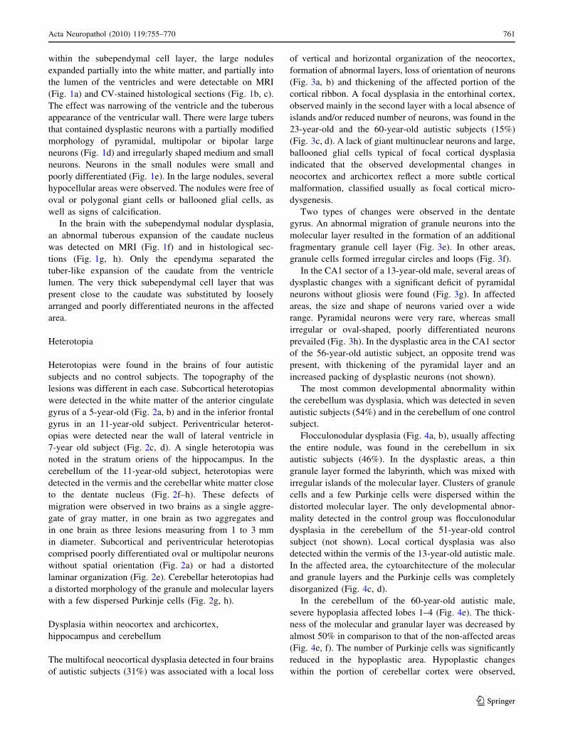
within the subependymal cell layer, the large nodules
expanded partially into the white matter, and partially into
the lumen of the ventricles and were detectable on MRI
(Fig. 1a) and CV-stained histological sections (Fig. 1b, c).
The effect was narrowing of the ventricle and the tuberous
appearance of the ventricular wall. There were large tubers
that contained dysplastic neurons with a partially modified
morphology of pyramidal, multipolar or bipolar large
neurons (Fig. 1d) and irregularly shaped medium and small
neurons. Neurons in the small nodules were small and
poorly differentiated (Fig. 1e). In the large nodules, several
hypocellular areas were observed. The nodules were free of
oval or polygonal giant cells or ballooned glial cells, as
well as signs of calcification.
In the brain with the subependymal nodular dysplasia,
an abnormal tuberous expansion of the caudate nucleus
was detected on MRI (Fig. 1f) and in histological sec-
tions (Fig. 1g, h). Only the ependyma separated the
tuber-like expansion of the caudate from the ventricle
lumen. The very thick subependymal cell layer that was
present close to the caudate was substituted by loosely
arranged and poorly differentiated neurons in the affected
area.
Heterotopia
Heterotopias were found in the brains of four autistic
subjects and no control subjects. The topography of the
lesions was different in each case. Subcortical heterotopias
were detected in the white matter of the anterior cingulate
gyrus of a 5-year-old (Fig. 2a, b) and in the inferior frontal
gyrus in an 11-year-old subject. Periventricular heterot-
opias were detected near the wall of lateral ventricle in
7-year old subject (Fig. 2c, d). A single heterotopia was
noted in the stratum oriens of the hippocampus. In the
cerebellum of the 11-year-old subject, heterotopias were
detected in the vermis and the cerebellar white matter close
to the dentate nucleus (Fig. 2f–h). These defects of
migration were observed in two brains as a single aggre-
gate of gray matter, in one brain as two aggregates and
in one brain as three lesions measuring from 1 to 3 mm
in diameter. Subcortical and periventricular heterotopias
comprised poorly differentiated oval or multipolar neurons
without spatial orientation (Fig. 2a) or had a distorted
laminar organization (Fig. 2e). Cerebellar heterotopias had
a distorted morphology of the granule and molecular layers
with a few dispersed Purkinje cells (Fig. 2g, h).
Dysplasia within neocortex and archicortex,
hippocampus and cerebellum
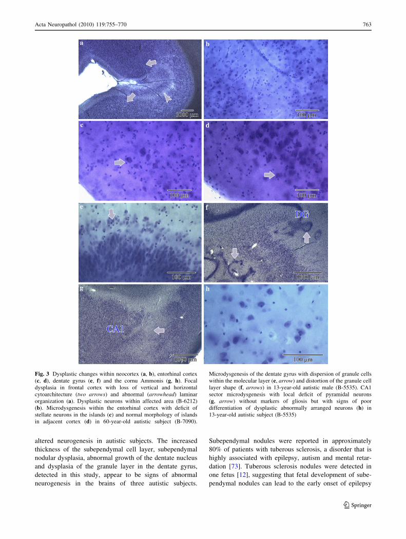
The multifocal neocortical dysplasia detected in four brains
of autistic subjects (31%) was associated with a local loss
of vertical and horizontal organization of the neocortex,
formation of abnormal layers, loss of orientation of neurons
(Fig. 3a, b) and thickening of the affected portion of the
cortical ribbon. A focal dysplasia in the entorhinal cortex,
observed mainly in the second layer with a local absence of
islands and/or reduced number of neurons, was found in the
23-year-old and the 60-year-old autistic subjects (15%)
(Fig. 3c, d). A lack of giant multinuclear neurons and large,
ballooned glial cells typical of focal cortical dysplasia
indicated that the observed developmental changes in
neocortex and archicortex reflect a more subtle cortical
malformation, classified usually as focal cortical micro-
dysgenesis.
Two types of changes were observed in the dentate
gyrus. An abnormal migration of granule neurons into the
molecular layer resulted in the formation of an additional
fragmentary granule cell layer (Fig. 3e). In other areas,
granule cells formed irregular circles and loops (Fig. 3f).
In the CA1 sector of a 13-year-old male, several areas of
dysplastic changes with a significant deficit of pyramidal
neurons without gliosis were found (Fig. 3g). In affected
areas, the size and shape of neurons varied over a wide
range. Pyramidal neurons were very rare, whereas small
irregular or oval-shaped, poorly differentiated neurons
prevailed (Fig. 3h). In the dysplastic area in the CA1 sector
of the 56-year-old autistic subject, an opposite trend was
present, with thickening of the pyramidal layer and an
increased packing of dysplastic neurons (not shown).
The most common developmental abnormality within
the cerebellum was dysplasia, which was detected in seven
autistic subjects (54%) and in the cerebellum of one control
subject.
Flocculonodular dysplasia (Fig. 4a, b), usually affecting
the entire nodule, was found in the cerebellum in six
autistic subjects (46%). In the dysplastic areas, a thin
granule layer formed the labyrinth, which was mixed with
irregular islands of the molecular layer. Clusters of granule
cells and a few Purkinje cells were dispersed within the
distorted molecular layer. The only developmental abnor-
mality detected in the control group was flocculonodular
dysplasia in the cerebellum of the 51-year-old control
subject (not shown). Local cortical dysplasia was also
detected within the vermis of the 13-year-old autistic male.
In the affected area, the cytoarchitecture of the molecular
and granule layers and the Purkinje cells was completely
disorganized (Fig. 4c, d).
In the cerebellum of the 60-year-old autistic male,
severe hypoplasia affected lobes 1–4 (Fig. 4e). The thick-
ness of the molecular and granular layer was decreased by
almost 50% in comparison to that of the non-affected areas
(Fig. 4e, f). The number of Purkinje cells was significantly
reduced in the hypoplastic area. Hypoplastic changes
within the portion of cerebellar cortex were observed,
Acta Neuropathol (2010) 119:755–770 761
123
together with a significantly reduced convolution of the
dentate nucleus (Fig. 4g).
Discussion
This neuropathological study revealed a broad spectrum of
focal developmental abnormalities and pre- and perinatally
acquired lesions in 92% of the brains of autistic subjects
and striking inter-individual differences in the type and
topography of changes. Evidence that different features of
autism are caused by different genes associated with dif-
ferent brain regions [48] suggests a link between regional
developmental alterations in the brain and different com-
ponents of the autistic phenotype.
Altered neurogenesis in autism
Increased brain mass in autistic children and some autistic
adults [89], increase in the numerical density of neurons
[13, 14], reduced size of neurons [7] and brain structure-
specific delay of neuronal growth [111] indicate alterations
in neuronal and brain growth in autistic individuals. The
subventricular zone of the lateral ventricles [26] and the
dentate gyrus [33] are active sites of neurogenesis in adult
humans. Several of our findings support the hypothesis of
Fig. 2 Large subcortical heterotopia within anterior cingulate gyrus
in a 5-year-old autistic child (UMB-1627) (a) contained dysplastic
neurons without spatial orientation (b). Periventricular heterotopia
near the frontal horn of the lateral ventricle (c MRI, d, e CV-stained
section) shows a structure resembling molecular, granule and
pyramidal layers in a 7-year-old autistic subject (B-6403). MRI (f),low (g) and large (h) magnification of heterotopia (arrow) with
dysplastic granule (G) and molecular layer (M) detected within
cerebellar white matter in an 11-year-old autistic subject (B-5342)
762 Acta Neuropathol (2010) 119:755–770
123
altered neurogenesis in autistic subjects. The increased
thickness of the subependymal cell layer, subependymal
nodular dysplasia, abnormal growth of the dentate nucleus
and dysplasia of the granule layer in the dentate gyrus,
detected in this study, appear to be signs of abnormal
neurogenesis in the brains of three autistic subjects.
Subependymal nodules were reported in approximately
80% of patients with tuberous sclerosis, a disorder that is
highly associated with epilepsy, autism and mental retar-
dation [73]. Tuberous sclerosis nodules were detected in
one fetus [12], suggesting that fetal development of sube-
pendymal nodules can lead to the early onset of epilepsy
Fig. 3 Dysplastic changes within neocortex (a, b), entorhinal cortex
(c, d), dentate gyrus (e, f) and the cornu Ammonis (g, h). Focal
dysplasia in frontal cortex with loss of vertical and horizontal
cytoarchitecture (two arrows) and abnormal (arrowhead) laminar
organization (a). Dysplastic neurons within affected area (B-6212)
(b). Microdysgenesis within the entorhinal cortex with deficit of
stellate neurons in the islands (c) and normal morphology of islands
in adjacent cortex (d) in 60-year-old autistic subject (B-7090).
Microdysgenesis of the dentate gyrus with dispersion of granule cells
within the molecular layer (e, arrow) and distortion of the granule cell
layer shape (f, arrows) in 13-year-old autistic male (B-5535). CA1
sector microdysgenesis with local deficit of pyramidal neurons
(g, arrow) without markers of gliosis but with signs of poor
differentiation of dysplastic abnormally arranged neurons (h) in
13-year-old autistic subject (B-5535)
Acta Neuropathol (2010) 119:755–770 763
123

that was diagnosed at the age of 14 months in a neuropa-
thologically examined autistic male. The subependymal
nodules detected in this autistic male’s brain are partially
similar to tubers seen in subjects diagnosed with tuberous
sclerosis [24]. The cause of subependymal nodular dys-
plasia in the examined subject is unknown. In the reported
subjects, bilateral periventricular nodules are linked to
mutations of the filamin A (FLNA) gene located on chro-
mosome Xp28. Filamin A is an actin-crosslinking protein
that is essential for cell locomotion [16], and nodule for-
mation might be related to a defect in cell migration. The
presence of miniature nodules that were built of poorly
differentiated small neurons within the subependymal cell
layer and an increase in nodular size with signs of growth
and differentiation of neurons suggests that neurogenesis,
differentiation and maturation of neurons were in progress
within the subependymal germinal matrix of the 7-year-old
autistic child. This interpretation of subependymal nodule
genesis is consistent with lineage studies demonstrating
that cells in nodules express cellular markers that are
typical for progenitors derived from the subventricular
germinal zone [35, 67]. However, in contrast to the sube-
pendymal nodules seen in subjects with tuberous sclerosis,
in the examined autistic subject, the nodules seen were
small (from 258 to 3,310 lm in diameter), and did not have
the characteristic ovoid or polygonal giant cells, 80–
150 lm in diameter, giant cells with multiple and periph-
erally displaced nuclei [25], or balloon cells, which are
considered the sine qua non histopathological features of
the cortical tubers and subependymal nodules observed in
tuberous sclerosis [73].
The enlarged caudate nucleus detected in the brain of
the 7-year-old autistic subject is consistent with MRI
reports documenting an increased volume of basal ganglia,
including the caudate, in autism [54, 55, 66, 99]. A dis-
proportionate increase of the caudate nucleus volume [66]
suggests that in brains of some autistic individuals,
extended neurogenesis within the subependymal cell layer
may contribute to abnormal growth of the caudate nucleus.
A similar process has been observed in the brains of people
with Huntington disease, showing enhanced neurogenesis
in the subependymal layer and suggesting renewal of the
neuronal population in a degenerating caudate nucleus
[26]. The caudate nucleus is a part of the fronto-striatal
network involved in several functional domains that are
impaired in autism, including lower order repetitive motor
Fig. 4 Flocculonodular
dysplasia in cerebellum of
56-year-old autistic subject
(B-6276) (a) with thin irregular
granule (G) and molecular (M)
layer. b Dysplastic granule layer
(G), ectopic granule cells
(arrow) in the molecular layer,
and loosely dispersed Purkinje
cells (P) (B-6276). Cortical
dysplasia within vermis of 13-
year-old autistic male (c) with
dysplastic granule neurons
mixed with heterotopic (arrow)
large cells (d) (B-5535).
e Severe hypoplasia of
cerebellar lobe 3 and
unmodified lobe 6 (f),respectively, within the
cerebellum of a 60-year-old
autistic male (B-7090). In the
affected region, the thickness of
the hypoplastic molecular and
granule cell layer was reduced
by about 50%. Almost half of
the dentate nucleus (DN) was
less convoluted than the
unaffected part (g)
764 Acta Neuropathol (2010) 119:755–770
123

behavior; intense circumscribed patterns of interests and
higher order rituals and compulsions [41], and defects
in cognitive functions [19, 109], planning and problem-
solving skills [78, 98], short- and long-term memory [40]
and learning [88].
Defective migration in autism
Heterotopia is a sign of altered migration leading to an
abnormal distribution of gray matter nodular masses with
disorganized or rudimentary lamination within the peri-
ventricular area (periventricular heterotopia) or subcortical
white matter (subcortical heterotopia) [2]. In the examined
cohorts, heterotopias were detected in the brains of four
autistic subjects and in the brain of one control subject.
Heterotopias are associated with mutations in the filamin 1
gene (FLNA1) [39, 46] and the chromosome X-linked
DCX gene that codes for doublecortin, a protein expressed
during brain development in migrating neurons, and in
the cortical plate [29, 44, 45], which is involved in the
formation of the microtubules necessary for neuronal
migration [15]. Periventricular nodular heterotopia has
been reported to be associated with pharmaco-resistant
seizures in 80–90% of patients [31]. In the examined
cohort, two periventricular heterotopias were detected in
the brain of a child with subependymal nodular dysplasia
and seizures diagnosed at 14 months of age (B-6403).
Early onset epilepsy, diagnosed at the age of 4.5 months,
might be related to the multiple heterotopias found within
the frontal inferior gyrus, vermis and cerebellar white
matter, coexisting with a focal cortical dysplasia and
dentate gyrus dysplasia (B-5342).
Cortical, hippocampal and cerebellar dysplasia
in autism
The most common form of developmental changes detec-
ted in the examined brains was focal dysplasia, which was
observed in 11 (85%) of the autistic subjects. The mor-
phology of focal dysplasias appears to reflect signs of
abnormal migration, neuronal immaturity and altered cell
arrangement, resulting in focal distortion of cytoarchitec-
ture. In spite of similarities, the dysplastic changes in
the neocortex and archicortex, dentate gyrus and cornu
Ammonis and cerebellum also reveal a brain structure-
specific pattern of dysplastic changes in autism.
Dysplasias encompass a spectrum of changes ranging
from a mild form of cortical disruption, without cellular
abnormalities, to the most severe form with cortical dys-
lamination, with abnormal morphology of neurons and
astrocytes [93, 96, 107]. Focal cortical dysplasias with
giant neurons and balloon cells [107, 113] are histopatho-
logically similar to tubers containing giant cells in tuberous
sclerosis complex [25, 73], suggesting a common patho-
genic basis [113]. However, activation of the mammalian
target of rapamycin (mTOR) pathway observed in the
tuberous sclerosis complex is not present in focal cortical
dysplasia [8, 80]. The giant neurons and ballooned cells,
which are histopathological features of tuberous sclerosis
and focal cortical dysplasia, were absent both in the
subependymal nodules and in the focal cortical dysplasia
observed in the examined autistic cohort. These findings
suggest that in spite of similarities, the pathomechanisms of
developmental alterations are different in the examined
autistic subjects than those in tuberous sclerosis heterot-
opias or focal cortical dysplasia. The development of the
giant neuron- and balloon cell-free dysplasias observed in
the autistic subjects might be related to differences in cause
and/or mechanism. The detection of changes similar to
focal cortical dysplasia in association with prenatal ische-
mia [65] or in shaken infant syndrome [74] may support
these speculations.
Ectopias and dysplastic changes were reported in the
brains of autistic subjects, by several groups [4, 62–64, 91].
Bailey et al. [4] detected olivary dysplasia in the brain of
three of the five autistic subjects, and ectopic neurons
related to the olivary complex in two cases. Moreover, in
the brains of four autistic subjects, cortical dysgenesis was
found. In the brains of the autistic subjects, a strikingly
consistent finding was cingulate cortex disordered lami-
nation [62–64, 100]. A recent study of the cingulate cortex
of nine autistic subjects revealed a developmental malfor-
mation with irregular lamination in three cases, and an
increased number of neurons within the subcortical white
matter in two [100]. Simms et al. [100] suggest that the
excessive number of neurons in the subcortical white
matter reflects the lack of proper resolution of the transient
zone in the developing brain of autistic subjects. Studies by
Fatemi et al. [37, 38] link the migration and lamination
defects to a striking reduction of reelin (by 40%) and Bcl-2
(by 34–51%) in the brains of autistic subjects. Our studies
along with others’ suggest that in the majority of autistic
subjects, heterotopias and dysplastic changes are the local
sign of general developmental defects of migration with a
broad spectrum of topographic, morphological, and func-
tional outcomes.
In the examined brains of autistic subjects, signs of
neuronal immaturity were a common finding. Failure of
maturation of neuronal precursors caused by altered
expression of cytoskeletal proteins and loss of neuronal
polarity results in defects in migration to the destined layer
and in incorrect vertical and horizontal orientation [93].
The immaturity of dysplastic neurons is reflected in the
expression of a variety of proteins and mRNA that are not
present in mature neurons an altered expression of devel-
opmentally regulated cytoskeletal elements [3, 23, 61, 76],
Acta Neuropathol (2010) 119:755–770 765
123

which are known to be crucial for dendrite arborization,
spine formation, axon outgrowth and maintenance of cell
size and shape. Reduced cell size, dendritic arborization
and spine expression are characteristic of dysplastic neu-
rons [6, 93]. Cortical dysplasias are the most epileptogenic
lesions of the brain [107] and are observed in up to 25% of
all epileptic surgeries [102]. More subtle cortical malfor-
mations or dysgenesis encountered in adults with epilepsy
may lack the histological criteria for focal cortical dys-
plasia. They have been described as mild cortical dysplasia
or microdysgenesis [77].
Microdysgenesis within the entorhinal cortex of the 23-
and the 60-year-old autistic subjects in the examined cohort
is unique because the selective deficit of neurons was
limited almost exclusively to the stellate neurons in the
second layer. It is possible that the observed dysgenesis is a
result of defective migration of neurons to their intended
destinations. The presence of a thicker molecular layer and
the deeper location of islands in the entorhinal cortex of
subjects with schizophrenia were previously interpreted as
evidence that the stellate neurons do not reach their des-
tinations during development, probably due to abnormal
migration [36, 57]. Studies indicating the involvement of
reelin and Bcl2 genes in the pathogenesis of schizophrenia
[37, 47, 60] and the reduced expression of reelin and Bcl2
in people with autism suggest that these two genes play a
role in abnormal brain development and contribute to the
structural and functional anomalies seen in autism and
schizophrenia [37].
The distortion of dentate gyrus development detected in
two autistic subjects was reflected in granule cell migration
into the molecular layer and formation of an additional
granule cell layer. Distortion of the shape of the dentate
granule cell layer with the formation of irregular circles
and loops appears to be another piece of evidence sug-
gesting abnormal neuronal migration and networking.
Numerous factors up-regulate neurogenesis in the hippo-
campus [32], including seizures [70, 71], antidepressant
drugs [59, 72] and lithium [18]. Several areas of dysplastic
changes with significant deficits of pyramidal neurons were
found in the CA1 sector in three autistic subjects, but
thickening of the pyramidal layer and an increased packing
of dysplastic neurons in the CA1 sector of the 56-year-old
subject suggests a diversity of CA dysplasia patterns in
autism. The lack of gliosis indicates that the observed
pathology is a sign of microdysgenesis rather than an effect
of hypoxic neuronal loss. A significant deficit of mature
pyramidal neurons and the presence of small irregular or
poorly differentiated oval neurons suggest the defect of
neuronal maturation in autism.
We report a spectrum of focal developmental changes
seen in the cerebellum of eight autistic subjects, including
nodular (lobe X) [97] dysplasia in the cerebellum in five,
vermal dysplasia in one, severe focal hypoplasia in one,
and heterotopias in one other subject. The presence of
heterotopias only in one control subject is evidence of a
strong tendency for focal developmental changes of cere-
bellar microarchitecture that were present in 61% of the
autistic subjects. Flocculonodular dysplasia affecting
almost the entire lobe indicates that mechanisms leading to
focal dysplasia, which were present in five (38%) of the
autistic subjects, show extremely strong topographic pre-
dilection. The observed focal dysplasia was associated with
profound local disorganization of granule cells, Purkinje
cells and molecular layers limited to a small cerebellar
compartment receiving major projections from the vestib-
ular complex involved in the oculomotor and postural
system. Similar cerebellar dysplastic changes classified as
heterotaxias (clusters of poorly organized mixed cells)
were identified in 14% of normal infants but in 83% of
infants with trisomy of different chromosomes [92]. The
presence within the dysplastic nodule of both GABAergic
Purkinje cells produced from the cerebellar ventricular
zone, and the glutamatergic granule neurons produced from
the rhombic lip, and the preservation of the cytoarchitec-
ture in the adjacent cerebellar folia suggest that the final
steps of migration and networking are disturbed mainly or
exclusively in the nodule of the majority of autistic sub-
jects. The characteristic feature distinguishing lobule X
from the other lobules is the abundance of the transcription
factor Tbr2 positive unipolar brush cells (UBCs) [30, 34],
which amplify inputs from vestibular ganglia and nuclei,
by spreading and prolonging excitation within the internal
granular layer [84]. Abnormal networking of Purkinje cells,
granule neurons, and UBCs may contribute to altered
cerebellar coordination of locomotion and motor learning
and planning, as well as of higher cognitive processing
[58]. Flocculonodular dysplasia appears to be another sign
of the mosaic of local developmental defects, most likely
predetermined by the spatial patterning of germinal zones
in developing rhombic lip [110], and coexisting with more
general developmental defects resulting in the accelerated
growth of the brain in early childhood [89], minicolumn
pathology [13, 14], reduced neuron volume [7, 108, 111],
and desynchronized neuronal growth in many brain regions
[111] observed in autism.
Identification of sub-groups with signs of hyperplasia,
hypoplasia and normal-sized cerebellum [95] reflects the
heterogeneity of the autistic population. Piven et al. [87]
reported that cerebellar volume correlates with an
increased total brain volume. In the majority of autistic
subjects, reduced size of the cerebellar hemisphere is
observed [42, 82], but this trend is not detectable in cohorts
of high-functioning autistic individuals [56]. Regional
hypoplasia affects the vermis in autistic individuals rela-
tively often [20, 22, 52] and may be associated with the
766 Acta Neuropathol (2010) 119:755–770
123
deficits in attention-orienting [49, 104], stereotypic
behavior and reduced exploration observed in autism [86].
In the examined autistic cohort, selective and severe
hypoplasia of lobes 1–4 associated with hypoconvolution
of a large portion of the dentate nucleus appears to corre-
spond to clinically detected defects of movement
coordination. These findings suggest that differences in the
type, topography and severity of cerebellar developmental
defects may contribute to different clinical manifestations.
In the 4–7-year-old autistic children examined in this
study, the volume of the Purkinje cells was 38% smaller
than that of the age-matched control group [111]. More-
over, it has been reported that Purkinje cells of the autistic
subjects revealed a 40% decrease in the expression of
glutamic acid decarboxylase 67 (GAD67) mRNA [114]. In
autism, the basket cells provide an increased GABAergic
feed-forward inhibition to Purkinje cells. The result could
be disruption in the timing of Purkinje cell firings and
altered inhibition of the cerebellar nuclei, which could
directly affect cerebello-cortical output and contribute to
the changes in motor behavior and cognition observed in
autism [115]. These findings and the reduced volume (by
26%) of the neurons of the dentate nucleus seen in the 4–7-
year-old autistic children [111] suggest that in autism,
interactions between the Purkinje cells and dentate nucleus
are modified on the structural, molecular and functional
levels.
The (a) detected changes within the subependymal cell
layer with subependymal nodular dysplasia, (b) subcortical
and periventricular heterotopias and (c) neocortex, archi-
cortex, dentate gyrus, cornu Ammonis and cerebellar
dysplasia reflect focal modification of neurogenesis,
migration and alterations of the cytoarchitecture of brain
cortex, subcortical structures and cerebellum in autism.
Detection of dysplastic changes only in one control brain
and of the broad spectrum of focal developmental altera-
tions in the brains of 92% of the autistic subjects indicates
that focal changes are a reflection of global developmental
abnormalities and that regional changes may have their
own contribution to the clinical heterogeneity of autism.
Acknowledgments This study was supported in part by funds from
the New York State Office of Mental Retardation and Developmental
Disabilities, a grant from the Department of Defense Autism Spec-
trum Disorders Research Program (AS073234, Program Project; J.W.,
T.W., A.C.), a grant from Autism Speaks (Princeton, NJ), and grant
R01 HD43960 (J.W.) from the National Institutes of Health, National
Institute of Child Health and Human Development. Tissue and clin-
ical records’ acquisition was coordinated by the Autism Tissue
Program (Princeton, NJ; Directors: Jane Pickett, Ph.D. and Daniel
Lightfoot, Ph.D.). The tissue was obtained from the Harvard Brain
Tissue Resource Center, Belmont, MA, supported in part by PHS
grant number R24-MH 068855; the National Institute of Child Health
and Human Development Brain and Tissue Bank for Developmental
Disorders at the University of Maryland, Baltimore; and the Brain
Bank for Developmental Disabilities and Aging of the New York
State Institute for Basic Research in Developmental Disabilities,
Staten Island, NY. We thank Drs. Helmut Hainsen and Christoph
Schmitz for help in implementation of the celloidin protocol, and Mrs.
Jadwiga Wegiel, Cathy Wang and En Wu Zhang for histology. We
are deeply indebted to the families of the tissue donors who have
made this study possible.
Open Access This article is distributed under the terms of the
Creative Commons Attribution Noncommercial License which per-
mits any noncommercial use, distribution, and reproduction in any
medium, provided the original author(s) and source are credited.
References
1. American Psychiatric Association (2000) Diagnostic and sta-
tistical manual of mental disorders DSM-IV-TR. American
Psychiatric Association, Washington, DC
2. Andrade DM (2009) Genetic basis in epilepsies caused by
malformations of cortical development and in those with
structurally normal brain. Hum Genet 126:173–193
3. Avila J, Dominguez J, Diaz-Nido J (1994) Regulation of
microtubule dynamics by microtubule-associated protein
expression and phosphorylation during neuronal development.
Int J Dev Biol 38:13–25
4. Bailey AP, Luthert A, Dean B et al (1998) A clinicopathological
study of autism. Brain 121:889–905
5. Bailey AR, Giunta BN, Obregon D et al (2008) Peripheral
biomarkers in autism: secreted amyloid precursor protein-alpha
as a probable key player in early diagnosis. Int J Clin Exp Med
1:338–344
6. Barth PG (1987) Disorders of neuronal migration. Can J Neurol
Sci 14:1–16
7. Bauman ML, Kemper TL (1985) Histoanatomic observations of
the brain in early infantile autism. Neurology 35:866–867
8. Baybis M, Yu J, Lee A, Golden JA et al (2004) mTOR cascade
activation distinguishes tubers from focal cortical dysplasia.
Ann Neurol 56:478–487
9. Bobinski M, de Leon MJ, Convit A et al (1999) MRI of en-
torhinal cortex in mild Alzheimer’s disease. Lancet 353:38–40
10. Boddaert N, Zilbovicius M, Philipe A et al (2009) MRI findings
in 77 children with non-syndromic autistic disorder. PLoS One
4:e4415
11. Bruce S, Nyberg F, Melen E et al (2009) The protective effect of
farm animal exposure on childhood allergy is modified by
NPSR1 polymorphisms. J Med Genet 46:159–167
12. Carlson BA, Houser OW, Gomez MR (1999) Brain imaging
in the tuberous sclerosis complex. In: Gomez M, Sampson J,
Whittemore V (eds) Tuberous sclerosis complex, 3rd edn.
Oxford University Press, New York, pp 85–93
13. Casanova MF, Buxhoeveden DP, Switala AE, Roy E (2002)
Minicolumnar pathology in autism. Neurology 58:428–432
14. Casanova MF, van Kooten IAE, Switala EH et al (2006)
Minicolumnar abnormalities in autism. Acta Neuropathol
112:287–303
15. Caspi M, Atlas R, Kantor A, Sapir T, Reiner O (2000) Inter-
action between LIS1 and doublecortin, two lissencephaly gene
products. Hum Mol Genet 9:2205–2213
16. Chang BS, Walsh CA (2009) The genetic basis of human
cerebral cortical malformations. In: Runge MS, Patterson C
(eds) Principles of molecular medicine. Human Press Inc,
Totowa, NJ, pp 1073–1079
17. Chauhan A, Chauhan V (2006) Oxidative stress in autism.
Pathophysiology 13:171–181
Acta Neuropathol (2010) 119:755–770 767
123
18. Chen G, Rajkowska G, Du F, Seraji-Bozorgzad N, Manji NH
(2000) Enhancement of hippocampal neurogenesis by lithium.
J Neurochem 75:1729–1734
19. Chow TW, Cummings JL (1999) Frontal-subcortical circuits. In:
Miller BL, Cummings JL (eds) The human frontal lobes: func-
tions and disorders. Guilford Press, New York, pp 3–26
20. Ciesielski KT, Harris RJ, Hart BL, Pabst H (1997) Cerebellar
hypoplasia and frontal lobe cognitive deficits in disorders of
early childhood. Neuropsychologia 35:643–655
21. Courchesne E, Hesselink JR, Jernigan TL, Yeung-Courchesne R
(1987) Abnormal neuroanatomy in a nonretarded person with
autism. Unusual findings with magnetic resonance imaging.
Arch Neurol 44:335–341
22. Courchesne E, Yeung-Courchesne R, Press GA, Hesselink JR,
Jernigan TL (1988) Hypoplasia of cerebellar vermal lobules VI
and VII in autism. N Engl J Med 318:1349–1354
23. Crino PB, Trojanowski JQ, Eberwine J (1997) Internexin,
MAP1B, and nestin in cortical dysplasia as markers of devel-
opmental maturity. Acta Neuropathol 93:619–627
24. Crino PB, Henske EP (1999) New developments in the neuro-
biology of the tuberous sclerosis complex. Neurology 53:1384–
1390
25. Crino PB, Miyata H, Vinters HV (2002) Neurodevelopmental
disorders as a cause of seizures: neuropathologic, genetic, and
mechanistic considerations. Brain Pathol 12:212–233
26. Curtis MA, Penney EB, Pearson J, Dragunow M, Connor B,
Faull RL (2005) The distribution of progenitor cells in the
subependymal layer of the lateral ventricle in the normal and
Huntington’s disease human brain. Neuroscience 132:777–788
27. Damasio H, Maurer RG, Damasio AR, Chui HC (1980) Com-
puterized tomographic scan findings in patients with autistic
behavior. Arch Neurol 37:504–510
28. Department of Health and Human Services (2007) Morbidity
and mortality weekly report. In: Department of Health and
Human Services, Centers for Disease Control and Prevention,
pp 1–28
29. des Portes V, Francis F, Pinard JM et al (1998) Doublecortin is
the major gene causing X-linked subcortical laminar heterotopia
(SCLH). Hum Mol Genet 7:1063–1070
30. Dino MR, Willard FH, Mugnaini E (1999) Distribution of uni-
polar brush cells and other calretinin immunoreactive
components in the mammalian cerebellar cortex. J Neurocytol
28:99–123
31. Dubeau F, Tampieri D, Lee N et al (1995) Periventricular and
subcortical nodular heterotopia: a study of 33 patients. Brain
118:1273–1287
32. Duman RS, Nakagawa S, Malberg J (2001) Regulation of adult
neurogenesis by antidepressant treatment. Neuropsychophar-
macology 25:836–844
33. Eriksson PS, Perfilieva E, Bjork-Eriksson T et al (1998) Neu-
rogenesis in the adult human hippocampus. Nat Med 4:1313–
1317
34. Englund C, Kowalczyk T, Daza RAM et al (2006) Unipolar
brush cells of the cerebellum are produced in the rhombic lip
and migrate through developing white matter. J Neurosci
26:9184–9195
35. Ess KC, Kamp CA, Tu BP, Gutmann DH (2005) Developmental
origin of subependymal giant cell astrocytoma in tuberous
sclerosis complex. Neurology 64:1446–1449
36. Falkai P, Schneider-Axmann T, Honer WG (2000) Entorhinal
cortex pre-alpha cell clusters in schizophrenia: quantitative
evidence of a developmental abnormality. Biol Psychiatry
47:937–943
37. Fatemi SH, Kroll JL, Stary JM (2001) Altered levels of Reelin
and its isoforms in schizophrenia and mood disorders. Neuro-
report 12:3209–3215
38. Fatemi SH, Stary JM, Halth AR, Realmuto GR (2001) Dys-
regulation of reelin and Bcl-2 proteins in autistic cerebellum.
J Autism Dev Disord 31:529–535
39. Fox JW, Lamperti ED, Eksioglu YZ (1998) Mutations in filamin
1 prevent migration of cerebral cortical neurons in human
periventricular heterotopia. Neuron 21:1315–1325
40. Fuh JL, Wang SJ (1995) Caudate hemorrhage: clinical features,
neuropsychological assessments and radiological findings. Clin
Neurol Neurosurg 97:296–299
41. Gabriels RL, Cuccaro ML, Hill DE, Ivers BJ, Goldson E (2005)
Repetitive behaviors in autism: relationships with associated
clinical features. Res Dev Disabil 26:169–181
42. Gaffney GR, Tsai LY, Kuperman S, Minchin S (1987) Cere-
bellar structure in autism. Am J Dis Child 141:1330–1332
43. Gillberg C, Coleman M (1996) Autism and medical disorders:
a review of the literature. Dev Med Child Neurol 38:191–202
44. Gleeson JG, Allen KM, Fox JW (1998) Doublecortin, a brain-
specific gene mutated in human X-linked lissencephaly and
double cortex syndrome, encodes a putative signaling protein.
Cell 92:63–72
45. Gleeson JG, Lin PT, Flanagan LA, Walsh CA (1999) Double-
cortin is a microtubule-associated protein and is expressed
widely by migrating neurons. Neuron 23:257–271
46. Gorlin JB, Henske E, Warren ST (1993) Actin-binding protein
(ABP-280) filamin gene (FLN) maps telomeric to the color
vision locus (R/GCP) and centromeric to G6PD in Xq28.
Genomics 17:496–498
47. Guidotti A, Auta J, Davis JM (2000) Decrease in reelin and
glutamic acid decarboxylase67 (GAD67) expression in schizo-
phrenia and bipolar disorder: a postmortem brain study. Arch
Gen Psychiatry 57:1061–1069
48. Happe F, Ronald A, Plomin R (2006) Time to give up on a
single explanation for autism. Nat Neurosci 9:1218–1220
49. Harris NS, Courchesne E, Townsend J, Carper RA, Lord C
(1999) Neuroanatomic contributions to slowed orienting of
attention in children with autism. Brain Res Cogn Brain Res
8:61–71
50. Hashimoto T, Tayama M, Mori K, Fujino K, Miyazaki M,
Kuroda Y (1989) Magnetic resonance imaging in autism: pre-
liminary report. Neuropediatrics 20:142–146
51. Hashimoto T, Tayama M, Miyazaki M, Murakawa K, Kuroda Y
(1993) Brainstem and cerebellar vermis involvement in autistic
children. J Child Neurol 8:149–153
52. Hashimoto T, Tayama M, Murakawa K et al (1995) Develop-
ment of the brainstem and cerebellum in autistic patients.
J Autism Dev Disord 25:1–18
53. Heinsen H, Arzberger T, Schmitz C (2000) Celloidin mounting
(embedding without infiltration)—a new, simple and reliable
method for producing serial sections of high thickness through
complete human brains and its application to stereological and
immunohistochemical investigations. J Chem Neuroanat 20:49–
59
54. Herbert MR, Ziegler DA, Deutsch CK (2003) Dissociations of
cerebral cortex, subcortical and cerebral white matter volumes
in autistic boys. Brain 126:1182–1192
55. Hollander E, Anagnostou E, Chaplin W (2005) Striatal volume
on magnetic resonance imaging and repetitive behaviors in
autism. Biol Psychiatry 58:226–232
56. Holttum JR, Minshew NJ, Sanders RS, Phillips NE (1992)
Magnetic resonance imaging of the posterior fossa in autism.
Biol Psychiatry 32:1091–1101
57. Honer WG, Bassett AS, Falkai P, Beach TG, Lapointe JS (1996)
A case study of temporal lobe development in familial schizo-
phrenia. Psychol Med 26:191–195
58. Ito M (2008) Control of mental activities by internal models in
the cerebellum. Nat Rev Neurosci 9:304–313
768 Acta Neuropathol (2010) 119:755–770
123
59. Jacobs BL, Fornal CA (1999) Activity of serotonergic neurons
in behaving animals. Neuropsychopharmacology 21:9S–15S
60. Jarskog LF, Gilmore JH, Selinger ES, Lieberman JA (2000)
Cortical bcl-2 protein expression and apoptotic regulation in
schizophrenia. Biol Psychiatry 48:641–650
61. Kaplan MP, Chin SS, Fliegner KH, Liem RK (1990) Alpha-
internexin, a novel neuronal intermediate filament protein, pre-
cedes the low molecular weight neurofilament protein (NF-L) in
the developing rat brain. J Neurosci 10:2735–2748
62. Kemper TL (1988) Neuroanatomic studies of dyslexia and
autism. In: Disorders of the developing nervous system:
changing views on their origins, diagnosis, and treatments. Alan
R. Liss Inc, New York, pp 125–154
63. Kemper TL, Bauman ML (1993) The contribution of neuropa-
thologic studies to the understanding of autism. Behav Neurol
11:175–187
64. Kemper TL, Bauman M (1998) Neuropathology of infantile
autism. J Neuropathol Exp Neurol 57:645–652
65. Kremer S, De Saint MA, Minotti L et al (2002) Focal cortical
dysplasia possibly related to a probable prenatal ischemic injury.
J Neuroradiol 29:200–203
66. Langen M, Durston S, Staal WG, Palmen SJ, van Engelan H
(2007) Caudate nucleus is enlarged in high-functioning med-
ication-naive subjects with autism. Biol Psychiatry 62:262–
266
67. Lee A, Maldonado M, Baybis M et al (2003) Markers of cellular
proliferation are expressed in cortical tubers. Ann Neurol
53:668–673
68. Lopez-Hurtado E, Prieto JJ (2008) A microscopic study of
language-related cortex in autism. Am J Biochem Biotechnol
4:130–145
69. Lord C, Risi S, Lambrecht L et al (2000) The autism diagnostic
observation schedule-generic: a standard measure of social and
communication deficits associated with the spectrum of autism.
J Autism Dev Disord 30:205–223
70. Madsen TM, Treschow A, Bengzon J, Bolwig TG, Lindvall O,
Tingstrom A (2000) Increased neurogenesis in a model of
electroconvulsive therapy. Biol Psychiatry 47:1043–1049
71. Malberg JE, Eisch AJ, Nestler EJ, Duman RS (2000) Chronic
antidepressant treatment increases neurogenesis in adult rat
hippocampus. J Neurosci 20:9104–9110
72. Manev H, Uz T, Smalheiser NR, Manev R (2001) Antidepres-
sants alter cell proliferation in the adult brain in vivo and in
neural cultures in vitro. Eur J Pharmacol 411:67–70
73. Marcotte L, Crino PB (2006) The neurobiology of the tuberous
sclerosis complex. Neuromol Med 8:531–546
74. Marin-Padilla M, Parisi JE, Armstrong DL, Sargent SK, Kaplan
JA (2002) Shaken infant syndrome: developmental neuropa-
thology, progressive cortical dysplasia, and epilepsy. Acta
Neuropathol 103:321–332
75. Marshall CR, Noor A, Vincent JB et al (2008) Structural vari-
ation of chromosomes in autism spectrum disorder. Am J
Human Gen 82:477–488
76. Matus A (1988) Microtubule-associated proteins: their potential
role in determining neuronal morphology. Ann Rev Neurosci
11:29–44
77. Meencke HJ, Janz D (1985) The significance of microdysge-
nesia in primary generalized epilepsy: an answer to the
considerations of Lyon and Gastaut. Epilepsia 26:368–371
78. Mendez MF, Adams NL, Lewandowski KS (1989) Neurobe-
havioral changes associated with caudate lesions. Neurology
39:349–354
79. Miles JH, Takahashi TN, Bagby S et al (2005) Essential versus
complex autism: definition of fundamental prognostic subtypes.
Am J Med Genet A 135:171–180
80. Miyata H, Chiang AC, Vinters H (2004) Insulin signaling
pathways in cortical dysplasia and TSC-tubers: tissue micro-
array analysis. Ann Neurol 56:510–519
81. Muller RA (2007) The study of autism as a distributed disorder.
Ment Retard Dev Disabil Res Rev 13:85–95
82. Murakami W, Courchesne E, Press GA, Yeung-Courchesne R,
Hesselink JR (1989) Reduced cerebellar hemisphere size and its
relationship to vermal hypoplasia in autism. Arch Neurol
46:689–694
83. Newschaffer CJ, Fallin D, Lee NL (2002) Heritable and non-
heritable risk factors for autism spectrum disorders. Epidemiol
Rev 24:137–153
84. Nunzi MG, Birnstiel S, Bhattacharyya BJ, Slater NT, Mugnaini
E (2001) Unipolar brush cells form a glutamatergic projection
system within the mouse cerebellar cortex. J Comp Neurol
434:329–341
85. Palmen SJ, van Engelan H, Hof PR, Schmitz C (2004) Neuro-
pathological findings in autism. Brain 127:2572–2583
86. Pierce K, Courchesne E (2001) Evidence for a cerebellar role in
reduced exploration and stereotyped behavior in autism. Biol
Psychiatry 49:655–664
87. Piven J, Saliba K, Bailey J, Arndt S (1997) An MRI study of
autism: the cerebellum revisited. Neurology 49:546–551
88. Poldrack RA, Prabhakaran V, Seger CA, Gabrieli JD (1999)
Striatal activation during acquisition of a cognitive skill. Neu-
ropsychology 13:564–574
89. Redcay E, Courchesne E (2005) When is the brain enlarged in
autism? A meta-analysis of all brain size reports. Biol Psychiatry
58:1–9
90. Ritvo ER, Freeman BJ, Scheibel AB et al (1986) Lower Purkinje
cell counts in the cerebella of four autistic subjects: initial
findings of the UCLA-NSAC Autopsy Research Report. Am J
Psychiatry 143:862–866
91. Rodier PM, Ingram JL, Tisdale B, Nelson S, Romano J (1996)
Embryological origin of autism: developmental abnormalities of
the cranial nerve nuclei. J Comp Neurol 370:247–261
92. Rorke LB, Fogelson MH, Riggs HE (1968) Cerebellar hetero-
topia in infancy. Dev Med Child Neurol 10:644–650
93. Rorke LB (1994) A perspective: the role of disordered genetic
control of neurogenesis in the pathogenesis of migration disor-
ders. J Neuropathol Exp Neurol 53:105–117
94. Rutter M, Bailey A, Bolton P, Le Couteur A (1994) Autism and
known medical conditions: myth and substance. J Child Psychol
Psychiatry 35:311–322
95. Saitoh O, Courchesne E (1998) Magnetic resonance imaging
study of the brain in autism. Psychiatry Clin Neurosci 52(Sup-
pl):S219–S222
96. Sarnat HB, Benjamin DR, Siebert JR et al (1992) Cerebral
dysgenesis: embryology and clinical expression. Cell 69:581–
595
97. Schmahmann JD, Doyon J, McDonald D et al (1999) Three-
dimensional MRI atlas of the human cerebellum in proportional
stereotaxic space. Neuroimage 10:233–260
98. Schmidtke K, Manner H, Kaufmann R, Schmolck H (2002)
Cognitive procedural learning in patients with fronto-striatal
lesions. Learn Mem 9:419–429
99. Sears LL, Vest C, Mohamed S, Bailey J, Ranson BJ, Piven J
(1999) An MRI study of the basal ganglia in autism. Prog
Neuropsychopharmacol Biol Psychiatry 23:613–624
100. Simms ML, Kemper TL, Timbie CM, Bauman ML, Blatt GJ
(2009) The anterior cingulate cortex in autism: heterogeneity of
qualitative and quantitative cytoarchitectonic features suggests
possible subgroups. Acta Neuropathol 118:673–684
101. Sokol DK, Chen D, Farlow MR et al (2006) High levels of
Alzheimer beta-amyloid precursor protein (APP) in children
Acta Neuropathol (2010) 119:755–770 769
123
with severely autistic behavior and aggression. J Child Neurol
21:444–449
102. Tassi L, Colombo N, Garbelli R et al (2002) Focal cortical
dysplasia: neuropathological subtypes, EEG, neuroimaging and
surgical outcome. Brain 125:1719–1732
103. The Autism Genome Project Consortium (2007) Mapping autism
risk loci using genetic linkage and chromosomal rearrangements.
Nat Genet 39:319–328
104. Townsend J, Courchesne E, Covington J et al (1999) Spatial
attention deficits in patients with acquired or developmental
cerebellar abnormality. J Neurosci 19:5632–5643
105. Tuchman RF, Rapin I, Shinnar S (1991) Autistic and dysphasic
children. I. Clinical characteristics. Pediatrics 88:1211–1218
106. Tuchman RF, Rapin I (2002) Epilepsy in autism. Lancet Neurol
1:352–358
107. Usui N, Matsuda K, Mihara T et al (2001) MRI of cortical
dysplasia-correlation with pathological findings. Neuroradiology
43:830–837
108. van Kooten I, Palmen SJ, von Engelan CP et al (2008) Neurons
in the fusiform gyrus are fewer and smaller in autism. Brain
131:987–999
109. Voelbel GT, Bates ME, Buckman JF, Pandina G, Hendren RL
(2006) Caudate nucleus volume and cognitive performance: are
they related in childhood psychopathology? Biol Psychiatry
60:942–950
110. Volkmann K, Rieger S, Babaryka A, Koster RW (2008) The
zebrafish cerebellar rhombic lip is spatially patterned in pro-
ducing granule cell populations of different functional
compartments. Dev Biol 313:167–180
111. Wegiel J, Wisniewski T, Chauhan A (2010) Type, topography
and sequelae of neuropathological changes shaping clinical
phenotype of autism. In: Chauhan A, Chauhan V, Brown WT
et al (eds) Autism: oxidative stress, inflammation, and immune
abnormalities. Taylor & Francis/CRC Press, Boca Raton, FL, pp
1–34
112. Weiss LA, Arking DE, The Gene Discovery Project of Johns
Hopkins & the Autism Consortium (2009) A genome-wide
linkage and association scan reveals novel loci for autism.
Nature 461:802–811
113. Wolf HK, Normann S, Green AJ et al (1997) Tuberous sclerosis-
like lesions in epileptogenic human neocortex lack allelic loss at
the TSC1 and TSC2 regions. Acta Neuropathol 93:93–96
114. Yip J, Soghomonian JJ, Blatt GJ (2007) Decreased GAD67
mRNA levels in cerebellar Purkinje cells in autism: patho-
physiological implications. Acta Neuropathol 113:559–568
115. Yip JJ, Soghomonian J, Blatt GJ (2008) Increased GAD67
mRNA expression in cerebellar interneurons in autism: impli-
cations for Purkinje cell dysfunction. J Neurosci Res 86:525–
530
770 Acta Neuropathol (2010) 119:755–770
123