Embed Size (px)
Citation preview
Correspondence to
Jonathan M Schott, Reader in
Clinical Neurology, Dementia
Research Centre, UCL Institute of
Neurology, Queen Square,
London WC1N 3BG, UK;
Accepted 10 February 2017
Published Online First
21 February 2017
To cite: Schott JM. Pract
Neurol 2017;17:172–182.
The neurology of ageing: what is
normal?
Jonathan M Schott
ABSTRACT
Ageing is associated with changes in the nervous
system with consequent alterations in some
neurological examination findings: understanding
what is ‘normal’ at different ages is essential
when evaluating patients. In seminal papers
published in 1931, Dr MacDonald Critchley
summarised his observations and the prevailing
evidence on the effects of ageing on, among
others, sensation, reflexes, ocular function,
olfaction, movement and cognition. In this
review, these observations are re-evaluated in
light of contemporary evidence. Factors
influencing the measurement and interpretation
of these clinical findings are then discussed,
including reproducibility, the influence of
comorbidities, secular trends, how ‘normality’
should best be defined, the problems of
extrapolating group data to individuals and the
influence of presymptomatic neurodegenerative
disease states. The case is made that context is
critical, and that combining life course data with
detailed clinical and biomarker phenotyping is
required to understand the determinants of
normal neurological function associated with
ageing.
INTRODUCTION
As with all aspects of human physiology,
the nervous system alters with ageing.
Even in the healthiest elderly, there is
more neuronal loss, more vascular
pathology and numerous changes at the
cellular level compared with healthy
younger adults. These changes have
consequences, and all neurologists will
recognise that when examining a patient,
what can reasonably be determined to be
‘within normal limits’ for a healthy 70-
year-old differs from that for a healthy
20-year-old. Inherent therefore when
evaluating any patient is knowledge of
what can reasonably be considered to be
within the normal range in any given age
group. While there is considerable dogma
about what constitutes ‘normal’ neurolog-
ical ageing — there can be few
neurologists who will not recall being
told that one can disregard absent ankle
jerks or impaired distal vibration sense in
somebody in their 70s or 80s — the
evidence base for such assertions is rather
less clear.
While numerous papers have consid-
ered aspects of neurological ageing in
isolation, relatively few have attempted
an overview. A notable exception are the
three seminal papers in the Lancet in
1931 by Macdonald Critchley,1–3 based
on his Goulstonian Lectures to the Royal
College of Physicians that same year. In
these papers he covered the pathological,
clinical and cognitive changes associated
with ageing.
In Part 1 of this paper, Critchley’s orig-
inal observations are reviewed and
critiqued in light of contemporary
evidence. In Part 2, some factors relevant
to the interpretation of these findings are
discussed, with particular reference to
what might influence what is, or is not,
‘normal’ ageing.
Part 1: Macdonald Critchley revisited
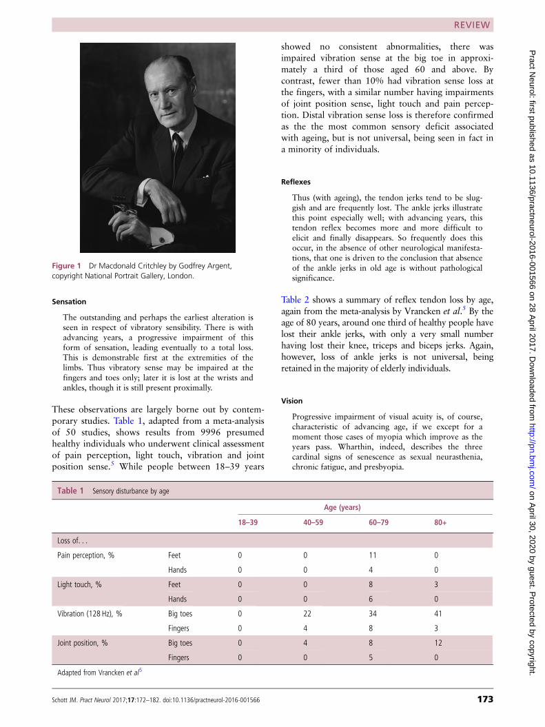
Dr Macdonald Critchley, CBE (1900–
1997) (figure 1), was one of the foremost
neurologists of his era, President of the
World Federation of Neurology, Vice-
President of the Royal College of Physi-
cians and author of over 200 books and
papers including seminal works on the
parietal lobes, aphasia and headache.4 His
prodigious output was based in large part
on his detailed and meticulous observa-
tions of patients. What follows are
selected quotations from some of his
writings on ageing including sensation,
reflexes, vision, hearing, taste and smell,
gait, hypokinetic and hyperkinetic move-
ments and cognition along with a brief
review of contemporary evidence for
each domain.
172 Schott JM. Pract Neurol 2017;17:172–182. doi:10.1136/practneurol-2016-001566
REVIEW
on April 30, 2020 by guest. P
rotected by copyright.http://pn.bm
j.com/
Pract N
eurol: first published as 10.1136/practneurol-2016-001566 on 28 April 2017. D
ownloaded from
Sensation
The outstanding and perhaps the earliest alteration isseen in respect of vibratory sensibility. There is withadvancing years, a progressive impairment of thisform of sensation, leading eventually to a total loss.This is demonstrable first at the extremities of thelimbs. Thus vibratory sense may be impaired at thefingers and toes only; later it is lost at the wrists andankles, though it is still present proximally.
These observations are largely borne out by contem-
porary studies. Table 1, adapted from a meta-analysis
of 50 studies, shows results from 9996 presumed
healthy individuals who underwent clinical assessment
of pain perception, light touch, vibration and joint
position sense.5 While people between 18–39 years
showed no consistent abnormalities, there was
impaired vibration sense at the big toe in approxi-
mately a third of those aged 60 and above. By
contrast, fewer than 10% had vibration sense loss at
the fingers, with a similar number having impairments
of joint position sense, light touch and pain percep-
tion. Distal vibration sense loss is therefore confirmed
as the the most common sensory deficit associated
with ageing, but is not universal, being seen in fact in
a minority of individuals.
Reflexes
Thus (with ageing), the tendon jerks tend to be slug-gish and are frequently lost. The ankle jerks illustratethis point especially well; with advancing years, thistendon reflex becomes more and more difficult toelicit and finally disappears. So frequently does thisoccur, in the absence of other neurological manifesta-tions, that one is driven to the conclusion that absenceof the ankle jerks in old age is without pathologicalsignificance.
Table 2 shows a summary of reflex tendon loss by age,
again from the meta-analysis by Vrancken et al.5 By the
age of 80 years, around one third of healthy people have
lost their ankle jerks, with only a very small number
having lost their knee, triceps and biceps jerks. Again,
however, loss of ankle jerks is not universal, being
retained in the majority of elderly individuals.
Vision
Progressive impairment of visual acuity is, of course,characteristic of advancing age, if we except for amoment those cases of myopia which improve as theyears pass. Wharthin, indeed, describes the threecardinal signs of senescence as sexual neurasthenia,chronic fatigue, and presbyopia.
Figure 1 Dr Macdonald Critchley by Godfrey Argent,
copyright National Portrait Gallery, London.
Table 1 Sensory disturbance by age
Age (years)
18–39 40–59 60–79 80+
Loss of. . .
Pain perception, % Feet 0 0 11 0
Hands 0 0 4 0
Light touch, % Feet 0 0 8 3
Hands 0 0 6 0
Vibration (128 Hz), % Big toes 0 22 34 41
Fingers 0 4 8 3
Joint position, % Big toes 0 4 8 12
Fingers 0 0 5 0
Adapted from Vrancken et al5
Schott JM. Pract Neurol 2017;17:172–182. doi:10.1136/practneurol-2016-001566 173
REVIEW
on April 30, 2020 by guest. P
rotected by copyright.http://pn.bm
j.com/
Pract N
eurol: first published as 10.1136/practneurol-2016-001566 on 28 April 2017. D
ownloaded from
Alterations in the pupillary reactions are important.With advancing years there is a progressive sluggish-ness in the response of the pupils, both to light andon accommodation, and ultimately a condition ofpupillary immobility may occur.
Still more often one sees an absence or impairment ofthe conjugate movements of upwards deviations ofthe eyeballs; lateral and downward movements rarelysuffer. Nystagmus does not occur except where focallesions within the cerebellum happen to be present.
Table 3 provides a summary of the prevalence of ocular
pathology in adults aged 40 years and older in the
USA.6 Blindness occurs in <1% until the age of 80
years, increasing to ~7% thereafter; a much greater
proportion have impaired vision. As Critchley
observed, myopia declines with increasing age, but
non-neurological causes of visual impairment including
cataract (affecting almost two thirds of individuals over
80) and glaucoma (nearly 10%) are clearly age related.
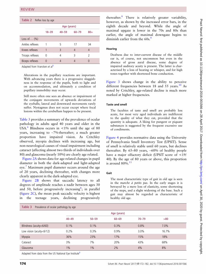
Figure 2A shows data for age-related changes in pupil
diameter in both the dark-adapted and light-adapted
eye.7 Maximum pupil diameter occurs around the age
of 20 years, declining thereafter, with changes more
clearly apparent in the dark-adapted eye.
Figure 2B shows that saccadic latency to all
degrees of amplitude reaches a nadir between ages 30
and 50, before progressively increasing8; in parallel
(figure 2C), the mean peak saccadic velocity is highest
in the teenage years, declining progressively
thereafter.8 There is relatively greater variability,
however, as shown by the increased error bars, in the
eighth decade and beyond. While the angle of
maximal upgaze is lower in the 70s and 80s than
earlier, the angle of maximal downgaze begins to
diminish earlier from the 60s.9
Hearing
Deafness due to inter-current disease of the middleear is, of course, not uncommon but even in theabsence of gross aural disease, some degree ofimpaired auditory acuity is present. The latter is char-acterised by a loss of hearing or whisper, and for hightones together with shortened bone conduction.
Figure 3 shows change in the ability to perceive
different frequencies between 18 and 55 years.10 As
noted by Critchley, age-related decline is much more
marked at higher frequencies.
Taste and smell
The faculties of taste and smell are probably lessacute, for most very aged individuals are indifferentto the quality of what they eat, provided that thequantity is adequate. A liking for pungent or piquantsubstances is suggested by the frequent excessive useof condiments.
Figure 4 provides normative data using the University
of Pennsylvania Smell Inventory Test (UPSIT). Sense
of smell is relatively stable until 60 years, but declines
thereafter. By 65–80 years, ~60% of healthy people
have a major olfactory deficit (UPSIT score of <19/
40). By the age of 80 years or above, this proportion
is around 80%.11
Gait
The most characteristic type of gait in old age is seenin the marche à petits pas. In the early stages it isbetrayed by a mere loss of elasticity, some shorteningof the steps, and a slight widening of the base. Such agait may almost be regarded as characteristic ofhealthy old age.
Table 2 Reflex loss by age
Age (years)
18–39 40–59 60–79 80+
Loss of. . . (%)
Ankles reflexes 1 5 17 34
Knees reflexes 1 3 4 4
Triceps reflexes 0 4 3 2
Biceps reflexes 0 4 4 0
Adapted from Vrancken et al5
Table 3 Prevalence of ocular pathology by age
Age (years)
40–49 50–59 60–69 70–79 >80
Blindness (acuity<6/60) 0.1% 0.1% 0.3% 0.8% 7.0%
Low vision (acuity<6/12) 0.2% 0.3% 0.9% 3.0% 16.7%
Myopia 36% 23% 17% 15% 18%
Cataract 3% 7% 20% 43% 68%
Glaucoma 1% 1% 2% 4% 8%
Adapted from data from the US National Eye Institute6
174 Schott JM. Pract Neurol 2017;17:172–182. doi:10.1136/practneurol-2016-001566
REVIEW
on April 30, 2020 by guest. P
rotected by copyright.http://pn.bm
j.com/
Pract N
eurol: first published as 10.1136/practneurol-2016-001566 on 28 April 2017. D
ownloaded from
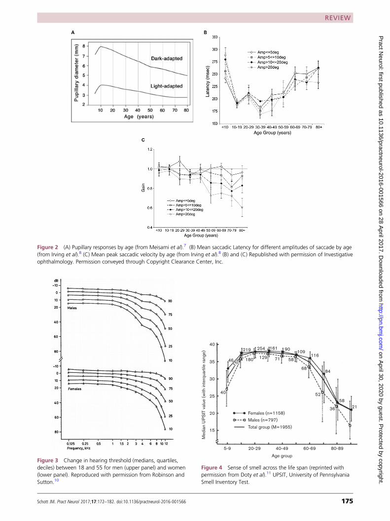
Figure 2 (A) Pupillary responses by age (from Meisami et al).7 (B) Mean saccadic Latency for different amplitudes of saccade by age
(from Irving et al).8 (C) Mean peak saccadic velocity by age (from Irving et al).8 (B) and (C) Republished with permission of Investigative
ophthalmology. Permission conveyed through Copyright Clearance Center, Inc.
Figure 3 Change in hearing threshold (medians, quartiles,
deciles) between 18 and 55 for men (upper panel) and women
(lower panel). Reproduced with permission from Robinson and
Sutton.10
40
35
30
25
Med
ian U
PS
IT v
alue (
with
inte
rquar
tile r
ang
e)
20
15
5-9
40
46155 180 129
219 254 161 90109
116
84
58
2136
52
68
5871
20-29 40-49
Age group
Females (n=1158)
Males (n=797)
Total group (M=1955)
60-69 80-89
Figure 4 Sense of smell across the life span (reprinted with
permission from Doty et al).11 UPSIT, University of Pennsylvania
Smell Inventory Test.
Schott JM. Pract Neurol 2017;17:172–182. doi:10.1136/practneurol-2016-001566 175
REVIEW
on April 30, 2020 by guest. P
rotected by copyright.http://pn.bm
j.com/
Pract N
eurol: first published as 10.1136/practneurol-2016-001566 on 28 April 2017. D
ownloaded from
. . .it must be remembered that an abnormal gait inthis age is frequently the result of disease outside thenervous system.
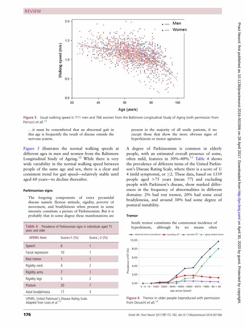
Figure 5 illustrates the normal walking speeds at
different ages in men and women from the Baltimore
Longitudinal Study of Ageing.12 While there is very
wide variability in the normal walking speed between
people of the same age and sex, there is a clear and
consistent trend for gait speed—relatively stable until
aged 60 years—to decline thereafter.
Parkinsonian signs
The forgoing components of extra pyramidaldisease namely flexion attitude, rigidity, poverty ofmovement, and bradykinesis when present in someintensity constitute a picture of Parkinsonism. But it isprobably that in some degree these manifestations are
present in the majority of all senile patients, if weexcept those that show the more obvious signs ofhyperkinesis or motor agitation.
A degree of Parkinsonism is common in elderly
people, with an estimated overall presence of some,
often mild, features in 30%–40%.13 Table 4 shows
the prevalence of different items of the United Parkin-
son’s Disease Rating Scale, where there is a score of 1/
4 (mild symptoms), or �2. These data, based on 1339
people aged >75 years (mean 77) and excluding
people with Parkinson’s disease, show marked differ-
ences in the frequency of abnormalities in different
domains: 2% had rest tremor, 20% had some axial
bradykinesia, and around 30% had some degree of
postural instability.
Tremor
Senile tremor constitutes the commonest incidence ofhyperkinesis, although by no means often
Figure 5 Usual walking speed in 711 men and 766 women from the Baltimore Longitudinal Study of Aging (with permission from
Ferrucci et al).12
Table 4 Prevalence of Parkinsonian signs in individuals aged 75years and older
UPDRS Item Score=1 (%) Score�2 (%)
Speech 6 1
Facial expression 10 1
Rest tremor 1 1
Rigidity neck 4 2
Rigidity arms 7 1
Rigidity legs 5 2
Posture 20 7
Axial bradykinesia 17 3
UPDRS, United Parkinson’s Disease Rating Scale.Adapted from Louis et al.13
observed tremor prevalence
10,00
8,00
6,00
Pre
vale
nce o
f E
T (
%)
4,00
2,00
0,000 - 9 10 - 1920 - 2930 - 3940 - 4950 - 5960 - 6970 - 7980 - 89 > 90
age group (years)
hereditary ET sporadic ET aging-related tremor
Figure 6 Tremor in older people (reproduced with permission
from Deuschl et al).14
176 Schott JM. Pract Neurol 2017;17:172–182. doi:10.1136/practneurol-2016-001566
REVIEW
on April 30, 2020 by guest. P
rotected by copyright.http://pn.bm
j.com/
Pract N
eurol: first published as 10.1136/practneurol-2016-001566 on 28 April 2017. D
ownloaded from
encountered. Its usual rate is 100 beats per minute,the common sites for its occurrence at the head andlower jaw, the hands and forearms.
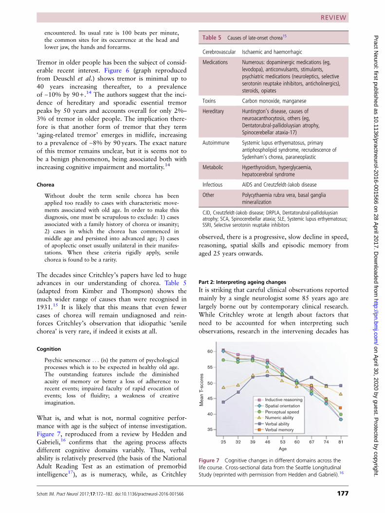
Tremor in older people has been the subject of consid-
erable recent interest. Figure 6 (graph reproduced
from Deuschl et al.) shows tremor is minimal up to
40 years increasing thereafter, to a prevalence
of ~10% by 90+.14 The authors suggest that the inci-
dence of hereditary and sporadic essential tremor
peaks by 50 years and accounts overall for only 2%–
3% of tremor in older people. The implication there-
fore is that another form of tremor that they term
‘aging-related tremor’ emerges in midlife, increasing
to a prevalence of ~8% by 90 years. The exact nature
of this tremor remains unclear, but it is seems not to
be a benign phenomenon, being associated both with
increasing cognitive impairment and mortality.14
Chorea
Without doubt the term senile chorea has beenapplied too readily to cases with characteristic move-ments associated with old age. In order to make thisdiagnosis, one must be scrupulous to exclude: 1) casesassociated with a family history of chorea or insanity;2) cases in which the chorea has commenced inmiddle age and persisted into advanced age; 3) casesof apoplectic onset usually unilateral in their manifes-tations. When these criteria rigidly apply, senilechorea is found to be a rarity.
The decades since Critchley’s papers have led to huge
advances in our understanding of chorea. Table 5
(adapted from Kimber and Thompson) shows the
much wider range of causes than were recognised in
1931.15 It is likely that this means that even fewer
cases of chorea will remain undiagnosed and rein-
forces Critchley’s observation that idiopathic ‘senile
chorea’ is very rare, if indeed it exists at all.
Cognition
Psychic senescence . . . (is) the pattern of psychologicalprocesses which is to be expected in healthy old age.The outstanding features include the diminishedacuity of memory or better a loss of adherence torecent events; impaired faculty of rapid evocation ofevents; loss of fluidity; a weakness of creativeimagination.
What is, and what is not, normal cognitive perfor-
mance with age is the subject of intense investigation.
Figure 7, reproduced from a review by Hedden and
Gabrieli,16 confirms that the ageing process affects
different cognitive domains variably. Thus, verbal
ability is relatively preserved (the basis of the National
Adult Reading Test as an estimation of premorbid
intelligence17), as is numeracy, while, as Critchley
observed, there is a progressive, slow decline in speed,
reasoning, spatial skills and episodic memory from
aged 25 years onwards.
Part 2: Interpreting ageing changes
It is striking that careful clinical observations reported
mainly by a single neurologist some 85 years ago are
largely borne out by contemporary clinical research.
While Critchley wrote at length about factors that
need to be accounted for when interpreting such
observations, research in the intervening decades has
Table 5 Causes of late-onset chorea15
Cerebrovascular Ischaemic and haemorrhagic
Medications Numerous: dopaminergic medications (eg,levodopa), anticonvulsants, stimulants,psychiatric medications (neuroleptics, selectiveserotonin reuptake inhibitors, anticholinergics),steroids, opiates
Toxins Carbon monoxide, manganese
Hereditary Huntington’s disease, causes ofneuroacanthocytosis, others (eg,Dentatorubral-pallidoluysian atrophy,Spinocerebellar ataxia-17)
Autoimmune Systemic lupus erthyematosus, primaryantiphospholipid syndrome, recrudescence ofSydenham’s chorea, paraneoplastic
Metabolic Hyperthyroidism, hyperglycaemia,hepatocerebral syndrome
Infectious AIDS and Creutzfeldt-Jakob disease
Other Polycythaemia rubra vera, basal gangliamineralization
CJD, Creutzfeldt-Jakob disease; DRPLA, Dentatorubral-pallidoluysianatrophy; SCA, Spinocerebellar ataxia; SLE, Systemic lupus erthyematosus;SSRI, Selective serotonin reuptake inhibitors
Inductive reasoning
Spatial orientation
Perceptual speedNumeric ability
Verbal abilityVerbal memory
25
35
40
45
50
Mean
T-s
core
s
55
60
32 39 46 53
Age
60 67 74 81
Figure 7 Cognitive changes in different domains across the
life course. Cross-sectional data from the Seattle Longitudinal
Study (reprinted with permission from Hedden and Gabrieli).16
Schott JM. Pract Neurol 2017;17:172–182. doi:10.1136/practneurol-2016-001566 177
REVIEW
on April 30, 2020 by guest. P
rotected by copyright.http://pn.bm
j.com/
Pract N
eurol: first published as 10.1136/practneurol-2016-001566 on 28 April 2017. D
ownloaded from
provided further insights, some of which are reviewed
below.
Reproducibility
Critical to comparing clinical signs between individuals
is an understanding of how reproducibly they can be
elicited. When comparing findings across the life
course, it is also vital to know if there is a systematic
reason why the method of assessment might be affected
by age per se. In some instances, e.g. with neuropsycho-
logical cognitive testing, standard methodologies allow
for (although not always perfect) between-subject and
within-subject comparisons to be made. In routine clin-
ical practice and for most of the domains discussed
previously, this is not the case and there is often huge
variability in how various tests are elicited and inter-
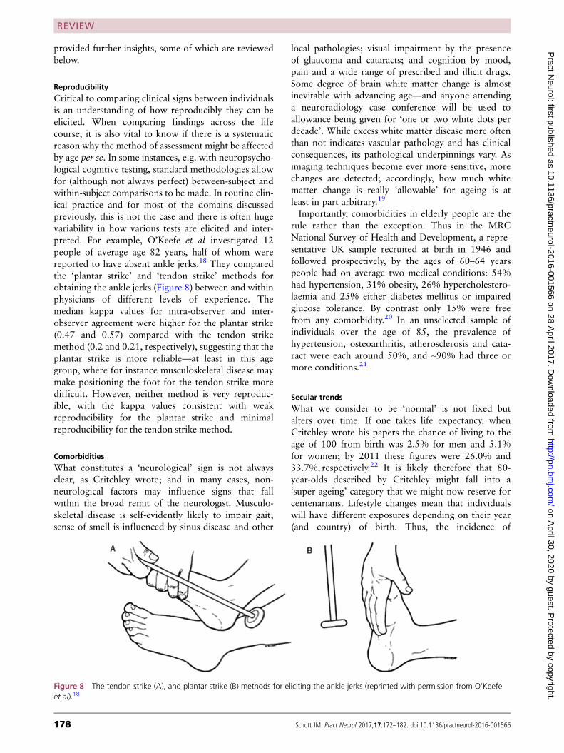
preted. For example, O’Keefe et al investigated 12
people of average age 82 years, half of whom were
reported to have absent ankle jerks.18 They compared
the ‘plantar strike’ and ‘tendon strike’ methods for
obtaining the ankle jerks (Figure 8) between and within
physicians of different levels of experience. The
median kappa values for intra-observer and inter-
observer agreement were higher for the plantar strike
(0.47 and 0.57) compared with the tendon strike
method (0.2 and 0.21, respectively), suggesting that the
plantar strike is more reliable—at least in this age
group, where for instance musculoskeletal disease may
make positioning the foot for the tendon strike more
difficult. However, neither method is very reproduc-
ible, with the kappa values consistent with weak
reproducibility for the plantar strike and minimal
reproducibility for the tendon strike method.
Comorbidities
What constitutes a ‘neurological’ sign is not always
clear, as Critchley wrote; and in many cases, non-
neurological factors may influence signs that fall
within the broad remit of the neurologist. Musculo-
skeletal disease is self-evidently likely to impair gait;
sense of smell is influenced by sinus disease and other
local pathologies; visual impairment by the presence
of glaucoma and cataracts; and cognition by mood,
pain and a wide range of prescribed and illicit drugs.
Some degree of brain white matter change is almost
inevitable with advancing age—and anyone attending
a neuroradiology case conference will be used to
allowance being given for ‘one or two white dots per
decade’. While excess white matter disease more often
than not indicates vascular pathology and has clinical
consequences, its pathological underpinnings vary. As
imaging techniques become ever more sensitive, more
changes are detected; accordingly, how much white
matter change is really ‘allowable’ for ageing is at
least in part arbitrary.19
Importantly, comorbidities in elderly people are the
rule rather than the exception. Thus in the MRC
National Survey of Health and Development, a repre-
sentative UK sample recruited at birth in 1946 and
followed prospectively, by the ages of 60–64 years
people had on average two medical conditions: 54%
had hypertension, 31% obesity, 26% hypercholestero-
laemia and 25% either diabetes mellitus or impaired
glucose tolerance. By contrast only 15% were free
from any comorbidity.20 In an unselected sample of
individuals over the age of 85, the prevalence of
hypertension, osteoarthritis, atherosclerosis and cata-
ract were each around 50%, and ~90% had three or
more conditions.21
Secular trends
What we consider to be ‘normal’ is not fixed but
alters over time. If one takes life expectancy, when
Critchley wrote his papers the chance of living to the
age of 100 from birth was 2.5% for men and 5.1%
for women; by 2011 these figures were 26.0% and
33.7%, respectively.22 It is likely therefore that 80-
year-olds described by Critchley might fall into a
‘super ageing’ category that we might now reserve for
centenarians. Lifestyle changes mean that individuals
will have different exposures depending on their year
(and country) of birth. Thus, the incidence of
Figure 8 The tendon strike (A), and plantar strike (B) methods for eliciting the ankle jerks (reprinted with permission from O’Keefe
et al).18
178 Schott JM. Pract Neurol 2017;17:172–182. doi:10.1136/practneurol-2016-001566
REVIEW
on April 30, 2020 by guest. P
rotected by copyright.http://pn.bm
j.com/
Pract N
eurol: first published as 10.1136/practneurol-2016-001566 on 28 April 2017. D
ownloaded from
smoking has radically declined in recent years in high-
income countries, and there have been major changes
in screening for, and treatment of, hypertension,
hypercholesterolaemia and diabetes mellitus.
Conversely, individuals now in their 70s were brought
up in an age of rationing, compared now to very
readily available high-calorie foods with consequent
rises in obesity.
Defining ‘normality’
How we define normality in the context of ageing is a
debatable question. One approach is to assess those
who are the best performers in their age group. This
approach confirms that there are limits to what can be
expected with age. Figure 9 illustrates the world
record time for running 5 km by age and by sex.12
While the best performing 80-year olds still run this
distance faster than the average runner of any age,
peak performance is still seen between the ages of
20 and 40 years, deteriorating slowly to the age of 80
years and rapidly thereafter.
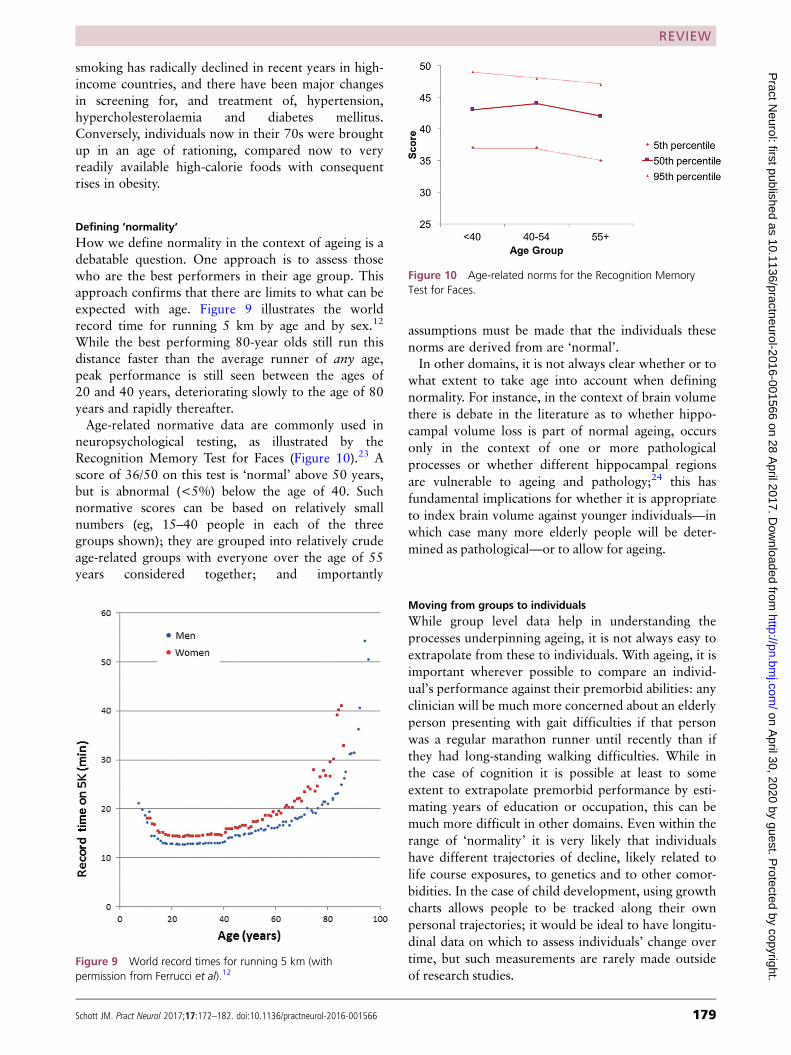
Age-related normative data are commonly used in
neuropsychological testing, as illustrated by the
Recognition Memory Test for Faces (Figure 10).23 A
score of 36/50 on this test is ‘normal’ above 50 years,
but is abnormal (<5%) below the age of 40. Such
normative scores can be based on relatively small
numbers (eg, 15–40 people in each of the three
groups shown); they are grouped into relatively crude
age-related groups with everyone over the age of 55
years considered together; and importantly
assumptions must be made that the individuals these
norms are derived from are ‘normal’.
In other domains, it is not always clear whether or to
what extent to take age into account when defining
normality. For instance, in the context of brain volume
there is debate in the literature as to whether hippo-
campal volume loss is part of normal ageing, occurs
only in the context of one or more pathological
processes or whether different hippocampal regions
are vulnerable to ageing and pathology;24 this has
fundamental implications for whether it is appropriate
to index brain volume against younger individuals—in
which case many more elderly people will be deter-
mined as pathological—or to allow for ageing.
Moving from groups to individuals
While group level data help in understanding the
processes underpinning ageing, it is not always easy to
extrapolate from these to individuals. With ageing, it is
important wherever possible to compare an individ-
ual’s performance against their premorbid abilities: any
clinician will be much more concerned about an elderly
person presenting with gait difficulties if that person
was a regular marathon runner until recently than if
they had long-standing walking difficulties. While in
the case of cognition it is possible at least to some
extent to extrapolate premorbid performance by esti-
mating years of education or occupation, this can be
much more difficult in other domains. Even within the
range of ‘normality’ it is very likely that individuals
have different trajectories of decline, likely related to
life course exposures, to genetics and to other comor-
bidities. In the case of child development, using growth
charts allows people to be tracked along their own
personal trajectories; it would be ideal to have longitu-
dinal data on which to assess individuals’ change over
time, but such measurements are rarely made outside
of research studies.Figure 9 World record times for running 5 km (with
permission from Ferrucci et al).12
Figure 10 Age-related norms for the Recognition Memory
Test for Faces.
Schott JM. Pract Neurol 2017;17:172–182. doi:10.1136/practneurol-2016-001566 179
REVIEW
on April 30, 2020 by guest. P
rotected by copyright.http://pn.bm
j.com/
Pract N
eurol: first published as 10.1136/practneurol-2016-001566 on 28 April 2017. D
ownloaded from
Furthermore, in many clinical settings it is often
necessary to define what are inevitably arbitrary cut-
offs for normality or abnormality. Thus, to define
somebody as having amnestic mild cognitive
impairment—an intermediate state between normal
ageing and dementia—requires both decline from pre-
morbid functioning and ‘objective memory impairment
for age’.25 If the latter is operationalised, as is often
done, to define performance below a pre-specified
score (eg, <1.5 SD below age norms), this will inevi-
tably be a lower bar for individuals with premorbid
weak memories than those with superior abilities. An
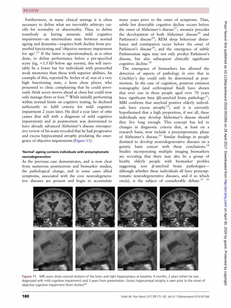
example of this, reported by Archer et al, was of a very
high functioning man, a keen chess player, who
presented to clinic complaining that he could previ-
ously think seven moves ahead at chess but could now
only manage three or four.26 While initially performing
within normal limits on cognitive testing, he declined
sufficiently to fulfil criteria for mild cognitive
impairment 2 years later. He died a year later of othe
causes (but still with a diagnosis of mild cognitive
impairment) and at postmortem was determined to
have already advanced Alzheimer’s disease retrospec-
tive review of his scans revealed that he had progressive
and excess hippocampal atrophy predating the emer-
gence of objective impairments (Figure 11).
‘Normal’ ageing contains individuals with presymptomatic
neurodegeneration
As the previous case demonstrates, and is now clear
from numerous postmortem and biomarker studies,
the pathological change, and in some cases allied
symptoms, associated with the core neurodegenera-
tive diseases are present and begin to accumulate
many years prior to the onset of symptoms. Thus,
subtle but detectable cognitive decline occurs before
the onset of Alzheimer’s disease27; anosmia precedes
the development of both Alzheimer disease28 and
Parkinson’s disease29; REM sleep behaviour distur-
bance and constipation occur before the onset of
Parkinson’s disease29; and the emergence of subtle
Parkinsonian signs may not only predict Parkinson’s
disease, but also subsequent clinically significant
cognitive decline.30
The emergence of biomarkers has allowed the
detection of aspects of pathology in vivo that in
Critchley’s day could only be determined at post-
mortem. In the case of cognition, positron emission
tomography (and cerbrospinal fluid) have shown
that over one in three people aged over 70 years
have significant beta (b)-amyloid brain pathology31;
MRI confirms that amyloid positive elderly individ-
uals have excess atrophy32; and it is currently
hypothesised that a high proportion, if not all, these
indivdiuals may develop Alzheimer’s disease should
they live long enough. This concept has led to
changes in diagnostic criteria that, at least on a
research basis, now include a presymptomatic phase
of Alzheimer’s disease.33 Similar findings in people
destined to develop neurodegenerative diseases on a
genetic basis concur with these conclusions.34
Studies incorporating multiple imaging biomarkers
are revealing that there may also be a group of
heathy elderly people with biomarker profiles
suggesting non b-amyloid brain pathologies—
although whether these individuals all have presymp-
tomatic neurodegenerative diseases, and if so which
one(s), is the subject of considerable debate.35 36
Figure 11 MRI scans show coronal sections of the brain and right hippocampus at baseline, 9 months, 2 years (when he was
diagnosed with mild cognitive impairment) and 3 years from presentation. Excess hippocampal atrophy is seen prior to the onset of
objective cognitive impairment (from Archer)26
180 Schott JM. Pract Neurol 2017;17:172–182. doi:10.1136/practneurol-2016-001566
REVIEW
on April 30, 2020 by guest. P
rotected by copyright.http://pn.bm
j.com/
Pract N
eurol: first published as 10.1136/practneurol-2016-001566 on 28 April 2017. D
ownloaded from
These uncertainties aside, the inevitable conclusion
from such studies is that a significant proportion of
apparently healthy people who may be recruited as,
or considered to be, ‘normal’, will be in the early
stages of developing disease.
Clinical relevance?
Although it is important for a neurologist to deter-
mine if the clinical signs they elicit are or are not
within the normal limits for age, it is also important
that the clinical context is taken into account. This
relates to many aspects including whether the
patient is symptomatic or not; whether signs occur
in isolation or combination; where there are
biomarkers available, whether they are positive or
not; and what are the consequences of finding
pathology. In a recent population study investigating
polyneuropathy, the combinations of signs—that is,
not just the prevalence of isolated vibration sense
loss or decreased tendon reflexes—and (in all but a
few case) nerve conduction studies abnormalities
were required to define the presence of a definite
polyneuropathy. Using these criteria, 13.2% of those
over the age of 80 years had a polyneuropathy.
While this represents a considerably smaller propor-
tion of those with either absent ankle jerks or
sensory disturbance alone (tables 1 and 2), impor-
tantly, 50% of this group were previously
undiagnosed, and a potentially treatable cause was
determined in a proportion.37
CONCLUSIONS
As Critchley elegantly delineated some 85 years ago,
ageing is associated with changes within the nervous
system, and some aspects of the nervous system are
more vulnerable than others. Ageing is associated
with accumulation of many pathologies, notably
cerebrovascular disease, but also with the emergence
of neurodegeneration, and often the signs typically
associated with ‘normal’ ageing and pathology can
overlap. In general, ‘within-individual’ changes are
likely to be more powerful than comparisons
between individuals; and longitudinal research
studies incorporating life course data with detailed
clinical phenotyping and biomarkers provide a
powerful paradigm for understanding the complexity
of the ageing process.38 As and when disease-modi-
fying therapies for neurodegenerative diseases
become available, using biomarkers to determine the
presence of specific pathologies will become increas-
ingly important, both to ensure that treatments are
given to symptomatic people with the pathology in
question and also to determine which elderly people
with presymptomatic disease may benefit most from
treatment.
Acknowledgements This paper was based on a lecture given to the
Association of British Neurologists annual meeting in 2016. The
author is grateful to Dr Geoffrey Schott, Professor Anette Schrag,
Professor Jason Warren, Professor Diana Kuh and Professor Marcus
Richards for their helpful comments and suggestions, and to Kirsty
MacPherson for help in preparing Figure 10.
Funding The author acknowledges the support of the NIHR Queen
Square Dementia BRU, the NIHR UCL/H Biomedical Research
Centre, Wolfson Foundation, EPSRC (EP/J020990/1), MRC
(CSUB19166), ARUK (ARUK-Network 2012-6-ICE; ARUK-
PG2014-1946; ARUK-PG2017-2016), Brain Research Trust
(UCC14191) and European Union’s Horizon 2020 research and
innovation programme (Grant 666992).
Competing interests None declared.
Provenance and peer review Commissioned; externally peer
reviewed. This paper was reviewed by David Burn, Newcastle Upon
Tyne, UK.
© Article author(s) (or their employer(s) unless otherwise stated in
the text of the article) 2017. All rights reserved. No commercial use
is permitted unless otherwise expressly granted.
REFERENCES
1 Critchley M. The neurology of old age. The Lancet
1931;217:1119–27.
2 Critchley M. The neurology of old age. The Lancet
1931;217:1221–31.
3 Critchley M. The neurology of old age. The Lancet
1931;217:1331–7.
4 McDonald I. Short memoir of Dr MacDonald critchley. Cortex
2006;42:782–3.
5 Vrancken AF, Kalmijn S, Brugman F, et al. The meaning of
distal sensory loss and absent ankle reflexes in relation to age: a
meta-analysis. J Neurol 2006;253:578–89.
6 Statistics and data, prevalence of blindness data. https://nei.nih.
gov/eyedata/pbd_tables (Accessed 4 Nov 2016).
7 Meisami E, Brown CM, Emerle HF. Sensory systems: normal
aging, disorders, and treatments of vision and hearing in
humans In: Timiras PS, (editor). Physiological basis of aging and
geriatrics. New York: CRC Press, 2007:109–37.
8 Irving EL, Steinbach MJ, Lillakas L, et al. Horizontal saccade
dynamics across the human life span. Invest Ophthalmol Vis Sci
2006;47:2478–84.
9 Oguro H, Okada K, Suyama N, et al. Decline of vertical gaze
and convergence with aging. Gerontology 2004;50:177–81.
Key points
" Ageing is associated with changes in the nervous system:understanding what is ‘normal’ at different ages is importantwhen evaluating patients.
" As Macdonald Critchley recognised over 85 years ago, someaspects of the neurological examination are morevulnerable to ageing than others.
" Interpretation of neurological signs in the elderly depends oncontext: factors that must be taken into account include theinfluence of comorbidities, the problems of extrapolatinggroup data to individuals and the influence of presymptom-atic or asymptomatic disease.
" Longitudinal studies incorporating detailed phenotyping,biomarkers and life course data are starting to reveal thecomplexities of the ageing process.
Schott JM. Pract Neurol 2017;17:172–182. doi:10.1136/practneurol-2016-001566 181
REVIEW
on April 30, 2020 by guest. P
rotected by copyright.http://pn.bm
j.com/
Pract N
eurol: first published as 10.1136/practneurol-2016-001566 on 28 April 2017. D
ownloaded from
10 Robinson DW, Sutton GJ. Age effect in hearing: a comparative
analysis of published threshold data. Audiology
1979;18:320–34.
11 Doty RL, Shaman P, Applebaum SL, et al. Smell identification
ability: changes with age. Science 1984;226:1441–3.
12 Ferrucci L, Cooper R, Shardell M, et al. Age-related change in
mobility: perspectives from life course epidemiology and
geroscience. J Gerontol A Biol Sci Med Sci 2016;71:1184–94.
13 Bennett DA, Beckett LA, Murray AM, et al. Prevalence of
Parkinsonian signs and associated mortality in a community
population of older people. N Engl J Med 1996;334:71–6.
14 Deuschl G, Petersen I, Lorenz D, et al. Tremor in the elderly:
essential and aging-related tremor. Mov Disord
2015;30:1327–34.
15 Kimber TE, Thompson PD Senile chorea. Handb Clin Neurol
2011;100:213–7.
16 Hedden T, Gabrieli JD. Insights into the ageing mind:
a view from cognitive neuroscience. Nat Rev Neurosci
2004;5:87–96.
17 Nelson HE. The national adult reading Test (NART): test
manual. Windsor: NFER-Nelson, 1982.
18 O’Keeffe ST, Smith T, Valacio R, et al. A comparison of two
techniques for ankle jerk assessment in elderly subjects. Lancet
1994;344:1619–20.
19 Schmidt R, Schmidt H, Haybaeck J, et al. Heterogeneity in age-
related white matter changes. Acta Neuropathol
2011;122:171–85.
20 Pierce MB, Silverwood RJ, Nitsch D, et al; NSHD Scientific
and Data Collection Teams. Clinical disorders in a post war
British cohort reaching retirement: evidence from the first
national birth cohort study. PLoS One 2012;7:e44857.
21 Collerton J, Davies K, Jagger C, et al. Health and disease in 85
year olds: baseline findings from the newcastle 85+ cohort
study. BMJ 2009;339:b4904.
22 Differences in life expectancy between those aged 20, 50 and
80 – in 2011 and at birth. https://www.gov.uk/government/
uploads/system/uploads/attachment_data/file/223114/diffs_life_
expectancy_20_50_80.pdf.
23 Warrington EK. Recognition memory test: manual. Berkshire,
UK: NFER-Nelson, 1984.
24 Jagust W. Vulnerable neural systems and the borderland of brain
aging and neurodegeneration. Neuron 2013;77:219–34.
25 Petersen RC. Mild cognitive impairment as a diagnostic entity.
J Intern Med 2004;256:183–94.
26 Archer HA, Schott JM, Barnes J, et al. Knight’s move thinking?
Mild cognitive impairment in a chess player. Neurocase
2005;11:26–31.
27 Donohue MC, Sperling RA, Salmon DP, et al; Australian
Imaging, Biomarkers, and Lifestyle Flagship Study of Ageing,
Alzheimer’s Disease Neuroimaging Initiative, Alzheimer’s
Disease Cooperative Study. The preclinical Alzheimer cognitive
composite: measuring amyloid-related decline. JAMA Neurol
2014;71:961–70.
28 Wilson RS, Arnold SE, Schneider JA, et al. Olfactory
impairment in presymptomatic Alzheimer’s disease. Ann N Y
Acad Sci 2009;1170:730–5.
29 Noyce AJ, Lees AJ, Schrag AE. The prediagnostic phase of
Parkinson’s disease. J Neurol Neurosurg Psychiatry
2016;87:871–8.
30 Richards M, Stern Y, Mayeux R. Subtle extrapyramidal signs
and incident dementia: a followup analysis. Neurology
1942;1995:45.
31 Jansen WJ, Ossenkoppele R, Knol DL, et al; Amyloid
Biomarker Study Group. Prevalence of cerebral amyloid
pathology in persons without dementia: a meta-analysis. JAMA
2015;313:1924–38.
32 Schott JM, Bartlett JW, Fox NC, et al; Alzheimer’s Disease
Neuroimaging Initiative Investigators. Increased brain atrophy
rates in cognitively normal older adults with low cerebrospinal
fluid Ab1-42. Ann Neurol 2010;68:825–34.
33 Sperling RA, Aisen PS, Beckett LA, et al. Toward defining the
preclinical stages of Alzheimer’s disease: recommendations from
the National Institute on Aging-Alzheimer’s Association
workgroups on diagnostic guidelines for Alzheimer’s disease.
Alzheimers Dement 2011;7:280–92.
34 Bateman RJ, Xiong C, Benzinger TL, et al; Dominantly
Inherited Alzheimer Network. Clinical and biomarker changes
in dominantly inherited Alzheimer’s disease. N Engl J Med
2012;367:795–804.
35 Jack CR, Therneau TM, Wiste HJ, et al. Transition rates
between amyloid and neurodegeneration biomarker states and
to dementia: a population-based, longitudinal cohort study.
Lancet Neurol 2016;15:56–64.
36 Mormino EC, Papp KV, Rentz DM, et al. Heterogeneity in
suspected non-Alzheimer disease pathophysiology among
clinically normal older individuals. JAMA Neurol
2016;73:1185–91.
37 Hanewinckel R, Drenthen J, van Oijen M, et al. Prevalence of
polyneuropathy in the general middle-aged and elderly
population. Neurology 2016;87:1892–8.
38 Lane CA, Parker TD, Cash DM, et al. Study protocol: Insight
46 - a neuroscience sub-study of the MRC National Survey of
Health and Development. BMC Neurol 2017;17:75.
182 Schott JM. Pract Neurol 2017;17:172–182. doi:10.1136/practneurol-2016-001566
REVIEW
on April 30, 2020 by guest. P
rotected by copyright.http://pn.bm
j.com/
Pract N
eurol: first published as 10.1136/practneurol-2016-001566 on 28 April 2017. D
ownloaded from