Embed Size (px)
Citation preview
The Neurological examination
General frame
Reduced your neurological differential diagnosis
• How the symptoms started?• How they progressed over time?• What is the localization?
• Mental status, cognition & language• Signs of meningeal irritation.• Cranial nerves• Motor system• Sensory system • Cerebellar functions• Gait and balance
• Remember that normal range of findings dynamically changes with age.
• We all have some soft neurological signs – be symptoms oriented and try to look at the entire picture and not on a soft abnormal finding.

The technique and expected
findings

Mental status, cognition & language
• Define the patient mental status - alert? Confused? Lethargic? comatose?
• Be sure your patient is oriented to time and place.
• If during conversation you suspect any language abnormality test spontaneous conversation (fluency, paraphasia), understanding commands, low and high frequency naming, repetition, reading, writing. Distinguished and define dysarthria.
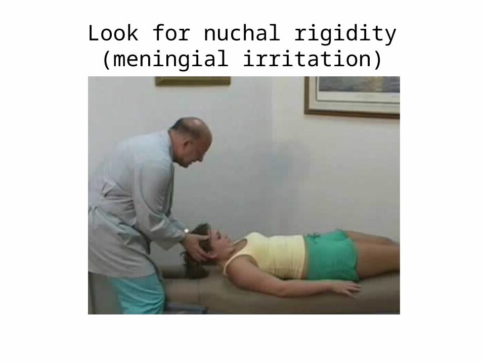
Look for nuchal rigidity (meningial irritation)
Cranial Nerves
•(I), II-XII
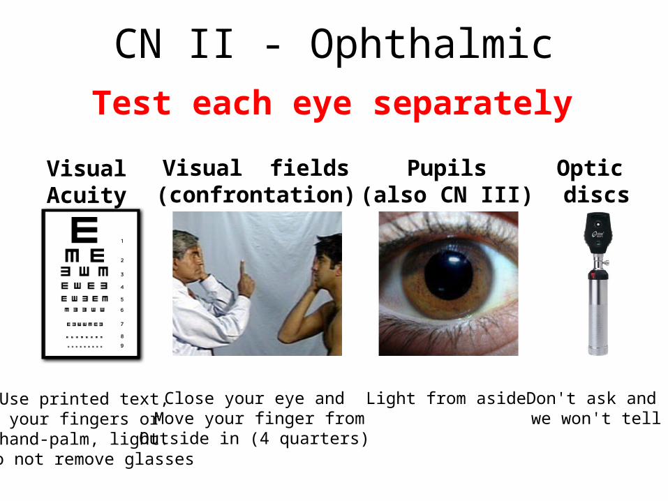
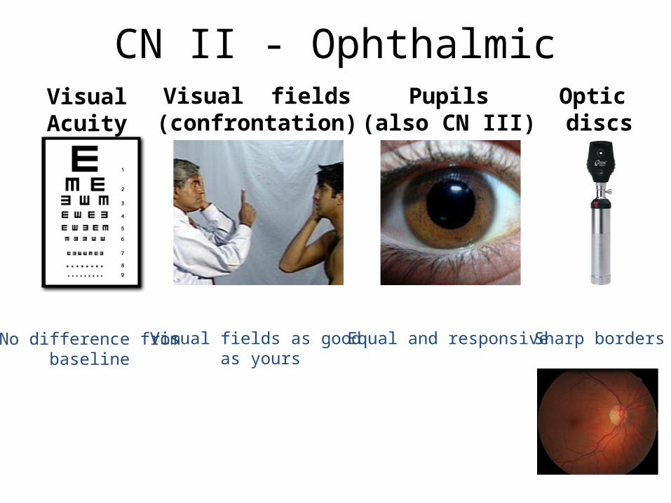
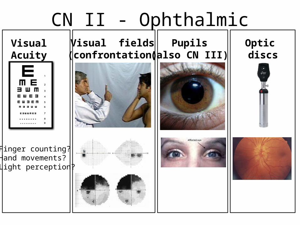
CN II - OphthalmicTest each eye separately
Visual Acuity
Optic discs
Visual fields(confrontation)
Pupils(also CN III)
Use printed text, your fingers or
hand-palm, light. Do not remove glasses
Don't ask and we won't tell
Light from asideClose your eye and Move your finger fromOutside in (4 quarters)
CN II - OphthalmicVisual Acuity
Optic discs
Visual fields(confrontation)
Pupils(also CN III)
No difference frombaseline
Equal and responsiveVisual fields as good as yours
Sharp borders
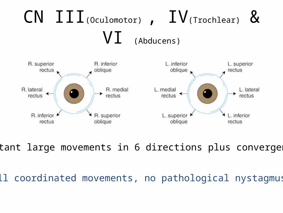
CN III(Oculomotor) , IV(Trochlear) & VI (Abducens)
Distant large movements in 6 directions plus convergence
Full coordinated movements, no pathological nystagmus
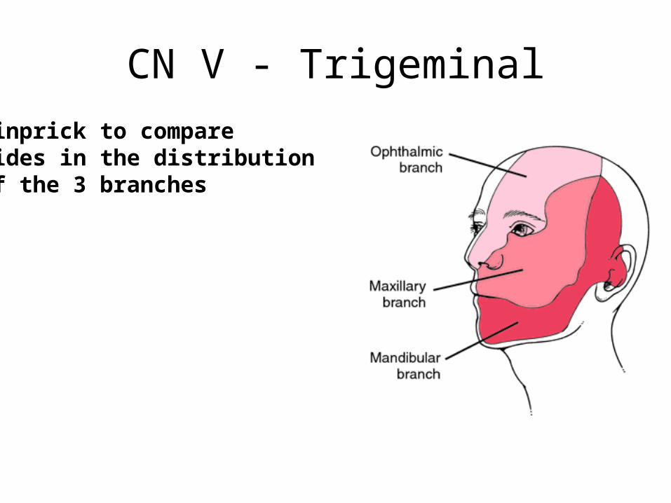
CN V - TrigeminalPinprick to compare sides in the distribution of the 3 branches
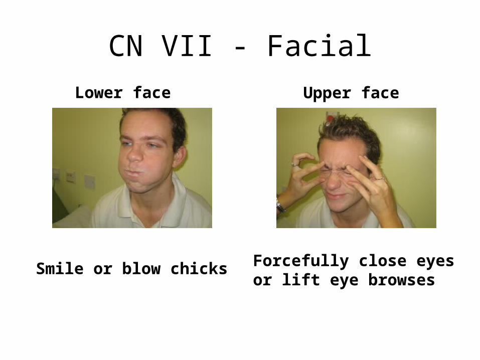
CN VII - FacialLower face Upper face
Smile or blow chicks Forcefully close eyes or lift eye browses
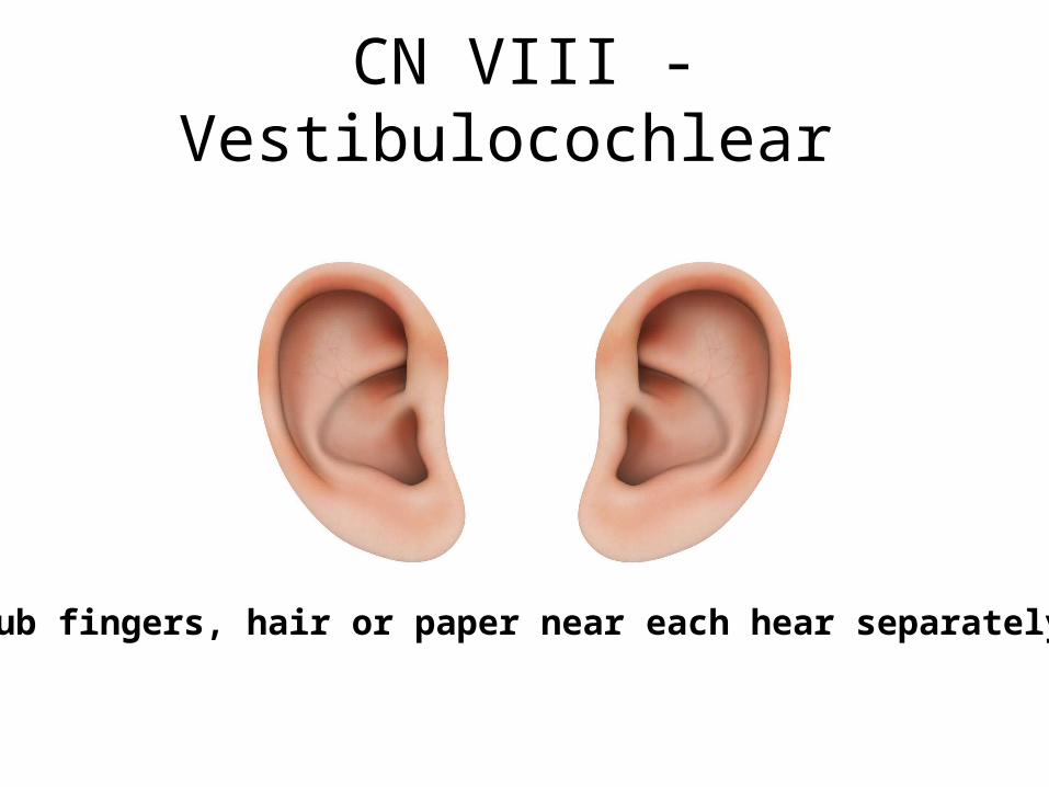
CN VIII - Vestibulocochlear
Rub fingers, hair or paper near each hear separately
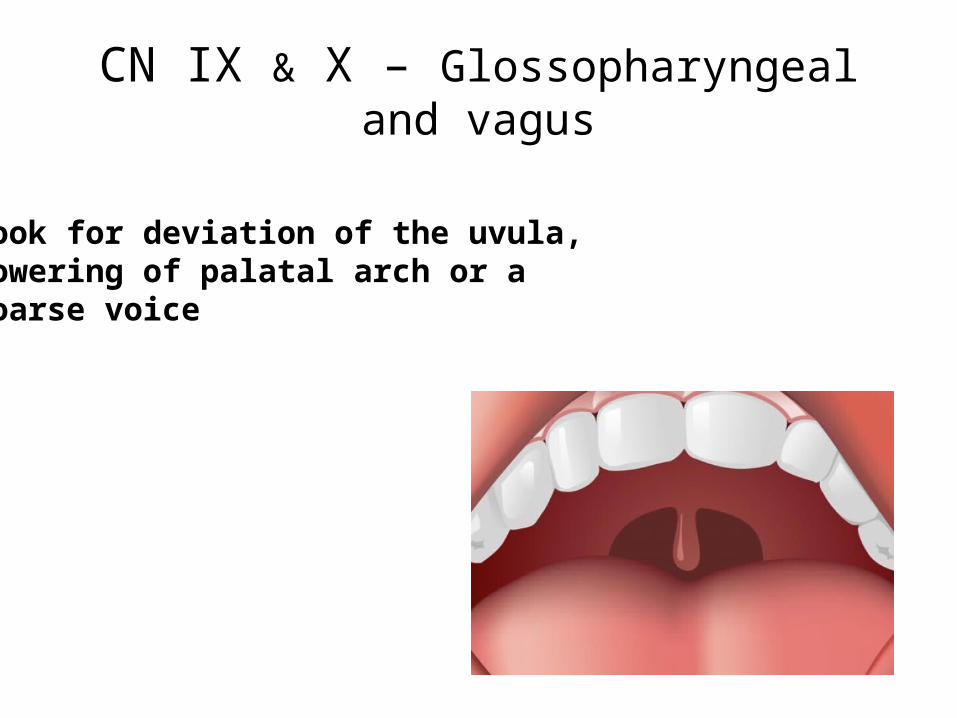
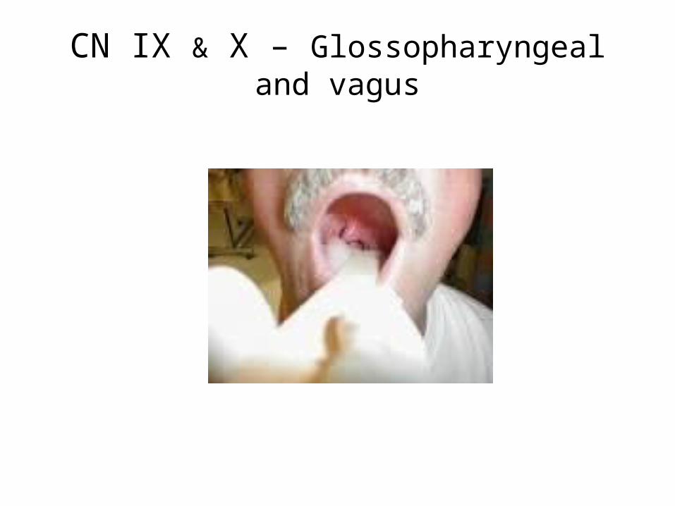
CN IX & X – Glossopharyngeal and vagus
Look for deviation of the uvula, lowering of palatal arch or a coarse voice
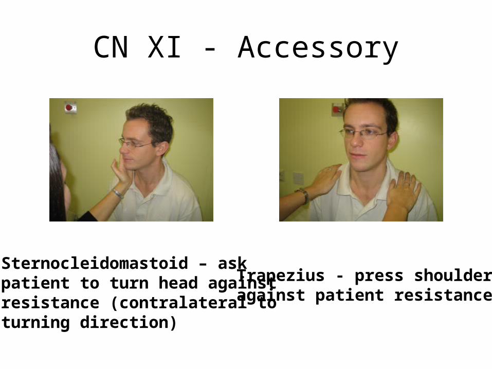
CN XI - Accessory
Sternocleidomastoid – ask patient to turn head against resistance (contralateral to turning direction)
Trapezius - press shoulderagainst patient resistance
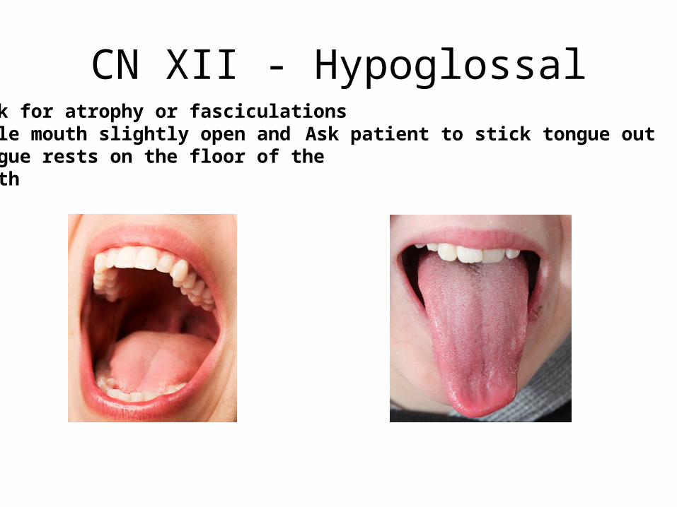
CN XII - HypoglossalLook for atrophy or fasciculationswhile mouth slightly open andtongue rests on the floor of the mouth
Ask patient to stick tongue out

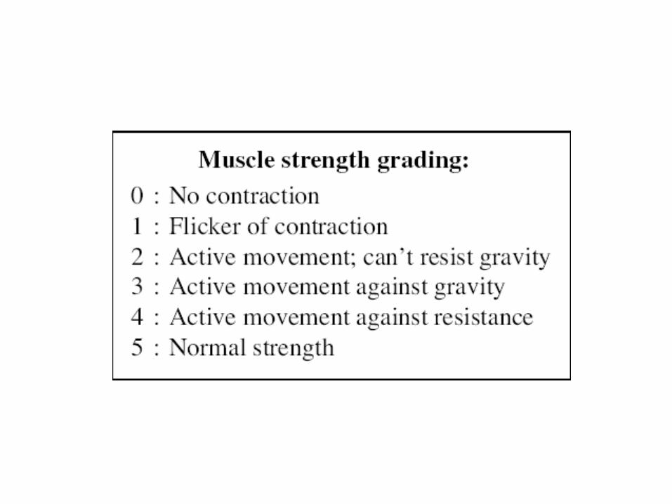
MotorSystem• Observe• Passive tone• Muscle force• Reflexes• Babinski
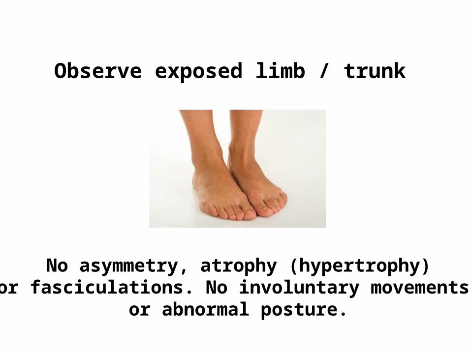
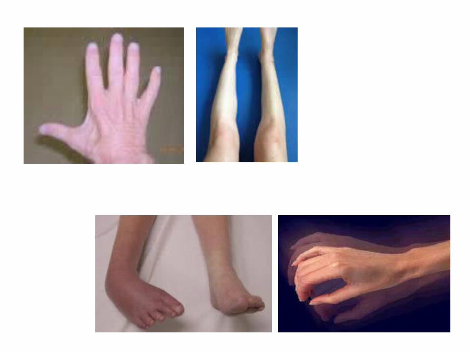
No asymmetry, atrophy (hypertrophy) or fasciculations. No involuntary movements,
or abnormal posture.
Observe exposed limb / trunk
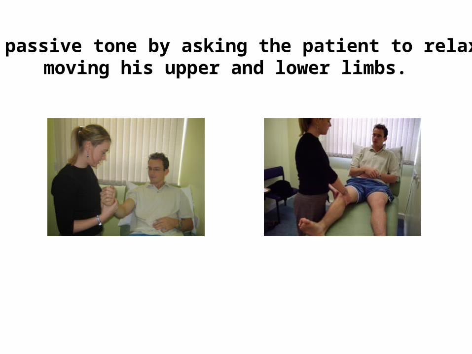
Test passive tone by asking the patient to relax andmoving his upper and lower limbs.
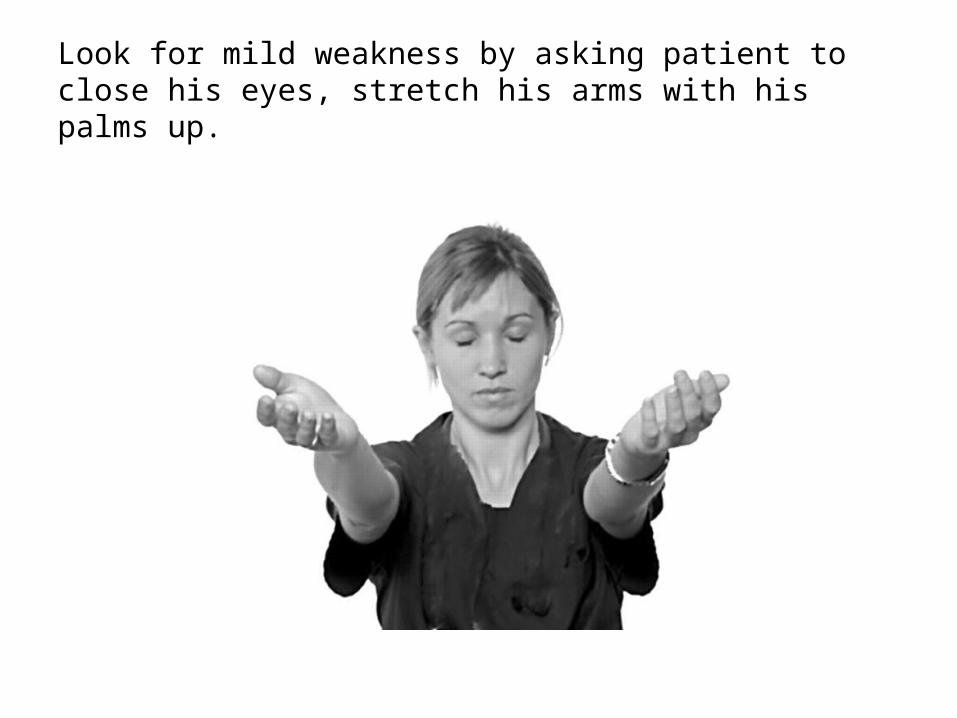
Look for mild weakness by asking patient to close his eyes, stretch his arms with his palms up.
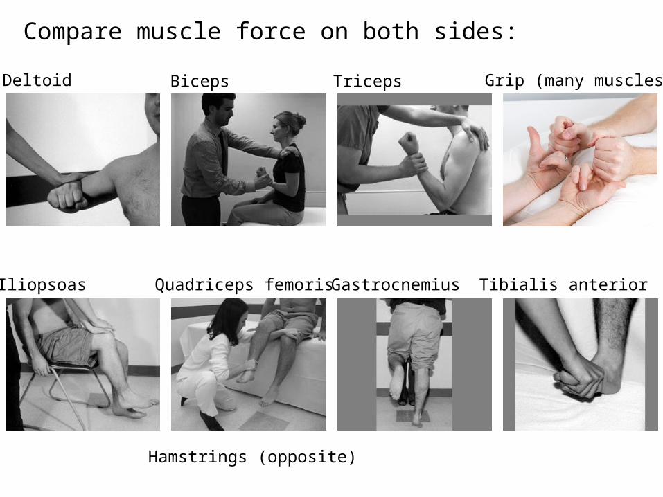
Biceps Triceps
Gastrocnemius Tibialis anteriorIliopsoas Quadriceps femoris
Deltoid Grip (many muscles)
Compare muscle force on both sides:
Hamstrings (opposite)
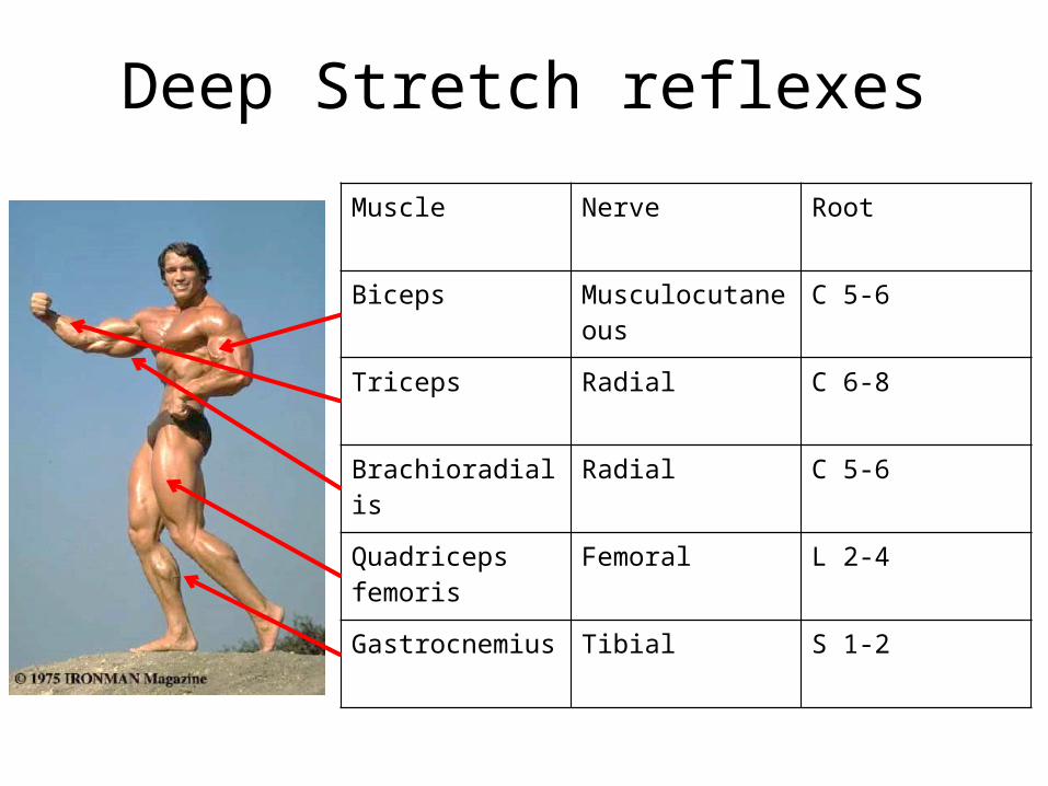
Deep Stretch reflexes
Muscle Nerve Root
Biceps Musculocutaneous C 5-6
Triceps Radial C 6-8
Brachioradialis Radial C 5-6
Quadriceps femoris Femoral L 2-4
Gastrocnemius Tibial S 1-2
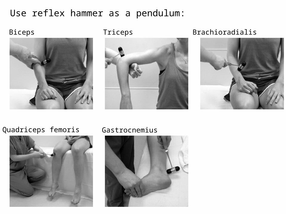
Use reflex hammer as a pendulum: Biceps Triceps Brachioradialis
Quadriceps femoris Gastrocnemius
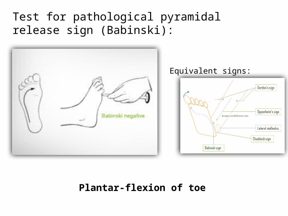
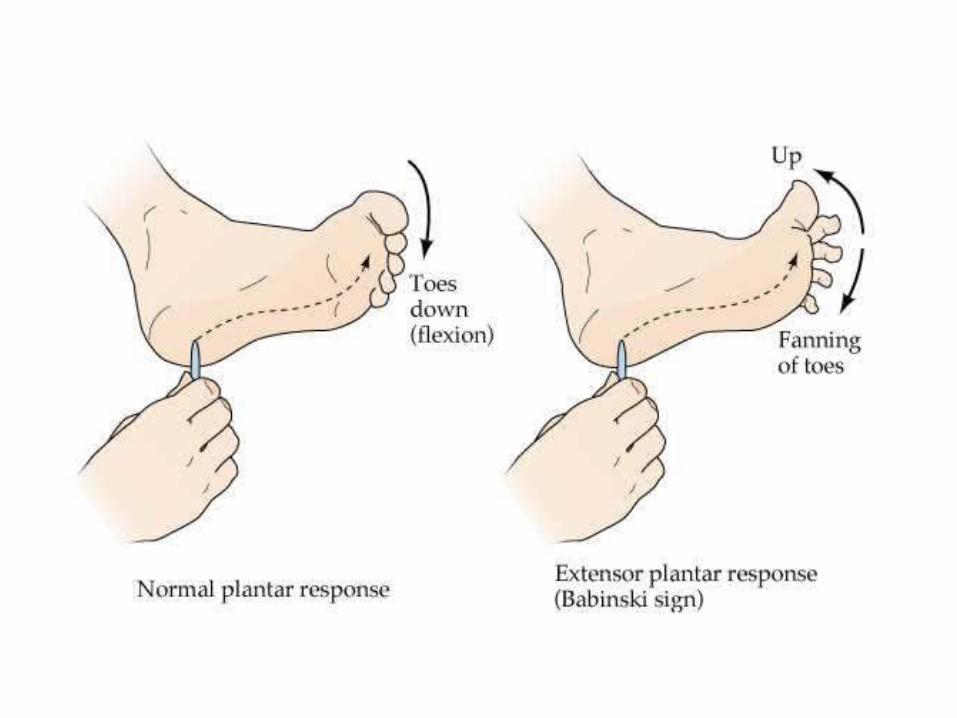
Test for pathological pyramidal release sign (Babinski):
Plantar-flexion of toe
Equivalent signs:

SensoryExam
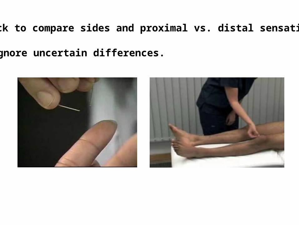
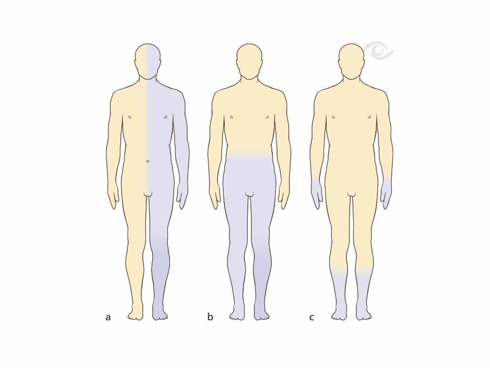
Pinprick to compare sides and proximal vs. distal sensation. Ignore uncertain differences.
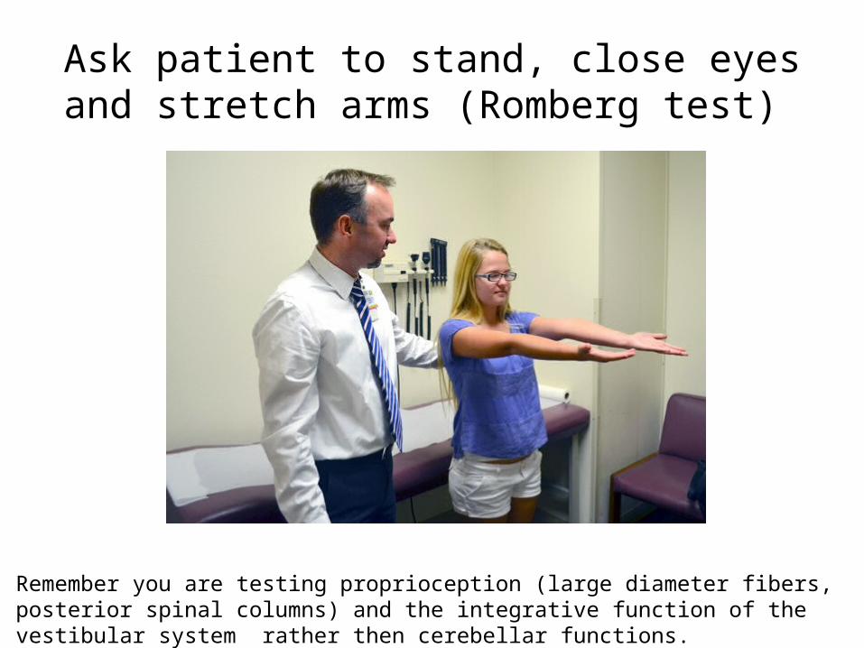
Ask patient to stand, close eyes and stretch arms (Romberg test)
Remember you are testing proprioception (large diameter fibers, posterior spinal columns) and the integrative function of the vestibular system rather then cerebellar functions.

Cerebellar functions
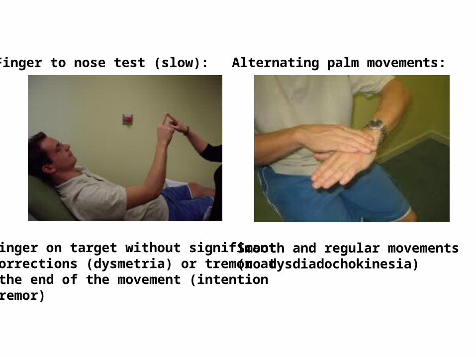
Smooth and regular movements(no dysdiadochokinesia)
Finger on target without significantcorrections (dysmetria) or tremor at the end of the movement (intentiontremor)
Alternating palm movements: Finger to nose test (slow):
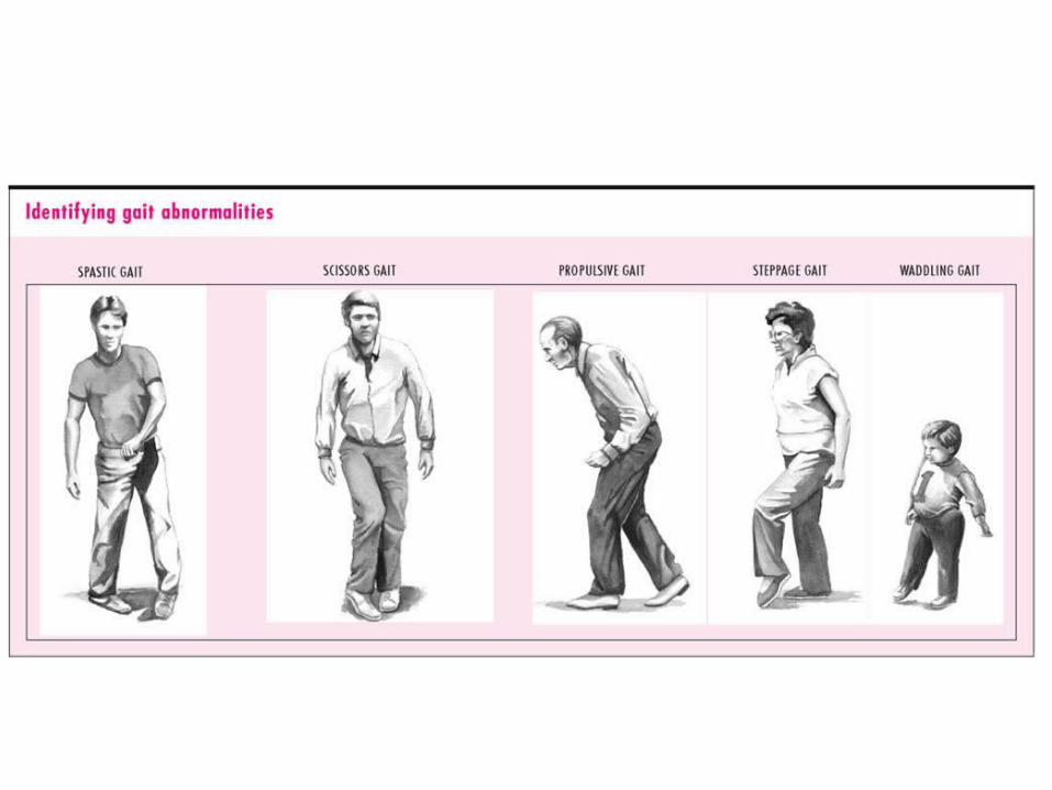
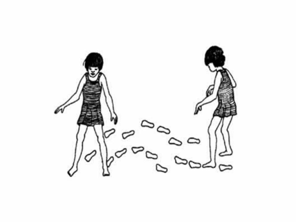
Gait & Balance
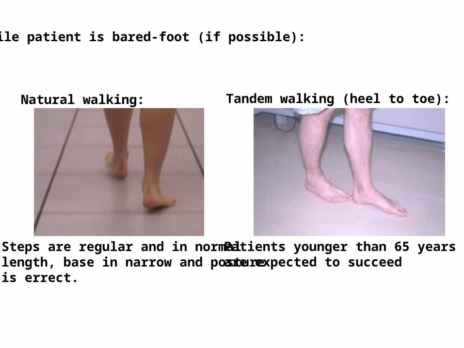
Steps are regular and in normal length, base in narrow and postureis errect.
Patients younger than 65 years are expected to succeed
Natural walking: Tandem walking (heel to toe):
While patient is bared-foot (if possible):
Abnormalfindings

Cranial Nerves
•(I), II-XII
Visual Acuity
Optic discs
Visual fields(confrontation)
Pupils(also CN III)
CN II - Ophthalmic
Finger counting?Hand movements?Light perception?
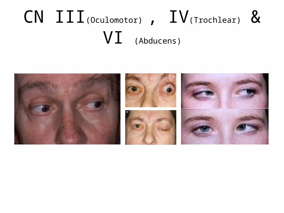
CN III(Oculomotor) , IV(Trochlear) & VI (Abducens)
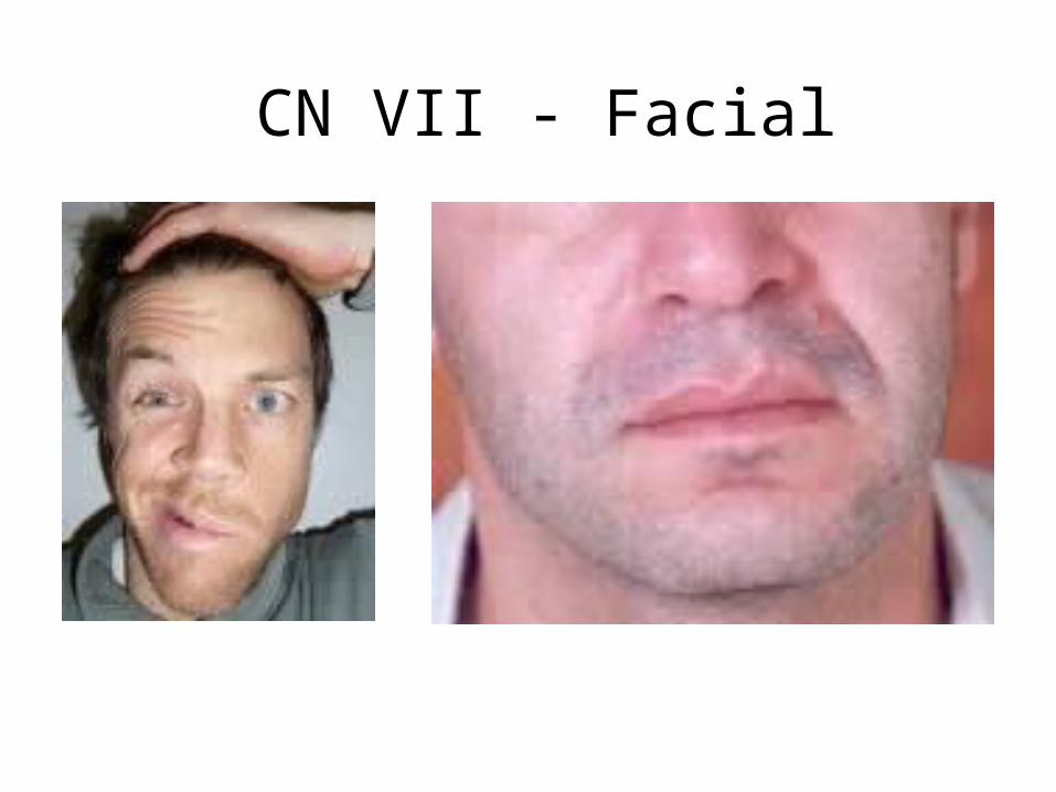
CN VII - Facial
CN IX & X – Glossopharyngeal and vagus
MotorSystem
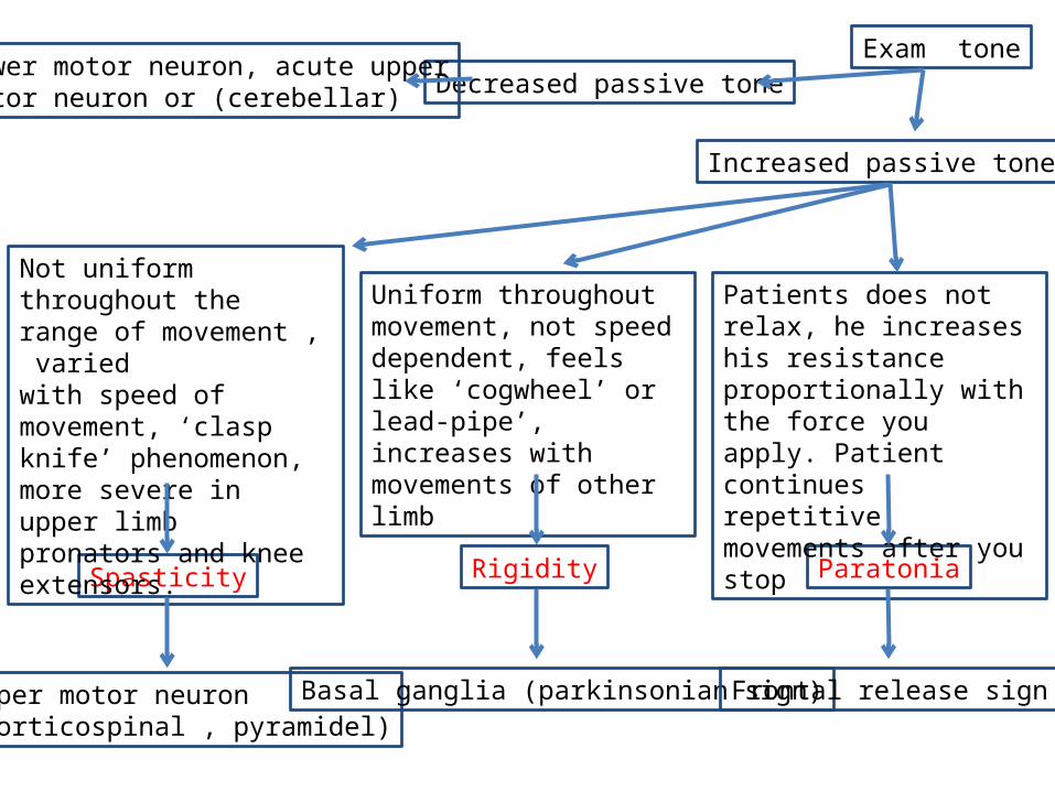
Decreased passive tone
ParatoniaRigiditySpasticity
Uniform throughout movement, not speed dependent, feels like ‘cogwheel’ or lead-pipe’, increases with movements of other limb
Not uniform throughout the range of movement , varied with speed of movement, ‘clasp knife’ phenomenon, more severe in upper limb pronators and knee extensors.
Increased passive tone
Patients does not relax, he increases his resistance proportionally with the force you apply. Patient continues repetitive movements after you stop
Lower motor neuron, acute upperMotor neuron or (cerebellar)
Upper motor neuron (corticospinal , pyramidel)
Basal ganglia (parkinsonian sign) Frontal release sign
Exam tone
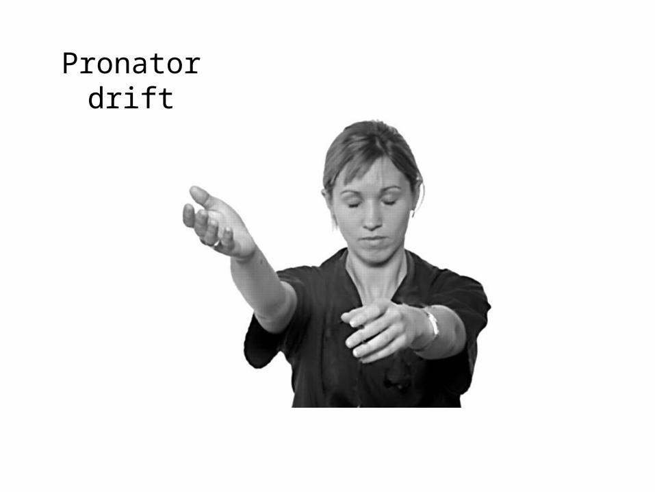
Pronator drift
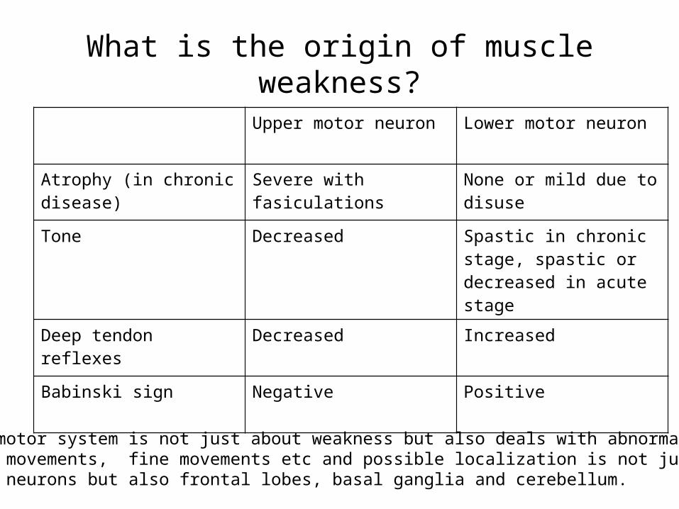
What is the origin of muscle weakness?
Upper motor neuron Lower motor neuron
Atrophy (in chronic disease) Severe with fasiculations None or mild due to disuse
Tone Decreased Spastic in chronic stage, spastic or decreased in acute stage
Deep tendon reflexes Decreased Increased
Babinski sign Negative Positive
Remember – motor system is not just about weakness but also deals with abnormal involuntary movements, fine movements etc and possible localization is not just upper vs. lower motor neurons but also frontal lobes, basal ganglia and cerebellum.
SensoryExam

Cerebellar functions

Gait & Balance

Few more tests you should apply
when relevant
Mental status, cognition & language
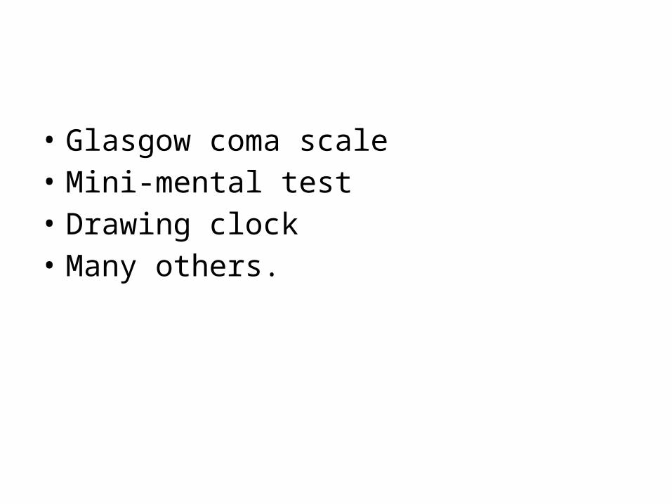
• Glasgow coma scale• Mini-mental test• Drawing clock• Many others.
Cranial Nerves
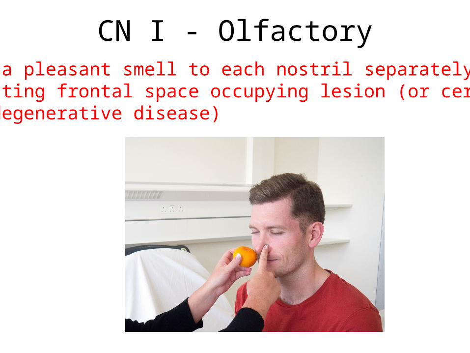
CN I - OlfactoryApply a pleasant smell to each nostril separately when suspecting frontal space occupying lesion (or certain neurodegenerative disease)
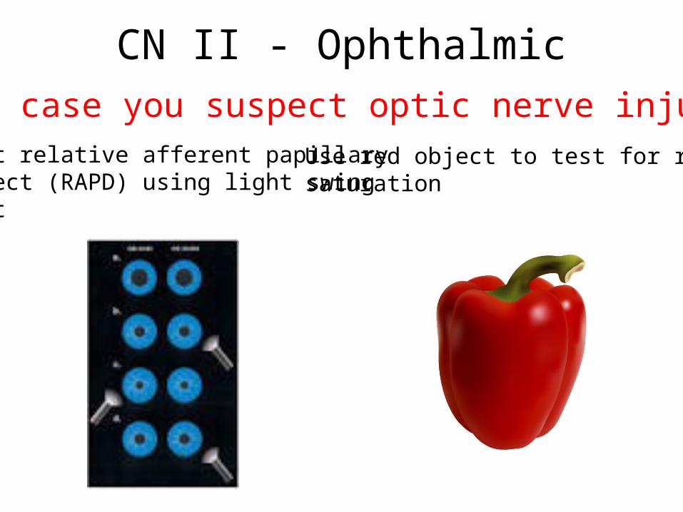
CN II - OphthalmicIn case you suspect optic nerve injury:
Test relative afferent papillary defect (RAPD) using light swing test
Use red object to test for red saturation
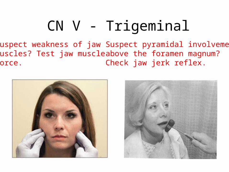
CN V - TrigeminalSuspect weakness of jaw muscles? Test jaw muscle Force.
Suspect pyramidal involvement above the foramen magnum? Check jaw jerk reflex.

MotorSystem
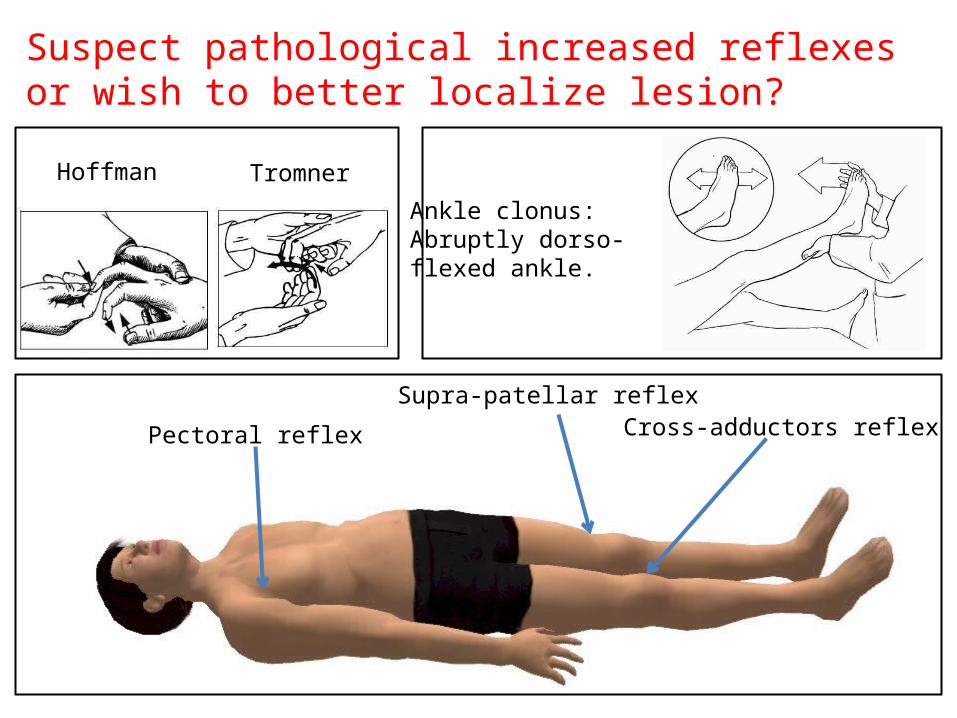
Suspect pathological increased reflexes or wish to better localize lesion?
Pectoral reflex
Supra-patellar reflexCross-adductors reflex
Hoffman Tromner
Ankle clonus:Abruptly dorso-flexed ankle.
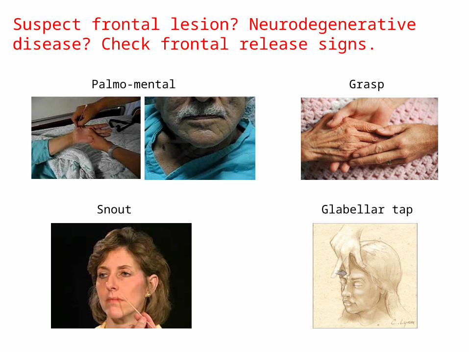
Suspect frontal lesion? Neurodegenerative disease? Check frontal release signs.
Palmo-mental Grasp
Snout Glabellar tap

SensoryExam
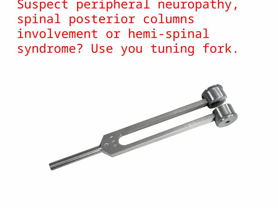
Suspect peripheral neuropathy, spinal posterior columns involvement or hemi-spinal syndrome? Use you tuning fork.

Test for extinction when you suspect non-dominant parietal involvement. You can apply the test while checking visual fields
Visual extinction Tactile extinction
Cerebellar functions
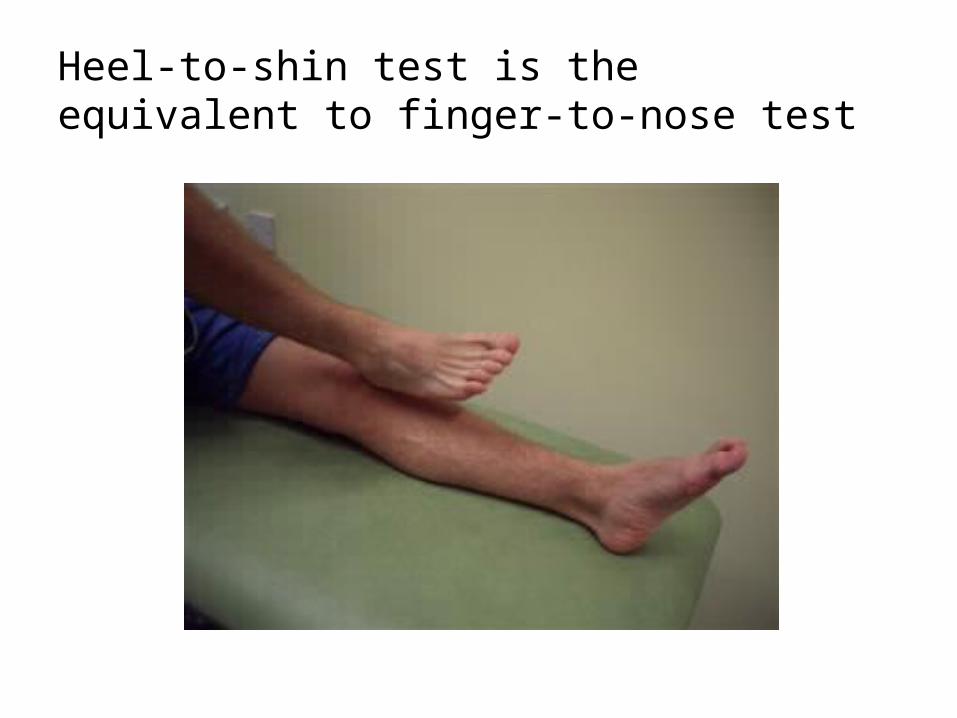
Heel-to-shin test is the equivalent to finger-to-nose test

Gait & Balance
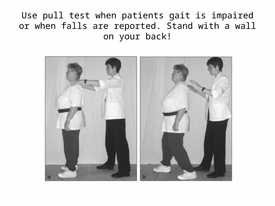
Use pull test when patients gait is impaired or when falls are reported. Stand with a wall on your back!
Thanks,