Embed Size (px)
Citation preview
The Neural Correlates of Religious and NonreligiousBeliefSam Harris1,7,10., Jonas T. Kaplan2., Ashley Curiel3, Susan Y. Bookheimer4,5,6,7,9, Marco Iacoboni1,4,6,7,
Mark S. Cohen5,6,7,8,9*
1 UCLA Ahmanson-Lovelace Brain Mapping Center, David Geffen School of Medicine, University of California Los Angeles (UCLA), Los Angeles, California, United States of
America, 2 Brain and Creativity Institute and Department of Psychology, University of Southern California (USC), Los Angeles, California, United States of America,
3 Department of Clinical Psychology, Pepperdine University, Los Angeles, California, United States of America, 4 Semel Institute for Neuroscience and Human Behavior,
University of California Los Angeles (UCLA), Los Angeles, California, United States of America, 5 Center for Cognitive Neuroscience, University of California Los Angeles, Los
Angeles (UCLA), Los Angeles, California, United States of America, 6 Departments of Psychiatry and Biobehavioral Sciences, University of California Los Angeles (UCLA), Los
Angeles, California, United States of America, 7 The Brain Research Institute, University of California Los Angeles (UCLA), Los Angeles, California, United States of America,
8 Departments of Neurology, Radiological Sciences, Biomedical Engineering, and Biomedical Physics, University of California Los Angeles (UCLA), Los Angeles, California,
United States of America, 9 Department of Psychology, University of California Los Angeles (UCLA), Los Angeles, California, United States of America, 10 The Reason
Project, Santa Monica, California, United States of America
Abstract
Background: While religious faith remains one of the most significant features of human life, little is known about itsrelationship to ordinary belief at the level of the brain. Nor is it known whether religious believers and nonbelievers differ inhow they evaluate statements of fact. Our lab previously has used functional neuroimaging to study belief as a generalmode of cognition [1], and others have looked specifically at religious belief [2]. However, no research has compared thesetwo states of mind directly.
Methodology/Principal Findings: We used functional magnetic resonance imaging (fMRI) to measure signal changes in thebrains of thirty subjects—fifteen committed Christians and fifteen nonbelievers—as they evaluated the truth and falsity ofreligious and nonreligious propositions. For both groups, and in both categories of stimuli, belief (judgments of ‘‘true’’ vsjudgments of ‘‘false’’) was associated with greater signal in the ventromedial prefrontal cortex, an area important for self-representation [3,4,5,6], emotional associations [7], reward [8,9,10], and goal-driven behavior [11]. This region showedgreater signal whether subjects believed statements about God, the Virgin Birth, etc. or statements about ordinary facts. Acomparison of both stimulus categories suggests that religious thinking is more associated with brain regions that governemotion, self-representation, and cognitive conflict, while thinking about ordinary facts is more reliant upon memoryretrieval networks.
Conclusions/Significance: While religious and nonreligious thinking differentially engage broad regions of the frontal,parietal, and medial temporal lobes, the difference between belief and disbelief appears to be content-independent. Ourstudy compares religious thinking with ordinary cognition and, as such, constitutes a step toward developing aneuropsychology of religion. However, these findings may also further our understanding of how the brain acceptsstatements of all kinds to be valid descriptions of the world.
Citation: Harris S, Kaplan JT, Curiel A, Bookheimer SY, Iacoboni M, et al. (2009) The Neural Correlates of Religious and Nonreligious Belief. PLoS ONE 4(10):e0007272. doi:10.1371/journal.pone.0007272
Editor: Olaf Sporns, Indiana University, United States of America
Received June 3, 2009; Accepted September 7, 2009; Published October 1, 2009
Copyright: � 2009 Harris et al. This is an open-access article distributed under the terms of the Creative Commons Attribution License, which permitsunrestricted use, distribution, and reproduction in any medium, provided the original author and source are credited.
Funding: For generous support the authors wish to thank the Brain Mapping Medical Research Organization, Brain Mapping Support Foundation, Pierson-Lovelace Foundation, The Ahmanson Foundation, William M. and Linda R. Dietel Philanthropic Fund at the Northern Piedmont Community Foundation, TamkinFoundation, Jennifer Jones-Simon Foundation, Capital Group Companies Charitable Foundation, Robson Family and Northstar Fund. The project described wassupported in part by Grant Numbers RR12169, RR13642 and RR00865 from the National Center for Research Resources (NCRR), a component of the NationalInstitutes of Health (NIH) and by a grant from The Reason Project; its contents are solely the responsibility of the authors and do not necessarily represent theofficial views of NCR, NIH, or those of any other funding source. Sam Harris (joint first author) is the Co-founder and CEO of The Reason Project (www.reasonproject.org). The Reason Project is a 501(c) (3) nonprofit foundation whose mission includes conducting original scientific research related to human values,cognition, and reasoning. This affiliation does not alter the authors’ adherence to all PLoS ONE policies on the sharing of data for the purpose of academic, non-commercial research. For this study, The Reason Project provided partial funding for MRI scanner use, subject recruitment, and psychological testing. The othersources of funding had no role in study design, data collection and analysis, decision to publish, or preparation of the manuscript.
Competing Interests: The authors have declared that no competing interests exist.
* E-mail: [email protected]
. These authors contributed equally to this work.
Introduction
Since the 19th century, it has been widely assumed that the
spread of industrialized society would spell the end of religion.
Marx [12], Freud [13,14], and Weber [15]—along with
innumerable anthropologists, sociologists, historians, and psychol-
ogists influenced by their work—expected religious belief to wither
in the light of modernity. It has not come to pass. Religion remains
PLoS ONE | www.plosone.org 1 October 2009 | Volume 4 | Issue 10 | e0007272
one of the most prominent features of human life in the 21st
century. While most developed societies have grown predomi-
nantly secular [16], with the curious exception of the United
States, orthodox religion is in full bloom throughout the
developing world. Indeed, humanity seems to becoming propor-
tionally more religious, as the combination of material advance-
ment and secularism is strongly correlated with decreased fertility
[17]. When one considers the rise of Islamism throughout the
Muslim world, the spread of Pentecostalism throughout Africa,
and the anomalous piety of the United States, it becomes clear that
religion will have geopolitical consequences well into the 21st
century.
Given the importance of religion in human life, surprisingly
little is known about its basis in the brain. The relevance of the
brain’s ventromedial dopaminergic systems to religious experi-
ence, belief and behavior is suggested by several lines of evidence,
including the fact that a variety of clinical conditions related to
dopaminergic dysfunction—mania, obsessive-compulsive disorder
(OCD), schizophrenia, and temporal-lobe epilepsy—are regularly
associated with hyperreligiosity [18]. The serotonergic system has
also been implicated, as drugs known to modulate it—like LSD,
psilocybin, mescaline, N,N-dimethyltryptamine (‘‘DMT’’), and
3,4-methylenedioxymethamphetamine (‘‘ecstasy’’)—seem espe-
cially potent drivers of religious/spiritual experience. In addition,
5-HT1A receptor densities have been inversely correlated with
high scores on the ‘‘spiritual acceptance’’ subscale of the
Temperament and Character Inventory [19].
There have been a number of neuroimaging and EEG studies
done on religious practice and experience—primarily focusing on
meditation [20,21,22,23,24] and prayer [25,26,27,28,29,30]. The
purpose of these studies has been to evoke spiritual/contemplative
experiences in religious subjects and to compare these states of
mind to a control condition. However, none of these studies were
designed to isolate the variable of belief itself, or to determine
whether religious belief differs from ordinary belief at the level of
the brain.
As many have noted, religion cannot be reduced to a mere
concatenation of religious beliefs. Every religion consists of rites,
rituals, prayers, social institutions, holidays, etc., that serve a wide
variety of purposes, explicit or otherwise [31,32]. However,
religious belief—that is, the acceptance of specific religious
propositions as being true—is generally what renders these
enterprises relevant, or even comprehensible. While there may
be many Catholics, for instance, who value the ritual of the Mass
without actually believing the doctrine of Transubstantiation, the
primacy of the Mass within the Church still hinges on the fact that
many Catholics do accept it as a metaphysical truth—a fact that
can be directly attributed to specific, doctrinal claims that are still
put forward by the Church. There is, of course, a distinction to be
made between mere profession of such beliefs and actual belief [33]—
a distinction that, while important, only makes sense in a world in
which some people actually believe what they say they believe.
There seems little reason to doubt that a significant percentage of
human beings, likely a majority, falls into this latter category with
respect one or another religious creed.
Our lab published the first neuroimaging study of belief as a
general mode of cognition [1], and another group has looked
specifically at religious conviction [2]. However, no research has
compared these two states of mind directly. Here we show that
while religious and nonreligious thinking differentially engage
broad regions of the frontal, parietal, and medial temporal lobes—
and, hence, appear quite distinct as modes of thought—the
difference between belief and disbelief appears to be content-
independent.
Results
We used functional magnetic resonance imaging (fMRI) to
measure signal changes in the brains of thirty subjects—fifteen
committed Christians and fifteen nonbelievers—as they evaluated
the truth and falsity of religious and nonreligious propositions. For
each trial either a religious statement (e.g., ‘‘Jesus Christ really
performed the miracles attributed to him in the Bible’’) or a
nonreligious statement (e.g., ‘‘Alexander the Great was a very
famous military leader’’) appeared, and participants pressed a
button to indicate whether the statement was true or false. Our
stimuli were designed to produce roughly equal numbers of
believed and disbelieved trials in each category.
Behavioral dataResponse time data were submitted to a repeated-measures
ANOVA with belief (true, false) and statement content (religious,
nonreligious) as within-subject variables, and group (nonbeliever,
Christian) as a between-subject variable. Response times were
significantly longer for false (3.95 s) compared to true (3.70 s)
responses (F (1,28) = 33.4, p,.001), and also significantly longer
for religious (3.99 s) compared with nonreligious (3.66 s) stimuli (F
(1,28) = 18, p,.001). The two-way interaction between belief and
content type did not reach significance, but there was a three-way
interaction between belief, content type, and group (F
(1,28) = 6.06, p,.05). While both groups were quicker to respond
‘‘true’’ than ‘‘false’’ on both categories of stimuli, the effect of truth
was especially pronounced for nonbelievers when responding to
religious statements (see Supplementary Information: Table S1
and Figure S1).
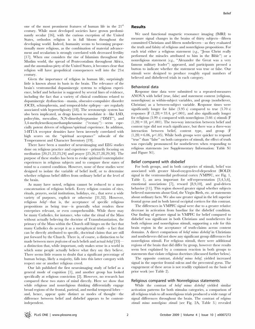
Belief compared with disbeliefFor both groups, and in both categories of stimuli, belief was
associated with greater blood-oxygen-level-dependent (BOLD)
signal in the ventromedial prefrontal cortex (VMPFC, see Fig. 1,
Table 1), an area important for self-representation [3,4,5,6],
emotional associations [7], reward [8,9,10], and goal-driven
behavior [11]. This region showed greater signal whether subjects
believed statements about God, the Virgin Birth, etc. or statements
about ordinary facts. We also saw greater signal in the left superior
frontal gyrus and in both lateral occipital cortices for this contrast.
The differences in VMPFC signal were due to a greater relative
decrease in activation from baseline for the disbelief condition.
Our finding of greater signal in VMPFC for belief compared to
disbelief was significant in both Christians and nonbelievers for
both religious and nonreligious stimuli, supporting a role for this
brain region in the acceptance of truth-claims across content
domains. A direct comparison of belief minus disbelief in Christians
and nonbelievers did not show any significant group differences for
nonreligious stimuli. For religious stimuli, there were additional
regions of the brain that did differ by group, however these results
seem best explained by a common reaction in both groups to
statements that violate religious doctrines (discussed further below).
The opposite contrast, disbelief minus belief, yielded increased
signal in the superior frontal sulcus and the precentral gyrus. The
engagement of these areas is not readily explained on the basis of
prior work (see Table 2).
Religious compared with Nonreligious statementsWhile the contrast of belief minus disbelief yielded similar
activation patterns for both stimulus categories, a comparison of
all religious trials to all nonreligious trials produced a wide range of
signal differences throughout the brain. The contrast of religious
stimuli minus nonreligious stimuli (see Fig. 2A, Table 3.) revealed
Belief
PLoS ONE | www.plosone.org 2 October 2009 | Volume 4 | Issue 10 | e0007272
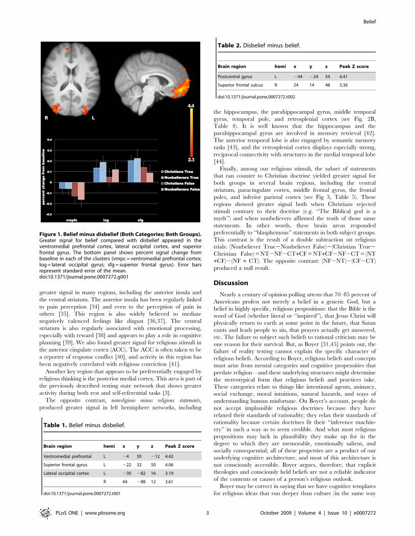
greater signal in many regions, including the anterior insula and
the ventral striatum. The anterior insula has been regularly linked
to pain perception [34] and even to the perception of pain in
others [35]. This region is also widely believed to mediate
negatively valenced feelings like disgust [36,37]. The ventral
striatum is also regularly associated with emotional processing,
especially with reward [38] and appears to play a role in cognitive
planning [39]. We also found greater signal for religious stimuli in
the anterior cingulate cortex (ACC). The ACC is often taken to be
a reporter of response conflict [40], and activity in this region has
been negatively correlated with religious conviction [41].
Another key region that appears to be preferentially engaged by
religious thinking is the posterior medial cortex. This area is part of
the previously described resting state network that shows greater
activity during both rest and self-referential tasks [3].
The opposite contrast, nonreligious minus religious statements,
produced greater signal in left hemisphere networks, including
the hippocampus, the parahippocampal gyrus, middle temporal
gyrus, temporal pole, and retrosplenial cortex (see Fig. 2B,
Table 4). It is well known that the hippocampus and the
parahippocampal gyrus are involved in memory retrieval [42].
The anterior temporal lobe is also engaged by semantic memory
tasks [43], and the retrosplenial cortex displays especially strong,
reciprocal connectivity with structures in the medial temporal lobe
[44].
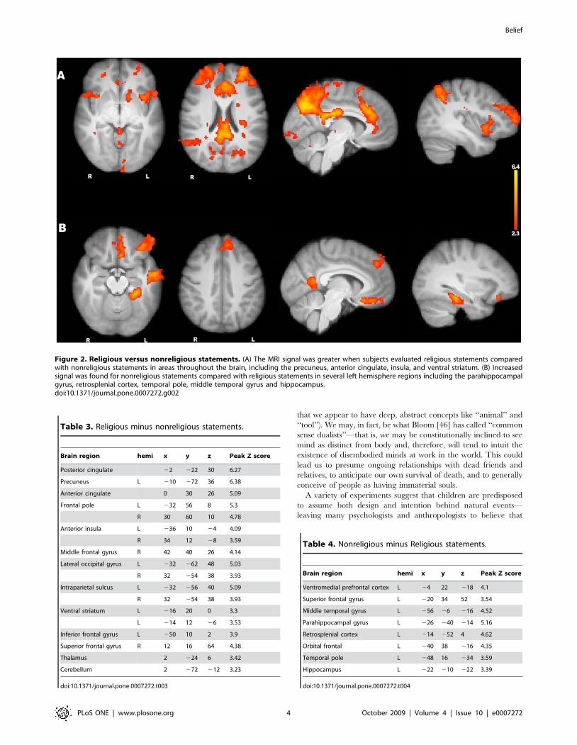
Finally, among our religious stimuli, the subset of statements
that ran counter to Christian doctrine yielded greater signal for
both groups in several brain regions, including the ventral
striatum, paracingulate cortex, middle frontal gyrus, the frontal
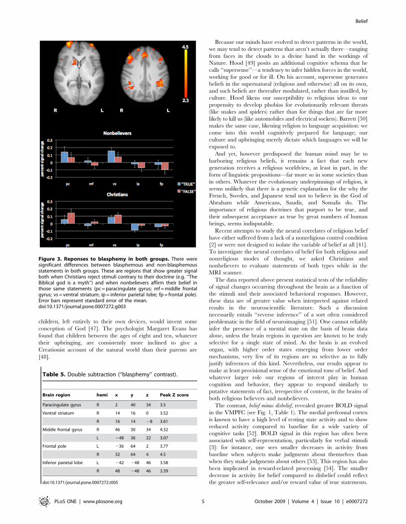
poles, and inferior parietal cortex (see Fig 3, Table 5). These
regions showed greater signal both when Christians rejected
stimuli contrary to their doctrine (e.g. ‘‘The Biblical god is a
myth’’) and when nonbelievers affirmed the truth of those same
statements. In other words, these brain areas responded
preferentially to ‘‘blasphemous’’ statements in both subject groups.
This contrast is the result of a double subtraction on religious
trials: (Nonbeliever True2Nonbeliever False)2(Christian True2
Christian False) = NT2NF2CT+CF = NT+CF2NF2CT = (NT
+CF)2(NF + CT). The opposite contrast: (NF2NT)2(CF2CT)
produced a null result.
Discussion
Nearly a century of opinion polling attests that 70–85 percent of
Americans profess not merely a belief in a generic God, but a
belief in highly specific, religious propositions: that the Bible is the
word of God (whether literal or ‘‘inspired’’), that Jesus Christ will
physically return to earth at some point in the future, that Satan
exists and leads people to sin, that prayers actually get answered,
etc. The failure to subject such beliefs to rational criticism may be
one reason for their survival. But, as Boyer [31,45] points out, the
failure of reality testing cannot explain the specific character of
religious beliefs. According to Boyer, religious beliefs and concepts
must arise from mental categories and cognitive propensities that
predate religion—and these underlying structures might determine
the stereotypical form that religious beliefs and practices take.
These categories relate to things like intentional agents, animacy,
social exchange, moral intuitions, natural hazards, and ways of
understanding human misfortune. On Boyer’s account, people do
not accept implausible religious doctrines because they have
relaxed their standards of rationality; they relax their standards of
rationality because certain doctrines fit their ‘‘inference machin-
ery’’ in such a way as to seem credible. And what most religious
propositions may lack in plausibility they make up for in the
degree to which they are memorable, emotionally salient, and
socially consequential; all of these properties are a product of our
underlying cognitive architecture, and most of this architecture is
not consciously accessible. Boyer argues, therefore, that explicit
theologies and consciously held beliefs are not a reliable indicator
of the contents or causes of a person’s religious outlook.
Boyer may be correct in saying that we have cognitive templates
for religious ideas that run deeper than culture (in the same way
Table 1. Belief minus disbelief.
Brain region hemi x y z Peak Z score
Ventromedial prefrontal L 24 50 212 4.42
Superior frontal gyrus L 222 32 50 4.06
Lateral occipital cortex L 230 282 16 3.19
R 44 288 12 3.61
doi:10.1371/journal.pone.0007272.t001
Table 2. Disbelief minus belief.
Brain region hemi x y z Peak Z score
Postcentral gyrus L 244 224 54 4.41
Superior frontal sulcus R 24 14 48 3.36
doi:10.1371/journal.pone.0007272.t002
Figure 1. Belief minus disbelief (Both Categories; Both Groups).Greater signal for belief compared with disbelief appeared in theventromedial prefrontal cortex, lateral occipital cortex, and superiorfrontal gyrus. The bottom panel shows percent signal change frombaseline in each of the clusters (vmpc = ventromedial prefrontal cortex;log = lateral occipital gyrus; sfg = superior frontal gyrus). Error barsrepresent standard error of the mean.doi:10.1371/journal.pone.0007272.g001
Belief
PLoS ONE | www.plosone.org 3 October 2009 | Volume 4 | Issue 10 | e0007272
that we appear to have deep, abstract concepts like ‘‘animal’’ and
‘‘tool’’). We may, in fact, be what Bloom [46] has called ‘‘common
sense dualists’’—that is, we may be constitutionally inclined to see
mind as distinct from body and, therefore, will tend to intuit the
existence of disembodied minds at work in the world. This could
lead us to presume ongoing relationships with dead friends and
relatives, to anticipate our own survival of death, and to generally
conceive of people as having immaterial souls.
A variety of experiments suggest that children are predisposed
to assume both design and intention behind natural events—
leaving many psychologists and anthropologists to believe that
Figure 2. Religious versus nonreligious statements. (A) The MRI signal was greater when subjects evaluated religious statements comparedwith nonreligious statements in areas throughout the brain, including the precuneus, anterior cingulate, insula, and ventral striatum. (B) Increasedsignal was found for nonreligious statements compared with religious statements in several left hemisphere regions including the parahippocampalgyrus, retrosplenial cortex, temporal pole, middle temporal gyrus and hippocampus.doi:10.1371/journal.pone.0007272.g002
Table 3. Religious minus nonreligious statements.
Brain region hemi x y z Peak Z score
Posterior cingulate 22 222 30 6.27
Precuneus L 210 272 36 6.38
Anterior cingulate 0 30 26 5.09
Frontal pole L 232 56 8 5.3
R 30 60 10 4.78
Anterior insula L 236 10 24 4.09
R 34 12 28 3.59
Middle frontal gyrus R 42 40 26 4.14
Lateral occipital gyrus L 232 262 48 5.03
R 32 254 38 3.93
Intraparietal sulcus L 232 256 40 5.09
R 32 254 38 3.93
Ventral striatum L 216 20 0 3.3
L 214 12 26 3.53
Inferior frontal gyrus L 250 10 2 3.9
Superior frontal gyrus R 12 16 64 4.38
Thalamus 2 224 6 3.42
Cerebellum 2 272 212 3.23
doi:10.1371/journal.pone.0007272.t003
Table 4. Nonreligious minus Religious statements.
Brain region hemi x y z Peak Z score
Ventromedial prefrontal cortex L 24 22 218 4.1
Superior frontal gyrus L 220 34 52 3.54
Middle temporal gyrus L 256 26 216 4.52
Parahippocampal gyrus L 226 240 214 5.16
Retrosplenial cortex L 214 252 4 4.62
Orbital frontal L 240 38 216 4.35
Temporal pole L 248 16 234 3.59
Hippocampus L 222 210 222 3.39
doi:10.1371/journal.pone.0007272.t004
Belief
PLoS ONE | www.plosone.org 4 October 2009 | Volume 4 | Issue 10 | e0007272
children, left entirely to their own devices, would invent some
conception of God [47]. The psychologist Margaret Evans has
found that children between the ages of eight and ten, whatever
their upbringing, are consistently more inclined to give a
Creationist account of the natural world than their parents are
[48].
Because our minds have evolved to detect patterns in the world,
we may tend to detect patterns that aren’t actually there—ranging
from faces in the clouds to a divine hand in the workings of
Nature. Hood [49] posits an additional cognitive schema that he
calls ‘‘supersense’’—a tendency to infer hidden forces in the world,
working for good or for ill. On his account, supersense generates
beliefs in the supernatural (religious and otherwise) all on its own,
and such beliefs are thereafter modulated, rather than instilled, by
culture. Hood likens our susceptibility to religious ideas to our
propensity to develop phobias for evolutionarily relevant threats
(like snakes and spiders) rather than for things that are far more
likely to kill us (like automobiles and electrical sockets). Barrett [50]
makes the same case, likening religion to language acquisition: we
come into this world cognitively prepared for language; our
culture and upbringing merely dictate which languages we will be
exposed to.
And yet, however predisposed the human mind may be to
harboring religious beliefs, it remains a fact that each new
generation receives a religious worldview, at least in part, in the
form of linguistic propositions—far more so in some societies than
in others. Whatever the evolutionary underpinnings of religion, it
seems unlikely that there is a genetic explanation for the why the
French, Swedes, and Japanese tend not to believe in the God of
Abraham while Americans, Saudis, and Somalis do. The
importance of religious doctrines that purport to be true, and
their subsequent acceptance as true by great numbers of human
beings, seems indisputable.
Recent attempts to study the neural correlates of religious belief
have either suffered from a lack of a nonreligious control condition
[2] or were not designed to isolate the variable of belief at all [41].
To investigate the neural correlates of belief for both religious and
nonreligious modes of thought, we asked Christians and
nonbelievers to evaluate statements of both types while in the
MRI scanner.
The data reported above present statistical tests of the reliability
of signal changes occurring throughout the brain as a function of
the stimuli and their associated behavioral responses. However,
these data are of greater value when interpreted against related
results in the neuroscientific literature. Such a discussion
necessarily entails ‘‘reverse inference’’ of a sort often considered
problematic in the field of neuroimaging [51]. One cannot reliably
infer the presence of a mental state on the basis of brain data
alone, unless the brain regions in question are known to be truly
selective for a single state of mind. As the brain is an evolved
organ, with higher order states emerging from lower order
mechanisms, very few of its regions are so selective as to fully
justify inferences of this kind. Nevertheless, our results appear to
make at least provisional sense of the emotional tone of belief. And
whatever larger role our regions of interest play in human
cognition and behavior, they appear to respond similarly to
putative statements of fact, irrespective of content, in the brains of
both religious believers and nonbelievers.
The contrast, belief minus disbelief, revealed greater BOLD signal
in the VMPFC (see Fig. 1, Table 1). The medial prefrontal cortex
is known to have a high level of resting state activity and to show
reduced activity compared to baseline for a wide variety of
cognitive tasks [52]. BOLD signal in this region has often been
associated with self-representation, particularly for verbal stimuli
[3]: for instance, one sees smaller decreases in activity from
baseline when subjects make judgments about themselves than
when they make judgments about others [53]. This region has also
been implicated in reward-related processing [54]. The smaller
decrease in activity for belief compared to disbelief could reflect
the greater self-relevance and/or reward value of true statements.
Figure 3. Reponses to blasphemy in both groups. There weresignificant differences between blasphemous and non-blasphemousstatements in both groups. These are regions that show greater signalboth when Christians reject stimuli contrary to their doctrine (e.g. ‘‘TheBiblical god is a myth’’) and when nonbelievers affirm their belief inthose same statements (pc = paracingulate gyrus; mf = middle frontalgyrus; vs = ventral striatum; ip = inferior parietal lobe; fp = frontal pole).Error bars represent standard error of the mean.doi:10.1371/journal.pone.0007272.g003
Table 5. Double subtraction (‘‘blasphemy’’ contrast).
Brain region hemi x y z Peak Z score
Paracingulate gyrus R 2 40 34 3.5
Ventral striatum R 14 16 0 3.52
R 16 14 28 3.61
Middle frontal gyrus R 46 30 34 4.32
L 248 36 22 3.07
Frontal pole L 236 64 2 3.77
R 32 64 4 4.5
Inferior parietal lobe L 242 248 46 3.58
R 48 248 46 3.39
doi:10.1371/journal.pone.0007272.t005
Belief
PLoS ONE | www.plosone.org 5 October 2009 | Volume 4 | Issue 10 | e0007272
Our study was designed to produce high concordance on
nonreligious stimuli (e.g., ‘‘Eagles really exist’’) and high discor-
dance on religious stimuli (e.g., ‘‘Angels really exist’’). The fact that
we found essentially the same signal maps for belief minus disbelief in
both groups, on both categories of content, argues strongly for the
content-independence of belief and disbelief as cognitive processes.
Despite the fact that religious believers and nonbelievers accepted
and rejected diametrically opposite statements in half of our
experimental trials, the same neural systems were engaged in both
groups throughout. This would seem to rule out the possibility that
these results could be explained by any property of the stimuli
apart from their being deemed ‘‘true’’ or ‘‘false’’ by the subjects in
our study. The involvement of the VMPFC for belief is consistent
with our earlier findings [1].
In our earlier study of belief, we found anterior insula signal to
be associated with the contrast disbelief minus belief. Kapogiannis et
al. [2] also found signal in the insula to be correlated with the
rejection of religious statements deemed false. The significance of
the anterior insula for negative affect/appraisal has been discussed
above. Because Kapogiannis et al. did not include a nonreligious
control condition in their experiment, they interpreted the insula’s
recruitment as a sign that violations of religious doctrine might
provoke ‘‘aversion, guilt, or fear of loss’’ in people of faith.
Reducing the statistical thresholding in our present study did
nominate the insula as a region of interest for disbelief, in both
groups and on both categories of stimuli. However, these areas of
signal did not survive our cluster thresholding.
Our previous study of belief, in which we explicitly modeled
uncertainty, revealed greater signal in the ACC and adjacent
regions of the superior frontal gyrus in the uncertainty condition.
Given that our signal maps in the contrast religious minus nonreligious
elicited this same pattern, we speculate that both groups
experienced greater cognitive conflict and uncertainty while
evaluating religious statements. In support of this conjecture, we
also note that our religious stimuli, while semantically and
grammatically well matched to our nonreligious stimuli, incurred
longer response times for both groups. This contrast also showed
bilateral signal in the striatum and the anterior insulae. It is
perhaps not surprising that the evaluation of religious statements
would more fully engage regions of the brain responsive to
emotional salience, both positive and negative.
The contrast religious minus nonreligious also showed increased
signal in the medial parietal regions regularly associated with self-
referential tasks. We note that a possible difference between
responding to our religious and nonreligious stimuli is that, for
both groups, a person’s answers could serve to affirm his or her
identity: i.e. for every religious trial, Christians were explicitly
affirming their religious worldview, while nonbelievers were
explicitly denying the truth-claims of religion.
The opposite contrast, nonreligious minus religious, showed
increased signal in left hemisphere memory networks. Thus,
judgments about the nonreligious stimuli presented in our study
seemed more dependent upon those brain systems involved in
accessing stored knowledge.
Finally, there were several regions that showed greater signal in
both groups in response to ‘‘blasphemous’’ statements (i.e. those
that ran counter to Christian doctrine). The ventral striatum signal
in this contrast suggests that decisions about these stimuli may
have been more rewarding for both groups: Nonbelievers may
take special pleasure in making assertions that explicitly negate
religious doctrine, while Christians may enjoy rejecting such
statements as false.
There is, of course, no reason to expect that any regions of the
human brain are dedicated solely to belief and disbelief.
Nevertheless, our work suggests that these opposing states of
cognition can be discriminated by functional neuroimaging and
are intimately tied to networks involved in self-representation and
reward. Despite vast differences in the underlying processing
responsible for religious and nonreligious modes of thought, the
distinction between believing and disbelieving a proposition
appears to transcend content. These results may have many areas
of application—ranging from the neuropsychology of religion, to
the use of ‘‘belief-detection’’ as a surrogate for ‘‘lie-detection,’’ to
understanding how the practice of science itself, and truth-claims
generally, emerge from the biology of the human brain.
Materials and Methods
Experimental SubjectsWe enrolled 54 subjects who were (1) between the ages of 18–
30, (2) not taking anti-depressants, (3) neurologically healthy, (4)
free of obvious psychiatric illness or suicidal ideation, and (5)
native speakers of English as their first language. These inclusion/
exclusion criteria sought to remove confounding effects of (1&2)
age- or drug-related hypometabolism in the brain, (3) structural
and functional anomalies due to illness or injury, (4) differences in
psychological health, and (5) differences in linguistic processing.
Subjects with implanted metal are routinely excluded from
experiments using magnetic resonance imaging (MRI) for reasons
of safety. All subjects gave written, informed consent according to
the guidelines of the UCLA Human Subjects Protection
Committee.
In order to implement these inclusion/exclusion criteria,
subjects were screened by means of a telephone questionnaire.
This questionnaire allowed us to isolate the variable of religious
belief, in an effort to admit only dedicated Christians and
nonbelievers into the protocol.
Once we had two groups of subjects (Christians and
Nonbelievers), we attempted to balance these groups with respect
to 1) general reasoning ability, 2) age, and 3) years of education.
We also sought to exclude all subjects who exhibited signs of
psychopathology. To this end we assessed subjects’ general
intelligence using the Weschler Abbreviated Scale of Intelligence
(WASI) and screened for psychopathology using the Brief
Psychiatric Rating Scale (BPRS). Subjects were not given the
results of these tests.
Thirteen subjects were excluded on the basis of these
psychological assessments. This left us with 41 subjects (19 female,
22 male; 20 Christians; 21 Nonbelievers). Forty of these
participated in the fMRI portion of our study, but ten were later
dropped, and their data excluded from subsequent analysis, due to
technical difficulties with their scans (2 subjects), or to achieve a
gender balance between the two groups (1 subject), or because
their responses to our experimental stimuli indicated that they did
not actually meet the criteria for inclusion in our study as either
nonbelievers or committed Christians (7 subjects).
While gradations of belief are certainly worth investigating, our
experiment sought to characterize belief and disbelief in their
purest form. It was, therefore, essential that we exclude subjects
who could not consistently respond ‘‘true’’ or ‘‘false’’ with
conviction. Our decision to exclude data from subjects whose
answers were not consistent with our pre-screening criteria was
part of our original design and was not made based on any
evaluation of the scanning data (the fMRI data from these subjects
were never analyzed). While we adopted the criteria of excluding
anyone who responded to one category of statements with less
than 90% predictability, the 7 subjects who were excluded on this
basis had responses that ranged from 22% to 43% discord with the
Belief
PLoS ONE | www.plosone.org 6 October 2009 | Volume 4 | Issue 10 | e0007272
expected responses. (For instance, one subject who passed our
initial screening as a nonbeliever actually agreed with 43% of the
religious Christian statements once inside the scanner.) Because
our telephone questionnaire needed to screen for all relevant
variables (age, native language, MRI safety issues, etc.), it
contained only a very abbreviated assessment of belief. Thus,
the high exclusion rate at this later stage of the experiment
represents the failure of our brief screening procedure to
accurately assess a person’s religious beliefs, rather than a bias in
our approach to data analysis. These exclusions ensured that our
final group of subjects did, in fact, strongly believe/disbelieve our
religious stimuli. We note, however, that the subjects retained in
this experiment do not represent the full range of religious
commitment found in the general population.
Our final study consisted of data acquired from 30 subjects (15
Christians; 15 Nonbelievers; 7 men and 8 women in each group).
The mean full-scale WASI scores, years of education, and ages for
the groups appear in Table 6.
Experimental designOnce inside the scanner, subjects were presented with a series of
short statements through a video-goggle display (Resonance
Technology, Inc). After reading each statement, they were asked
to evaluate its truth content with the press of a button, indicating
‘‘true’’ (belief), ‘‘false’’ (disbelief), and ‘‘undecidable’’ (uncertainty).
The presentation of stimuli was self-paced. Stimuli were drawn
from two categories, religious and nonreligious. All statements
were designed to be judged easily as ‘‘true’’ or ‘‘false’’ (the response
of ‘‘undecidable,’’ while available to subjects, was not expected).
Within each category, we attempted to balance the stimuli with
respect to semantic structure and content. Strict balancing across
categories was not possible, however, as the two categories differ
with respect to content, in principle. For the purposes of stimulus
design (not presentation) we generated our statements in groups of
four (true and false; religious and nonreligious):
The Biblical God really exists. (Christian true/nonbe-
liever false)
The Biblical God is a myth. (Christian false/nonbeliever
true)
Santa Claus is a myth. (Both groups true)
Santa Claus really exists. (Both groups false)
Christians and Nonbelievers were expected to respond identi-
cally to nonreligious stimuli and to be discordant for all religious
trials. The nature of the questions, along with a telephone
screening protocol that selected for nonbelievers and committed
Christians, more or less ensured that subjects’ responses would
segregate in this way (see Supplementary Information: Experi-
mental Stimuli S1).
Prior to scanning, all stimuli were tested to ensure that they
would function appropriately in our experiment. For this purpose,
we created several sets of candidate stimuli and solicited responses
from the nonbelievers and Christians on the Internet. For each
statement the number of respondents averaged around 5000, 80–
90% of whom were nonbelievers. The numbers of committed
Christians responding to each statement ranged from 254–787.
Participants were asked to judge the veracity of each statement
using a Likert scale (ranging from 1-‘‘strongly disbelieve’’ to 5-
‘‘strongly believe’’). In selecting stimuli for this study, we retained
only those statements that reliably elicited ratings of 1 or 5 in these
surveys. We kept only those religious statements that segregated
along the lines of stated belief (Christian v. nonbeliever), and only
those nonreligious statements that showed no such interaction.
Each functional scan was balanced with respect to category
content (religious/nonreligous) and response valence (true/false).
After scanning, subjects were asked to review their recorded
responses to all statements to ensure that they reflected their actual
beliefs at the time of scanning. Erroneous responses, responses of
‘‘undecided,’’ or those statements which, upon debriefing, could
not be clearly judged by subjects to be ‘‘true’’ or ‘‘false’’ were
excluded from subsequent data analysis.
The stimuli were presented in an order optimized to produce
maximal signal differentiation and to ensure temporal jitter between
trials using a genetic optimization algorithm [55]. Jitter was
achieved by interspersing the task trials with fixation trials in an
order determined by the genetic algorithm. The presentation of
each of three stimulus sets was randomized for each subject. For the
purposes of data analysis, an experimental trial began the moment a
statement appeared and ended with each subject’s response.
Functional MRI Data AcquisitionAll scanning was performed on a Siemens Trio 3T scanner. Each
subject received three functional scans of approximately 6 to
10 minutes in length. Functional images were acquired in the
AC-PC orientation using T2*-weighted echo-planar scans
(TR = 2000 ms, TE = 35 ms, flip angle = 80 degrees, FOV =
1926192 mm, slice thickness = 3 mm, number of slices = 29, inter-
slice gap = 1 mm, bandwidth = 3256 Hz/pixel). FMRI data process-
ing was carried out using FEAT (FMRI Expert Analysis Tool)
Version 5.98, part of FSL (FMRIB’s Software Library, www.fmrib.
ox.ac.uk/fsl). Registration to high resolution structural and to
standard space images was carried out using FLIRT [56,57,58].
We used FLIRT to register the functional data to the atlas space in
three stages. First, functional images were aligned with the high-
resolution co-planar T2-weighted image (TR = 5000 ms, TE =
31 ms, flip angle = 90 degrees, FOV = 2006200 mm, slice thick-
ness = 3 mm, slices = 29, inter-slice gap = 1 mm, bandwidth = 1628)
using 6 degrees of freedom rigid-body warping procedure. Next, the
co-planar volume was registered to the T1-weighted MP-RAGE
(TR = 1900 ms, TE = 3.43 ms, TI = 900 ms, flip angle = 9 degrees,
FOV = 2566256 mm, slice thickness = 1 mm, number of slic-
es = 160, inter-slice gap = .5 mm, bandwidth = 180 Hz/pixel) using
a six degrees of freedom rigid-body warp. Finally, the MP-RAGE was
registered to the standard MNI atlas with a twelve degrees of freedom
affine transformation. Registration from high resolution structural to
standard space was then further refined using FNIRT nonlinear
registration [59,60].
Functional MRI Data AnalysisAll functional data were analyzed using FSL. We performed
standard preprocessing—slice timing correction, motion correc-
Table 6. Subject Data: The mean full-scale WASI scores, yearsof education, and ages for all subjects retained in thisexperiment.
GROUP WASI EDUCATION AGE
Christians (all): 125.6 15.1 22.0
Nonbelievers (all): 124.7 15.1 21.6
Christians (male): 127.6 15.3 22.7
Christian (female): 123.9 14.9 21.4
Nonbelievers (male): 123.7 14.6 21.3
Nonbelievers (female): 125.5 15.6 21.9
doi:10.1371/journal.pone.0007272.t006
Belief
PLoS ONE | www.plosone.org 7 October 2009 | Volume 4 | Issue 10 | e0007272
tion, brain extraction, spatial smoothing (using a 5 mm kernel),
high-pass filtering, and pre-whitening—prior to contrast modeling.
Individual responses were analyzed in an event-related manner.
We modeled four types of trials with separate regressors:
nonreligious true, nonreligious false, religious true, and religious
false. Since response time varied among conditions, we also
included in our model an additional regressor to account for the
effects of response time. This regressor had a height equal to the
response time for each trial, and was orthogonalized with respect
to the other four regressors. The six motion correction parameters
were also included as additional regressors. Our maps of blood
oxygen level dependant (BOLD) signal changes were the result of
pairwise contrasts between each of the task conditions. Statistical
images were thresholded using clusters determined by Z .2.3 and
a corrected cluster size significance threshold of p = 0.05.
Supporting Information
Experimental Stimuli S1 The full set of stimuli used in this
experiment.
Found at: doi:10.1371/journal.pone.0007272.s001 (0.05 MB
RTF)
Table S1
Found at: doi:10.1371/journal.pone.0007272.s002 (0.04 MB
DOC)
Figure S1
Found at: doi:10.1371/journal.pone.0007272.s003 (0.08 MB TIF)
Author Contributions
Conceived and designed the experiments: SH JTK MI MSC. Performed
the experiments: JTK. Analyzed the data: SH JTK MI MSC. Contributed
reagents/materials/analysis tools: MI MSC. Wrote the paper: SH JTK.
Performed all subject recruitment, telephone screenings, and psychometric
assessments prior to scanning: AC. Supervised our psychological
assessment procedures and consulted on subject exclusions: SB. Gave
extensive notes on the manuscript: MSC MI.
References
1. Harris S, Sheth SA, Cohen MS (2008) Functional neuroimaging of belief,
disbelief, and uncertainty. Ann Neurol 63: 141–147.
2. Kapogiannis D, Barbey AK, Su M, Zamboni G, Krueger F, et al. (2009) Cognitiveand neural foundations of religious belief. Proc Natl Acad Sci U S A 106: 4876–4881.
3. Northoff G, Heinzel A, de Greck M, Bermpohl F, Dobrowolny H, et al. (2006)
Self-referential processing in our brain–a meta-analysis of imaging studies on theself. Neuroimage 31: 440–457.
4. D’Argembeau A, Feyers D, Majerus S, Collette F, Van der Linden M, et al.
(2008) Self-reflection across time: cortical midline structures differentiate
between present and past selves. Soc Cogn Affect Neurosci 3: 244–252.
5. Moran JM, Macrae CN, Heatherton TF, Wyland CL, Kelley WM (2006)Neuroanatomical evidence for distinct cognitive and affective components of
self. J Cogn Neurosci 18: 1586–1594.
6. Schneider F, Bermpohl F, Heinzel A, Rotte M, Walter M, et al. (2008) Theresting brain and our self: self-relatedness modulates resting state neural activity
in cortical midline structures. Neuroscience 157: 120–131.
7. Bechara A, Damasio H, Damasio AR (2000) Emotion, decision making and theorbitofrontal cortex. Cereb Cortex 10: 295–307.
8. Hornak J, O’Doherty J, Bramham J, Rolls ET, Morris RG, et al. (2004) Reward-
related reversal learning after surgical excisions in orbito-frontal or dorsolateral
prefrontal cortex in humans. J Cogn Neurosci 16: 463–478.
9. Rolls ET, Grabenhorst F, Parris BA (2008) Warm pleasant feelings in the brain.Neuroimage 41: 1504–1513.
10. O’Doherty J, Winston J, Critchley H, Perrett D, Burt DM, et al. (2003) Beauty
in a smile: the role of medial orbitofrontal cortex in facial attractiveness.Neuropsychologia 41: 147–155.
11. Matsumoto K, Tanaka K (2004) The role of the medial prefrontal cortex in
achieving goals. Curr Opin Neurobiol 14: 178–185.
12. Marx K ([1843] 1971) Critique of Hegel’s Philosophy of Right O’Malley AJaJ,translator; O’Malley J, ed. Cambridge, UK: Cambridge University Press.
13. Freud S ([1930] 1994) Civilization and its discontents. New York: Dover
Publications. v, 70 p.
14. Freud S, Strachey J ([1927] 1975) The future of an illusion. New York: Norton.
63 p.
15. Weber M ([1922] 1993) The sociology of religion. Boston: Beacon Press. lxxvii,304 p.
16. Zuckerman P (2008) Society Without God. New York: New York University
Press.
17. Norris P, Inglehart R (2004) Sacred and secular : religion and politics worldwide.Cambridge, UK ; New York: Cambridge University Press. xv, 329 p.
18. Previc FH (2006) The role of the extrapersonal brain systems in religious activity.
Conscious Cogn 15: 500–539.
19. Borg J, Andree B, Soderstrom H, Farde L (2003) The serotonin system andspiritual experiences. Am J Psychiatry 160: 1965–1969.
20. Lutz A, Brefczynski-Lewis J, Johnstone T, Davidson RJ (2008) Regulation of the
neural circuitry of emotion by compassion meditation: effects of meditative
expertise. PLoS ONE 3: e1897.
21. Lutz A, Slagter HA, Dunne JD, Davidson RJ (2008) Attention regulation andmonitoring in meditation. Trends Cogn Sci 12: 163–169.
22. Brefczynski-Lewis JA, Lutz A, Schaefer HS, Levinson DB, Davidson RJ (2007)
Neural correlates of attentional expertise in long-term meditation practitioners.Proc Natl Acad Sci U S A 104: 11483–11488.
23. Lutz A, Greischar LL, Rawlings NB, Ricard M, Davidson RJ (2004) Long-term
meditators self-induce high-amplitude gamma synchrony during mentalpractice. Proc Natl Acad Sci U S A 101: 16369–16373.
24. Newberg A, Alavi A, Baime M, Pourdehnad M, Santanna J, et al. (2001) The
measurement of regional cerebral blood flow during the complex cognitive taskof meditation: a preliminary SPECT study. Psychiatry Res 106: 113–122.
25. Azari NP, Nickel J, Wunderlich G, Niedeggen M, Hefter H, et al. (2001) Neural
correlates of religious experience. Eur J Neurosci 13: 1649–1652.
26. Schjoedt U, Stodkilde-Jorgensen H, Geertz AW, Roepstorff A (2009) Highly
religious participants recruit areas of social cognition in personal prayer. SocCogn Affect Neurosci 4: 199–207.
27. Schjoedt U, Stodkilde-Jorgensen H, Geertz AW, Roepstorff A (2008) Rewarding
prayers. Neurosci Lett 443: 165–168.
28. Newberg A, Pourdehnad M, Alavi A, d’Aquili EG (2003) Cerebral blood flow
during meditative prayer: preliminary findings and methodological issues.Percept Mot Skills 97: 625–630.
29. Anastasi MW, Newberg AB (2008) A preliminary study of the acute effects of
religious ritual on anxiety. J Altern Complement Med 14: 163–165.
30. Newberg AB, Wintering NA, Morgan D, Waldman MR (2006) The
measurement of regional cerebral blood flow during glossolalia: a preliminarySPECT study. Psychiatry Res 148: 67–71.
31. Boyer P (2001) Religion explained: The evolutionary orgins of religious thought.
New York: Basic Books.
32. Durkheim E, Cosman Ct (2001 [1912]) The elementary forms of religious life.
Oxford New York: Oxford University Press. xli, 358 p.
33. Dennett DC (2006) Breaking the spell: religion as a natural phenomenon.London: Allen Lane. xiv, 448 p.
34. Wager TD, Rilling JK, Smith EE, Sokolik A, Casey KL, et al. (2004) Placebo-induced changes in FMRI in the anticipation and experience of pain. Science
303: 1162–1167.
35. Singer T, Seymour B, O’Doherty J, Kaube H, Dolan RJ, et al. (2004) Empathyfor pain involves the affective but not sensory components of pain. Science 303:
1157–1162.
36. Wicker B, Keysers C, Plailly J, Royet JP, Gallese V, et al. (2003) Both of us
disgusted in My insula: the common neural basis of seeing and feeling disgust.Neuron 40: 655–664.
37. Royet JP, Plailly J, Delon-Martin C, Kareken DA, Segebarth C (2003) fMRI of
emotional responses to odors: influence of hedonic valence and judgment,
handedness, and gender. Neuroimage 20: 713–728.
38. Izuma K, Saito DN, Sadato N (2008) Processing of social and monetary rewardsin the human striatum. Neuron 58: 284–294.
39. Monchi O, Petrides M, Strafella AP, Worsley KJ, Doyon J (2006) Functional
role of the basal ganglia in the planning and execution of actions. Ann Neurol
59: 257–264.
40. Carter CS, Braver TS, Barch DM, Botvinick MM, Noll D, et al. (1998) Anteriorcingulate cortex, error detection, and the online monitoring of performance.
Science 280: 747–749.
41. Inzlicht M, McGregor I, Hirsh JB, Nash K (2009) Neural markers of religious
conviction. Psychol Sci 20: 385–392.
42. Diana RA, Yonelinas AP, Ranganath C (2007) Imaging recollection andfamiliarity in the medial temporal lobe: a three-component model. Trends Cogn
Sci 11: 379–386.
43. Patterson K, Nestor PJ, Rogers TT (2007) Where do you know what you know?
The representation of semantic knowledge in the human brain. Nat RevNeurosci 8: 976–987.
44. Buckner RL, Andrews-Hanna JR, Schacter DL (2008) The brain’s default
network: anatomy, function, and relevance to disease. Ann N Y Acad Sci 1124:
1–38.
Belief
PLoS ONE | www.plosone.org 8 October 2009 | Volume 4 | Issue 10 | e0007272
45. Boyer P (2003) Religious thought and behaviour as by-products of brain
function. Trends Cogn Sci 7: 119–124.46. Bloom P (2004) Descartes’ baby: how the science of child development explains
what makes us human. New York: Basic Books. xv, 271 p.
47. Brooks M (2009) Born believers: How your brain creates God. New Scientist.48. Evans EM (2001) Cognitive and contextual factors in the emergence of diverse
belief systems: creation versus evolution. Cogn Psychol 42: 217–266.49. Hood BM (2009) Supersense: Why we believe in the unbelievable. New York:
HarperOne.
50. Barrett JL (2000) Exploring the natural foundations of religion. Trends Cogn Sci4: 29–34.
51. Poldrack RA (2006) Can cognitive processes be inferred from neuroimagingdata? Trends Cogn Sci 10: 59–63.
52. Raichle ME, MacLeod AM, Snyder AZ, Powers WJ, Gusnard DA, et al. (2001)A default mode of brain function. Proc Natl Acad Sci U S A 98: 676–682.
53. Kelley WM, Macrae CN, Wyland CL, Caglar S, Inati S, et al. (2002) Finding
the self? An event-related fMRI study. J Cogn Neurosci 14: 785–794.
54. O’Doherty J, Kringelbach ML, Rolls ET, Hornak J, Andrews C (2001) Abstract
reward and punishment representations in the human orbitofrontal cortex. Nat
Neurosci 4: 95–102.
55. Wager TD, Nichols TE (2003) Optimization of experimental design in fMRI: a
general framework using a genetic algorithm. Neuroimage 18: 293–309.
56. Jenkinson M, Bannister P, Brady M, Smith S (2002) Improved optimization for
the robust and accurate linear registration and motion correction of brain
images. Neuroimage 17: 825–841.
57. Jenkinson M, Smith S (2001) A global optimisation method for robust affine
registration of brain images. Med Image Anal 5: 143–156.
58. Woolrich MW, Ripley BD, Brady M, Smith SM (2001) Temporal autocorre-
lation in univariate linear modeling of FMRI data. Neuroimage 14: 1370–1386.
59. Andersson JLR, Jenkinson M, Smith SM (2007) Non-linear registration, aka
Spatial normalisation. FMRIB technical report TR07JA2.
60. Andersson JLR, Jenkinson M, Smith SM (2007) Non-linear optimisation.
FMRIB technical report TR07JA1.
Belief
PLoS ONE | www.plosone.org 9 October 2009 | Volume 4 | Issue 10 | e0007272


![Supreme Court, U.$. ]In $1~ OFFICE OF THE CLERK • eu t el ...€¦ · 09/03/2010 · provide scholarships for children to attend private schools, including both religious and nonreligious](https://img.pdfslide.us/doc/110x75/5f55839cf51d22012d527de4/supreme-court-u-in-1-office-of-the-clerk-a-eu-t-el-09032010-provide.jpg)