Embed Size (px)
Citation preview

BRAINA JOURNAL OF NEUROLOGY
The neural basis of deictic shifting in linguisticperspective-taking in high-functioning autismAkiko Mizuno,1 Yanni Liu,1 Diane L. Williams,2 Timothy A. Keller,1 Nancy J. Minshew3,4 andMarcel Adam Just1
1 Centre for Cognitive Brain Imaging, Department of Psychology, Baker Hall 327G, Carnegie Mellon University, Pittsburgh, PA 15213-3890, USA
2 Department of Speech Language Pathology, Duquesne University, 403 Fisher Hall, Pittsburgh, PA 15282, USA
3 Department of Psychiatry, University of Pittsburgh School of Medicine, Pittsburgh, PA 15260, USA
4 Department of Neurology, University of Pittsburgh School of Medicine, Pittsburgh, PA 15260, USA
Correspondence to: Akiko Mizuno,
Centre for Cognitive Brain Imaging,
Department of Psychology,
Carnegie Mellon University,
5000 Forbes Avenue,
Pittsburgh, PA 15213-3890,
USA
E-mail: [email protected]
Personal pronouns, such as ‘I’ and ‘you’, require a speaker/listener to continuously re-map their reciprocal relation to their
referent, depending on who is saying the pronoun. This process, called ‘deictic shifting’, may underlie the incorrect production
of these pronouns, or ‘pronoun reversals’, such as referring to oneself with the pronoun ‘you’, which has been reported in
children with autism. The underlying neural basis of deictic shifting, however, is not understood, nor has the processing of
pronouns been studied in adults with autism. The present study compared the brain activation pattern and functional connect-
ivity (synchronization of activation across brain areas) of adults with high-functioning autism and control participants using
functional magnetic resonance imaging in a linguistic perspective-taking task that required deictic shifting. The results revealed
significantly diminished frontal (right anterior insula) to posterior (precuneus) functional connectivity during deictic shifting in
the autism group, as well as reliably slower and less accurate behavioural responses. A comparison of two types of deictic
shifting revealed that the functional connectivity between the right anterior insula and precuneus was lower in autism while
answering a question that contained the pronoun ‘you’, querying something about the participant’s view, but not when
answering a query about someone else’s view. In addition to the functional connectivity between the right anterior insula
and precuneus being lower in autism, activation in each region was atypical, suggesting over reliance on individual regions as a
potential compensation for the lower level of collaborative interregional processing. These findings indicate that deictic shifting
constitutes a challenge for adults with high-functioning autism, particularly when reference to one’s self is involved, and that
the functional collaboration of two critical nodes, right anterior insula and precuneus, may play a critical role for deictic shifting
by supporting an attention shift between oneself and others.
Keywords: autism; functional connectivity; pronoun reversal; precuneus; insula
doi:10.1093/brain/awr151 Brain 2011: 134; 2422–2435 | 2422
Received October 5, 2010. Revised April 26, 2011. Accepted May 6, 2011. Advance Access publication July 6, 2011
� The Author (2011). Published by Oxford University Press on behalf of the Guarantors of Brain. All rights reserved.
For Permissions, please email: [email protected]
Dow
nloaded from https://academ
ic.oup.com/brain/article/134/8/2422/355749 by guest on 16 D
ecember 2021

Introduction‘Personal pronouns are repeated just as heard, with no change to
suit the altered situation. The child, once told by his mother,
“Now I will give you your milk”, expresses the desire for milk in
exactly the same words. Consequently, he comes to speak of
himself always as “you”, and of the person addressed as “I”’
Kanner (1943, p. 244).
Efficient communicators achieve an understanding of
another’s knowledge, information and emotion, and use this
understanding to make appropriate adjustments when delivering
their messages. The human neural system conducts this intricate
process in an online and relatively spontaneous manner.
Dysfunction of such dynamic flexibility in reciprocal communica-
tion has been delineated as a characteristic of Autism Spectrum
Disorder. For example, as described in Leo Kanner’s seminal
documentation of autism above, children with autism sometimes
incorrectly refer to themselves by using the second-person
pronoun, ‘you’, instead of the first-person pronoun, ‘I’, by repeat-
ing the pronoun they heard someone else use when referring to
them. Such atypical production of personal pronouns, called
‘pronoun reversals’, has long been recognized as a common
impairment in autism.
Interestingly, it is also not uncommon to observe pronoun
reversals among typically developing children (Dale and Crain-
Thoreson, 1993). These authors posited that the substantial
processing demand of updating the anchoring site of an utterance,
and shifting the relationship between an utterance-generating
speaker and a referred-to listener, a process called ‘deictic
shifting’, challenges children and triggers pronoun reversals in
early development. In autism, several studies have described the
decrease or even cessation of the use of pronoun reversals in later
childhood (Kanner, 1943, 1971; Cantwell et al., 1989). Both typ-
ically developing children and children with autism eventually
master correct pronominal deixis, but it is undetermined whether
a neural trace of such dexis difficulty in autism remains in
adulthood. Thus, although pronoun reversals are often
thought of as an issue in early development, adults with
high-functioning autism may also experience difficulty in
transforming a personal pronoun to an appropriate form in a
linguistic task that requires deictic shifting. The present study
aimed to examine this process of deictic shifting in a linguistic
perspective-taking task among adults with high-functioning
autism whose verbal IQ is within the normal range. We
employed a computerized perspective-taking task that required
deictic shifting of the personal pronouns ‘I’ and ‘you’, similar
to a paradigm reported by Lee et al. (1994), and we col-
lected both behavioural and neuroimaging measures of the
process.
Another important component of deictic shifting, involving
personal pronouns such as ‘I’ or ‘you’, is relating oneself to an-
other person, depending on who is speaking. As indicated in the
term ‘autism’, derived from the Greek word autos meaning ‘self’,
‘concept of self’ is a central element of this disorder, particularly
the relation between the self and others. Individuals with autism
exhibit atypical behaviour regarding themselves, namely extreme
self-focus and lack of higher order understanding of self, and this
phenomenon may have something in common with the deictic
shifting problem (Frith and de Vignemont, 2005; Lombardo and
Baron-Cohen, 2010).
Based on accumulating brain imaging studies with functional
MRI in autism, which indicate frontal–posterior functional under
connectivity theory (a lower level of synchronization of functional
MRI-measured activation between brain areas) (Just et al., 2004,
2007; Koshino et al., 2005; Villalobos et al., 2005; Kana et al.,
2006, 2007, 2009; Mason et al., 2008), the present study
hypothesized that diminished functional communication between
frontal and posterior brain regions may constrain the process of
deictic shifting in autism. One of the brain areas believed to be
involved in the representation of self, and hence in deictic shifts
involving the self, is the precuneus (Ruby and Decety, 2001; Farrer
and Frith, 2002; Vogeley et al., 2004; Frings et al., 2006; Zaehle
et al., 2007), situated in the medial posterior region of the parietal
cortex and adjoining the posterior cingulate cortex. Regions of the
posterior parietal region are believed to contribute to the dorsal
visual pathway by processing both egocentric (body-dependent)
and allocentric (body-independent) spatial information (Culham
and Kanwisher, 2001; Marshall and Fink, 2001), and by playing
a role in selective attention (Behrmann et al., 2004). Cavanna and
Trimble (2006) suggest that the integration of those functions
allows the precuneus to play an essential role in shifting attention
between targets of attention. For deictic shifting, involvement of
the precuneus may be critical for shifting attention between two
people: a speaker and a listener. Whitney and colleagues (2009)
support this view by reporting the involvement of the precuneus
in shifting of person, time, location or action in a narrative
comprehension task.
Another region that may be involved in deictic shifting is the
anterior insula, believed to be a neural substrate of self-awareness
(Critchley, 2005; Craig, 2009). The anterior insula exhibits this role
in various ways, such as right-lateralized activation for interocep-
tive signals (temperature: Craig et al., 2000; heartbeat: Critchley
et al., 2004; pain: Wager et al., 2004), visual recognition of one’s
own face (Uddin et al., 2005; Devue et al., 2007) and subjective
feelings (Damasio et al., 2000; Jabbi et al., 2007). By contributing
to the intrinsic understanding of one’s own position in the sche-
matic space, the right anterior insula may provide a central axis for
self- and other-representations. Thus, interregional communication
between the right anterior insula and precuneus might underpin a
functional network involved in deictic shifting, computing where
one’s self stands in the reciprocal communication with another
person.
In summary, we predicted that adults with high-functioning
autism would exhibit poorer behavioural performance for deictic
shifting in the perspective-taking task. We employed functional
connectivity analysis by using functional MRI to examine the
degree of blood oxygen level-dependent signal synchronization
between the precuneus and right anterior insula, the postulated
underlying neural basis for suboptimal performance of deictic shift-
ing in autism, and expected to observe diminished synchron-
ization. This frontal–posterior network may be supporting
interpersonal attention shifting.
Neural basis of deictic shifting Brain 2011: 134; 2422–2435 | 2423
Dow
nloaded from https://academ
ic.oup.com/brain/article/134/8/2422/355749 by guest on 16 D
ecember 2021

Materials and methods
ParticipantsParticipants were 15 adults (14 males and 1 female) with high-
functioning autism and 15 matched controls (all males), and all
participants were native English speakers. All the analyses were re-
peated with the one female participant in the autism group excluded,
and resulted in the same conclusions for all behavioural and functional
MRI measures. Both groups were matched for age, full scale IQ, per-
formance IQ and verbal IQ scores, which were determined by the
Wechsler Adult Intelligence Scale-Revised (WAIS-R). There were no
significant group differences in age or in any of the IQ measures
(Table 1).
The diagnosis of autism was determined using the Autism Diagnostic
Observation Schedule (ADOS) (Lord et al., 2000) and the Autism
Diagnostic Interview-Revised (ADI-R) (Lord et al., 1994), supple-
mented with confirmation by expert clinical opinion. Potential partici-
pants with autism were excluded if they had an identifiable cause of
autism, such as fragile-X syndrome, tuberous sclerosis and foetal cyto-
megalovirus infection. Potential control and autism participants were
also excluded if there was evidence of birth asphyxia, head injury or a
seizure disorder. Exclusionary criteria were based on neurological
history and examination, and chromosomal analysis or metabolic
testing, if indicated.
The control participants were community volunteers recruited to
match the autism participants on age, full scale IQ, race and socio-
economic status of family of origin, as measured by the Hollingshead
method. Potential control participants were screened by questionnaire,
telephone, face-to-face interview and observation during screening
psychometric tests. Exclusionary criteria, evaluated through these pro-
cedures, included current or past psychiatric and neurological dis-
orders, birth injury, developmental delay, school problems, acquired
brain injury, learning disabilities, substance abuse and medical dis-
orders with implications for the CNS or those requiring regular medi-
cation. Potential control participants were also screened to exclude
those with a family history (in parents, siblings and offspring) of
autism, developmental cognitive disorders, affective disorders, anxiety
disorders, schizophrenia, obsessive compulsive disorder, substance
abuse or other neurological or psychiatric disorders thought to have
a genetic component. Handedness was determined with the Lateral
Dominance Examination from the Halstead–Reitan Neuropsychological
Test Battery (Reitan, 1985), revealing that four participants in the
autism group and one in the control group were left handed. The
brain activation data from these left handers were clearly similar to
their respective groups, and therefore, the data were not separated by
handedness.
This study was approved by the Institutional Review Boards of the
University of Pittsburgh and Carnegie Mellon University. Participants
were recruited from the participant pool of the Collaborative Program
for the Autism Centres for Excellence at the University of Pittsburgh.
Experimental paradigmFor each participant, the stimulus texts were customized to use the
participant’s first name, such as ‘John’, to refer to the participant in the
stimulus sentences (Table 2 and Fig. 1). The other character depicted
in the pictorial part of the stimuli was referred to as ‘Sarah’ when a
proper name was used. All participants received written instructions
from the experimenter and then participated in a practice session to
become familiar with the task.
In the perspective-taking task, the participants were asked to
generate a response from either a first- (SELF) or second-person
Table 2 Summary of conditions
Type of Task Deixis
SHIFT (Pronoun) FIXED (Name)
Main Task (‘What’ question)
Target
SELF (Participant’s view) ‘What can you see now?’ ‘What can John see now?’
Answer choice Carrot House Carrot House
OTHER (Depicted person’s view) ‘What can I see now?’ ‘What can Sarah see now?’
Answer choice Carrot House Carrot House
Pronoun Name
Manipulation Check (‘Who’ question)
SELF (Participant’s view) ‘Who can see the carrot now?’ ‘Who can see the carrot now?’
Answer choice You can I can Sarah can John can
OTHER (Depicted person’s view) ‘Who can see the house now?’ ‘Who can see the house now?’
Answer choice You can I can Sarah can John can
Examples of experimental conditions where the participant’s name was John. Both upper and lower panels describe the cases introduced: a picture of ‘house’ on the frontcover (right side) and a picture of ‘carrot’ on the back cover (left side) are displayed during the 2 s of ‘open book,’ while the carrot is displayed on the back cover for 5 s whenthe book is closed (Fig. 1).
Table 1 Participant characteristics
Demographic Information Autism Control t (28) P
Age (years) 24.7 � 7.8 24.7 � 7.7 0.00 1.00
Full scale IQ 106.3 � 10.7 108.7 � 5.1 0.81 0.43
Performance IQ 106.9 � 16.1 107.4 � 6.1 0.12 0.91
Verbal IQ 104.4 � 12.7 108.0 � 5.8 1.00 0.33
Handedness (right:left) 11:4 14:1
Gender (male:female) 14: 1 15:0
Values are represented as mean � SD.
2424 | Brain 2011: 134; 2422–2435 A. Mizuno et al.
Dow
nloaded from https://academ
ic.oup.com/brain/article/134/8/2422/355749 by guest on 16 D
ecember 2021

(OTHER) perspective. The task was composed of two different scenes
(Fig. 1): an opened book and a closed book. First, the opened book
scene was displayed for 2 s, with Sarah (the depicted person in the
scene) holding a book with two objects visible, one on the front and
one on the back cover. The depicted objects were vegetables and
buildings, and one of four items from each category was shown on
either side. The number of appearances for each object was equated.
The position (front or back cover) of the object was
pseudo-randomized, as well as the pairing of the two objects. This
scene acquainted the participant with the two objects involved in
the task.
Following the opened book scene, the closed book scene, displayed
for 5 s, illustrated Sarah holding the closed book, with only one object
visible and a question about the picture with two possible answer
choices. The participant could only see one side of the book with
one of the two objects, while Sarah was able to see the other side
of the book showing the other object. A word balloon read, ‘What can
[the participant (SELF) or Sarah (OTHER)] see now?’ The two possible
answer choices were displayed on the bottom of the screen and the
participants were instructed to answer by pressing one of the two
buttons as quickly and accurately as possible.
The questions were constructed using personal pronouns or proper
names. Therefore, there were two within-subject independent vari-
ables: Target (SELF versus OTHER) and Deixis (SHIFT versus FIXED),
resulting in four conditions: SHIFT-TO-SELF, SHIFT-TO-OTHER,
FIXED-SELF and FIXED-OTHER (summarized in the upper panel of
Table 2). For example, in the SHIFT-TO-SELF condition, the question
was ‘What can you see now?’ The participant had to comprehend that
the personal pronoun ‘you’ referred to him/her (SELF) and select the
object that s/he could see. In the SHIFT-TO-OTHER condition, Sarah
asked ‘What can I see now?’ The participant had to comprehend that
the pronoun ‘I’ referred to Sarah (OTHER), and select the object that
she was facing, but which the participant could not see. For the FIXED
conditions, the questions used proper names, such as ‘What can John
see now?’ for the SELF condition, and ‘What can Sarah see now?’ for
the OTHER condition. These four conditions were pseudo-randomly
presented 12 times in two separate blocks of 24 trials.
Manipulation checkIn addition, participants answered 48 questions that used a ‘Who’
question form in order to confirm the manipulation for deictic shifting
(the lower panel of the Table 2). These questions asked who could see
a particular object, the participant or Sarah, e.g. ‘Who can see the
carrot now?’ The participant indicated an answer using either pro-
nouns or proper nouns: ‘I can’ or ‘You can,’ or ‘John can’ or ‘Sarah
can.’ The aim of having two different question forms (‘What’ and
‘Who’) was to compare the conditions with and without deictic shift-
ing (see details in the Results and Discussion sections). The ‘Who’
questions were only employed for a manipulation check and were
presented separately from the ‘What’ questions. They were excluded
from the main analyses. In sum, the participants completed a total of
96 trials throughout the experiment, which alternated between 24-trial
blocks of ‘What’ and ‘Who’ questions. After each trial, the participants
Fixation(6 s)
Only one object visible (5 s)
Two objects visible (2 s)
What can you see now?
Carrot House
*
Time
Figure 1 Schematic diagram of the experimental stimuli.
Neural basis of deictic shifting Brain 2011: 134; 2422–2435 | 2425
Dow
nloaded from https://academ
ic.oup.com/brain/article/134/8/2422/355749 by guest on 16 D
ecember 2021

saw a fixation ‘X’ in the middle of the screen for 6 s, with instructions
to fixate on the ‘X’ while relaxing their minds and waiting for the next
question.
Data acquisitionThe scanning was conducted on a 3.0T Siemens Allegra scanner at the
Brain Imaging Research Centre (BIRC), jointly owned by Carnegie
Mellon University and the University of Pittsburgh. Activation was
measured using blood oxygen level-dependent contrast. The stimuli
were rear projected onto a semi-translucent plastic screen and partici-
pants viewed the screen through a mirror attached to the head coil.
The study was performed with a gradient echo, echo planar imaging
sequence with repetition time = 1000 ms, echo time = 30 ms and a 60�
flip angle. Seventeen oblique axial slices were acquired; each slice was
5 mm thick with a gap of 1 mm between slices. The acquisition matrix
was 64 � 64 with 3.125 � 3.125 � 5 mm voxels.
Functional magnetic resonance imaging analyses
The data were analysed using SPM2. Images were corrected for
slice acquisition timing and head motion, and were normalized to
the Montreal Neurological Institute (MNI) template, resampled to
2 � 2 � 2 mm voxels and smoothed with an 8 mm Gaussian kernel
to decrease spatial noise. The time-series data for each participant
was high-pass filtered with a 128 s cut-off to remove low frequency
drifts. Statistical analysis was performed on individual and group data
by using the general linear model as implemented in SPM2 (Friston
et al., 1995). Group analyses were performed using a random-effects
model. Within-group and between-group t-maps at P5 0.001
(uncorrected) and an extent threshold of ten 8 mm3 voxels was used.
Functional region of interest definitionThe central analyses focused on the two regions of the self-processing
network that we hypothesized to be critically involved in deictic shift-
ing: the precuneus and the right anterior insula. Both functional
regions of interest (precuneus and right anterior insula) were defined
to encompass the main clusters of activation in the group activation
map for each group in the overall task versus fixation contrast. The
defined centres of the regions of interest (in MNI coordinates) were
(x = 0, y = �64, z = 50) for the precuneus, and (x = 32, y = 26, z = 6)
for the right anterior insula, and the locations were verified with
reference to the parcellation of the MNI single subject T1-weighted
dataset carried out by Tzourio-Mazoyer and colleagues (2002).
A sphere was defined for each cluster with a radius of 12 mm that
best captured the cluster of activation in the map for each group.
Percentage change in signal intensityThe average per cent signal change across all voxels in the right
anterior insula and precuneus regions of interest was computed for
participants and each experimental condition relative to the fixation
condition. The mean per cent signal change of the eight images
acquired with an offset of 5 s from the stimulus onset (to account
for the delay in haemodynamic response) is reported. These averaged
data for each participant were submitted to separate mixed ANOVAs
for each region of interest. For these region of interest-based analyses,
effects were considered significant at P5 0.05 (uncorrected).
Functional connectivityThe functional connectivity was computed (separately for each partici-
pant) as a correlation between the average time-course of signal
intensity of all the activated voxels of regions of interest. The activa-
tion time-course extracted for each participant over the activated
voxels within each region of interest originated from the normalized
and smoothed images, which were high-pass filtered and had the
linear trend removed. One participant in the control group who did
not have activation in a given functional region of interest was
excluded from further analysis involving that region of interest.
The functional connectivity correlation was computed on the images
belonging only to the experimental conditions, so it reflects the
synchronization of the activation between two areas while the partici-
pant is performing the task and not during the baseline condition.
Fisher’s r to z transformation was applied to the correlation coefficients
for each participant prior to averaging and statistical comparison of
the two groups. The transformed values for each participant were
submitted to mixed ANOVAs and effects were considered significant
at P5 0.05.
In order to assess the robustness of any group differences in
functional connectivity, two anatomical regions of interest were also
defined, based on peak activations reported in previous relevant
self-processing experiments (for the precuneus: Ruby and Decety,
2001; Farrer and Frith, 2002; Vogeley et al., 2004; Frings et al.,
2006; Zaehle et al., 2007; Whitney et al., 2009 and for the right
anterior insula: Craig et al., 2000; Damasio, et al., 2000; Critchley
et al., 2004; Uddin et al., 2005; Devue et al., 2007). The criteria for
these two anatomical regions of interest were that they encompass the
targeted anatomical regions comprehensively (capturing all of the
activation therein), yet exclusively (excluding neighbouring anatomical
areas). In order to restrict the insula region of interest to the anterior
segment, peak-activations located posterior to y = 0 in MNI space
were excluded from the calculation to determine the centroid (Craig,
2010). The average reported peak-activations of these studies were
(x = 0, y = �62, z = 54) for the precuneus, and (x = 36, y = 20, z = 8)
for the anterior insula. A sphere was defined around each of the
centroids, and then further refined by masking out adjacent cortical
and subcortical regions using anatomical regions of interest defined by
Tzourio-Mazoyer et al. (2002), in order to limit the spherical regions of
interest exclusively to the precuneus and anterior insula. For the pre-
cuneus, this involved removal of anterior somatosensory association
cortex [Brodmann area (BA) 5], and for the right anterior insula, this
involved removal of areas identified as the caudate, putamen, lateral
inferior frontal cortex and the posterior insula. In order to approxi-
mately equate the total volume of the two regions of interest after
removal of adjacent cortical and subcortical areas, an initial sphere
with a radius of 14 mm was used for the precuneus, and 18 mm
was used for the right anterior insula (where the activation volume
was larger).
ResultsBehavioural and neural activation measures provided converging
evidence of the autism group’s greater difficulty in conditions
involving deictic shifting. First, participants with high-functioning
autism showed reliably slower and less accurate responses than
the control group for the items requiring a deictic shift (SHIFT)
compared with items using a fixed label (FIXED). Second, these
slower and less accurate responses of the autism group were
2426 | Brain 2011: 134; 2422–2435 A. Mizuno et al.
Dow
nloaded from https://academ
ic.oup.com/brain/article/134/8/2422/355749 by guest on 16 D
ecember 2021

accompanied by lower functional connectivity between the right
anterior insula and precuneus only for the SHIFT condition.
Functional connectivity was reliably greater for the SHIFT than
FIXED condition among controls, suggesting that the autism
group failed to show a typical adaptive change of insula–
precuneus communication. In addition, activation in the right an-
terior insula was significantly greater for the SHIFT than FIXED
conditions in the autism group only. Further analyses indicated
that underconnectivity between the right anterior insula and
precuneus was observed when transforming ‘you’ to ‘I’ (SHIFT-
TO-SELF), but not when transforming ‘I’ to ‘you’ (SHIFT-TO-
OTHER), and that activation in the precuneus did not change
between these two conditions in the autism group, but was sig-
nificantly lower for the SHIFT-TO-SELF than the SHIFT-TO-
OTHER conditions in the control group. Finally, for the
SHIFT-TO-SELF condition, only the autism group showed a reli-
able positive correlation between right anterior insula–precuneus
functional connectivity and verbal IQ, and a negative correlation
between right anterior insula–precuneus functional connectivity
and reaction time. All the analyses of functional connectivity
above were repeated using the anatomical regions of interest
and the results remained the same.
Behavioural results
Response time
The results supported the hypothesis that deictic shifting should
slow the response time of autism participants more than control
participants. A 2 � 2 � 2 (Group: Autism, Control � Deixis:
SHIFT, FIXED � Target: SELF, OTHER) mixed ANOVA showed
an interaction for Group and Deixis [F (1,28) = 6.46, P = 0.02],
as expected (Fig. 2A), along with the main effects of Deixis
(taking longer than the FIXED condition), [F(1,28) = 47.99,
P50.01] and Target (slower for SHIFT-TO-OTHER relative to
SHIFT-TO-SELF) [F(1,28) = 33.76, P50.01]. Additional tests of
the simple effect of Group within level of Deixis yielded a
marginally slower response for the Autism group (2593 ms) rela-
tive to the Control group (2295 ms) in the SHIFT condition
[F(1,28) = 3.28, P = 0.08], but no group difference in the FIXED
condition (P = 0.35). Thus, the Group � Deixis interaction resulted
from a relatively greater disadvantage among participants with
autism when the task required processing a deictic shift, but not
when the task used proper names.
Accuracy
The accuracy scores showed a very similar pattern to the response
times. Both groups responded less accurately for SHIFT relative to
FIXED and for OTHER relative to SELF. A 3-way ANOVA yielded
a Group by Deixis interaction [F(1,28) = 11.05, P50.01], a main
effect of Deixis [F(1,28) = 6.32, P = 0.02] and a main effect of
Target [F(1,28) = 22.48, P50.01]. Tests for the simple main
effect of Group within level of Deixis indicated that accuracy
was reliably lower for the autism group relative to the control
group in the SHIFT [F(1,28) = 5.12, P = 0.03] but not in the
FIXED condition, and tests for the simple main effect of Deixis
within each group showed that accuracy was reliably lower for
SHIFT (0.91) than FIXED (0.97) in the Autism group
1800
2000
2200
2400
2600
2800
3000
Res
po
nse
tim
e (m
s)
1800
2000
2200
2400
2600
2800
3000
Resp
on
se t
ime (
ms)
Fixed Shift
A B
‘Who’ with nameresponse
‘Who’ withpronoun response
What can ‘I’ see now?
What can ‘you’ see now?
What can John see now?
What can Sarah see now?
Who can see the carrot now? Who can see the house now?
Who can see the carrot now? Who can see the house now?
Carrot or House John can or Sarah can I can or You can Carrot or House
Qu
esti
on
Autism
Control Autism
Control
An
swer
ch
oic
es
Main task(What questions)
Manipulation check(Who questions)
Figure 2 Mean reaction time. (A) A reliable interaction between the Group (Autism, Control) and Deixis (SHIFT, FIXED) (P = 0.02) for
‘What can X see now?’. (B) No reliable Deixis and Group interaction for ‘Who can see the Y now?’. The error bars represent the 95%
confidence interval for the within-subject effect in each condition (Loftus and Masson, 1994).
Neural basis of deictic shifting Brain 2011: 134; 2422–2435 | 2427
Dow
nloaded from https://academ
ic.oup.com/brain/article/134/8/2422/355749 by guest on 16 D
ecember 2021

[F(1,14) = 12.84, P50.01]. The accuracy results again indicate
that the autism group had more difficulty with deictic shifting
than controls.
Manipulation check for deictic shifting
Increased processing requirements associated with deictic shifting
are postulated to be the cause of pronoun reversals (Dale and
Crain-Thoreson, 1993). To compare the groups’ performance in
the absence of a deictic shift, a control condition used a ‘Who’
question form, with pronouns and names (the lower panel of the
Table 2), to probe the understanding of the depicted situation
(who can see what) without requiring any deictic shift. In this
manipulation check, the use of pronouns per se should impose
no greater burden on the autism group than on the control
group, so that no Group (Autism, Control) by Label (Pronoun,
Name) interaction would be expected. Supporting this, the
two-way ANOVAs for the ‘Who’ questions conducted for both
reaction time and accuracy revealed no reliable interaction be-
tween these factors (Fig. 2B). For completeness, we report that
the three-way ANOVAs [Group (Autism, Control) � Question
(What, Who) � Label (Pronoun, Name)] show significant
three-way interactions for reaction time [F(1,28) = 6.58,
P = 0.02] and accuracy [F(1,28) = 9.89, P5 0.01], resulting from
the difference between the ‘What’ and ‘Who’ conditions. Figure 2
displays the reaction time data for both conditions. Thus, the
observed increase in reaction time and decreased accuracy for
‘What’ questions in the main conditions with pronouns (SHIFT),
compared with those with names (FIXED), is consistent with the
contention that the poorer performance in pronoun use in the
autism group was due to deictic shifting.
Functional magnetic resonance imagingresults
Activation distribution
The two groups showed similar cortical activation locations
(Table 3). Both groups showed a large activation cluster in
posterior cortical regions, extending from the occipital cortex to
the posterior parietal lobule, and including the precuneus, inferior
parietal lobule, posterior middle temporal gyrus, inferior temporal
gyrus and cerebellum. Although frontal activation in the right
hemisphere was similar for both groups, the autism group
exhibited more activation in the left frontal cortex. A direct
group comparison indicated that the autism group exhibited
significantly greater activation in the right frontal and parietal
areas; however, there was no area that showed greater activation
for the control group than the autism group. In addition to these
whole brain analyses with uncorrected P5 0.001, in multiple com-
parisons based on Gaussian random field theory across all voxels
P = 0.01, only one cluster in each group survives a family-wise error
correction [Autism: cerebellum (x = � 32, y = �56, z = �32);
Control: left hippocampus (x = �24, y = �32, z = �2)], and
none of the clusters survived, even with a more liberal threshold
(P = 0.05, family-wise error) for the group comparison.
Additionally, no contrasts between experimental conditions
showed significant differences in either group at this threshold.
Signal change in precuneus
A three-way (Group � Deixis � Target) mixed ANOVA yielded a
marginally significant main effect for Target [F(1,28) = 3.78,
P = 0.06], such that the mean per cent signal change in the
precuneus was marginally greater for OTHER (0.38) than for
SELF (0.32), and there was a reliable three-way interaction
[F(1,28) = 5.21, P = 0.03]. However, no significant interaction
between Group and Deixis was found [F(1,28) = 1.21, P = 0.28]
(Fig. 3A), indicating that both groups exhibited similar precuneus
activation overall for the SHIFT condition relative to the FIXED
conditions.
The three-way interaction was explored by examining the
simple Group � Target interactions within each Deixis condition
(SHIFT and FIXED), and a significant interaction was found only
for the SHIFT condition [F(1,28) = 6.54, P = 0.02] (Fig. 4A). Tests
of the main effect of Target within each group showed that the
mean per cent signal change in the precuneus reliably increased
when the shift was to the other person (SHIFT-TO-OTHER)
(0.39), relative to the shift to the self (SHIFT-TO-SELF) (0.26),
only in the control group [F(1,14) = 10.57, P50.01], as shown
in Fig. 4A. Whereas the controls showed significantly lower
per cent signal change in precuneus for SHIFT-TO-SELF relative
to SHIFT-TO-OTHER, there was little difference between
SHIFT-TO-SELF and SHIFT-TO-OTHER for the autism group.
Signal change in right anterior insula
A similar three-way mixed ANOVA was conducted for the per
cent signal change of the right anterior insula. This analysis yielded
a reliable Group � Deixis interaction [F(1, 28) = 4.80, P = 0.04],
and tests of the simple main effect of Deixis condition within
each group revealed significantly greater mean per cent signal
change in the right anterior insula for SHIFT compared with
FIXED only in the autism group, as seen in Fig. 3B [Autism:
SHIFT (0.14)4 FIXED (0.09), F(1,14) = 7.27, P = 0.02; Control:
F(1,14) = 0.15, P = 0.71]. The simple main effect of Group
within each Deixis condition yielded a trend of greater per cent
signal change in the autism group than the control group only for
the SHIFT condition [F(1,28) = 3.09, P = 0.09].
Additional tests of the simple Group � Target interaction for
each Deixis condition were not significant, and there was only a
marginal simple main effect of the Group for the SHIFT condition
[F(1,28) = 3.24, P = 0.09] (Fig. 4B). Therefore, these results
indicate that the activation in the right anterior insula tended to
be greater in autism than in controls for both targets of the SHIFT
condition (SHIFT-TO-SELF and SHIFT-TO-OTHER).
Functional connectivity
As predicted, underconnectivity between the right anterior
insula and precuneus in autism was observed for deictic shifting,
particularly when answering about oneself. A 2 � 2 � 2
(Group � Deixis � Target) mixed ANOVA was conducted on
these functional connectivity measures, and the results showed a
significant Group by Deixis interaction [F(1,27) = 4.92, P = 0.04].
Tests of the simple main effect of Group within each Deixis
condition indicated significantly reduced functional connectivity
in autism (0.41), relative to controls (0.58), for the SHIFT condi-
tion [F(1,27) = 4.27, P = 0.05]. In addition, the simple effect
2428 | Brain 2011: 134; 2422–2435 A. Mizuno et al.
Dow
nloaded from https://academ
ic.oup.com/brain/article/134/8/2422/355749 by guest on 16 D
ecember 2021

of Deixis was reliable only in the control group, with increased
functional connectivity for the SHIFT compared with the FIXED
condition [F(1,13) = 5.44, P = 0.04] (Fig. 3C).
There was also a reliable three-way interaction for the functional
connectivity between the right anterior insula and precuneus
[F(1,27) = 8.18, P50.01]. Tests of the simple effect of target
conditions, when a deictic shift was required, showed reliably
lower functional connectivity in the autism group (0.39) than in
the control group (0.63) only for questions in the SHIFT-TO-SELF
conditions (i.e. with the pronoun ‘you’) [SHIFT-TO-SELF:
F(1,27) = 6.68, P = 0.02], and not in the SHIFT-TO-OTHER con-
dition (i.e. with the pronoun ‘I’) [F(1,27) = 1.16, P = 0.29]. Tests
of the simple effect of target for trials requiring a shift within each
group, indicated a trend of increased functional connectivity for
the SHIFT-TO-SELF condition compared with the SHIFT-TO-
OTHER condition in the control group [F(1,13) = 3.35, P = 0.09],
and no difference between those conditions in the autism group
[F(1,14) = 0.80, P = 0.39]. These results, displayed in Fig. 4C,
suggest that the autism group failed to exhibit an adaptive
increase of functional connectivity between the right anterior
insula and precuneus in the SHIFT-TO-SELF condition relative to
the SHIFT-TO-OTHER condition.
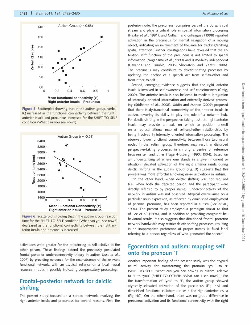
Correlation between functional connectivity andverbal IQ
Among the participants with autism, the functional connectivity
between the right anterior insula and precuneus during the
SHIFT-TO-SELF (What can you see now?) condition was positively
correlated with verbal IQ [r = 0.66, t(14) = 3.17, P50.01], as
shown in Fig. 5, whereas the correlation was not significant in
the control group [r = 0.31, t(13) = 1.18, P = 0.26]. Although
both groups exhibited a positive correlation that did not differ
statistically (z = 1.13, P = 0.26), inspection of the data indicated
that the positive relationship among controls was due to one par-
ticipant with low verbal IQ and a negative functional connectivity
score. When this outlier was removed from the control group, the
Table 3 Areas of activation for the contrasts of all task conditions minus fixation
Region Cluster size t(14) MNI coordinates
x y z
Control
Occipital/inferior parietal/superior parietal/inferior temporal/middle temporal/cerebellum 45 755 16.91 �26 �62 42
Right middle frontal (BA9) 121 6.08 50 34 36
Anterior cingulate (BA32) 162 5.97 12 22 38
Right insula (BA13) 565 5.91 20 30 6
Right superior frontal (BA10) 34 5.38 40 60 �2
Right middle frontal/precentral (BA6) 109 4.98 36 4 62
Right amygdala 57 4.62 30 0 �12
Right middle frontal (BA10) 13 4.62 34 58 26
Right middle frontal (BA6) 19 4.44 24 �12 40
Autism
Occipital/inferior parietal/superior parietal/inferior temporal/middle temporal/cerebellum 43 906 16.53 �34 �56 �32
Left middle frontal/precentral (BA6/9) 2536 9.26 �34 6 64
Right middle frontal/precentral (BA6/9) 1282 7.01 34 4 66
Posterior cingulate 357 6.14 �2 �36 24
Right superior frontal (BA10) 42 5.51 40 60 2
Right inferior frontal (BA45) 72 5.19 56 32 28
Left superior frontal (BA10) 18 4.75 �20 60 0
Cerebellum 18 4.37 4 �54 �26
Right insula (BA13) 26 4.33 36 26 �4
Right middle frontal (BA46) 22 4.32 32 34 24
Right caudate 13 4.30 14 16 20
Right middle frontal (BA10) 13 4.18 34 50 16
Left caudate 26 4.10 �10 12 24
Left postcentral 10 3.92 �54 �18 22
Autism`Control
Right middle frontal/precentral/postcentral (BA6/4/3) 467 4.67 44 �18 60
Right postcentral (BA5) 45 4.41 44 �44 60
Right precuneus (BA19) 12 4.04 22 �86 38
Post cingulate 28 3.97 �6 �36 28
Right posterior middle temporal (BA21) 11 3.92 58 �48 �8
Right middle frontal (BA6) 64 3.92 38 10 36
The threshold for significant activation was P5 0.001 for a spatial extent of at least 10 voxels, uncorrected for multiple comparisons. Region labels apply to the entire extent
of the cluster. T-values and MNI coordinates are displayed for the peak activated voxel in each cluster. For group comparison, there was no area that showed greateractivation for the control group than the autism group.
Neural basis of deictic shifting Brain 2011: 134; 2422–2435 | 2429
Dow
nloaded from https://academ
ic.oup.com/brain/article/134/8/2422/355749 by guest on 16 D
ecember 2021

moderate positive correlation disappeared [r = �0.04,
t(12) = �0.13, P = 0.45], and there was a significant difference
between correlations of the two groups (z = 1.95, P = 0.05).
Thus, the significant correlation in the autism group indicates
that the participants with autism who exhibit greater functional
connectivity between the right anterior insula and precuneus,
when the task required deictic shifting to refer to oneself,
tended to have higher language skill (indicated by high verbal
IQ scores). Other psychometric measures (full scale IQ, perform-
ance IQ, and Autism Diagnostic Observation Schedule scores) did
not show a significant correlation with the functional connectivity
for either group.
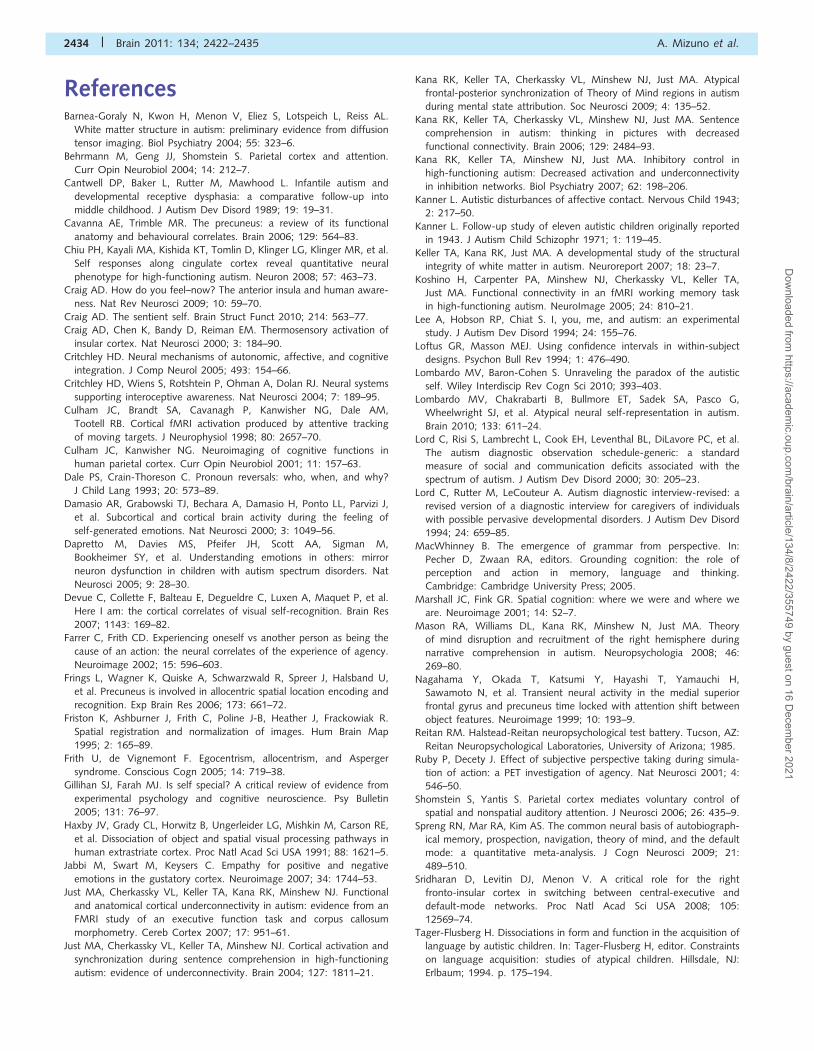
Correlation between functional connectivity andreaction time
Among the participants with autism, the functional connectivity
between the right anterior insula and precuneus during the
SHIFT-TO-SELF (What can you see now?) condition showed a
significant correlation with reaction time [r = �0.51,
t(14) = �2.14, P = 0.03], as shown in Fig. 6, whereas the correl-
ation was not significant in the control group [r = 0.18,
t(13) = 0.63, P = 0.27], and there was a significant difference
between correlations of the two groups (z = 1.78, P = 0.04). Thus,
the correlation in the autism group indicates that the participants
with autism who had lower functional connectivity between the
0.00
0.05
0.10
0.15
0.20
0.25
0.30
0.35
0.40
0.45
0.50
0.00
0.05
0.10
0.15
0.20
0.25
0.30
0.35
0.40
0.45
0.50
0
0.1
0.2
0.3
0.4
0.5
0.6
0.7
SHIFTFIXED
*P = 0.05
*P = 0.04
What can ‘you’ see now?What can ‘I’ see now?
What can John see now?What can Sarah see now?
Mea
n f
un
ctio
nal
co
nn
ecti
vity
(z’)
ControlAutism Autism Control
*P = 0.02
Sig
nal
ch
ang
e (%
)
A
SHIFTFIXEDControlAutism Autism Control
B
C
Sig
nal
ch
ang
e (%
)
SHIFTControlAutism Autism Control
FIXED
Figure 3 FIXED versus SHIFT. Per cent signal change in the precuneus (A) and right anterior insula (B). Functional connectivity between
the right anterior insula and precuneus (C). The error bars represent the 95% confidence interval for the within-subject effect in each
condition. The image (centre) indicates the location of the region of interest (radius = 12 mm) for the precuneus [0, �64, 50] and right
anterior insula [32, 26, 6] on the MNI coordinates.
2430 | Brain 2011: 134; 2422–2435 A. Mizuno et al.
Dow
nloaded from https://academ
ic.oup.com/brain/article/134/8/2422/355749 by guest on 16 D
ecember 2021

right anterior insula and precuneus tended to show a slower re-
sponse when the task required deictic shifting to refer to oneself.
DiscussionThe aim of the study was to evaluate behavioural performance
and compare neural activity between adults with high-functioning
autism and neurotypical adults during deictic shifting (updating
the relationship of generated and referred agents) in a
perspective-taking task. The primary finding was diminished func-
tional connectivity between the right anterior insula and precuneus
in autism when the task required deictic shifting. Although the
functional connectivity between two neural nodes was lower
during a deictic shift in autism, the activation of the frontal
node of the network, the right anterior insula, was increased rela-
tive to when the task used fixed labels of people (i.e. proper
names). Another contribution of the present study was that the
functional connectivity of the same neural network was particu-
larly low when recognizing ‘you’ as referring to the self in autism.
Activations of the posterior neural node, the precuneus, in autism,
were similar for both directions of deictic shifting (transforming
‘you’ to ‘I’ for referencing one’s self, and ‘I’ to ‘you’ for referen-
cing the other person), although the control participants’
0.00
0.05
0.10
0.15
0.20
0.25
0.30
0.35
0.40
0.45
0.50
0.00
0.05
0.10
0.15
0.20
0.25
0.30
0.35
0.40
0.45
0.50
0
0.1
0.2
0.3
0.4
0.5
0.6
0.7
FIXED-OTHERSHIFT-TO-SELFSHIFT-TO-OTHER
What can ‘you’ see now? What can ‘I’ see now?
Mea
n fu
nct
ion
al c
on
nec
tivi
ty(z
’)
ControlAutism Autism Control
Sig
nal
ch
ang
e (%
)
A
C
B
Sig
nal
ch
ang
e (%
)
*P= 0.02
ControlAutism Autism ControlSHIFT-TO-OTHER
ControlAutism Autism Control
*P< 0.01
SHIFT-TO-SELFSHIFT-TO-SELF SHIFT-TO-OTHER
Figure 4 SHIFT-TO-SELF versus SHIFT-TO-OTHER. Per cent signal change in the precuneus (A) and right anterior insula (B). Functional
connectivity between the right anterior insula and precuneus (C). The error bars represent the 95% confidence interval for the within-
subject effect in each condition.
Neural basis of deictic shifting Brain 2011: 134; 2422–2435 | 2431
Dow
nloaded from https://academ
ic.oup.com/brain/article/134/8/2422/355749 by guest on 16 D
ecember 2021
activations were greater for the referencing to self relative to the
other person. These findings extend the previously postulated
frontal–posterior underconnectivity theory in autism (Just et al.,
2007) by providing evidence for the near-absence of the relevant
functional network, with an atypical reliance on a local neural
resource in autism, possibly indicating compensatory processing.
Frontal–posterior network for deicticshiftingThe present study focused on a cortical network involving the
right anterior insula and precuneus for several reasons. First, the
posterior node, the precuneus, comprises part of the dorsal visual
stream and plays a critical role in spatial information processing
(Haxby et al., 1991), and Culham and colleagues (1998) reported
activation in the precuneus for mental navigation of a moving
object, indicating an involvement of the area for tracking/shifting
spatial attention. Further investigations have revealed that the at-
tention shift function of the precuneus is not limited to spatial
information (Nagahama et al., 1999) and is modality independent
(Cavanna and Trimble, 2006; Shomstein and Yantis, 2006).
The precuneus may contribute to deictic shifting processes by
updating the anchor of a speech act from self-to-other and
from other-to-self.
Second, emerging evidence suggests that the right anterior
insula is involved in self-awareness and self-consciousness (Craig,
2009). The anterior insula is also believed to mediate integration
of internally oriented information and externally derived process-
ing (Sridharan et al., 2008). Uddin and Menon (2009) proposed
that there is dysfunctional connectivity of the anterior insula in
autism, lowering its ability to play the role of a network hub.
For deictic shifting in the perspective-taking task, the right anterior
insula may provide an axis on which to position oneself
on a representational map of self-and-other relationships by
being involved in internally oriented information processing. The
observed lower functional connectivity between those two neural
nodes in the autism group, therefore, may result in disturbed
perspective-taking processes in shifting a centre of reference
between self and other (Tager-Flusberg, 1990, 1994), based on
an understanding of where one stands in a given moment or
situation. Elevated activation of the right anterior insula during
deictic shifting in the autism group (Fig. 3) suggests that this
process was more effortful (showing more activation) in autism.
On the other hand, when deictic shifting was not required
(i.e. when both the depicted person and the participant were
directly referred to by proper name), underconnectivity of the
network in autism was not observed. Atypical overreliance on a
particular noun expression, as reflected by diminished employment
of personal pronouns, has been reported in autism (Lee et al.,
1994). The present study employed a paradigm similar to that
of Lee et al. (1994), and in addition to providing congruent be-
havioural results, it also suggests that diminished frontal–posterior
network bandwidth may restrict deictic shifting processes, resulting
in an inappropriate preference of proper names (a fixed label
referring to a person regardless of who generated the speech).
Egocentrism and autism: mapping selfonto the pronoun ‘I’Another important finding of the present study was the atypical
neural activity for transforming the pronoun ‘you’ to ‘I’
(SHIFT-TO-SELF: ‘What can you see now?’) in autism, relative
to ‘I’ to ‘you’ (SHIFT-TO-OTHER: ‘What can I see now?’). For
the transformation of ‘you’ to ‘I’, the autism group showed
atypically elevated activation of the precuneus (Fig. 4A) and
diminished functional collaboration with the right anterior insula
(Fig. 4C). On the other hand, there was no group difference in
precuneus activation and its functional connectivity with the right
80
90
100
110
120
130
140
0 0.2 0.4 0.6 0.8 1
Verb
al IQ
sco
res
Autism Group (r = 0.66)
Mean functional connectivity (z’)Right anterior insula – Precuneus
Figure 5 Scatterplot showing that in the autism group, verbal
IQ increased as the functional connectivity between the right
anterior insula and precuneus increased for the SHIFT-TO-SELF
condition (What can you see now?).
1400
1600
1800
2000
2200
2400
2600
2800
3000
3200
3400
0 0.2 0.4 0.6 0.8 1
Rea
ctio
n t
ime
(ms)
Mean Functional Connectivity (z’)Right anterior insula – Precuneus
Autism Group (r = -0.51)
Figure 6 Scatterplot showing that in the autism group, reaction
time for the SHIFT-TO-SELF condition (What can you see now?)
decreased as the functional connectivity between the right an-
terior insula and precuneus increased.
2432 | Brain 2011: 134; 2422–2435 A. Mizuno et al.
Dow
nloaded from https://academ
ic.oup.com/brain/article/134/8/2422/355749 by guest on 16 D
ecember 2021

anterior insula when reversing in the other direction (i.e. ‘I’ to
‘you’, as in ‘What can I see now?’). In order to answer this ques-
tion, the participant could determine that the pronoun ‘I’ referred
to the experimenter independently from one’s own
representational position. These findings indicate that the critical
disturbance in the successful operation of deictic shifting in autism
may be dysfunctional processing when recognizing the self as
a referent of ‘you’, and shifting to map self onto the pronoun
‘I’. An observed positive correlation between functional connect-
ivity and verbal IQ (Fig. 5), and a negative correlation between
functional connectivity and reaction time in autism (Fig. 6), may
also indicate that diminished interregional synchronization
between the right anterior insula and precuneus restricts the
neural communication underlying the shift of a deictic centre
from another person to oneself. Furthermore, the present study
did not require participants to say their responses, but pronoun
reversals in autism may entail the same underlying basis and
reflect a consequence of unsuccessful reversal, resulting in produ-
cing an overt statement of ‘you’ (e.g. saying ‘You can see the
carrot’ when expressing that the participant him/herself is able
to see the carrot).
The recent functional MRI studies of self- and other-
representation in autism provide complementary evidence: a
greater group difference in brain activity has been found for
self-related processing relative to other-related (Chiu et al.,
2008; Lombardo et al., 2010). Therefore, as suggested by Frith
and de Vignemont (2005), idiosyncratic egocentrism in autism
may be characterized as dysfunction of representing the external
world on the basis of understanding its relation to oneself.
Pronoun reversals in autism may reflect a disturbed processing
of understanding of self and other in the reciprocal relationship,
rather than a semantic error to adjust pronominal forms.
LimitationsThe reported under-connectivity in autism in the present study
was derived from measures of synchronization of activation
between the right anterior insula and precuneus, but did not in-
clude measures of white matter tissues that provide anatomical
connectivity (that can be measured by diffusion tensor imaging).
Diffusion tensor imaging studies in autism have previously
reported reduced white matter integrity (Barnea-Goraly et al.,
2004; Keller et al., 2007). Further investigation of the white
matter tracts connecting the right anterior insula and precuneus
may enhance our understanding of the perspective-taking issues in
autism.
Conclusion and future directionsOver six decades ago, Kanner (1946) documented unique refer-
ential expressions with personal pronouns, referred to as pronoun
reversals, among young individuals with autism. The current
state-of-the-art tool, functional MRI, allowed us to assess the
underlying neural basis of deictic shifting as a critical component
of pronoun reversals, and found an elevated level of neural activity
with lower coordination of relevant brain centres. Pronoun rever-
sals are described as idiosyncratic language impairment in autism,
but the findings suggest that they may also characterize an
atypical understanding of the social world because deictic shifting
is embedded in understanding the self- and other-relationship,
which requires the recognition of the self-stance relative to the
other’s existence. MacWhinney (2005) advocates this view by
emphasizing the significance of the ability to flexibly shift the view-
point in social communication in stating that ‘perspective-taking is
at the very core of language structure and higher-cognition’ (p.
198). If the system of perceiving an external world were rooted in
the understanding of self-stance, dysfunction of the system would
affect not only interpersonal interactions but also intrapersonal
cognitive states, such as memory, temporal and spatial mental
navigation, and Theory of Mind, all possibly sharing a common
neural basis (Spreng et al., 2009). Although our findings may be
limited to the disturbed fundamental understanding of self and
other among individuals in autism, such disturbances are apparent
in many different tasks, such as difficulty with motor mirroring
between self and other that may result from mirror neuron
system dysfunction (Dapretto et al., 2005; Williams et al.,
2006), as well as problems with more abstract cognitive self-
and other-representations, possibly resulting from atypical cingu-
late activation that is associated with an altered default-mode
system (Chiu et al., 2008; Lombardo et al., 2010). In particular,
the frontal portion of the system, including medial prefrontal and
anterior cingulate cortex, has shown activation for both self- and
other-processing in the neurotypical population across different
domains (Gillihan and Farah, 2005). An investigation of a potential
differential involvement and an interaction of the medial frontal
regions and insula for self-related processing and social cognition
in autism could enhance our understanding of this neurodevelop-
mental disorder. Furthermore, it may be useful for future
investigations to examine the neural basis of various coordinate
systems within which the various aspects of the world are mentally
represented in autism and control groups. These coordinate
systems include space (here/there), time (now/then), memory
(semantic/episodic), referential frame (egocentrism/allocentrism)
and meta-representation or Theory of Mind (self/other).
AcknowledgementsWe would like to express our sincere appreciation to the individ-
uals and families who generously gave their time and courage to
participate in this research. We also appreciate the assistance of
the members of the Centre of Cognitive Brain Imaging, particularly
Kara Cohen and Jennifer Moore for editorial comments on the
article.
FundingThis research was supported by the Autism Centers of Excellence
Grant HD055748 from the National Institute of Child Health and
Human Development, and the Pre-Doctoral Fellowship 4868 from
the Autism Speaks Foundation.
Neural basis of deictic shifting Brain 2011: 134; 2422–2435 | 2433
Dow
nloaded from https://academ
ic.oup.com/brain/article/134/8/2422/355749 by guest on 16 D
ecember 2021
ReferencesBarnea-Goraly N, Kwon H, Menon V, Eliez S, Lotspeich L, Reiss AL.
White matter structure in autism: preliminary evidence from diffusion
tensor imaging. Biol Psychiatry 2004; 55: 323–6.
Behrmann M, Geng JJ, Shomstein S. Parietal cortex and attention.
Curr Opin Neurobiol 2004; 14: 212–7.
Cantwell DP, Baker L, Rutter M, Mawhood L. Infantile autism and
developmental receptive dysphasia: a comparative follow-up into
middle childhood. J Autism Dev Disord 1989; 19: 19–31.
Cavanna AE, Trimble MR. The precuneus: a review of its functional
anatomy and behavioural correlates. Brain 2006; 129: 564–83.Chiu PH, Kayali MA, Kishida KT, Tomlin D, Klinger LG, Klinger MR, et al.
Self responses along cingulate cortex reveal quantitative neural
phenotype for high-functioning autism. Neuron 2008; 57: 463–73.Craig AD. How do you feel–now? The anterior insula and human aware-
ness. Nat Rev Neurosci 2009; 10: 59–70.
Craig AD. The sentient self. Brain Struct Funct 2010; 214: 563–77.Craig AD, Chen K, Bandy D, Reiman EM. Thermosensory activation of
insular cortex. Nat Neurosci 2000; 3: 184–90.
Critchley HD. Neural mechanisms of autonomic, affective, and cognitive
integration. J Comp Neurol 2005; 493: 154–66.
Critchley HD, Wiens S, Rotshtein P, Ohman A, Dolan RJ. Neural systems
supporting interoceptive awareness. Nat Neurosci 2004; 7: 189–95.Culham JC, Brandt SA, Cavanagh P, Kanwisher NG, Dale AM,
Tootell RB. Cortical fMRI activation produced by attentive tracking
of moving targets. J Neurophysiol 1998; 80: 2657–70.
Culham JC, Kanwisher NG. Neuroimaging of cognitive functions in
human parietal cortex. Curr Opin Neurobiol 2001; 11: 157–63.
Dale PS, Crain-Thoreson C. Pronoun reversals: who, when, and why?
J Child Lang 1993; 20: 573–89.
Damasio AR, Grabowski TJ, Bechara A, Damasio H, Ponto LL, Parvizi J,
et al. Subcortical and cortical brain activity during the feeling of
self-generated emotions. Nat Neurosci 2000; 3: 1049–56.
Dapretto M, Davies MS, Pfeifer JH, Scott AA, Sigman M,
Bookheimer SY, et al. Understanding emotions in others: mirror
neuron dysfunction in children with autism spectrum disorders. Nat
Neurosci 2005; 9: 28–30.
Devue C, Collette F, Balteau E, Degueldre C, Luxen A, Maquet P, et al.
Here I am: the cortical correlates of visual self-recognition. Brain Res
2007; 1143: 169–82.
Farrer C, Frith CD. Experiencing oneself vs another person as being the
cause of an action: the neural correlates of the experience of agency.
Neuroimage 2002; 15: 596–603.
Frings L, Wagner K, Quiske A, Schwarzwald R, Spreer J, Halsband U,
et al. Precuneus is involved in allocentric spatial location encoding and
recognition. Exp Brain Res 2006; 173: 661–72.
Friston K, Ashburner J, Frith C, Poline J-B, Heather J, Frackowiak R.
Spatial registration and normalization of images. Hum Brain Map
1995; 2: 165–89.
Frith U, de Vignemont F. Egocentrism, allocentrism, and Asperger
syndrome. Conscious Cogn 2005; 14: 719–38.
Gillihan SJ, Farah MJ. Is self special? A critical review of evidence from
experimental psychology and cognitive neuroscience. Psy Bulletin
2005; 131: 76–97.
Haxby JV, Grady CL, Horwitz B, Ungerleider LG, Mishkin M, Carson RE,
et al. Dissociation of object and spatial visual processing pathways in
human extrastriate cortex. Proc Natl Acad Sci USA 1991; 88: 1621–5.
Jabbi M, Swart M, Keysers C. Empathy for positive and negative
emotions in the gustatory cortex. Neuroimage 2007; 34: 1744–53.Just MA, Cherkassky VL, Keller TA, Kana RK, Minshew NJ. Functional
and anatomical cortical underconnectivity in autism: evidence from an
FMRI study of an executive function task and corpus callosum
morphometry. Cereb Cortex 2007; 17: 951–61.
Just MA, Cherkassky VL, Keller TA, Minshew NJ. Cortical activation and
synchronization during sentence comprehension in high-functioning
autism: evidence of underconnectivity. Brain 2004; 127: 1811–21.
Kana RK, Keller TA, Cherkassky VL, Minshew NJ, Just MA. Atypical
frontal-posterior synchronization of Theory of Mind regions in autism
during mental state attribution. Soc Neurosci 2009; 4: 135–52.
Kana RK, Keller TA, Cherkassky VL, Minshew NJ, Just MA. Sentence
comprehension in autism: thinking in pictures with decreased
functional connectivity. Brain 2006; 129: 2484–93.Kana RK, Keller TA, Minshew NJ, Just MA. Inhibitory control in
high-functioning autism: Decreased activation and underconnectivity
in inhibition networks. Biol Psychiatry 2007; 62: 198–206.
Kanner L. Autistic disturbances of affective contact. Nervous Child 1943;
2: 217–50.
Kanner L. Follow-up study of eleven autistic children originally reported
in 1943. J Autism Child Schizophr 1971; 1: 119–45.
Keller TA, Kana RK, Just MA. A developmental study of the structural
integrity of white matter in autism. Neuroreport 2007; 18: 23–7.
Koshino H, Carpenter PA, Minshew NJ, Cherkassky VL, Keller TA,
Just MA. Functional connectivity in an fMRI working memory task
in high-functioning autism. NeuroImage 2005; 24: 810–21.
Lee A, Hobson RP, Chiat S. I, you, me, and autism: an experimental
study. J Autism Dev Disord 1994; 24: 155–76.
Loftus GR, Masson MEJ. Using confidence intervals in within-subject
designs. Psychon Bull Rev 1994; 1: 476–490.
Lombardo MV, Baron-Cohen S. Unraveling the paradox of the autistic
self. Wiley Interdiscip Rev Cogn Sci 2010; 393–403.
Lombardo MV, Chakrabarti B, Bullmore ET, Sadek SA, Pasco G,
Wheelwright SJ, et al. Atypical neural self-representation in autism.
Brain 2010; 133: 611–24.Lord C, Risi S, Lambrecht L, Cook EH, Leventhal BL, DiLavore PC, et al.
The autism diagnostic observation schedule-generic: a standard
measure of social and communication deficits associated with the
spectrum of autism. J Autism Dev Disord 2000; 30: 205–23.
Lord C, Rutter M, LeCouteur A. Autism diagnostic interview-revised: a
revised version of a diagnostic interview for caregivers of individuals
with possible pervasive developmental disorders. J Autism Dev Disord
1994; 24: 659–85.
MacWhinney B. The emergence of grammar from perspective. In:
Pecher D, Zwaan RA, editors. Grounding cognition: the role of
perception and action in memory, language and thinking.
Cambridge: Cambridge University Press; 2005.
Marshall JC, Fink GR. Spatial cognition: where we were and where we
are. Neuroimage 2001; 14: S2–7.
Mason RA, Williams DL, Kana RK, Minshew N, Just MA. Theory
of mind disruption and recruitment of the right hemisphere during
narrative comprehension in autism. Neuropsychologia 2008; 46:
269–80.
Nagahama Y, Okada T, Katsumi Y, Hayashi T, Yamauchi H,
Sawamoto N, et al. Transient neural activity in the medial superior
frontal gyrus and precuneus time locked with attention shift between
object features. Neuroimage 1999; 10: 193–9.Reitan RM. Halstead-Reitan neuropsychological test battery. Tucson, AZ:
Reitan Neuropsychological Laboratories, University of Arizona; 1985.Ruby P, Decety J. Effect of subjective perspective taking during simula-
tion of action: a PET investigation of agency. Nat Neurosci 2001; 4:
546–50.
Shomstein S, Yantis S. Parietal cortex mediates voluntary control of
spatial and nonspatial auditory attention. J Neurosci 2006; 26: 435–9.
Spreng RN, Mar RA, Kim AS. The common neural basis of autobiograph-
ical memory, prospection, navigation, theory of mind, and the default
mode: a quantitative meta-analysis. J Cogn Neurosci 2009; 21:
489–510.Sridharan D, Levitin DJ, Menon V. A critical role for the right
fronto-insular cortex in switching between central-executive and
default-mode networks. Proc Natl Acad Sci USA 2008; 105:
12569–74.
Tager-Flusberg H. Dissociations in form and function in the acquisition of
language by autistic children. In: Tager-Flusberg H, editor. Constraints
on language acquisition: studies of atypical children. Hillsdale, NJ:
Erlbaum; 1994. p. 175–194.
2434 | Brain 2011: 134; 2422–2435 A. Mizuno et al.
Dow
nloaded from https://academ
ic.oup.com/brain/article/134/8/2422/355749 by guest on 16 D
ecember 2021

Tager-Flusberg H, Calkins S, Nolin T, Baumberger T, Anderson M,Chadwick-Dias A. A longitudinal study of language acquisition in
autistic and Down syndrome children. J Autism Dev Disord 1990;
20: 1–21.
Tzourio-Mazoyer N, Landeau B, Papathanassiou D, Crivello F, Etard O,Delcroix N, et al. Automated anatomical labeling of activations in SPM
using a macroscopic anatomical parcellation of the MNI MRI
single-subject brain. NeuroImage 2002; 15: 273–89.
Uddin LQ, Kaplan JT, Molnar-Szakacs I, Zaidel E, Iacoboni M. Self-facerecognition activates a frontoparietal ‘mirror’ network in the right
hemisphere: an event-related fMRI study. NeuroImage 2005; 25:
926–35.Uddin LQ, Menon V. The anterior insula in autism: under-
connected and under-examined. Neurosci Biobehav Rev 2009; 33:
1198–203.
Villalobos ME, Mizuno A, Dahl BC, Kemmotsu N, Muller RA. Reducedfunctional connectivity between V1 and inferior frontal cortex
associated with visuomotor performance in autism. NeuroImage2005; 25: 916–25.
Vogeley K, May M, Ritzl A, Falkai P, Zilles K, Fink GR. Neural correlates
of first-person perspective as one constituent of human
self-consciousness. J Cogn Neurosci 2004; 16: 817–27.Wager TD, Rilling JK, Smith EE, Sokolik A, Casey KL, Davidson RJ, et al.
Placebo-induced changes in FMRI in the anticipation and experience
of pain. Science 2004; 303: 1162–7.
Whitney C, Huber W, Klann J, Weis S, Krach S, Kircher T. Neuralcorrelates of narrative shifts during auditory story comprehension.
Neuroimage 2009; 47: 360–6.
Williams JH, Waiter GD, Gilchrist A, Perrett DI, Murray AD, Whiten A.Neural mechanisms of imitation and ‘mirror neuron’ functioning in
autistic spectrum disorder. Neuropsychologia 2006; 44: 610–21.
Zaehle T, Jordan K, Wustenberg T, Baudewig J, Dechent P, Mast FW.
The neural basis of the egocentric and allocentric spatial frame ofreference. Brain Res 2007; 1137: 92–103.
Neural basis of deictic shifting Brain 2011: 134; 2422–2435 | 2435
Dow
nloaded from https://academ
ic.oup.com/brain/article/134/8/2422/355749 by guest on 16 D
ecember 2021