Embed Size (px)
Citation preview

The Nervous System
Human Physiology

Nervous System Overview
• Nervous system = communication system of the body
• Divided into 2 main parts:
– Central nervous system (CNS) – brain and spinal cord
– Peripheral nervous system (PNS) – peripheral nerves through the body
• 31 pairs of spinal nerves
• 12 pairs of cranial nerves
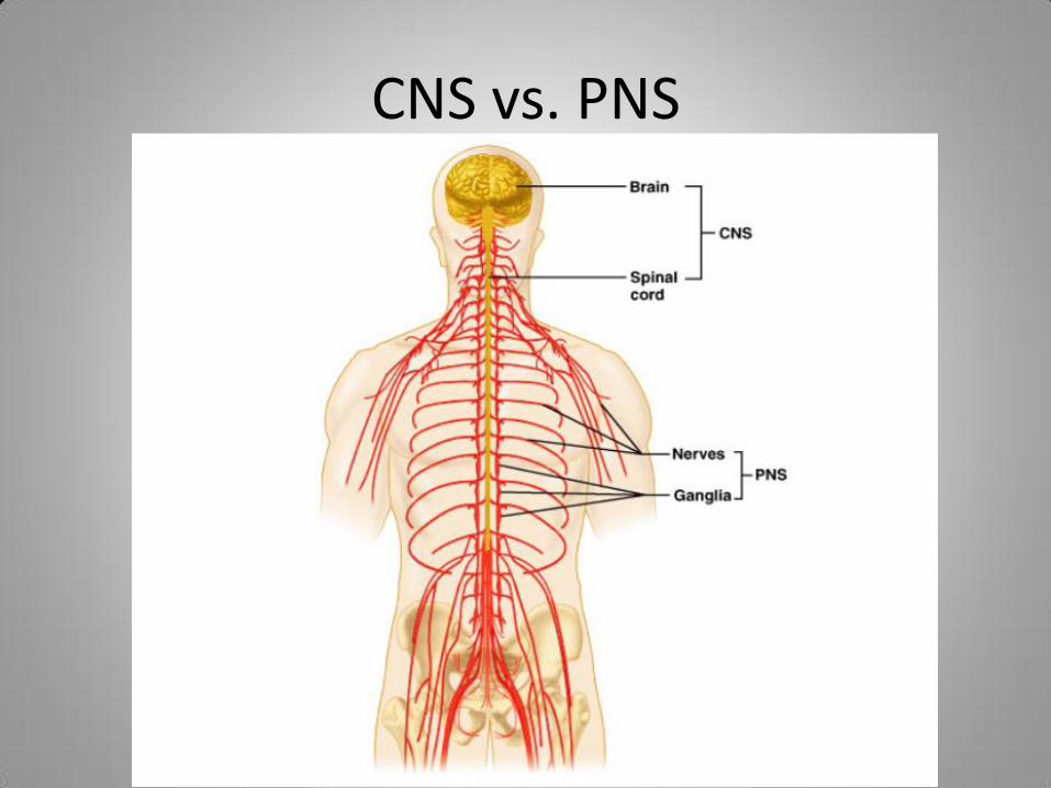
CNS vs. PNS
Nervous System Overview, cont.
• 3 basic functions – Sensory
• Gathers information
– Integrative • Information is “brought together”
– Motor • Responses to signals (impulses)
• Overall Function: Communication of body systems
Nervous System Overview, cont.
• Translates environmental stimuli into messages understood by cells
• Best defined as system of cells, tissues, and organs that regulate the body’s responses to external and internal stimuli
– External – signal from outside the body
– Internal – signal from inside the body

Peripheral Nervous System
• Somatic
– Skeletal muscles
– Under voluntary control
• Autonomic
– Smooth muscles, glands
– involuntary

Neural Tube
• Nervous system starts out as a hollow neural tube of ectoderm that runs lengthwise along the back of the developing fetus – Produces stem cells that form the two main cell lines
of the nervous system • Neurons
– Operational component of nervous system
– Excitable cells that receive, interpret, and transmit information
• Neuroglia (later in chapter) – Not directly involved in nervous system communication
– Important for development and repair of nervous system

Neurons • Four common features:
– Nerve cell body (soma) • Houses nucleus and organelles needed for neuron function
– Dendrite • “antennae” of neuron • Carry information to the nerve cell body
– Axon • Fiber-like extension of a neuron that sends information to terminus • Comes off nerve cell body at region termed axon hillock
– Axon hillock – swelling at cell body where axon begins
• Usually 1 per neuron, some can have branches or “collaterals” that reach to other neurons
– Terminus • Releases neurotransmitters that transmit information from neurons to
other cells
• Identified primarily by arrangement of 4 components

Neurons

Neuron Shape
• Bipolar (Interneurons) – 1 dendrite, 1 axon
• Sometimes called interneurons – communicate information only from one neuron to another
• Multipolar (motor) – Many dendrites that attach directly to the nerve body
• Most numerous type of neuron • Communicate info from brain to glands and muscles
• Unipolar (sensory) – Long axon that connects dendrites to terminus – Nerve cell body positioned to the side of axon – Carry sensory information


Synapse
• Neurons do not make direct contact with cells
– Synapse – junction where impulses are transmitted between neurons
• Synaptic cleft – gap at junction
– Presynaptic cleft
» Sends signal across synapse
– Postsynaptic cleft
» Receives signal across synapse

SYNAPSE

Neurotransmitters
• Sensitivity of postsynaptic cleft depends on number of neurotransmitter receptors – Neurons with fewer receptors are less sensitive and
vice versa
• Neurons can adjust # of neurotransmitters in response to variety of conditions
• Drugs, such as morphine, mimic natural neurotransmitters – alter brain cells – After long-term use, brain reduces # of receptors to
desensitize the neurons to continuous stimulation • Person develops addiction to morphine because body needs
it for new level of normal receptor function
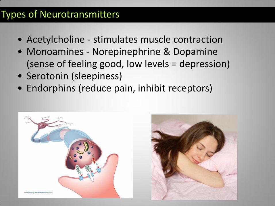
Types of Neurotransmitters
• Acetylcholine - stimulates muscle contraction • Monoamines - Norepinephrine & Dopamine
(sense of feeling good, low levels = depression) • Serotonin (sleepiness) • Endorphins (reduce pain, inhibit receptors)

8.2 Neuroglia and Stem Cells
• Make up bulk of cell types in nervous system
• Most found in the brain
• Carry out crucial roles in regulating environment of neurons
• Closely assist neurons with their function
• Have high lipid content
– Give them shiny, white appearance
– Also makes them vulnerable to deterioration with poor diet

Astrocytes
• Or, macroglia – largest class of neuroglia
• Connect neurons to blood vessels
• Star-like shape
• Maintain chemical environment of neurons
• Helps form protective features called blood-brain barrier – Restricts passage of materials from the blood into the
brain
• Mostly found in brain and spinal cord
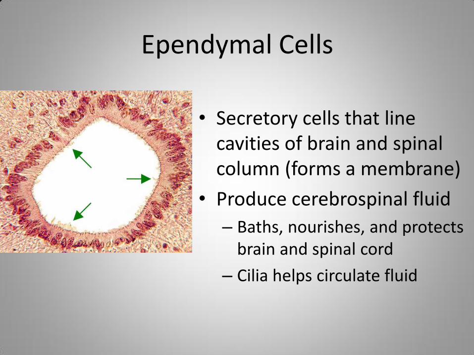
Ependymal Cells
• Secretory cells that line cavities of brain and spinal column (forms a membrane)
• Produce cerebrospinal fluid
– Baths, nourishes, and protects brain and spinal cord
– Cilia helps circulate fluid

Microglia
• Highly variable
• Carry out phagocytosis – removes infectious agents and repairs nervous system damage
– Digest debris or bacteria
• respond to immunological alarms
• Microglia malfunctions = bases of many nervous system disorders

Oligodendrites
• Large – many branching processes
• Form insulating cover called myelin sheath
– Sheath facilitates transmission of stimuli along the axon
– Limited to neurons in the brain and spinal cord

Radial Glia
• Found in developing nervous system
• Provide framework that organizes connections of neurons
• Communicate needs of certain neurons

Satellite Cells
• Very numerous, small cells that cover the surface of neurons outside of the brain and spinal cord
• Help maintain chemical environment of neurons
• May also assist with nerve cell repair

Schwann Cells
• Form a myelin sheath around neurons located outside of the brain and spinal cord
• Slight gaps between Schwann cells are called nodes of Ranvier
– Responsible for carrying stimuli along the axon’s length


Neural Crest Cells
• New discovery, type of stem cell in adults
• Confirmed in rats
– May also be found in hair follicles
• Capable of producing neuroglia, neurons, and more stem cells
• Not yet possible to relocate neural crest cells to repair nervous system damage
Reflexes
• Defined as involuntary responses to a stimulus
• Reflex arc – neuron arrangement that links a stimulus to a response without requiring the intervention of conscious control

Afferent and Efferent
• Afferent neuron - neuron conducts impulses inward to a nerve center in brain or spinal cord (transmits sensory info
• Efferent neuron – conducts impulses outwards from nerve center in brain or spinal cord (transmits motor information)
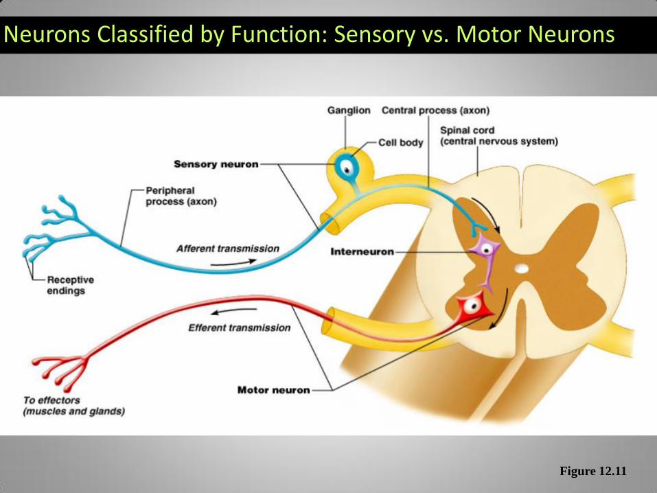
Neurons Classified by Function: Sensory vs. Motor Neurons
Figure 12.11

Generating a Nerve Impulse
• Inner surface of cell membrane has an electrical charge different from the outside of the cells, this difference is the membrane potential
– Expressed in voltage
• To communicate nerve impulses changes in these electrical charges occur and send an electrical signal called an action potential
Resting Potential
• When a neuron is not conducting a nerve impulse
– It is negative
• Sodium-potassium pump (3 Na+ out, 2 K+ in)
• Cell membrane allows K+ ion to diffuse, removes positive charge
• Neurons contain negatively charged molecules too big to diffuse

Action Potential
• When stimulus is received, cell membrane potential changes
• Na+ channels will open, allowing Na+ to flow into the cell
– Makes cell less negative (so more positive)
• Triggers K+ channels to open, and K+ flows out of cell making cell return to resting state
– Resting state is negative again

Threshold
• Cells receive stimuli of different strength
• Action potential is “all or nothing”
– Stimulus either will or will not produce action potential
• When a stimulus causes an action potential we say its reached the threshold
– If stimulus is too weak to trigger threshold an action potential will not start

Neuron Physiology
• Neurons at resting potential – Higher concentration of sodium ions outside cell
than inside, potassium ions are higher inside the cell, creates diffusion gradient
– Stimulus received and sodium channels open, allows neuron to reach threshold, -55mV
• Internal charge of neuron falls below -70mV and starts chain reaction called action potential – Travels from the dendrites down axon to terminus

Neuron Physiology
• At threshold more sodium channels start to open changes cytoplasm to +30mV called Depolarization
• Sends stimulus down length of axon
• Sodium ions channels close and potassium channels open and positive potassium will flow out, loss of K+ negative charge returns, neuron repolarizes, and returns to resting potential

Hyperpolarization
• When the repolarizing stage goes beyond the resting potential of -70mV and reaches -90mV
• 2 purposes:
– Prevents another stimulus during repolarization stage
– Prevents action potential from traveling in the opposite direction once it reaches terminus

8.3 Pathology of Nervous System Function
• Nerve cells can be injured by infectious agents or by damage to other body parts
• Also subject to disorders caused by DNA defects and poisoning
• Neuroglia as well as neuron components, particularly the axon, are subject to nervous system pathologies

Nerve Cell Disease Categories
• Infectious – caused by microorganisms capable of invading or infecting nervous system cells
• Degenerative – progressive deterioration of a cell or tissue over time
• Congenital – diseases caused by embryological and maturation errors
• Toxicological – caused by poisons
• Traumatic – injuries resulting from a wound that was caused by external force or violence

Infectious Diseases
• Botulism – bacteria that grows in spoiled foods
– Causes flaccid paralysis
• Tetanus – bacteria that prevents muscles from relaxing
• Endotoxins – toxic substance found in certain bacteria
– Diseases caused by endotoxins
• Encephalitis – inflammation of brain
• Meningitis – inflammation of the membranes surrounding the brain and spinal cord

Infant Botulism: •Infant botulism occurs when spores from C. botulinum are swallowed from either ingestion or breathing. •Usually occurs in otherwise healthy children under the age of one and rarely affects older children and adults due to stronger immune systems •One of the only foods founds to have a relatively high occurrence of C. botulinum spores is honey.
Wound Botulism: •occurs when the spores of C. botulinum manage to enter an open wound to enter the body. • Because of the general difficulty of this, wound botulism is the rarest of all cases, only counting for 3% of Botulism cases around the globe.
Foodborne Botulism: • occurs after ingesting food contaminated with C. botulinum. •Nearly all cases throughout the world can be directly correlated to improper food preservation. •It can be experienced as a minor illness, or a disease that could be fatal within a day’s time.

Tetanus
• Preventing is easier than treating
• Babies are vaccinated against tetanus from the time they are two months old until they reach the age of 6 or 7, sometimes as late as age 10. – Thereafter adults should receive
their tetanus vaccination every ten years.
Infectious diseases, cont.
• Neurotrophic – refers to an organism that infects nerve tissue
– Neurotrophic diseases
• Herpesvirus – inflammatory virus that grows in nervous tissue
• Rabies – spread through saliva of infected animals
• Mad cow disease (bovine spongiform encephalopathy & Creutzfeldt-Jacob disease) – causes degeneration of the nervous system

Herpes • 1 in 9 adults in USA currently has the
virus that can cause genital herpes. • More than 90% of the people who are
infected with genital herpes don’t even realise they have the virus in the first place.
• The symptoms of herpes can vary widely from person to person. Some people will suffer sores, rashes or blisters. This can occur on the genitals, thighs, back, chest or even hands.
• There is currently no cure for Herpes – but if diagnosed your doctor can give you anti-viral treatments to keep it under control.
• Most people who have a slight rash do not even realise they are infected.
• Herpes spreads easily as it spreads best when no symptoms are obvious.

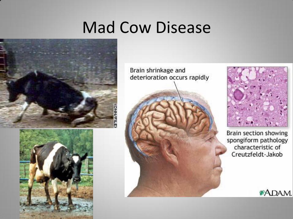
Mad Cow Disease

Genetic Degenerative Disorders
• Amyotrophic lateral sclerosis (ALS) or Lou Gehrigs disease – caused by faulty mitochondria passed down by the egg – Affects motor nerves, causing gradual cessation of
muscle function
• Demyelination – loss of neuroglia around the axons and bodies of neurons – Causes slower neural impulses and eventual
degeneration of neuron
• Multiple Sclerosis (MS) – prominent demyelinating disease whose cause is unknown

ALS – Lou Gehrigs
• A person with ALS usually presents with problems in dexterity or gait resulting from muscle weakness, or with difficulty speaking or swallowing. Sphincter control, sensory function, intellectual ability, and skin integrity are preserved. Patients become paralyzed and often require ventilation and surgery to provide a new opening in the stomach (gastrostomy). Loss of respiratory function is ultimately the cause of death.
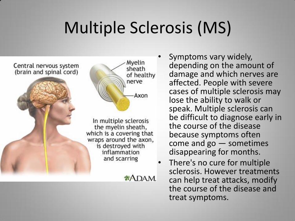
Multiple Sclerosis (MS)
• Symptoms vary widely, depending on the amount of damage and which nerves are affected. People with severe cases of multiple sclerosis may lose the ability to walk or speak. Multiple sclerosis can be difficult to diagnose early in the course of the disease because symptoms often come and go — sometimes disappearing for months.
• There's no cure for multiple sclerosis. However treatments can help treat attacks, modify the course of the disease and treat symptoms.
Congenital Disorders
• Can alter the metabolism of the nervous system cells
• Galactosylceramide beta-galactosidase – enzyme that prevents the accumulation of toxic wastes in nerve cells
• Krabbe’s disease – starts out in the embryo, causes buildup of harmful fats – Causes abnormal functioning of neurons and diminishes
neuroglia maturation
• Hirschsprung’s disease – develops before child is born – Specific to neurons of large intestine
– Nerve cells stop growing, causing a loss of large intestine function

Krabbe’s Disease
• Krabbe’s disease is a rare, inherited degenerative disorder of the central and peripheral nervous systems.
• The disease most often affects infants, with onset before age 6 months, but can occur in adolescence or adulthood.
• Symptoms include irritability, unexplained fever, limb stiffness, seizures, feeding difficulties, vomiting, and slowing of mental and motor development. – Other symptoms include muscle weakness, spasticity,
deafness, and blindness.
• There is no cure for Krabbe’s disease.
• Infantile Krabbe disease is generally fatal before age 2

Hirschsprung’s Disease • Condition that affects the large intestine
(colon) and causes problems with passing stool.
• Present when a baby is born (congenital) and results from missing nerve cells in the muscles of a portion of the baby's colon.
• Children with Hirschsprung's disease are often constipated. – In severe cases of Hirschsprung's disease, a
newborn child experiences an obstructed colon and is unable to have a bowel movement. • In mild cases, doctors may not detect
Hirschsprung's disease until later in a child's life.
• Hirschsprung's disease is treated with surgery to remove the affected portion of the colon. After surgery, most children pass stool normally.

Toxicological
• Nervous system poisoning
• Metals, such as lead (lead poisoning), slow neuron function – Can accumulate to toxic levels
• Arsenic and cyanide are found in pesticides – Can enter body through food, water, etc
• Block cellular respiration and disable neurons
• Tetrodotoxin – exotic form of neuron poison – Found in tropical frogs and the puffer fish
• Prevents the flow of sodium cells into nerve cells

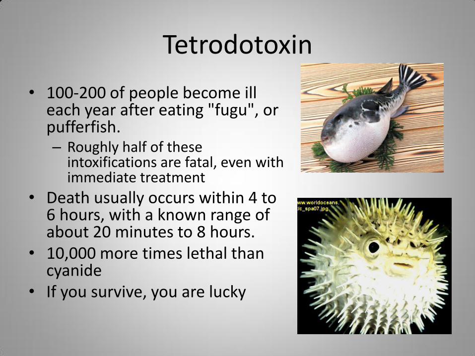
Tetrodotoxin
• 100-200 of people become ill each year after eating "fugu", or pufferfish. – Roughly half of these
intoxifications are fatal, even with immediate treatment
• Death usually occurs within 4 to 6 hours, with a known range of about 20 minutes to 8 hours.
• 10,000 more times lethal than cyanide
• If you survive, you are lucky

Traumatic Damage
• Athletic injuries, automobile accidents, and work-related falls are common causes
• Neurons cannot be replaced once they die
– Injured neurons can be repaired as long as intact neuroglia are nearby

9.1 Overview
• Central nervous system (CNS)
– Brain and spinal cord
– Control network for entire body
• Peripheral nervous system (PNS)
– Provide motor and sensory communication between CNS and body
• Sensory information travels from PNS to CNS
• Motor information travels from CNS to PNS

Overview, cont.
• In fetus, CNS first component of nervous system to develop
– Starts out as hollow tube called neural tube
• Neuroglia spread to various parts of the body to form PNS
• Organs of nervous system = nerves

Central Nervous System
• CNS isolated from rest of body by layers of membranes encased in bony covering
– Brain lies within cranial cavity
• Directly connects to spinal cord through foramen magnum in occipital bone – Spinal cord runs through flexible
tunnel called vertebral canal
» Major blood vessels enter and exit here

Meninges • Three layers of tissue called meninges surround the
brain and spinal cord – Dura mater – outermost
• Pressed tightly against bony surface of cranium
• Acts as barrier against trauma
– Arachnoid mater • Thin, delicate
• “like a spiders web”
• Cushion the CNS from rapid movements and blunt hits – Subarachnoid space – cavity between arachnoid matter and pia mater
» Filled with cerebrospinal fluid, many blood vessels
– Pia mater • Makes direct contact with brain and spinal cord
• Carrying the blood vessels into CNS
• Forms sheaths over nerves passing through outer meninges layers
• Assists in production of cerebrospinal fluid

Meninges of Brain

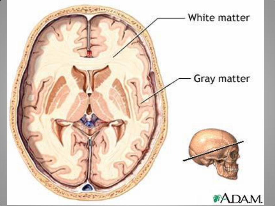
CNS, cont. • Noted for patterns for gray and white color
when viewed without microscope
• Gray matter – Composed of concentrated areas of neuron cell
bodies
– Gray because neurons accumulate the dark-pigmented fat called lipfuscin
– Mostly on surface, or cortex
• White matter – Color comes from light-color fats making up
myelin cell membranes

Brain
• Developmentally divided into three parts – Forebrain
• Largest division of embryonic brain
– Midbrain • Neurons that connects with the forebrain and organizes
sensory information
– Hindbrain • Lowermost portion of embryonic brain, just above spinal
cord
• All develop specific functions as they develop – Parts work closely together to coordinate control

Forebrain…..
• Forms the cerebrum and diencephalon (largest and most anterior portion of brain)
• Responsible for emotions, memory, motor development, and thought
• Cerebral cortex – gray matter covering the brain – If stretched out, would cover an area 18 sq. ft.
– Nourished by blood vessels that form blood-brain barrier • Prevents harmful substances from entering brain
– Also selective against many medications… a constant problem.
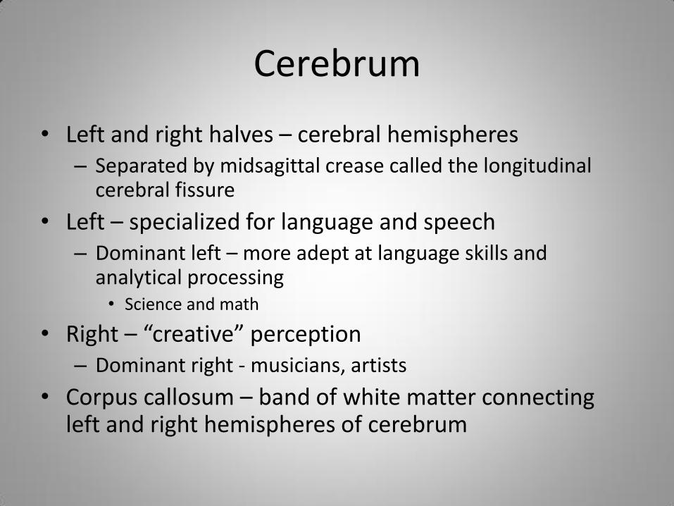
Cerebrum
• Left and right halves – cerebral hemispheres – Separated by midsagittal crease called the longitudinal
cerebral fissure
• Left – specialized for language and speech – Dominant left – more adept at language skills and
analytical processing • Science and math
• Right – “creative” perception – Dominant right - musicians, artists
• Corpus callosum – band of white matter connecting left and right hemispheres of cerebrum

Brain Lobes • Hemispheres further divided into four distinct
anatomically regions called lobes – Frontal lobe - Processes intellectual information
and helps organize thoughts
– Parietal lobe – involved mostly in emotions and sensory information
– Temporal lobe – organizes and stores the memories of sounds and vision
– Occipital lobe – interprets vision and assists with eye function


Midbrain…
• Small strip of neurons that connects the cerebrum to the hindbrain
– Posses auditory and visual reflex areas
– Controls ability of the eye to adjust to changes
– Coordinates motor function
• Produce neurotransmitter dopamine
– Decrease responsible for Parkinson’s
• Directly attached to midbrain is the pons

Hindbrain…
• Pons is the first component of hindbrain
– Medulla oblongata – connects pons to hindbrain
• Regulates involuntary body functions – Blood pressure, breathing, heart rate, and swallowing
• Cerebellum – posterior to pons – 3rd component of hindbrain
– Balance, posture, coordination of body movements
• Brainstem – midbrain and hindbrain collectively….
Spinal Cord
• Extends from base of medulla oblongata
• Spinal cord is nervous tissue enclosed in vertebral column
• Meninges cover outer surface
• Contains hollow core called central canal – Canal contains cerebral spinal fluid
• Spinal cord narrows down to loose strands of nerve tracts that exit the sacral and coccygeal vertebrae
9.2 Peripheral Nervous System
• Composed of nerves that branch out from brain and spinal cord – Not covered with meninges – Do not have cavity containing cerebrospinal fluid
• 2 categories: – Somatic
• Enable voluntary control of the body – Afferent neurons – collect sensory information – Efferent neurons – relay commands for muscle action
– Autonomic • Controls involuntary body functions • Maintain stable internal environment for body

Neurons Classified by Function: Sensory vs. Motor Neurons
Figure 12.11

Cranial Nerves
• Carry out specialized tasks associated with the somatic and autonomic nervous system
• Identified by a Roman numeral and an anatomical name
• Head injuries can hinder action of cranial nerves
– Locate nerve damage by the evaluating body functions affected

Cranial Nerves
I. Olfactory – transmits smell to brain II. Optic – transmits vision to the brain III. Oculomotor – controls eye and eyelid
movement IV. Trochlear – controls downward and lateral eye
movement V. Trigeminal – transmits sensory information from
face and mouth to brain VI. Abducens – tarnsmits signals for lateral eye
movement

Cranial Nerves, cont.
VII. Facial – controls facial expressions, and mouth and eye secretions
VIII.Vestibulocochlear – transmits sensations of balance and hearing to brain
IX. Glossopharyngeal – sensory information from skin to tongue – controls swallowing and movement of food through dig. System
X. Vagus – transmits cardiovascular reflexes – controls heart rate and digestion
XI. Accessory – controls swallowing, movements of neck and shoulder
XII. Hypoglossal – controls tongue movement

Spinal Nerves
• 31 branch off a various regions of the spinal cord
• Carry motor and sensory info for reflex control and two-way communication with the brain
• Dorsal root – sensory branch – goes into dorsal portion of spinal column
– Forms bulge called dorsal root ganglion
• Ventral root – motor nerves

Autonomic Nervous System
• Uses cranial and spinal nerves to perform tasks
• Activities do not require conscious thought
• 2 anatomically and functionally distinct regions – Parasympathetic nerves
• Emerge from cranial and sacral spinal nerves
– Sympathetic nerves • Arise from thoracic and lumbar spinal cord
– Each counteract each other to fine tune the body’s responses to internal changes and environmental stimuli

Autonomic Nervous System, cont.
• Autonomic – referred to the “fight or flight, and rest to digest” system
– Prepares body to react to stress
• Parasympathetic – promotes relaxation and digestion
• Know the table on page 346
– Autonomic nervous system
• Sypmathetic vs. parasympathetic action