Embed Size (px)
Citation preview

The Nervous System
The Spinal Cord-part of the CNS found within the spinal column
The spinal cord communicates with the sense organs and muscles below the level of the head
Bell-Magendie Law-the entering dorsal roots carry sensory information and the exiting ventral roots carry motor information to the muscles and glands
Dorsal Root Ganglia-clusters of neurons outside the spinal cord

Spinal Cord
• Extends from foramen magnum to second lumbar vertebra
• Segmented– Cervical
– Thoracic
– Lumbar
– Sacral
• Gives rise to 31 pairs of spinal nerves
• Not uniform in diameter throughout length

The spinal cord and spinal nerves

Gross anatomical structures of the spinal cord
A. Two enlargements of the spinal cord occur in
regions where the amount of gray matter is greater than
others; regions of sensory/motor control of the limbs.
1. Cervical enlargement - nerves supply the shoulder and
upper limbs;
2. Lumbar enlargement - nerves supply the pelvis and the
lower limb.
B. Conus medullaris: region at the end of the spinal cord
where the tissue tapers into a cone shape; approximately
in region of L2.
C. Filum terminale: connective tissue, continuous with
the pia mater of the spinal cord.
D. Cauda equina: extension of nerves exiting from the
base of the spinal cord; appears like a horse's tail.

Cross Section of Spinal Cord

Cross Section of Spinal Cord
• White matter:• Myelinated axons
forming nerve tracts• Fissure and sulcus• Three columns:
– Ventral – Dorsal– Lateral
• Gray matter:• Neuron cell cell bodies,
dendrites, axons• ‘Horns’:
– Posterior (dorsal)– Anterior (ventral)– Lateral
• Commissures:– Gray: Central canal – White
(see later for white matter pathways)

Spinal CordIn the spinal cord, the grey matter is found in the centre (the butterfly shape) and the white matter surrounds it. The opposite arrangement is found in the cortex of the brain. Notice the surrounding dura mater .

See ANS lecture

‘Plexus’:intermingling / merging of nerves and subsequent re-organisation--> distribution of peripheral nerves is different from that of spinal nerves


Dermatomal Map• Skin area supplied with sensory innervation by spinal nerves
Equivalent for motor output to muscle groups:Myotome

CNS
PNSsensory motor motor sensory
spinal nerves (31p) cranial nerves (12p)
spinal cord brain
The Organisation of the Nervous System
• Sensory information has to be passed on from the spinal cord to the brain ascending pathways (red)
• Commands from the brain have to be sent out to the PNSdescending pathways (green)

Ascending (afferent) spinal tracts:
• Pathways that carry sensory information to a conscious level
Basic principle of information flow:
• receptor(e.g. pain receptor in skin)
• primary sensory neurone (cell body in dorsal root ganglia)
• second order neurone (in the spinal cord or brainstem)
• third order neurone (in thalamus)
• target area: cortexsomatosensory (somatic sensory) area (postcentral gyrus) of the cortex

Light touch
Conscious perception
Example

Sensory information travels to the brain via 3 main pathways:
To somatosensory cortex (see lecture 3!):1) Spinothalamic system (lateral and anterior tract):somatosensory information to brain:pain and temperature, light touch, pressure, tickle, itch
2) Dorsal column / medial lemniscus:Two-point discrimination, proprioception, pressure, vibration
To cerebellum:3) Spinocerebellar system (posterior and anterior):proprioception, for comparator function (see lecture 2!)
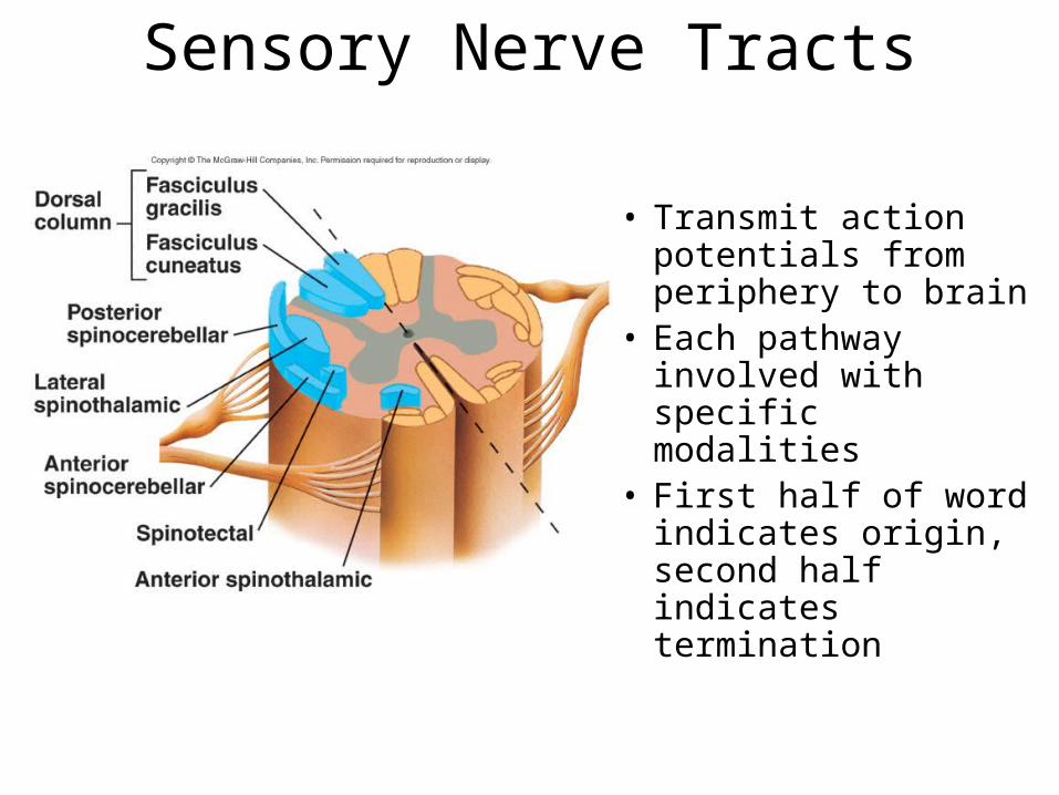
Sensory Nerve Tracts
• Transmit action potentials from periphery to brain
• Each pathway involved with specific modalities
• First half of word indicates origin, second half indicates termination
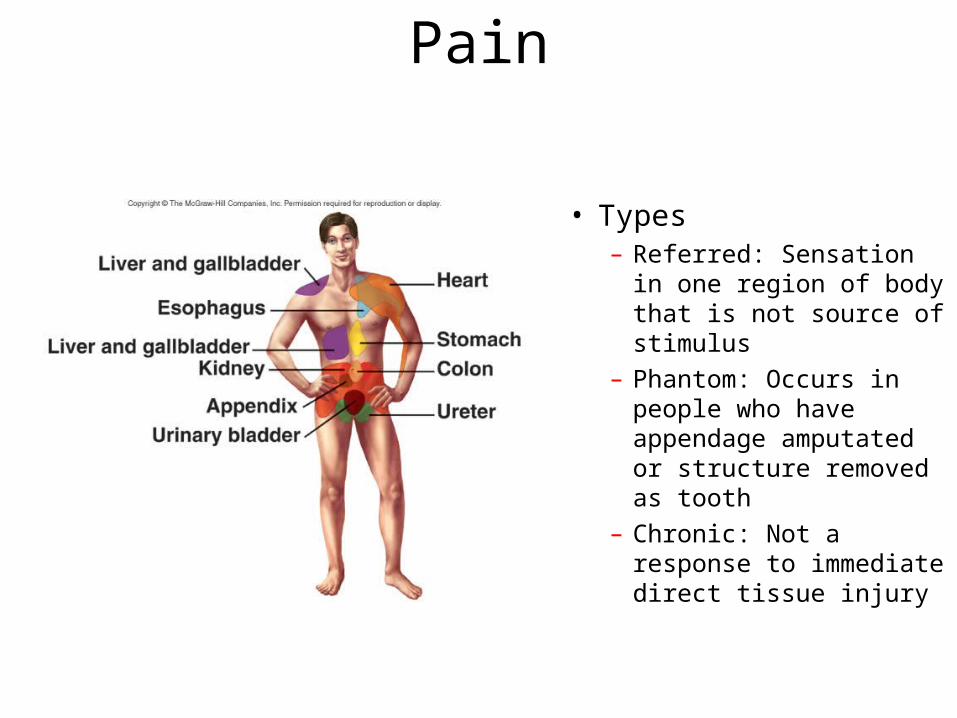
Pain
• Types– Referred: Sensation in one
region of body that is not source of stimulus
– Phantom: Occurs in people who have appendage amputated or structure removed as tooth
– Chronic: Not a response to immediate direct tissue injury

Descending (efferent) spinal tracts:
• Pathways that carry motor commands from the brain to the muscle
• 2 Systems: direct (pyramidal) and indirect (extrapyramidal)
Basic principle of information flow:
1) Pyramidal System• initiation in (pre-) motor cortex
• upper motor neurone (from motor cortex to brain stem or spinal cord via pyramids) • lower motor neurone (target: muscle)
2) Extrapyramidal System• originate in motor cortex and cerebellum(4 different pathways)
• involve projections via brainstem nuclei
• lower motor neurones(target: muscle)
Unconscious movements(posture, balance, reflexes)
Functions:Muscle tone,voluntary movement

Pyramidal system

SUMMARY: PATHWAYS IN THE SPINAL CORD
Ascending (afferent) pathways (sensation) Descending (efferent) pathways (motor commands)
*
*

Spinal Cord AnatomySpinal Cord Anatomy
Slide 7.53c
Copyright © 2003 Pearson Education, Inc. publishing as Benjamin Cummings
Central canal filled with cerebrospinal fluid
Figure 7.19

Spinal Cord AnatomySpinal Cord Anatomy
Slide 7.54Copyright © 2003 Pearson Education, Inc. publishing as Benjamin Cummings
Meninges cover the spinal cord
Nerves leave at the level of each vertebrae Dorsal root
Associated with the dorsal root ganglia – collections of cell bodies outside the central nervous system
Ventral root

Peripheral Nervous SystemPeripheral Nervous System
Slide 7.55Copyright © 2003 Pearson Education, Inc. publishing as Benjamin Cummings
Nerves and ganglia outside the central nervous system
Nerve = bundle of neuron fibers
Neuron fibers are bundled by connective tissue

Structure of a NerveStructure of a Nerve
Slide 7.56Copyright © 2003 Pearson Education, Inc. publishing as Benjamin Cummings
Endoneurium surrounds each fiber
Groups of fibers are bound into fascicles by perineurium
Fascicles are bound together by epineurium
Figure 7.20

Classification of NervesClassification of Nerves
Slide 7.57Copyright © 2003 Pearson Education, Inc. publishing as Benjamin Cummings
Mixed nerves – both sensory and motor fibers
Afferent (sensory) nerves – carry impulses toward the CNS
Efferent (motor) nerves – carry impulses away from the CNS

Spinal NervesSpinal Nerves
Slide 7.63Copyright © 2003 Pearson Education, Inc. publishing as Benjamin Cummings
There is a pair of spinal nerves at the level of each vertebrae for a total of 31 pairs

Spinal NervesSpinal Nerves
Slide 7.64Copyright © 2003 Pearson Education, Inc. publishing as Benjamin CummingsFigure 7.22a

Autonomic Nervous SystemAutonomic Nervous System
Slide 7.67Copyright © 2003 Pearson Education, Inc. publishing as Benjamin Cummings
The involuntary branch of the nervous system
Consists of only motor nerves
Divided into two divisions
Sympathetic division
Parasympathetic division

Comparison of Somatic and Comparison of Somatic and Autonomic Nervous SystemsAutonomic Nervous Systems
Slide 7.69Copyright © 2003 Pearson Education, Inc. publishing as Benjamin Cummings Figure 7.24

Anatomy of the Autonomic Nervous Anatomy of the Autonomic Nervous SystemSystem
Slide 7.73Copyright © 2003 Pearson Education, Inc. publishing as Benjamin Cummings
Figure 7.25

Autonomic FunctioningAutonomic Functioning
Slide 7.74a
Copyright © 2003 Pearson Education, Inc. publishing as Benjamin Cummings
Sympathetic – “fight-or-flight”
Response to unusual stimulus
Takes over to increase activities
Remember as the “E” division = exercise, excitement, emergency, and embarrassment

Autonomic FunctioningAutonomic Functioning
Slide 7.74b
Copyright © 2003 Pearson Education, Inc. publishing as Benjamin Cummings
Parasympathetic – housekeeping activites
Conserves energy
Maintains daily necessary body functions
Remember as the “D” division - digestion, defecation, and diuresis

Development Aspects of the Development Aspects of the Nervous SystemNervous System
Slide 7.75a
Copyright © 2003 Pearson Education, Inc. publishing as Benjamin Cummings
The nervous system is formed during the first month of embryonic development
Any maternal infection can have extremely harmful effects
The hypothalamus is one of the last areas of the brain to develop

Development Aspects of the Development Aspects of the Nervous SystemNervous System
Slide 7.75b
Copyright © 2003 Pearson Education, Inc. publishing as Benjamin Cummings
No more neurons are formed after birth, but growth and maturation continues for several years (new evidence!)
The brain reaches maximum weight as a young adult
However, we can always grow dendrites!