Embed Size (px)
Citation preview

THE NERVOUS SYSTEM I

The study of nervous system histology could easily comprise the entire subject matter of this course and a couple more. Thus, we will only be considering a broad overview of the subject.
I. Two major components of nervous system.
A. central nervous system (CNS) - brain, spinal cord
http://www.student.loretto.org/anatomyphys/Coordination-%20nervous%20system.htm
B. peripheral nervous system (PNS)
1) axonal processes extending toward or away from CNS (nerves)
2) ganglia (aggregations of nerve cell bodies outside CNS).

III. If unstained brain or spinal cord are sectioned, we find that two major areas of brain tissue may be defined on the basis of color.
A. gray matter - neuron perikarya (cell bodies), glial cells, axons, dendrites, synapses
B. white matter - axons + myelin sheaths, oligodendroglia, other glial cells, no neuron perikarya, no synapses.

II. Nervous tissue consists of two major types of cells
1. neurons - responsible for conduction, propagation, and reception of nervous impulses. Processes called axons or dendrites extend from these cells.
2. glial cells - (neuroglia) cells associated with neurons. No axons or dendrites. These cells are involved in nutrition, support, insulation, protection of neurons.
Copyright Dennis Kunkelhttp://www.pbrc.hawaii.edu/~kunkel/gallery

IV. BASIC NEURON STRUCTURE

IV. BASIC NEURON STRUCTUREA. Perikaryon - nerve cell body, contains nucleus and typical cell organelles
1. nucleus - large, central in most, large amount of euchromatin (intense synthetic activity), nucleolus, Barr body in females (Dormant X chromosome of females).
http://www.udel.edu/Biology/Wags/histopage/colorpage/cne/cnemnns.GIF
2. rough endoplasmic reticulum (RER) - lots of RER for synthesis of structural and transport proteins, Nissl bodies/substance seen with light microscope are condensations of this RER and free ribosomes.
3. golgi apparatus - only found near nucleus in perikaryon. Expected, since intense synthetic activity of neurotransmitters and/or neurohormones that must be packaged in vesicles.
Nissl bodies

4. mitochondria - abundant for high energy requirements
Copyright Dennis Kunkelhttp://www.pbrc.hawaii.edu/~kunkel/gallery
5. Neurofilaments - intermediate filaments (10 nm)
7. inclusions - pigment deposits - function unknown. Lipofuscin deposits - residual bodies from autophagosome activity. Increase with age.
IV. BASIC NEURON STRUCTURE
A. Perikaryon
6. Microtubules - important in transport of materials (e.g. neurotransmitters)

IV. BASIC NEURON STRUCTURE
B. Dendrite - cell process extending away from the perikaryon
2. forms receptive area for synaptic contacts from other neurons
3. has tiny rough projectons or spines called gemmules or dendritic spines that are points of synaptic contact
4. dendrites from larger neurons may be lightly myelinated by oligodendroglia
5. Cytoplasm in these processes similar to that of perikaryon, but no golgi bodies or nucleus. Copyright Dennis Kunkel
http://www.pbrc.hawaii.edu/~kunkel/gallery
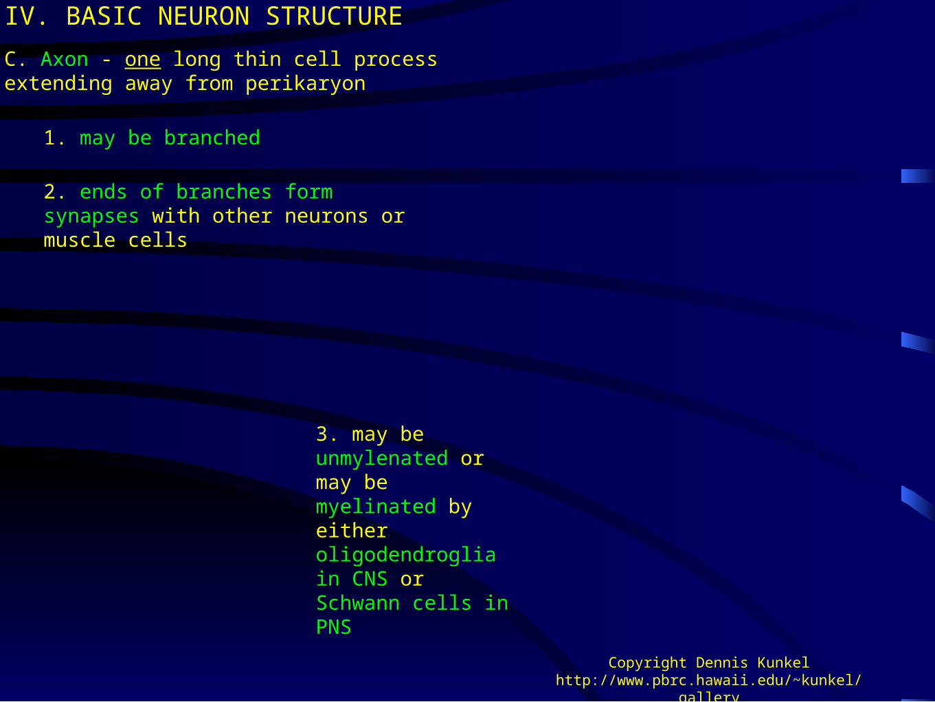
1. may be branched
1. may be branched
C. Axon - one long thin cell process extending away from perikaryon
IV. BASIC NEURON STRUCTURE
2. ends of branches form synapses with other neurons or muscle cells
3. may be unmylenated or may be myelinated by either oligodendroglia in CNS or Schwann cells in PNS
Copyright Dennis Kunkelhttp://www.pbrc.hawaii.edu/~kunkel/gallery

IV. BASIC NEURON STRUCTURE
4. Axon structure
b. initial segment (unmyelinated intitial portion of axon) between axon hillock and beginning of myelination.
c. remainder of axon (may be myelinated, may be branched)
d. axons carry electrical impulses (action potentials) to synapses at end of axon.
e. accept for the synapse, the axon cytoplasm (axoplasm) has few organelles. Not much synthetic activity in this part of neuron.
http://vv.carleton.ca/~neil/neural/neuron-a.html
a. axon hillock (pyramid shaped region associated with perikaryon) - where axon begins - cytoplasm in this region lacks ribosomes and organelles; however, neurofilaments and microtubles are present.
http://www.lab.anhb.uwa.edu.au/mb140/CorePages/Nervous/Nervous.htm
f. Microtubules and neurofilaments present.

D. Synapse
IV. BASIC NEURON STRUCTURE
1. specialized junctions with other cells that are along the length or at end of an axon.
2. act as transmission points for electrical impulses or chemical (ionic) changes.
3. synapse can transmit action potential, or can polarize or depolarize the postsynaptic cell.
4. synapses at the end of an axon or axon branches that are swollen into a club shape are called boutons terminaux.
5. a synapse along length of an axon results in varicosities (swellings) in the axon that are called boutons en passage. http://www.nature.com/neuro/journal/v4/n11/full/nn74
4.html
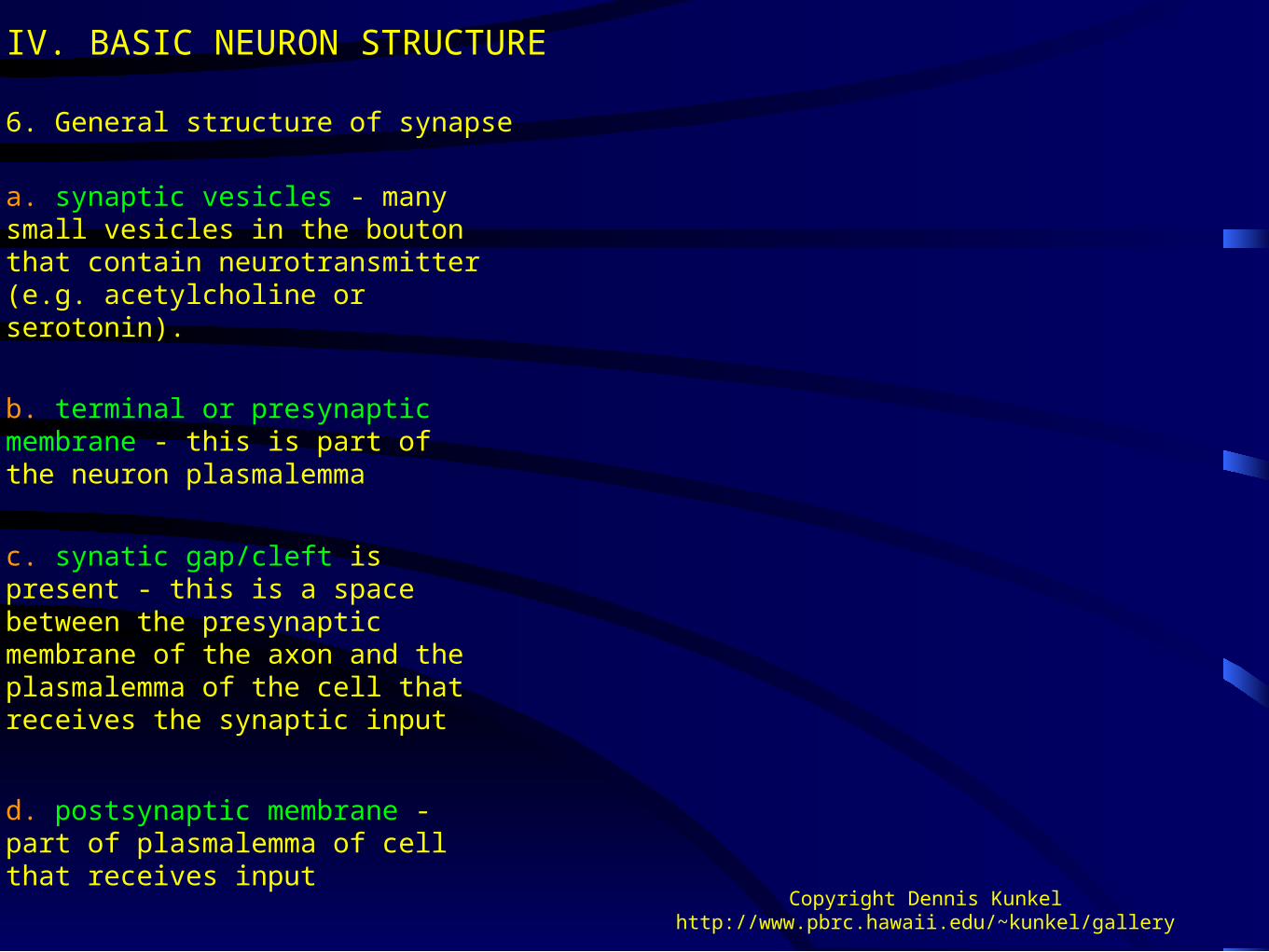
6. General structure of synapse
IV. BASIC NEURON STRUCTURE
b. terminal or presynaptic membrane - this is part of the neuron plasmalemma
Copyright Dennis Kunkelhttp://www.pbrc.hawaii.edu/~kunkel/gallery
c. synatic gap/cleft is present - this is a space between the presynaptic membrane of the axon and the plasmalemma of the cell that receives the synaptic input
d. postsynaptic membrane - part of plasmalemma of cell that receives input
a. synaptic vesicles - many small vesicles in the bouton that contain neurotransmitter (e.g. acetylcholine or serotonin).

e. when action potential reaches synapse, the synaptic vesicles are exocytosed at the presynaptic membrane and their contents (neurotransmitter) are released into the synaptic gap.
6. General structure of synapse
IV. BASIC NEURON STRUCTURE
Copyright Dennis Kunkelhttp://www.pbrc.hawaii.edu/~kunkel/gallery
f. neurotransmitter binds to receptors on postsynaptic membrane and propagates polarity change in post-synaptic cell.

V. TYPES OF NEURONS
A. Types of neurons based on shape/morphology
1. multipolar - more than two processes arising from perikaryon, most of neurons in brain and spinal cord are of this type.
http://www.lab.anhb.uwa.edu.au/mb140/

2. bipolar - two processes arising from perikaryon, may be branched at ends, some sensory neurons in retina and cochlea, are of this type.
http://www.csus.edu/org/nrg/carter/NeurosylActive/histology/neuron/bipolar.htm
Bipolar neurons in vestibular ganglion

3. Unipolar or pseudounipolar - one process arising from perikaryon, really two processes that are fused along portions closest to perikaryon - found in spinal ganglia and some crainial ganglia. Developmentally, this type of neuron starts out as a bipolar neuron.
4. Regardless of the type of neuron, the general structure dendrite(s)-perikaryon-axon-synapse is the same. Appearance may differ due number of processes and branching or fusion of processes.

B. Types of neurons based on function
A. Motor neurons - efferent, axon extends out of CNS to an effector organ/tissue (e.g. muscle) in peripheral regions
B. Sensory neurons - afferent, dendrite/axon extends from peripheral sensory structure (e.g. pacinian corpuscles, touch, pressure) into CNS
C. Interneurons - form connections between neurons

VI. GLIAL CELLS
•More glial cells in the nervous system than there are neurons.
http://www.lab.anhb.uwa.edu.au/mb140/
•Cells are situated among the neurons and are generally smaller.
•Special staining techniques needed to differentiate from each other and from surrounding neurons.
•With hematoxylin - eosin, only the glial cell nuclei show up.

VI. GLIAL CELLS - major types
A. Astrocytes
a. provide physical support for neurons
b. store glycogen
c. isolate synaptic areas from one and other
d. In the brain, processes abut against the basement membrane of capillary endothelium (pedicles) forming the blood-brain barrier
f. may form a conduit for nutrients from blood vessels to neurons
e. other processes are closely applied to neurons (pedicles)
http://members.tripod.com/blustein/Astrocytes/astrocytes.htm
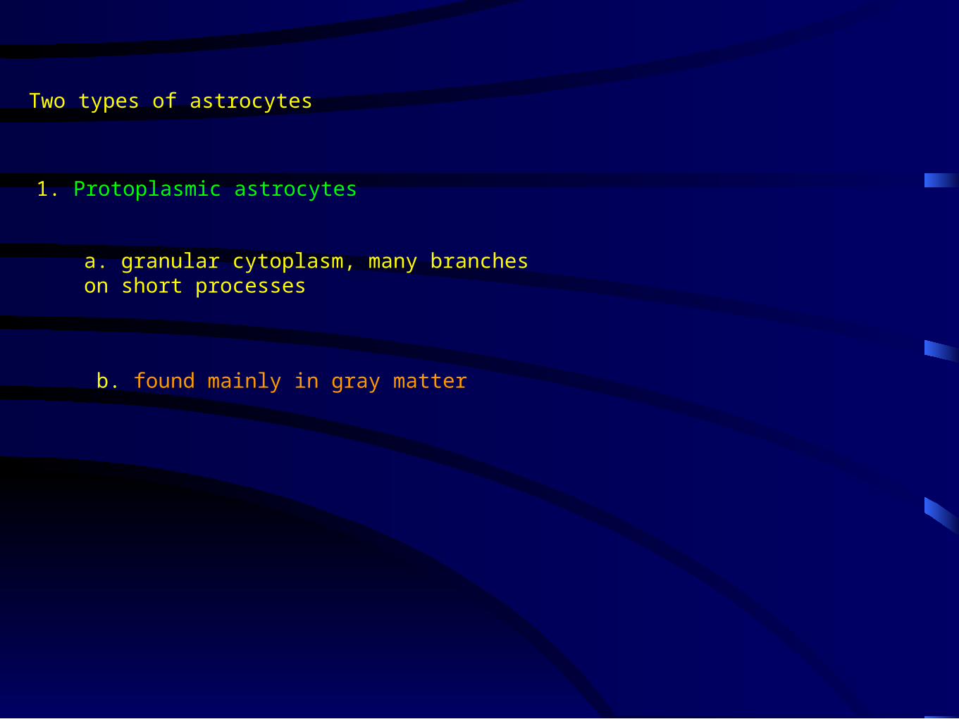
1. Protoplasmic astrocytes
a. granular cytoplasm, many branches on short processes
b. found mainly in gray matter
Two types of astrocytes

2. Fibrous astrocytes
a. have longer slender processes
b. found mainly in white matter (but also occur in gray matter).
http://www.benbest.com/cryonics/protocol.html
VI. GLIAL CELLS - major types
B. Oligodendroglia (plural), also called oligodendria (plural) or oligodendrocytes (plural). oligodendrocyte (singular)
1. smaller than astrocytes, fewer processes
2. found in both grey and white matter of CNS
3. particularly in white matter, processes from these cells form the myelin sheaths that are around many axons
4. analogous to Schwann cells of peripheral nervous system
5. these cells must be cultured with neurons in order to get neurons to grow in tissue culture. Suggests intimate interactive association http://members.tripod.com/blustein/Oligodendrocytes/oligodendrocytes.htm

C. Microglia
4. microglial cells are derived from mesoderm.
5. microglial cells function in phagocytosis - components of immune system, act as brain macrophages.
6. known to migrate and accumulate at the site of nerve damage within the central nervous system.
2. sometimes an elongate nucleus with mostly heterochromatin (Other glia have spherical nucleus)
3. many of what were thought to be microglia under the light microscope, have turned out to be oligodendroglia when cells were examined with the electron microscope.
1. small cell body that is usually elongated and stains densely
http://members.tripod.com/blustein/Microglia/microglia.htm

http://www.biology.uiowa.edu/daileylab/movies.html
Microglial movement in brain slice culture
Microglial cell engulfing nucleus of dead cell in brain slice culture.

D. Ependymal cells
VII. GLIAL CELLS - major types
1. ciliated cells forming single layer of simple cuboidal to low columnar epithelium that lines the entire neurocoel
2. ciliary action acts to circulate cerebral spinal fluid.
http://www.lab.anhb.uwa.edu.au/mb140/CorePages/Nervous/Nervous.htm
http://members.tripod.com/blustein/Oligodendrocytes/oligodendrocytes.htm
E. Schwann cells - peripheral nervous system
Peripheral Nervous System Glial Cells
http://www.siumed.edu/~dking2/ssb/neuron.htm
Animation of myelination

1. Provide myelin sheaths for axons in the PNS
Schwann cells
http://education.vetmed.vt.edu/Curriculum/VM8054/Labs/Lab9/Lab9.htm
F. satellite cells
1. Found surrounding neurons in ganglia.
Peripheral Nervous System Glial Cells

VIII. STRUCTURE OF NERVES
A. Nerves are surrounded by a thick connective tissue sheath composed of collagenous fibers and containing small blood vessels that is called the epineurium - also extends into the nerve between the fascicles of axons.
B. Within the epineurium are bundles of nerve fibers called fascicles.
1. Nerve fibers - axons, each one of which is ensheathed by a single or multiple layers of plasmalemma from a Schwann cell.
C. Each bundle (or fascicle) of nerve fibers is surrounded by a layer of connective tissue called the perineurium
D. Each nerve fiber within each bundle is individually surrounded by a layer of reticular connective tissue called the endoneurium
1. Small diameter axons usually ensheathed by a single layer of Schwann cell plasmalemma and are not myelinated.
2. Larger axons are myelinated - ensheathed by multiple layers of specialized Schwann cell plasma membrane called myelin.

Nerve Histology
http://www.meddean.luc.edu/lumen/medEd/Histo/frames/h_frame6.html

VIII. STRUCTURE OF NERVES
fascicle)
epineurium extending between fascicles
nerve fibers