Embed Size (px)
DESCRIPTION
THE NERVOUS SYSTEM. Divisions of the nervous system. Anatomical Organization of the Nervous System. Major Regions of the Brain. Figure 15.1 Major Divisions of the Brain. Neuronal Organization: CNS. Two kinds of neural tissue found in both brain and spinal cord: - PowerPoint PPT Presentation
Citation preview
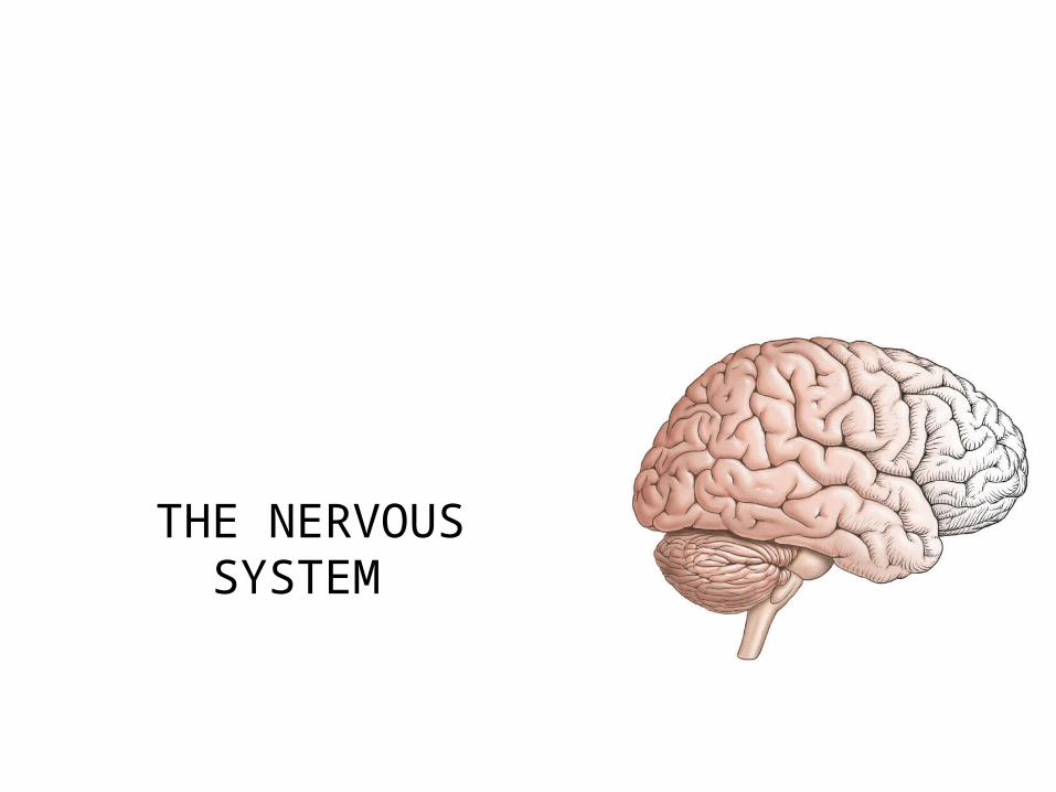
THE NERVOUS SYSTEM
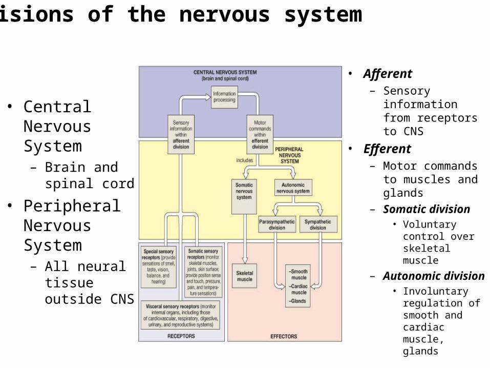
Divisions of the nervous system
• Central Nervous System– Brain and spinal
cord
• Peripheral Nervous System– All neural tissue
outside CNS
• Afferent– Sensory
information from receptors to CNS
• Efferent – Motor commands
to muscles and glands
– Somatic division• Voluntary
control over skeletal muscle
– Autonomic division• Involuntary
regulation of smooth and cardiac muscle, glands
Neuronal Organization: CNS
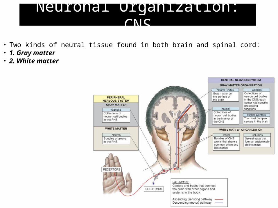
• Two kinds of neural tissue found in both brain and spinal cord:• 1. Gray matter• 2. White matter
Neuronal Organization: CNS
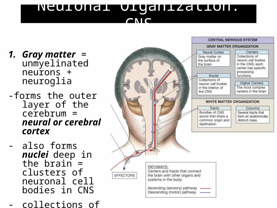
1. Gray matter = unmyelinated neurons + neuroglia
-forms the outer layer of the cerebrum = neural or cerebral cortex
- also forms nuclei deep in the brain = clusters of neuronal cell bodies in CNS
- collections of nuclei can form a center (higher brain function)
Neuronal Organization: CNS
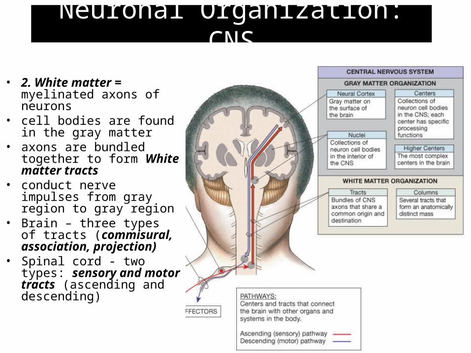
• 2. White matter = myelinated axons of neurons
• cell bodies are found in the gray matter
• axons are bundled together to form White matter tracts
• conduct nerve impulses from gray region to gray region
• Brain – three types of tracts (commisural, association, projection)
• Spinal cord - two types: sensory and motor tracts (ascending and descending)
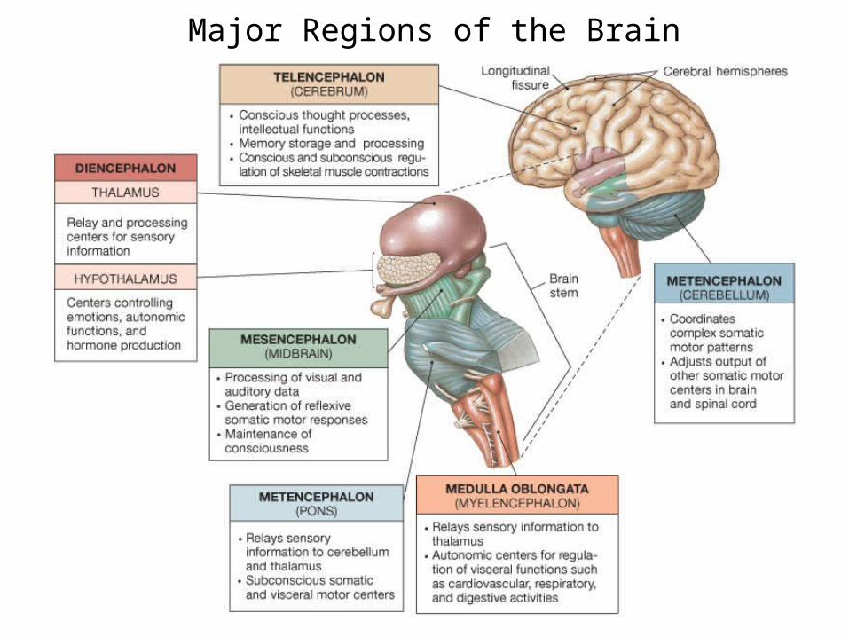
Figure 15.1 Major Divisions of the Brain
Major Regions of the Brain
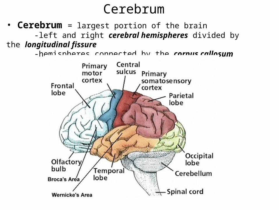
• Cerebrum = largest portion of the brain-left and right cerebral hemispheres divided by the longitudinal
fissure-hemispheres connected by the corpus callosum
Cerebrum
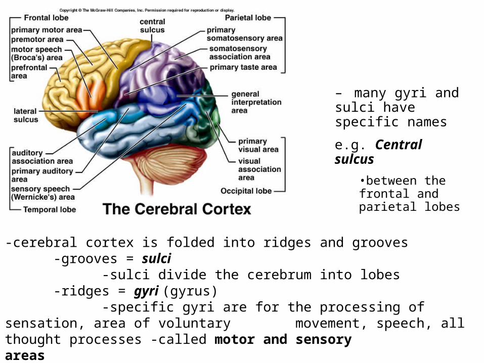
-cerebral cortex is folded into ridges and grooves-grooves = sulci
-sulci divide the cerebrum into lobes-ridges = gyri (gyrus)
-specific gyri are for the processing of sensation, area of voluntary movement, speech, all thought processes -called motor and sensory areas
– many gyri and sulci have specific names
e.g. Central sulcus
•between the frontal and parietal lobes
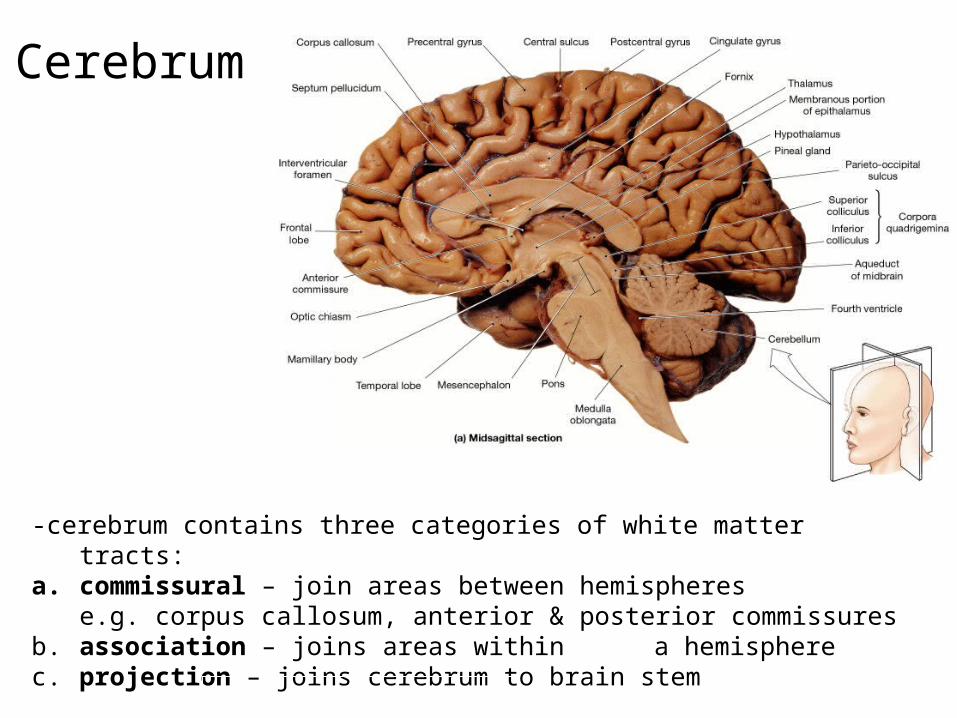
-cerebrum contains three categories of white matter tracts:a. commissural – join areas between hemispheres
e.g. corpus callosum, anterior & posterior commissuresb. association – joins areas within a hemispherec. projection – joins cerebrum to brain stem
Cerebrum
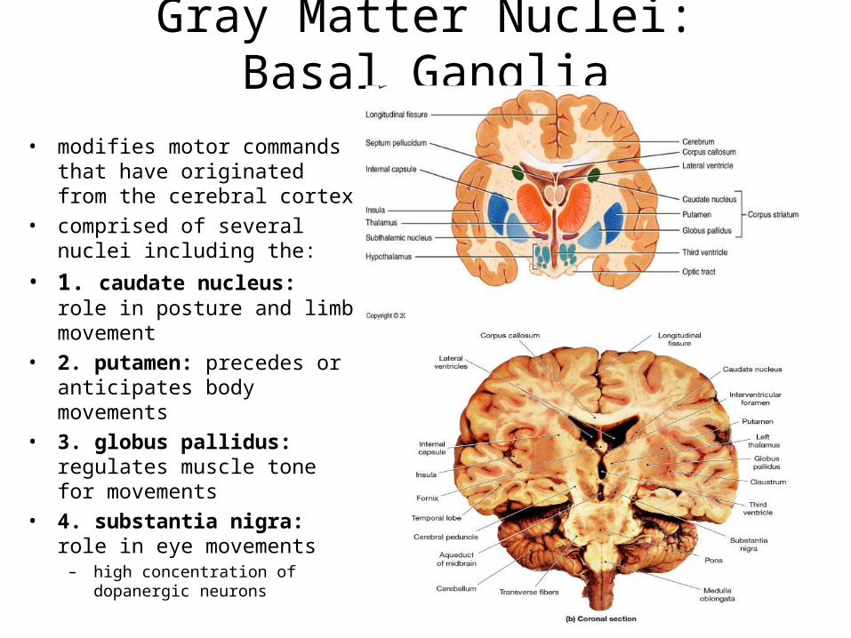
Gray Matter Nuclei: Basal Ganglia
• modifies motor commands that have originated from the cerebral cortex
• comprised of several nuclei including the:
• 1. caudate nucleus: role in posture and limb movement
• 2. putamen: precedes or anticipates body movements
• 3. globus pallidus: regulates muscle tone for movements
• 4. substantia nigra: role in eye movements
– high concentration of dopanergic neurons
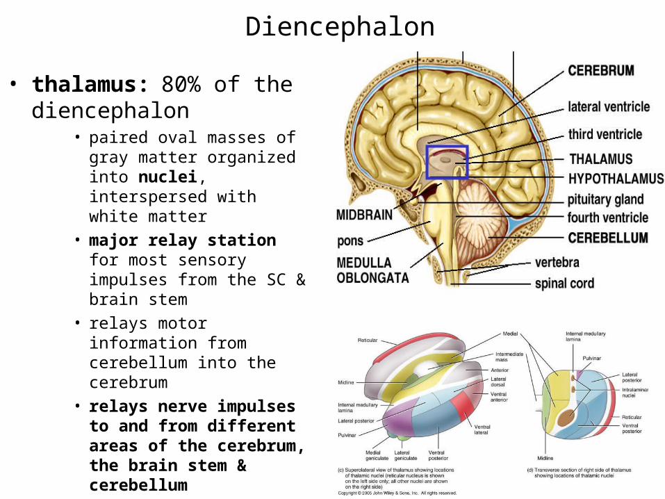
• thalamus: 80% of the diencephalon
• paired oval masses of gray matter organized into nuclei, interspersed with white matter
• major relay station for most sensory impulses from the SC & brain stem
• relays motor information from cerebellum into the cerebrum
• relays nerve impulses to and from different areas of the cerebrum, the brain stem & cerebellum
Diencephalon
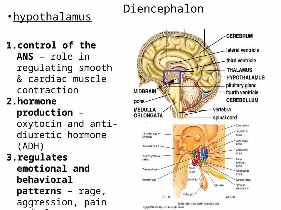
Diencephalon•hypothalamus
1. control of the ANS – role in regulating smooth & cardiac muscle contraction
2. hormone production – oxytocin and anti-diuretic hormone (ADH)
3. regulates emotional and behavioral patterns – rage, aggression, pain and pleasure + sexual arousal
4. regulates eating & drinking
5. controls body temp
•epithalamus – consists of the pineal gland and habenular nuclei-pineal gland – part of the endocrine system
-secretes the hormone melatonin-increased secretion in dark-promote sleepiness and helps set the circadianrhythms of the body (awake/sleep period)
•subthalamus – works with the cerebrum and cerebellum to control bodymovements
Diencephalon
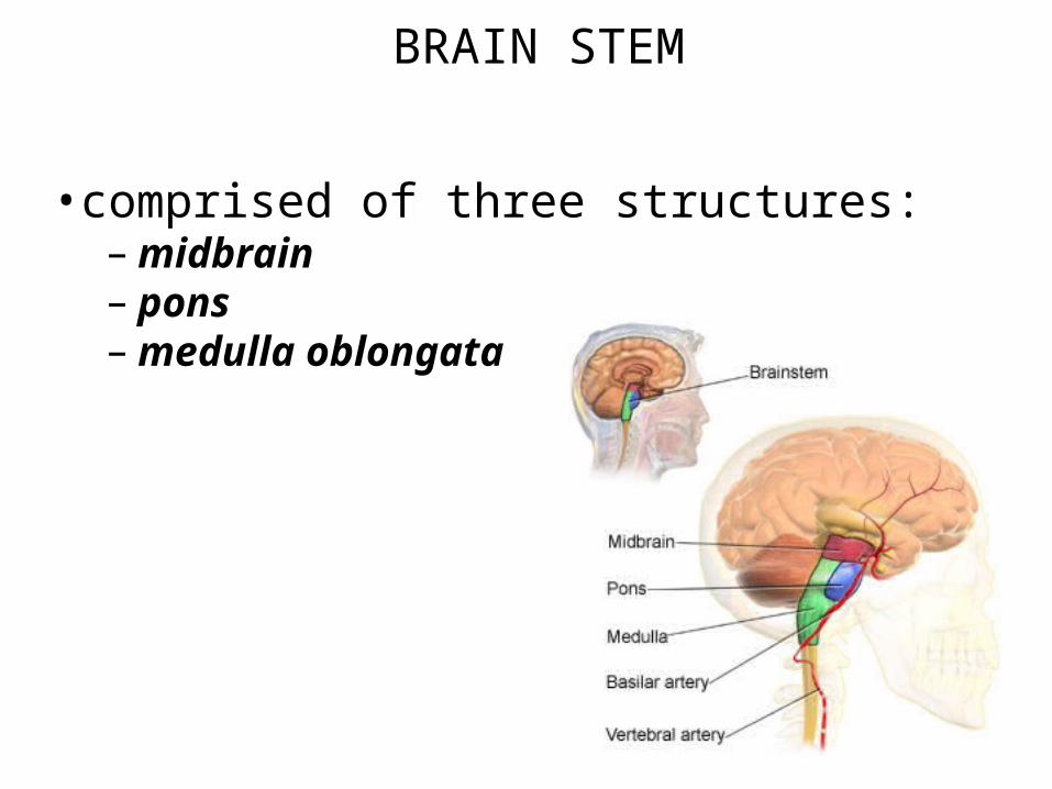
BRAIN STEM
•comprised of three structures:– midbrain– pons – medulla oblongata
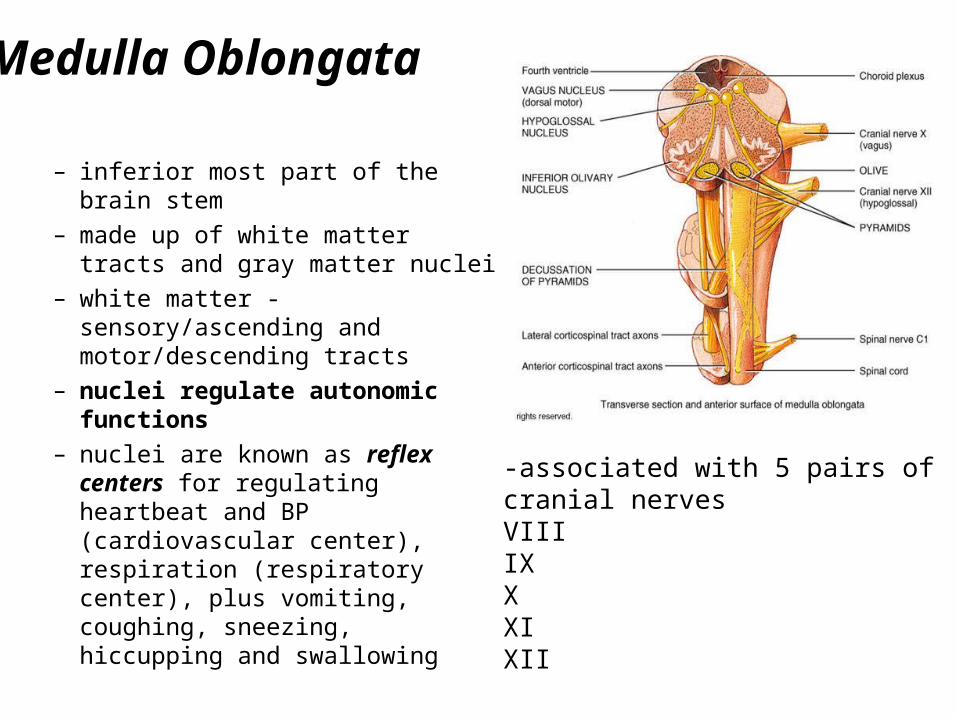
Medulla Oblongata
– inferior most part of the brain stem
– made up of white matter tracts and gray matter nuclei
– white matter - sensory/ascending and motor/descending tracts
– nuclei regulate autonomic functions
– nuclei are known as reflex centers for regulating heartbeat and BP (cardiovascular center), respiration (respiratory center), plus vomiting, coughing, sneezing, hiccupping and swallowing
-associated with 5 pairs ofcranial nervesVIIIIXXXIXII
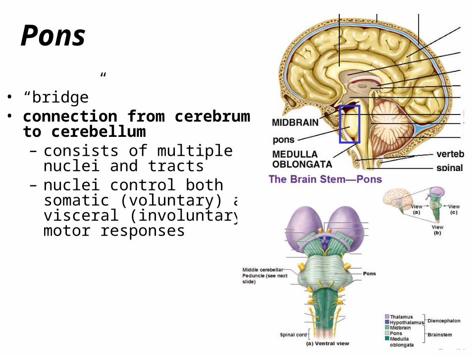
• “bridge”• connection from cerebrum to
cerebellum– consists of multiple nuclei and
tracts– nuclei control both somatic
(voluntary) and visceral (involuntary) motor responses
Pons
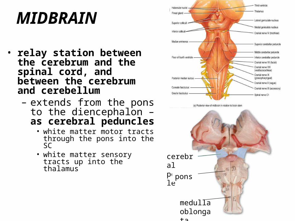
• relay station between the cerebrum and the spinal cord, and between the cerebrum and cerebellum– extends from the pons to the
diencephalon – as cerebral peduncles
• white matter motor tracts through the pons into the SC
• white matter sensory tracts up into the thalamus
MIDBRAIN
cerebral peduncle
pons
medullaoblongata
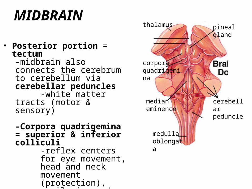
• Posterior portion = tectum-midbrain also connects the cerebrum to cerebellum via cerebellar peduncles
-white matter tracts (motor & sensory)
-Corpora quadrigemina = superior & inferior colliculi
-reflex centers for eye movement, head and neck movement (protection), pupil size and eye tracking
MIDBRAIN
cerebellar peduncle
corporaquadrigemina
medianeminence
pinealgland
thalamus
medullaoblongata
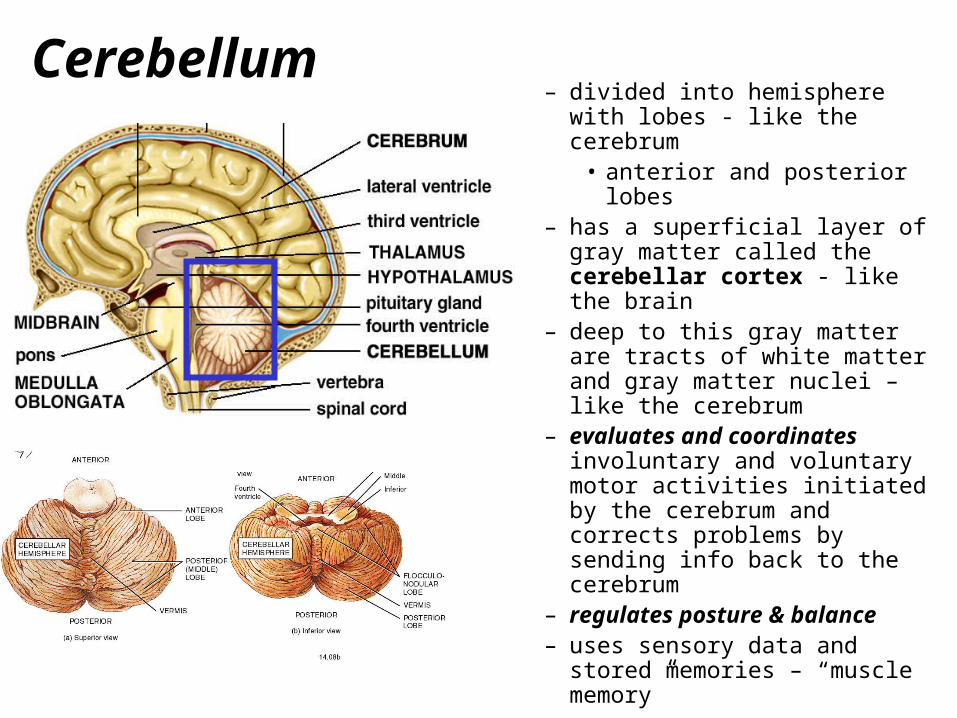
– divided into hemisphere with lobes - like the cerebrum
• anterior and posterior lobes– has a superficial layer of gray
matter called the cerebellar cortex - like the brain
– deep to this gray matter are tracts of white matter and gray matter nuclei – like the cerebrum
– evaluates and coordinates involuntary and voluntary motor activities initiated by the cerebrum and corrects problems by sending info back to the cerebrum
– regulates posture & balance– uses sensory data and stored
memories – “muscle memory”
Cerebellum
The Limbic System
olfactory tract
amygdala
hippocampus
anterior thalmic nuclei
fornix
mamillary body
parahippocampal gyrus
hypothalmic nuclei
corpus callosum
cingulate gyrus
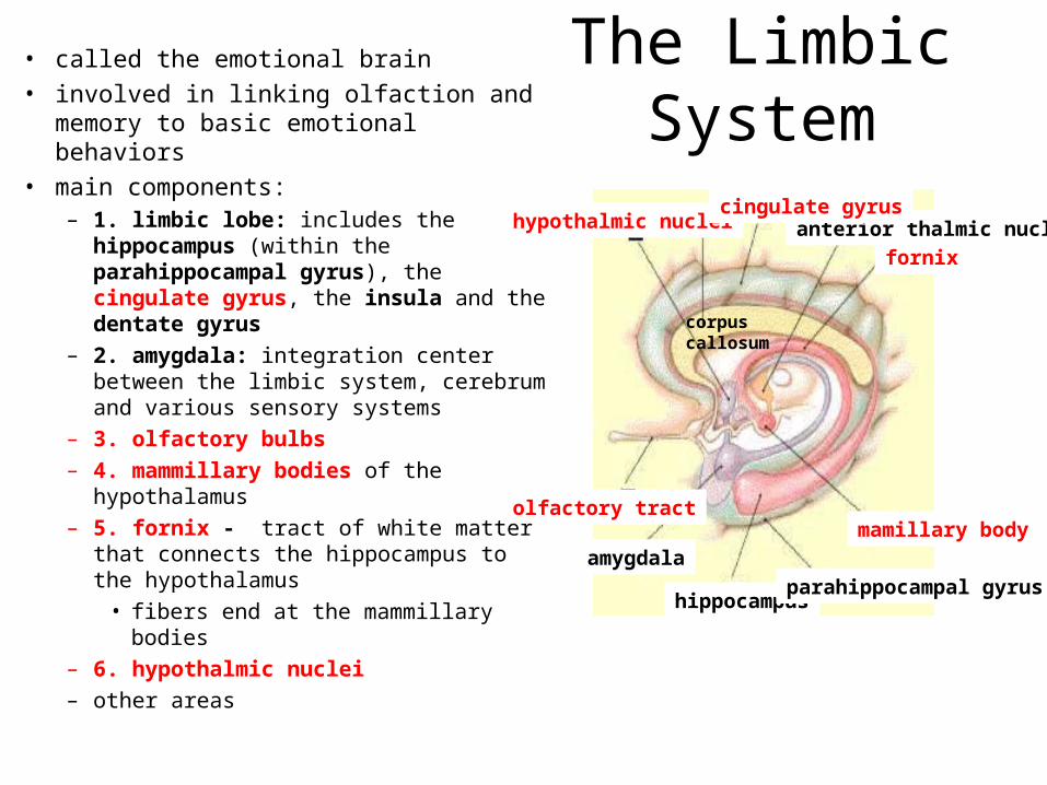
• called the emotional brain
• involved in linking olfaction and memory to basic emotional behaviors
• main components:– 1. limbic lobe: includes the
hippocampus (within the parahippocampal gyrus), the cingulate gyrus, the insula and the dentate gyrus
– 2. amygdala: integration center between the limbic system, cerebrum and various sensory systems
– 3. olfactory bulbs
– 4. mammillary bodies of the hypothalamus
– 5. fornix - tract of white matter that connects the hippocampus to the hypothalamus
• fibers end at the mammillary bodies
– 6. hypothalmic nuclei
– other areas
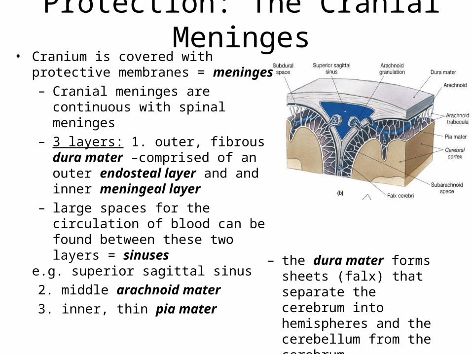
Protection: The Cranial Meninges• Cranium is covered with protective
membranes = meninges
– Cranial meninges are continuous with spinal meninges
– 3 layers: 1. outer, fibrous dura mater –comprised of an outer endosteal layer and and inner meningeal layer
– large spaces for the circulation of blood can be found between these two layers = sinuses
e.g. superior sagittal sinus
2. middle arachnoid mater
3. inner, thin pia mater – the dura mater forms sheets (falx) that separate the cerebrum into hemispheres and the cerebellum from the cerebrum
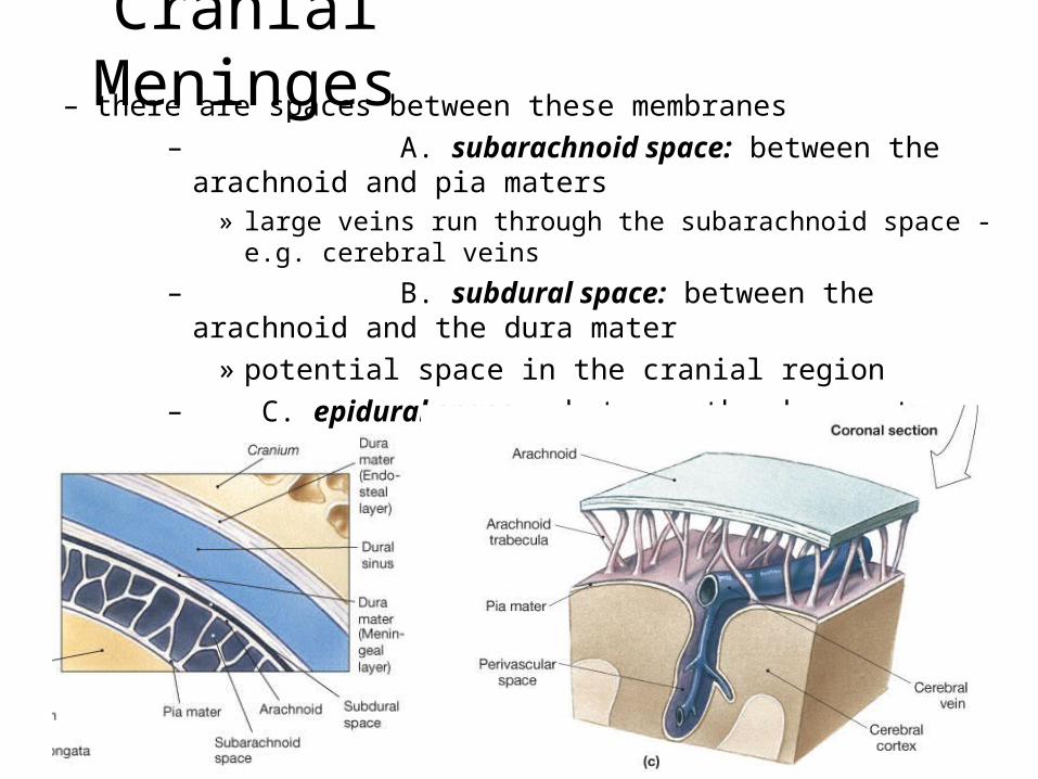
Cranial Meninges– there are spaces between these membranes
– A. subarachnoid space: between the arachnoid and pia maters
» large veins run through the subarachnoid space - e.g. cerebral veins
– B. subdural space: between the arachnoid and the dura mater
» potential space in the cranial region
– C. epidural space – between the dura mater and the vertebral canal in the spinal column
» potential space in the cranial region
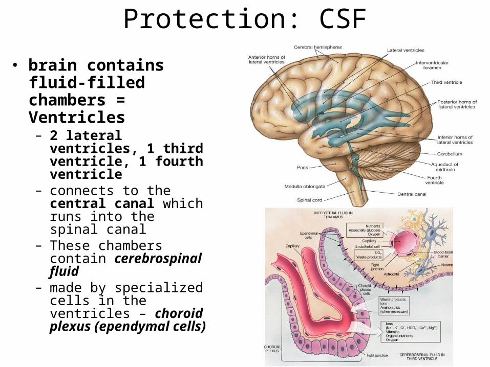
Protection: CSF
• brain contains fluid-filled chambers = Ventricles– 2 lateral ventricles, 1 third
ventricle, 1 fourth ventricle
– connects to the central canal which runs into the spinal canal
– These chambers contain cerebrospinal fluid
– made by specialized cells in the ventricles – choroid plexus (ependymal cells)
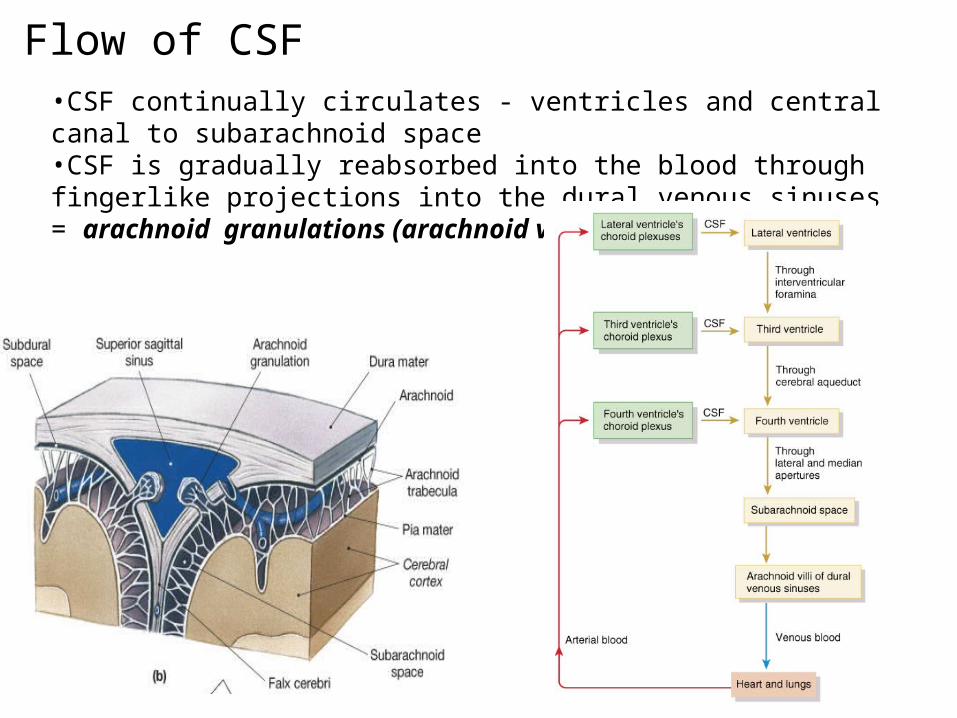
•CSF continually circulates - ventricles and central canal to subarachnoid space•CSF is gradually reabsorbed into the blood through fingerlike projections into the dural venous sinuses = arachnoid granulations (arachnoid villi)
Flow of CSF
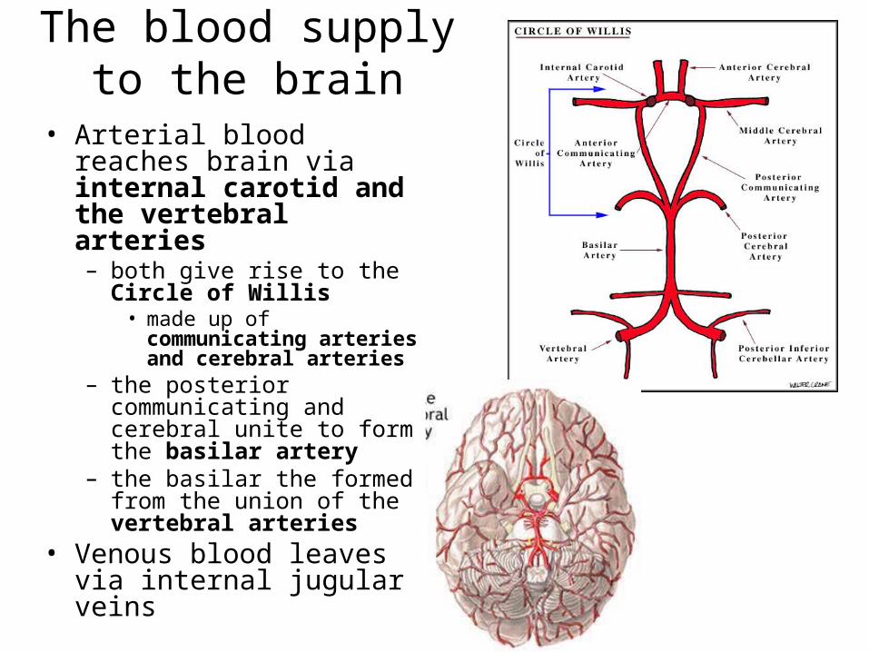
The blood supply to the brain
• Arterial blood reaches brain via internal carotid and the vertebral arteries– both give rise to the Circle of
Willis• made up of communicating
arteries and cerebral arteries– the posterior communicating and
cerebral unite to form the basilar artery
– the basilar the formed from the union of the vertebral arteries
• Venous blood leaves via internal jugular veins
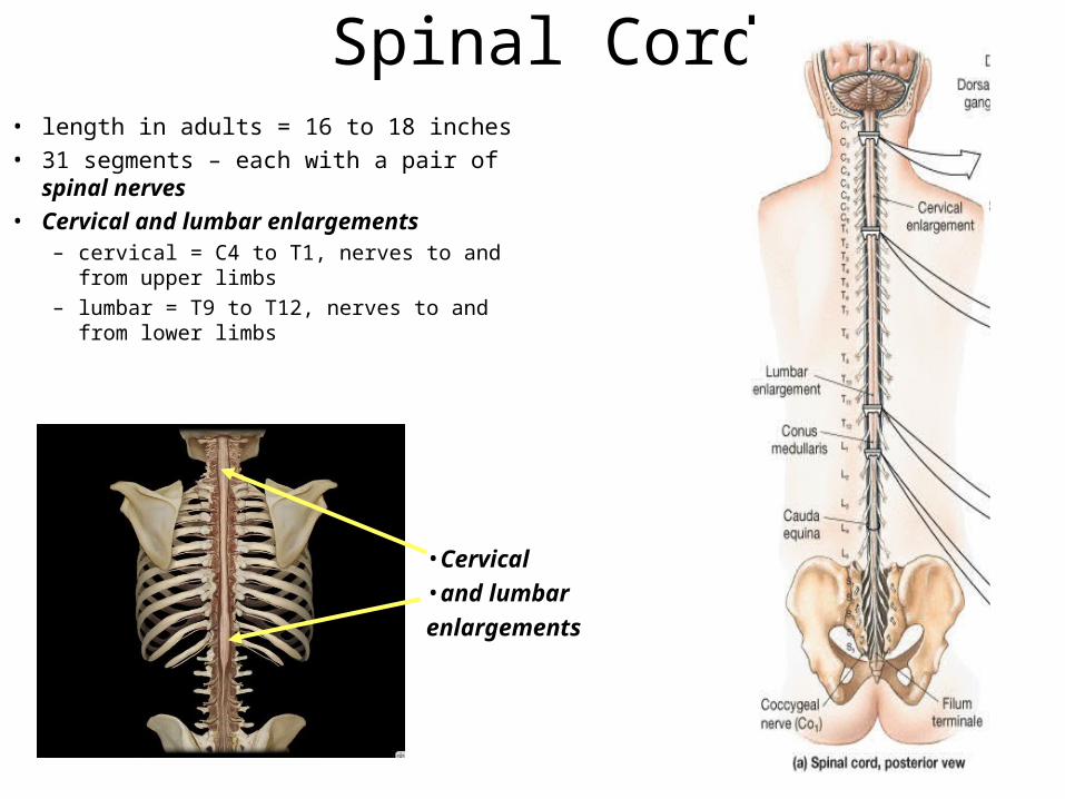
Spinal Cord• length in adults = 16 to 18 inches• 31 segments – each with a pair of spinal nerves• Cervical and lumbar enlargements
– cervical = C4 to T1, nerves to and from upper limbs
– lumbar = T9 to T12, nerves to and from lower limbs
•Cervical
•and lumbar
enlargements
Spinal Cord
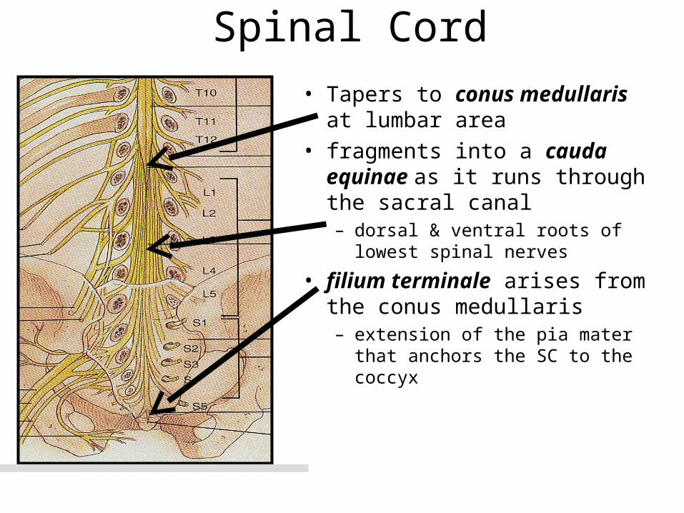
• Tapers to conus medullaris at lumbar area
• fragments into a cauda equinae as it runs through the sacral canal– dorsal & ventral roots of lowest
spinal nerves
• filium terminale arises from the conus medullaris – extension of the pia mater that
anchors the SC to the coccyx
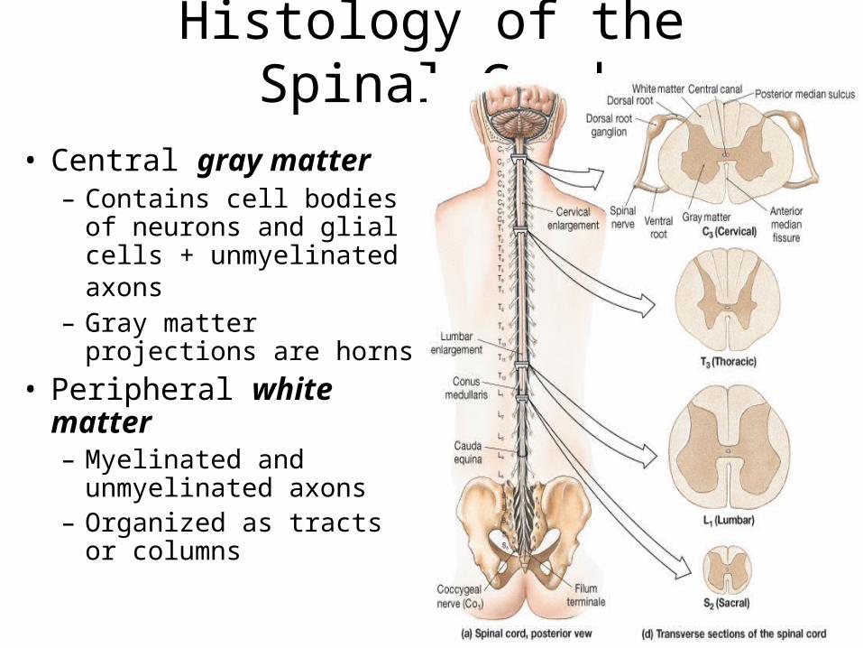
Histology of the Spinal Cord
• Central gray matter– Contains cell bodies of
neurons and glial cells + unmyelinated axons
– Gray matter projections are horns
• Peripheral white matter– Myelinated and
unmyelinated axons– Organized as tracts or
columns
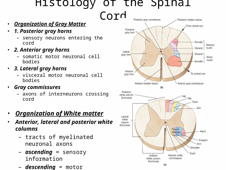
Histology of the Spinal Cord• Organization of Gray Matter• 1. Posterior gray horns
– sensory neurons entering the cord
• 2. Anterior gray horns– somatic motor neuronal cell bodies
• 3. Lateral gray horns– visceral motor neuronal cell bodies
• Gray commissures– axons of interneurons crossing cord
• Organization of White matter• Anterior, lateral and posterior white
columns
– tracts of myelinated neuronal axons
– ascending = sensory information
– descending = motor information
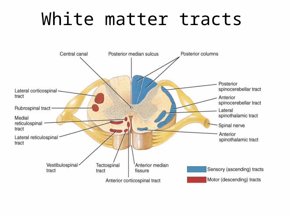
White matter tracts