Embed Size (px)
Citation preview
143
The Nerve-net of Metridium senile:Artifacts and the Nerve-net
By E. J. BATHAM, C. F. A. PANTIN, and E. A. ROBSON
(From the Zoological Laboratory, Cambridge)
With three plates (figs. 1-3)
SUMMARY
The present paper follows an account of the structure of the nervous system ofMetridium senile (L.). Conflicting statements about the actinian nervous system in theearlier literature made it necessary to assess the results of previous workers critically.Several of their methods have now been repeated and compared with our results afterusing more specific techniques. The criteria for distinguishing nerve-cells from non-nervous elements in actinians are discussed. Mesogloeal fibres, amoebocytes, nemato-cyst threads, and muscle-fibres may on occasion be confused with nerve-cells, anddeteriorating nerve-cells may also have a misleading appearance. Gross artifacts maybe reduced by the use of special staining methods, and on the basis of this work theresults of several earlier authors have been re-interpreted. It is concluded that thenervous system in the mesenteries and column of Metridium follows the epitheliallayers and does not penetrate the mesogloea.
ARTIFACTS AND THE NERVE-NET
RECENT work (Pantin, 1952; Batham, Pantin, and Robson, i960) has,. emphasized the importance of exact information about the organization
of the nerve-net of actinians for understanding the machinery of theirelementary nervous system. Existing knowledge began with the studies of theHertwigs of 1879. ^ n e c a n 0V^Y ^e filled with admiration for the perspicacityand completeness of their work, and feel some regret that subsequent studies,upon which contemporary ideas of this sort of nervous system are based,sometimes fall short of the standards of these workers of 80 years ago.
In relating nervous organization to the activities displayed by actiniansthere is one major problem. According to the Hertwigs the nerve-net isessentially intra-epithelial, being confined to the two layers of ectoderm andendoderm (Hertwigs, 1879). They found some connexion between theectodermal and endodermal nets only where the two layers join at the baseof the pharynx. There may possibly be other connexions of the same sort,however, as at the pore-like cinclides that pierce the column of some anemonesand pores at the tips of the tentacles or in the foot in others. Our own studiesindicate that in localized places on the oral disk the mesogloea can be seen tobe interrupted, so that the ectoderm and endoderm occasionally meetdirectly; but this work will be reported later. We find that all such contactsbetween the ectodermal and endodermal nervous system of actinians arelimited; and the same is true of the nervous system of Hydra according to thevery thorough study of Hadzi (1909).
[Quarterly Journal of Microscopical Science, Vol. 102, part 2, pp. 143-56, June 1961.]2421.2 L
144 Batham, Pantin, and Robson—Artifacts and the Nerve-net
The separation of nerve-nets in different epithelia not only holds betweenectoderm and endoderm but also between the two sheets of endoderm thatare on each side of the mesogloea of the mesenteries. This accords with theessentially separate functional action of the muscle-sheets on the two sides ofthe mesogloea; as in the endodermal radial and retractor muscle-sheets ofthe mesenteries (Batham and Pantin, 1950), and in the muscle-sheets of theectoderm and endoderm of the tentacles and disk in actinians and in thewhole body of Hydra (Batham and Pantin, 1951).
The conclusion of the Hertwigs that the nerve-nets are intra-epithelial issupported by that of Groselj (1909) and by our own (Pantin, 1952; Batham,Pantin, and Robson, i960).
In sharp contrast to this picture of the organization of the nerve-net is thatderived from the studies of Havet (1901), and of Parker and Titus (1916) onMetridium, and those of Leghissa (1949, 1950). According to all these authors,there is a very well-developed system of nerve-fibres in the mesogloea—where the Hertwigs, Groselj, and we ourselves find none. This mesogloealsystem is said to supply both the ectoderm and endoderm and so wouldrepresent the primary nervous system. According to these authors the nervoussystem is therefore richer in nerve-cells than if it were restricted to theepithelial sense-cells and nerve-cells described by the Hertwigs, since thelatter would represent only the peripheral ramifications of a complex networkbased on the mesogloea.
Since the neurites of such a system are supposed to radiate in the thicknessof the mesogloea, the plan is not essentially two-dimensional as the intra-epithelial net of the Hertwigs would be (Batham, Pantin, and Robson, i960).The functionally separate muscle-sheets on each side of the mesogloea wouldthus not be controlled by functionally separate intra-epithelial nerve-nets,but would seem to depend for their independent contraction on somedifferential action within the common mesogloeal net. Although presentphysiological evidence does not support this idea, the presence or absence ofmesogloeal nerve-cells is clearly fundamental to our interpretation of actinianbehaviour in relation to the organization of the nervous system.
The question at issue is thus the presence or absence of a mesogloeal nerve-net in anemones. It is not an easy one to settle, for, if the Hertwigs are cor-rect, it involves the proof of a negative: a difficult matter where the capri-cious staining of nerve-cells is involved, notwithstanding the much simplerhistological picture which coelenterate epithelia present compared with thecomplex tissues of triploblastic animals. It is the primary object of this paperto consider the evidence about this problem.
The Hertwigs's work was based upon a very careful study of tissues treatedwith simple fixatives, such as strong alcohol and osmic oxide solutions andstained by such dyes as carmine derivatives (particularly alum and picro-carmine) and haematoxylin. Despite the simplicity of the methods, the reagentswere exhaustively tested. They prepared the tissues for examination bymaking hand-sections and by the painstaking and ingenious use of maceration
Bathatn, Pantin, and Robson—Artifacts and the Nerve-net 145
techniques. It is remarkable how much they discovered solely by meanswhich today would be dismissed as inadequate.
Groselj (1909), whose work substantially supports the Hertwigs, used vitalstaining with methylene blue. Batham, Pantin, and Robson (i960) have sinceconfirmed many results of the Hertwigs by their same methods. They alsoused vital staining with methylene blue, and made extensive use of a slightmodification of Holmes's method (1942, 1943) by which the nerve-cells arestained with silver. This silver method was developed by one of us (E. J. B.)after unsuccessful tests with several other silver impregnation methods.
It appears significant that all those who have, on the other hand, claimedthe presence of a mesogloeal nervous system in actinians have relied pri-marily upon silver impregnation methods. These are, unfortunately, liable toshow bad fixation and precipitation artifacts, and for this reason we decidedto make a fresh trial of several methods to see whether or not there wasevidence of objects in the mesogloea which could be shown quite unequivo-cally to be nerve-cells.
Parker and Titus (1916), for example, used a drastic method in which theanemone was rinsed in distilled water and then passed directly to 5% silvernitrate for impregnation. To expose living marine tissues to distilled waterand then to transfer them unfixed to silver nitrate might be expected to havedisastrous effects on their histology. Our experience of this method bears outthis expectation.
Havet (1901) used chiefly the rapid Golgi impregnation method by whichthe tissue is fixed by immersion in an osmic-bichromate mixture for severaldays and then impregnated in dilute silver nitrate. He notes the ease withwhich cells other than nerve and filamentous structures like nematocysts maytake the stain.
Havet's work is extensive and his drawings are clear. Whatever the natureof the objects he stained, their existence is convincingly demonstrated. Andmany of the objects he figures in the epithelia are, as he points out, identifiableas the nervous elements seen by the Hertwigs. It is the structures in themesogloea that are controversial.
Leghissa (1949, 1950) used a variety of methods which are not com-pletely specified, but his figures of what he identifies as mesogloeal nerve-cellsseem to be based mainly upon the Golgi-Kopsch silver method (Romeis,1948, No. 1772); upon fixation with potassium dichromate and formaldehyde,followed ultimately by impregnation with 0-75% silver nitrate; upon directimpregnation with silver nitrate (concentration not specified) after fixation inde Castro's (chloral / alcohol / nitric acid) mixture; and on certain goldchloride preparations.
The work of Woollard and Harpman (1939) should also briefly be men-tioned. After using a gold chloride / formic acid method on anemone andmedusa tissues, and some methylene blue, these authors concluded that thecoelenterate nervous system consisted of discontinuous cells and was nota fused network. But this support for our conclusions (Pantin, 1952; Batham,
146 Batham, Pantin, and Robson—Artifacts and the Nerve-net
Pantin, and Robson, i960) must be accepted with some reserve. In goldchloride / formic acid preparations, the formic acid distorts both muscle-fibres and mesogloea. Nor is it clear from their figures and their account thatthe structures studied by Woollard and Harpman were in fact nerve-cells.
Compared with all these, the modified Holmes's method used by Batham,Pantin, and Robson (i960) has a decided advantage in the quality of theinitial fixation and in the absence of excessive precipitation of silver, so thatmuch cytological detail can be seen compared with that found by more usualmethods of fixation and staining.
In addition to these, certain other silver methods were used in the presentinvestigation which, though they failed to reveal nerve-cells, yet gave goodinformation about tissue histology. Willis's (1945) method, for example, isdesigned to cause a reaction between silver and histones and protamines inthe nerve-cells. Tissue is fixed in ammoniacal formaldehyde in 70% alcoholand after paraffin sectioning passed again to formaldehyde, to acetic alcohol,and then to o-1 % silver, followed by hydroquinone and then gold toning andoxalate reduction. Peters's( 195 5e) method, involving fixation in formaldehyde /mercuric chloride followed by alkaline silver, sodium sulphite, and a glycinedeveloper, was also tried.
It has been noted by one of us (Pantin, 1952) that when trying to identifynerve-cells in coelenterates we are faced with the difficulty that histologicaltechniques giving good fixation rarely differentiate nerve-cells clearly fromthe surrounding tissues, while those which selectively stain nerves oftenproduce artifacts and may fix tissues very badly. To be cogent, the circum-stantial evidence in favour of the identification of nerve-cells must conformto the following criteria:
(1) The cells should stain by a variety of techniques in the same manner asnerve in other animals; and they should not stain in ways characteristicof other fibrous structures, such as collagen.
(2) Their cytological structure should be consistent with that of nerve-cells.
(3) Their anatomical relations should be consistent with the physiologicalevidence.
To this we may add:(4) The structure should if possible be identified in the living state.Applying these criteria Pantin (1952) and Batham, Pantin, and Robson
(i960) have shown by several methods that there are well-defined networksof nervous and sensory neurites in actinian tissues. The term 'neurite' is usedhere to denote the process of a nerve-cell or sensory cell. It has been explainedin a previous paper (Batham, Pantin, and Robson, i960) that as the moreusual terms 'axon' and 'nerve-fibre' are historically incorrect in this context,it is preferable to adopt the less specific word 'neurite' instead.
Unless such rules as these are applied there is a danger that other struc-tures, both cellular and non-cellular, may be identified as nerve-cells. This is
Batham, Pantin, and Robson—Artifacts and the Nerve-net 147
particularly the case with structures which, like neurites, are long and oftenfibrillar, such as muscle-fibres, the connective tissue-fibres in the mesogloea,nematocysts, and, as we shall show here, amoebocytes. As noted above, thedanger is greatest in those very useful methods which are intended to depositsilver in the nerve-cell and its processes, since if fixation is poor there may bestriking but false appearances due to precipitation of the metal on other fibrousstructures.
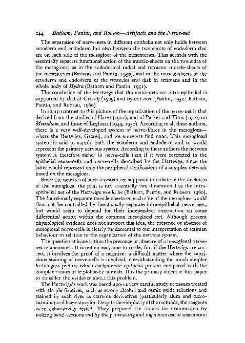
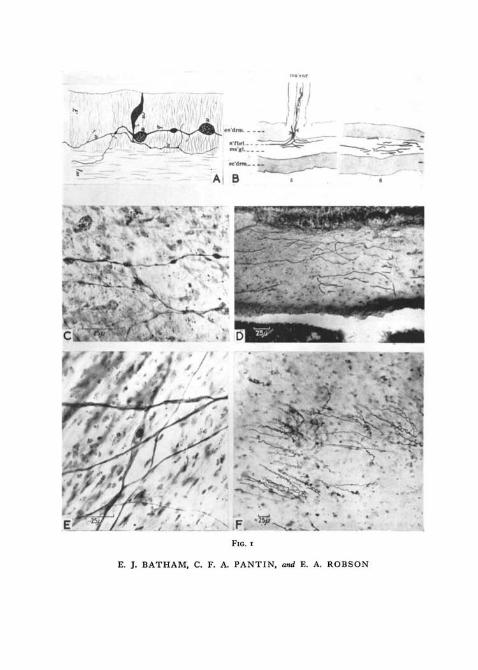
There is the further problem that nerve-cells which can be identified withcertainty may, nevertheless, be distorted on fixation so as to engender a mis-leading interpretation of their organization. Thus the neurites of the actiniannerve-net very easily become beaded during anaesthetization or fixation, orduring vital staining. In this they resemble the axons of other animals. If theydid not show such beading, the fact would cast doubt upon their being theprocesses of nerve-cells. Fig. 1, c shows a part of the retractor nerve-net ofMetridium which has become beaded in this way. Similar beading can beseen to occur in time-lapse motion pictures of living chick neurites in tissue-culture (Hughes, 1953), and it is in fact reversible. Young (1944) showed thatbeading takes place in myelinated nerve when cytoplasmic viscosity andintracellular pressure are insufficient. The fine terminals of living non-myelinated nerve in the cornea are morphologically unstable (Weddell andZander, 1951) and beading only becomes irreversible if it is caused by someadverse condition which has operated for too long (compare Bozler, 1927).
Beading is quite commonly seen in the neurites of Metridium after stainingby Holmes's silver method. The fixation used in this method also tends toprecipitate the axoplasm as neurofibrillar material, and lumps of such materialwithin the beads can then simulate nuclei, as in fig. 1, c. Comparison of suchappearances with other preparations leaves no doubt as to their true nature.Their close correspondence with certain similar structures figured by Havet(1901: compare, for example, fig. 1, A) in actinian nerve-cells indicates thatthe elements which he saw were also of this nature, and not chains of nerve-cells connected end to end as he suggests. Although such appearances may beparticularly striking after silver impregnation, beading can occur at or beforefixation.
Fig. 1, E shows a portion of nerve-net of the retractor face of a mesentery ofMetridium stained by Holmes's silver method, in which beading is absent.
We shall now consider some of the artifacts that can be produced, first inthe epithelio-muscular layer, and then in the mesogloea.
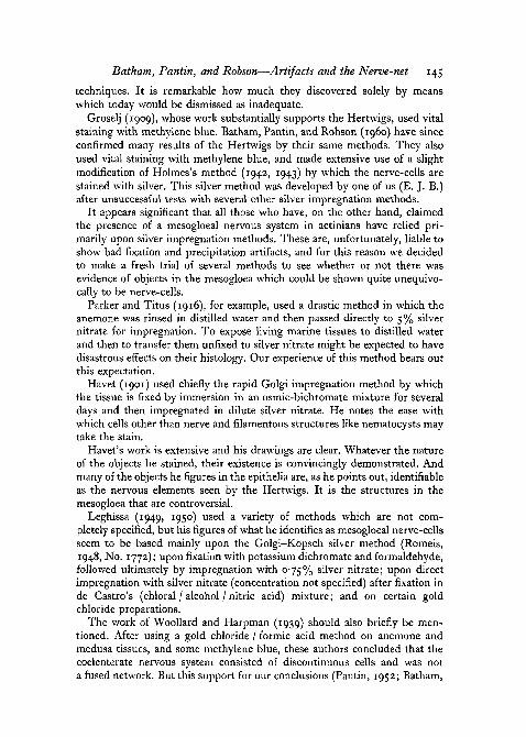
Mucus-cells. Fig. 2, D shows two mucus-cells in a mesentery stained withBatham-Holmes's silver method. These cells are common, and can beidentified by comparison with preparations fixed in Susa and stained bypolychrome staining methods, and by stains for mucin such as alcian blue.When fully developed they are goblet cells, and are anchored by a basalstrand to the mesogloea. Without such identification, badly fixed examplesmight possibly be confused with ganglion cells. In well-extended mesenteries,for instance, they sometimes appear to have stellate processes, as in fig. 2, D.
148 Batham, Pantin, and Robson—Artifacts and the Nerve-net
Amoebocytes are also numerous in the epithelium, but will be consideredlater (p. 150).
Nematocysts. Actinian tissues are often pierced by the animal's own nemato-cysts, which discharge on fixation. Fig. r, F shows nematocyst threadsstained by methylene blue which have pierced the mesogloea of Calliactis.The even diameter of a discharged thread, its frequently wavy course throughthe tissues, and origin at a capsule are characteristic. Havet (1901) pointedout that, on fixation by the Golgi method, nematocysts may be shot throughthe tissues, and their threads may be impregnated with silver. Parker andTitus (1916) using their own silver method recognize this, but state thatnerve-cells in the mesogloea can also be detected and can be distinguishedfrom nematocyst threads. But they could detect no cell-bodies. Preparationsmade by us following their methods show structures which seem identicalwith those which they draw (fig. 1, D). Examination of our preparations leadsus to conclude that in the great majority of cases these structures are withoutdoubt nematocyst threads. The violent osmotic changes in Parker and Titus'smethod inevitably cause a widespread discharge of nematocysts. Possiblysome of the structures seen may be due to deposition of silver on the collagenfibres of the mesogloea as well; we shall discuss this later (p. 151). We agreewith Parker and Titus in finding no evidence of nerve-cell bodies by thismethod. Nor is there clear evidence of other cellular structure. Fixation istoo poor, and the deposition of silver on fibrous remains is too great for anysound histological conclusions to be drawn from this method. The absence ofclear detailed information is seen if we compare Parker and Titus's figureswith those of the Hertwigs; or our fig. 1, D, repeating Parker and Titus'smethod, with our fig. 1, E by Holmes's silver method. The fibrous structureson which silver is deposited in the former do not appear to be nerve-cells,or even to be cellular, and in some cases the structures are unquestionablydischarged nematocyst threads. It is therefore impossible to conclude thatsuch preparations show nerve-cells in the mesogloea.
Muscle-cells. All silver methods we have tried are liable to stain the muscle-fibres (see fig. 3, A). These also stain vitally in due course with methyleneblue, though they take much longer than do nerve-cells. As the preparation
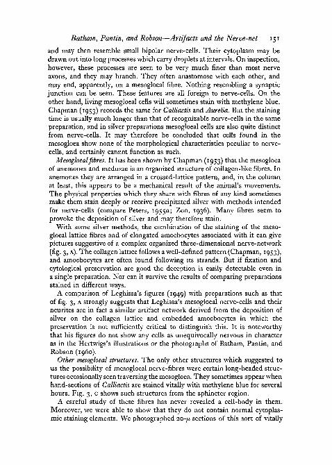
FIG. 1 (plate). A, one of Havet's illustrations (1901, pi. V, fig. 26) of a sectioned mesenteryfrom Metridium prepared by the Golgi method.
B, illustration from Parker and Titus (1916, pi. I, figs. 5 and 6), showing horizontal sectionsof Metridium column.
c, beading neurites in the nerve-net of a Metridium mesentery, stained with Batham'smodification of Holmes's silver method. Compare with Havet's figure above (A), and with E.
D, section of a Metridium mesentery stained with Parker and Titus's silver method. Thedark lines are the shafts of nematocysts which have penetrated the mesogloea. Compare withParker and Titus's illustrations above (B), and with F below.
E, part of a Metridium mesentery prepared by the Batham—Holmes silver method, in whichthe nerve-net shows little beading. Compare with c above.
F, section of Calliactis column, showing nematocyst threads discharged into the mesogloea.The tissues were stained for about an hour with methylene blue and fixed in ammoniummolybdate, as for fig. 3, c. Compare with D above.
• * • * .
FIG. I
E. J. BATHAM, C. F. A. PANTIN, and E. A. ROBSON

E. J. BATHAM, C. F. A. PANTIN, and E. A. ROBSON
Batham, Pantin, and Robson—Artifacts and the Nerve-net 149
ages, beading occurs, with drop-like aggregations of cytoplasm along themuscle-fibre (fig. 2, E), though the character of the beading is not like that ofneurites.
In all these features they show some resemblance to the histologicalbehaviour of nerve-cells, but here again, accurate identification depends uponthe avoidance of bad fixation and deformation. Provided this obtains, thereis no difficulty in identifying muscle-fibres stained by these or any of thestandard methods of polychrome staining. All give the same unmistakablepicture of a muscle-sheet of numerous parallel fibres in close apposition tothe surface of the mesogloea (Batham and Pantin, 1951; Grimstone, Home,Pantin, and Robson, T958). In agreement with the original observations ofthe Hertwigs (1879), it is important to realize how the layer of muscle-fibresis applied to the mesogloea at its surface. Fig. 2, A illustrates an electronmicrograph of this for a portion of the retractor muscle-sheet of the mesentery.The same is true of the circular muscle-layer of the column (Batham andPantin, 1951). Figures of this in some textbooks are misleading.
This point can be important particularly because with some methods, suchas that of Golgi impregnation, sometimes only individual muscle-fibres willtake up the stain, and it may be a matter of great difficulty to determine therelations of the muscle-cells to any mesogloea over which they lie. This isillustrated in fig. 2, c, a Golgi preparation from the sphincter region ofMetridium. As the Hertwigs showed, the endodermal muscle-cells are allcharacteristically epithelio-muscular cells, the muscle-fibre being connectedto the epithelial part of the cell by a cytoplasmic neck. In fig. 2, C this epithe-lial portion and part of the muscle-fibre of isolated muscle-cells have stainedwith silver and it is not easy to tell at just what level in the tissue they are.Havet (1901) illustrates just such structures by the same method, as shownin fig. 2, B. He, too, identifies them as epithelio-muscular cells, but figuresthe muscle-fibre as though it were embedded in the mesogloea. Carefulexamination of such cases convinces us that this is an optical misrepresen-tation due to isolated densely stained muscle-fibres of the muscle-sheet lyingabove the mesogloea and not within it. Like those of the Hertwigs, our ownvisual observations are entirely consistent with the muscle-fibre / mesogloearelation disclosed in fig. 2, A.
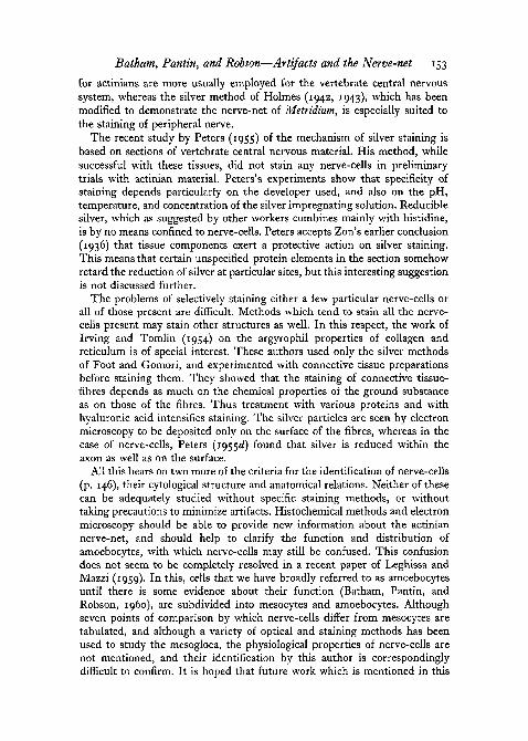
FIG. 2 (plate) A, electron micrograph of part of a transverse section of a Metridium mesentery.Retractor muscle-fibres (nil) are closely applied to the underlying mesogloea, within whichthe sectioned mesogloeal fibres form a distinct outer layer (ol).
B, one of Havet's figures of an epithelio-muscular cell from a Metridium mesentery (pi. V,fig. 34), from material sectioned after staining by the rapid Golgi method.
c, epithelio-muscular cell from the sphincter region of a small Metridium sectioned afterstaining by the rapid Golgi method. The epithelial part of the cell (ep) is connected bya cytoplasmic process (cyt) to the muscle-fibre (m/), only a short part of which occurs in thissection.
D, two mucus-cells in a mesentery of Metridium stained with Batham's modification ofHolmes's silver method. A neurite (left) and epithelial nuclei are also seen.
E, longitudinal muscle-fibres from a whole mesentery of Metridium showing beading dueto excessive stretching of the preparation. Compare fig. i, c. Susa, iron haematoxylin.
150 Batham, Pantin, and Robson—Artifacts and the Nerve-net
Amoebocytes. Among cells which will take up methylene blue in actiniansare amoebocytes, which are wandering cells found throughout the mesogloea,ectoderm, and endoderm. Their appearance is extremely variable but itseems to depend on external conditions, so that their form is consistent withinany preparation. Thus in one preparation all the amoebocytes may look moreor less rounded. In others they may appear elongated with fine pseudopodialextensions, often running in some common direction related to the fibrousstructure of the mesogloea. Although their functions are imperfectly known,it may be inferred that the amoebocytes are part of only one physiologicalsystem.
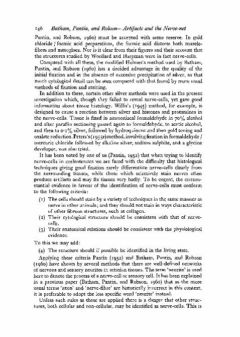
The amoebocytes often become elongated, especially in deteriorating tis-sues, and their long protoplasmic filaments may extend through the meso-gloea or epithelium. The cells may stain with silver or methylene blue andthus show a confusing resemblance to nerve-cells. In this guise they havemisled several authors who believed that nerve-fibres penetrated the meso-gloea. Sanchez (1918), however, pointed out the similarity between themesogloeal 'connective tissue cells' in his preparations of Sagartia ( = Calli-actis) parasitica and the mesogloeal 'nerve-cells' of Metridium described byHavet (1901). Havet's drawings are so clear that his so-called 'nerve-cells' inthe mesogloea can be identified as amoebocytes by their thin, branchingprocesses alone. If the figures denominated coelenterate nerve-cells byLeghissa (1949, 1950) and Torelli (1952) are compared with what aredemonstrably amoebocytes in fairly well-fixed tissue (fig. 3, B, D), the corre-spondence in form and situation is at once evident.
Amoebocytes stained with methylene blue can be distinguished from nerve-cells by their smaller size, and by the fact that their processes branch and mayapparently end on mesogloeal fibres, and are more tenuous than axons. Thelarge nucleolus of the nerve-cell is absent, the nucleus stains far less readily,and in well-fixed preparations the cytoplasm is consistently granular. Thegranules are strongly fuchsinophil (Chapman, 1953); their presence at onceidentifies amoebocytes in electron microscope sections (Grimstone and others,1958). When stained with silver by Holmes's method, their cytology ismanifestly different from that of nerve-cells. In living tissues amoebocytesmay aggregate locally, and individual cells mounted on a slide in sea-waterhave been observed to migrate. Unlike nerve-cells they are therefore mobile.
As mentioned above, these mesogloeal cells often assume a spindle-shape,
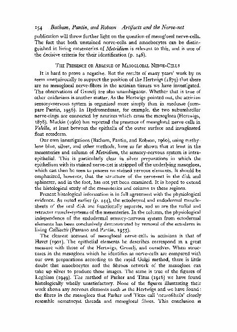
FIG. 3 (plate), A, transverse section of Calliactis column stained by Willis's silver method.Mesogloeal fibres and amoebocytes have stained, as well as muscle-fibres (upper left).
B, whole mount of Metridium mesentery, showing amoebocytes with irregular processes.Compare to A and D. Batham-Holmes silver method.
c, section showing beaded fibres in the mesogloea of the sphincter region in Calliactis.Slices of living tissue were treated with menthol overnight, stained with methylene blue forabout an hour, fixed in ammonium molybdate, and subsequently sectioned.
D, amoebocytes in a whole mesentery of Metridium. In this preparation they show fewstellate processes. Epithelial nuclei and muscle-fibres (right) are also visible. Baker'sformaldehyde-calcium / chlorazol black E.

» I i*
-> J •'.'• /
100/;
I iD
FIG. 3
E. J. BATHAM, C. F. A. PANTIN, and E. A. ROBSON
Bathatn, Pantin, and Robson—Artifacts and the Nerve-net 151
and may then resemble small bipolar nerve-cells. Their cytoplasm may bedrawn out into long processes which carry droplets at intervals. On inspection,however, these processes are seen to be very much finer than most nerveaxons, and they may branch. They often anastomose with each other, andmay end, apparently, on a mesogloeal fibre. Nothing resembling a synapticjunction can be seen. These features are all foreign to nerve-cells. On theother hand, living mesogloeal cells will sometimes stain with methylene blue.Chapman (1953) records the same for Calliactis and Aurelia. But the stainingtime is usually much longer than that of recognizable nerve-cells in the samepreparation, and in silver preparations mesogloeal cells are also quite distinctfrom nerve-cells. It may therefore be concluded that cells found in themesogloea show none of the morphological characteristics peculiar to nerve-cells, and certainly cannot function as such.
Mesogloeal fibres. It has been shown by Chapman (1953) that the mesogloeaof anemones and medusae is an organized structure of collagen-like fibres. Inanemones they are arranged in a crossed-lattice pattern, and, in the columnat least, this appears to be a mechanical result of the animal's movements.The physical properties which they share with fibres of any kind sometimesmake them stain deeply or receive precipitated silver with methods intendedfor nerve-cells (compare Peters, 1955a; Zon, 1936). Many fibres seem toprovoke the deposition of silver and may therefore stain.
With some silver methods, the combination of the staining of the meso-gloeal lattice fibres and of elongated amoebocytes associated with it can givepictures suggestive of a complex organized three-dimensional nerve-network(fig. 3, A). The collagen lattice follows a well-defined pattern (Chapman, 1953),and amoebocytes are often found following its strands. But if fixation andcytological preservation are good the deception is easily detectable even ina single preparation. Nor can it survive the results of comparing preparationsstained in different ways.
A comparison of Leghissa's figures (1949) with preparations such as thatof fig. 3, A strongly suggests that Leghissa's mesogloeal nerve-cells and theirneurites are in fact a similar artifact network derived from the deposition ofsilver on the collagen lattice and embedded amoebocytes in which thepreservation is not sufficiently critical to distinguish this. It is noteworthythat his figures do not show any cells as unequivocally nervous in characteras in the Hertwigs's illustrations or the photographs of Batham, Pantin, andRobson (1960).
Other mesogloeal structures. The only other structures which suggested tous the possibility of mesogloeal nerve-fibres were certain long-beaded struc-tures occasionally seen traversing the mesogloea. They sometimes appear whenhand-sections of Calliactis are stained vitally with methylene blue for severalhours. Fig. 3, c shows such structures from the sphincter region.
A careful study of these fibres has never revealed a cell-body in them.Moreover, we were able to show that they do not contain normal cytoplas-mic staining elements. We photographed 20-ju. sections of this sort of vitally

152 Batham, Pantin, and Robson—Artifacts and the Nerve-net
stained preparation fixed with ammonium molybdate, and identified land-marks in each section. We then re-stained them with Mallory's triple stainor Masson, and rephotographed the same area. We were able to identify partsof the fibre, but the structures stained identically with the surroundingcollagen and appeared to contain no cytoplasm.
A careful examination of silver sections sometimes shows beaded meso-gloeal structures which might possibly correspond to these methylene bluefibres.
THE VALIDITY OF SILVER METHODS
It is now appropriate to discuss certain aspects of the four criteria bywhich nerve-cells may be identified (p. 146).
The first, that they should stain like the nerve-cells of other animals aftera variety of methods, is the most difficult to establish, and this is not onlybecause special methods for nerve-cells tend to be capricious and may stainother structures of the kinds which have been discussed, but also because thereasons for which special methods may be successful are not yet fully under-stood. Recent studies of the use of methylene blue as a vital stain, and of themechanism of silver staining, have contributed several improvements intechnique; but it is not yet clear how the properties of nerve-cells areresponsible for their differential staining.
In a comprehensive review of peripheral nerve-terminations in mammaliantissues Weddell, Palmer, and Pallie (1955) have considered the problem ofartifacts in detail. These authors analyse the extensive literature, and are ableto infer much about peripheral nerve that is also demonstrated by their ownwork. They find that both methylene blue and silver staining are enhancedif the tissues are treated with hyaluronidase, which aids penetration of thedyes or fixative, and greatly reduces artifacts (Weddell and Pallie, 1954).Unstained nerve-cells studied under phase contrast provide a standard forreference. Although different techniques have been used to study the actiniannerve-net, and a trial of hyaluronidase did not improve staining with thismaterial, our conclusions and those of Weddell and others as to the origin ofcertain artifacts are much the same.
It is well known that the numerous silver methods available to histologistsare usually employed according to the type of structure that is to be stained.Some are more appropriate for the central nervous system, others for peri-pheral nerve (Carleton and Drury, 1957), and others are used for connectivetissue. Methods for the vertebrate central nervous system often aim at theselective staining of relatively few elements in a large mass of identifiednervous tissue. But when peripheral nerve is concerned, the aim is to stainthe system as completely as possible. The nerve-cells are relatively few innumber, and are usually scattered among other tissues. It is not yet under-stood why some empirical silver techniques can stain different types ofnerve-cell, apparently according to their function. It is worth noting, however,that the silver methods used by Havet, by Parker and Titus, and by Leghissa
Batham, Pantin, and Robson—Artifacts and the Nerve-net 153
for actinians are more usually employed for the vertebrate central nervoussystem, whereas the silver method of Holmes (1942, 1943), which has beenmodified to demonstrate the nerve-net of Metridium, is especially suited tothe staining of peripheral nerve.
The recent study by Peters (1955) of the mechanism of silver staining isbased on sections of vertebrate central nervous material. His method, whilesuccessful with these tissues, did not stain any nerve-cells in preliminarytrials with actinian material. Peters's experiments show that specificity ofstaining depends particularly on the developer used, and also on the pH,temperature, and concentration of the silver impregnating solution. Reduciblesilver, which as suggested by other workers combines mainly with histidine,is by no means confined to nerve-cells. Peters accepts Zon's earlier conclusion(1936) that tissue components exert a protective action on silver staining.This means that certain unspecified protein elements in the section somehowretard the reduction of silver at particular sites, but this interesting suggestionis not discussed further.
The problems of selectively staining either a few particular nerve-cells orall of those present are difficult. Methods which tend to stain all the nerve-cells present may stain other structures as well. In this respect, the work ofIrving and Tomlin (1954) on the argyrophil properties of collagen andreticulum is of special interest. These authors used only the silver methodsof Foot and Gomori, and experimented with connective tissue preparationsbefore staining them. They showed that the staining of connective tissue-fibres depends as much on the chemical properties of the ground substanceas on those of the fibres. Thus treatment with various proteins and withhyaluronic acid intensifies staining. The silver particles are seen by electronmicroscopy to be deposited only on the surface of the fibres, whereas in thecase of nerve-cells, Peters (1955c?) found that silver is reduced within theaxon as well as on the surface.
All this bears on two more of the criteria for the identification of nerve-cells(p. 146), their cytological structure and anatomical relations. Neither of thesecan be adequately studied without specific staining methods, or withouttaking precautions to minimize artifacts. Histochemical methods and electronmicroscopy should be able to provide new information about the actiniannerve-net, and should help to clarify the function and distribution ofamoebocytes, with which nerve-cells may still be confused. This confusiondoes not seem to be completely resolved in a recent paper of Leghissa andMazzi (1959). In this, cells that we have broadly referred to as amoebocytesuntil there is some evidence about their function (Batham, Pantin, andRobson, i960), are subdivided into mesocytes and amoebocytes. Althoughseven points of comparison by which nerve-cells differ from mesocytes aretabulated, and although a variety of optical and staining methods has beenused to study the mesogloea, the physiological properties of nerve-cells arenot mentioned, and their identification by this author is correspondinglydifficult to confirm. It is hoped that future work which is mentioned in this
154 Batham, Pantin, and Robson—Artifacts and the Nerve-net
publication will throw further light on the question of mesogloeal nerve-cells.The fact that both unstained nerve-cells and amoebocytes can be distin-guished in living mesenteries of Metridium is relevant to this, and is one ofthe decisive criteria for their identification (p. 146).
THE PRESENCE OR ABSENCE OF MESOGLOEAL NERVE-CELLS
It is hard to prove a negative. But the results of many years' work by usseem unequivocally to support the position of the Hertwigs (1879) that thereare no mesogloeal nerve-fibres in the actinian tissues we have investigated.The observations of Groselj are also unambiguous. Whether that is true ofother cnidarians is another matter. As the Hertwigs pointed out, the actiniansensory-nervous system is organized more simply than in medusae (com-pare Pantin, 1956). In Hydromedusae, for example, the two subumbrellarnerve-rings are connected by neurites which cross the mesogloea (Hertwigs,1878). Mackie (i960) has reported the presence of mesogloeal nerve-cells inVelella, at least between the epithelia of the outer surface and invaginatedfloat ectoderm.
Our own investigations (Batham, Pantin, and Robson, i960), using methy-lene blue, silver, and other methods, have so far shown that at least in themesenteries and column of Metridium, the sensory-nervous system is intra-epithelial. This is particularly clear in silver preparations in which theepithelium with its stained nerve-net is stripped off the underlying mesogloea,which can then be seen to possess no stained nervous elements. It should beemphasized, however, that the structure of the nerve-net in the disk andsphincter, and in the foot, has not yet been examined. It is hoped to extendthe histological study of the mesenteries and column to these regions.
Present histological information is in full agreement with the physiologicalevidence. As noted earlier (p. 144), the ectodermal and endodermal muscle-sheets of the oral disk are functionally separate, and so are the radial andretractor muscle-systems of the mesenteries. In the column, the physiologicalindependence of the endodermal sensory-nervous system from ectodermalelements has been conclusively demonstrated by removal of the ectoderm inliving Calliactis (Passano and Pantin, 1955).
The clearest account of mesogloeal nerve-cells in actinians is that ofHavet (1901). The epithelial elements he describes correspond in a greatmeasure with those of the Hertwigs, Groselj, and ourselves. When struc-tures in the mesogloea which he identifies as nerve-cells are compared withour own preparations according to the rapid Golgi method, there is littledoubt that amoebocytes and the fibrous network of the mesogloea cantake up silver to produce these images. The same is true of the figures ofLeghissa (1949). The method of Parker and Titus (1916) we have foundhistologically wholly unsatisfactory. None of the figures illustrating theirwork shows any nervous elements such as the Hertwigs and we have found:the fibres in the mesogloea that Parker and Titus call 'neurofibrils' closelyresemble nematocyst threads and mesogloeal fibres. This conclusion is
Batham, Pantin, and Robson—Artifacts and the Nerve-net 155
important because their work is cited in textbooks as evidence of mesogloealnerve-cells in actinians (Hyman, 1940).
Our conclusion thus wholly supports the intra-epithelial character of thenerve-net in the actinian material which we have studied. We also confirmthe essential separateness of the epithelia from the underlying mesogloea, andthe general absence of cells in that tissue other than amoebocytes. The onlyother cells which from time to time are truly to be found within the mesogloeaare the genital cells (Pantin, i960).
Much of this work was done at the Marine Biological Laboratory, Plymouth,to the Director and staff of which we are most grateful for the many facilitiesgiven us. We wish to thank the Department of Scientific aud IndustrialResearch for grants for the development of a special research which enabledthis work to be carried out, and the Council of Trinity College, Cambridge,for a contribution to the cost of the plates.
REFERENCESBATHAM, E. J., and PANTIN, C. F. A., 1950. 'Muscular and hydrostatic action in the sea-
anemone Metridium senile (L.).' J. exp. Biol., 27, 364.1951. 'The organisation of the muscular system of Metridium senile (L.).' Quart. J.
micr. Sci., 92, 27.and ROBSON, E. A. i960. 'The nerve-net of the sea-anemone Metridium senile
(L.): The mesenteries and the column.' Ibid., 101, 487.BOZLER, E., 1927. 'Untersuchungen iiber das Nervensystem der Coelenteraten. II. Uber die
Struktur der Ganglienzellen und die Function der Neurofibrillen nach Lebend-untersuchungen.' Z. vergl. Physiol., 6, 255.
CARLETON, H. M., and DKURY, R. A. B., 1957. Histological technique, 3rd ed. London(Oxford University Press).
CHAPMAN, G., 1953. 'Studies of the mesogloea of coelenterates. I. Histology and chemicalproperties.' Quart. J. micr. Sci., 94, 155.
GRIMSTONE, A. V., HORNE, R. W., PANTIN, C. F. A., and ROBSON, E. A., 1958. 'The finestructure of the mesenteries of the sea-anemone Metridium senile.' Ibid., 99, 523.
GROSELJ, P., 1909. 'Untersuchungen tiber das Nervensystem der Aktinien.' Arb. zool. Inst.Univ. Wien, 17, 269.
HADZI, J., 1909. 'Ober das Nervensystem von Hydra.' Ibid. 225.HAVET, J., 1901. 'Contribution a l'dtude du systeme nerveux des actinies.' Cellule, 18, 387.HERTWIG, O., and HERTWIG, R. 1878. Das Nervensystem und die Sinnesorgane der Medusen.
Leipzig (Vogel).HERTWIG, O., and HERTWIG, R., 1879. Studien zur Bldttertheorie. Heft I: Die Actinien.
Jena (Fischer).HOLMES, W., 1942. 'A new method for the impregnation of nerve axons in mounted paraffin
sections.' J. Path. Bact., 54, 132.'943. 'Silver staining of nerve axons in paraffin sections.' Anat. Rec, 86, 157.
HUGHES, A., 1953. 'The growth of embryonic neurites.' J. Anat., 87, 150.HYMAN, L. H., 1940. The invertebrates. I: Protozoa through Ctenopkora. New York
(McGraw-Hill).IRVING, E. A., and TOMLIN, S. G., 1954. 'Collagen, reticulum and their argyrophilic pro-
perties.' Proc. Roy. Soc. B, 142, 113.LEGHISSA, S., 1949. 'Contributo allo studio del tessuto e del sistema nervoso dei Celenterati.
i°. II tessuto nervoso delle Attinie.' Pubbl. staz. zool. Napoli, 21, 272.1950. 'L'Evoluzione morfologica del tessuto nervoso nei Celenterati fissi.' Boll. Zool.,17 suppl., 213.andMAZZi, S., 1959. 'Contributo ad una migliore conoscenza sulla struttura e compo-
sizione della mesodermide negli Antozoi.' Riv. Biol., 51, 293.

156 Batham, Pantin, and Robson—Artifacts and the Nerve-net
MACKIE, G. O., i960. 'The structure of the nervous system in Velella.' Quart. J. micr. Sci.,101, 119.
PANTIN, C. F. A., 1952. 'The elementary nervous system.' Proc. Roy. Soc. B, 140, 147.1956. 'The origin of the nervous system.' Pubbl. Staz. zool. Napoli, 38, 171.i960. 'Diploblastic animals.' Proc. Linn. Soc. Lond., 171, 1.
PARKER, G. H., and TITUS, E. G., 1916. 'The structure of Metridium (Actinoloba) marginatumMilne-Edwards with special reference to its neuromuscular mechanism.' J. exp. Zool.,21,433-
PASSANO, L. M., and PANTIN, C. F. A., 1955. 'Mechanical stimulation in the sea-anemoneCalliactis parasitica.' Proc. Roy. Soc. B, 143, 226.
PETERS, A., 1955a. 'Experiments on the mechanism of silver staining. I. Impregnation.'Quart. J. micr. Sci., 96, 84.
IO55&- 'Experiments on the mechanism of silver staining. II. Development.' Ibid.,96, 103.1955c. 'Experiments on the mechanism of silver staining. III. Quantitative studies.*
Ibid., 96, 301.ig5$d. 'Experiments on the mechanism of silver staining. IV. Electron microscope
studies.' Ibid., 96, 317.I955C- 'A general purpose method of silver staining.' Ibid., 96, 323.
ROMEIS, B., 1948. Mikroskopische Technih. Munich (Leibniz).SANCHEZ, M., 1918. 'Estudios sobre la histologia de las actinias.' Trab. Lab. Invest, biol.
Univ. Madrid, 16, fasc. 2, 1.TORELLI, B., 1952. 'Su alcuni particolari aspetti della istologia di Cerianthus.' Pubbl. Staz.
zool. Napoli, 23, 141.WEDDELL, G., and PALLIE, W., 1954. 'The value of "spreading factors" in the demonstration
of tissue neural elements.' Quart. J. micr. Sci., 95, 389.PALMER, E., and PALLIE, W., 1955. 'Nerve endings in mammalian skin.' Biol. Rev.,
30, 159-and ZANDER, E., 1951. 'The fragility of non-myelinated nerve terminals.' J. Anat.,
8s, 242.WILLIS, A. G., 1945. 'A new method for staining neurofibrillae and axis cylinders.' J. Roy.
micr. Soc, 15, 29.WOOLLARD, H. H., and HARPMAN, J. A., 1939. 'Discontinuity in the nervous system of
Coelenterates.' J. Anat., 73, 559.YOUNG, J. Z., 1944. 'Surface tension and the degeneration of nerve fibres.' Nature, 154, 521.ZON, L., 1936. 'Physical chemistry of silver staining.' Stain Tech., 11, 53.
![The ‘‘Naked Coral’’ Hypothesis Revisited – Evidence … et al...Order Species size (bp) GenBank accession # Reference Actiniaria Metridium senile 17,443 NC000933 [78] Nematostella](https://img.pdfslide.us/doc/110x75/5ec934cb1daf5f1d34431e11/the-aanaked-coralaa-hypothesis-revisited-a-evidence-et-al-order-species.jpg)


![Two mitochondrial group I introns a metazoan, Metridium ... · labeledusing[y-32P]ATP,andT4polynucleotidekinase(Unit-ed States Biochemical) was annealed with M. senile mtRNA (10,tg)](https://img.pdfslide.us/doc/110x75/5e19d953f7b2e93a05043a56/two-mitochondrial-group-i-introns-a-metazoan-metridium-labeledusingy-32patpandt4polynucleotidekinaseunit-ed.jpg)