Embed Size (px)
Citation preview
Biomedical imaging (BMI) is a strong and growingsubdiscipline of biomedical engineering (BME),with applications in basic science research, medicaldiagnosis, and the guidance of therapeutic interven-
tions. Of 112 academic programs in BME profiled on theWhitaker Foundation Web site [1], at least 67 involve imag-ing. The field of BMI is so important that, just two years ago,the National Institutes of Health broke from its traditional dis-ease and organ institute model and created the National Insti-tute for Biomedical Imaging and Bioengineering (NIBIB) tosupport research in this area. The recent Radiological Societyof North America meeting (December 2002) included on theorder of 2,600 papers with up to 30 parallel sessions, posters,specialized educational presentations, and 300 or more com-mercial exhibits. While this is largely a medical meeting, it isbased on imaging technologies developed and supported bybiomedical engineers and others from related disciplines. TheU.S. Department of Labor predicts a 31.4% increase in jobs inBME and a 23.1% increase in jobs for radiologic technolo-gists and technicians between 2000 and 2010 [2]. Since BMIis a subdiscipline of BME and a field closely related to thework of radiologic technologists, these employment statisticscan be used to infer a strong need for a continued supply of en-gineers well trained in the field of BMI.
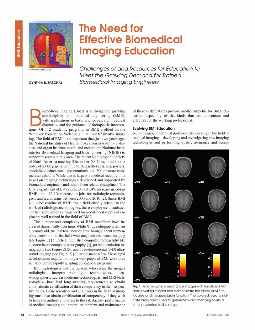
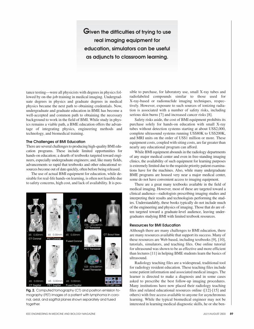
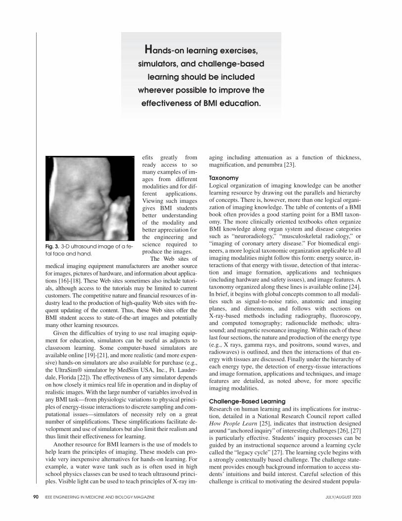
The number and complexity of BMI modalities have in-creased dramatically over time. While X-ray radiography is overa century old, the last few decades have brought about tremen-dous innovation in the field with magnetic resonance imaging(see Figure 1) [3], helical multislice computed tomography [4],electron beam computed tomography [4], positron emission to-mography (see Figure 2) [5], and three-dimensional (3-D) ultra-sound imaging (see Figure 3) [6], just to name a few. These rapiddevelopments require not only a well-prepared BMI workforcebut also require rapidly adapting educational programs.
Both radiologists and the persons who create the imagesradiologists interpret—radiologic technologists, ultra-sonographers, nuclear medicine technologists, and MRI tech-nologists—have had long-standing requirements to obtainand maintain certification of their competency in their respec-tive fields. Basic scientists and engineers in the field of imag-ing must also obtain certification of competency if they wishto have the authority to attest to the satisfactory performanceof medical imaging equipment. Attainment and maintenance
of these certifications provide another impetus for BMI edu-cation, especially of the kinds that are convenient andeffective for the working professional.
Evolving BMI EducationNot long ago, nonclinical professionals working in the field ofmedical imaging—developing and investigating new imagingtechnologies and performing quality assurance and accep-
IEEE ENGINEERING IN MEDICINE AND BIOLOGY MAGAZINE 0739-5175/03/$17.00©2003IEEE JULY/AUGUST 2003
BME
Educ
atio
n The Need forEffective BiomedicalImaging EducationChallenges of and Resources for Education toMeet the Growing Demand for TrainedBiomedical Imaging EngineersCYNTHIA B. PASCHAL
88
©1997 MASTER SERIES
Fig. 1. Axial magnetic resonance images with functional MRIdata overlaid in color that demonstrate the ability of MRI tolocalize and measure brain function. The colored regions indi-cate brain areas used to generate words that begin with aletter presented to the subject.
IEEE ENGINEERING IN MEDICINE AND BIOLOGY MAGAZINE JULY/AUGUST 2003 89
tance testing—were all physicists with degrees in physics fol-lowed by on-the-job training in medical imaging. Undergrad-uate degrees in physics and graduate degrees in medicalphysics became the next path to obtaining credentials. Now,undergraduate and graduate education in BME has become awell-accepted and common path to obtaining the necessarybackground to work in the field of BMI. While study in phys-ics remains a viable path, a BME education offers the advan-tage of integrating physics, engineering methods andtechnology, and biomedical training.
The Challenges of BMI EducationThere are several challenges to producing high-quality BMI edu-cation programs. These include limited opportunities forhands-on education; a dearth of textbooks targeted toward engi-neers, especially undergraduate engineers; and, like many fields,advancements so rapid that textbooks and other educational re-sources become out of date quickly, often before being released.
The use of actual BMI equipment for education, while de-sirable for real-life hands-on learning, is often not feasible dueto safety concerns, high cost, and lack of availability. It is pos-
sible to purchase, for laboratory use, small X-ray tubes andradiolabeled compounds similar to those used forX-ray-based or radionuclide imaging techniques, respec-tively. However, exposure to such sources of ionizing radia-tion is associated with a number of safety risks, includingserious skin burns [7] and increased cancer risks [8].
Safety risks aside, the cost of BMI equipment prohibits itspurchase solely for hands-on education with small X-raytubes without detection systems starting at about US$2,000,complete ultrasound systems running US$80K to US$200K,and MRI units on the order of US$1 million or more. Theseequipment costs, coupled with siting costs, are far greater thannearly any educational program can afford.
While BMI equipment abounds in the radiology departmentsof any major medical center and even in free-standing imagingclinics, the availability of such equipment for learning purposesis extremely limited due to the requisite priority patient examina-tions have for the machines. Also, while many undergraduateBME programs are housed very near a major medical center,some do not have convenient access to imaging equipment.
There are a great many textbooks available in the field ofmedical imaging. However, most of these are targeted toward aclinical audience—radiologists prescribing imaging studies andinterpreting their results and technologists performing the stud-ies. Understandably, these books typically do not include muchof the engineering and physics of imaging. Those that do are of-ten targeted toward a graduate-level audience, leaving under-graduates studying BMI with limited textbook resources.
Resources for BMI EducationAlthough there are many challenges to BMI education, thereare many resources available that support its success. Many ofthese resources are Web based, including textbooks [9], [10],tutorials, simulators, and teaching files. One online tutorialfor ultrasound was shown to be as effective and more efficientthan lectures [11] in helping BME students learn the basics ofultrasound.
Radiology teaching files are a widespread, traditional toolfor radiology resident education. These teaching files includesome patient information and associated medical images. Thelearner is directed to make a diagnosis and in some casesasked to prescribe the best follow-up imaging procedures.Many institutions have now placed their radiology teachingfiles and related educational resources online ([12]-[15] andothers) with free access available to anyone for asynchronouslearning. While the typical biomedical engineer may not beinterested in learning medical diagnostic skills, he or she ben-
CT Coronal
65 0.00mm
PET Coronal
CT Transaxial
65 0.00mm
65 0.00mm 65 0.00mm
65 0.00mm
65 0.00mm
CT Sagittal PET Sagittal Fused Sagittal
103 -474.00mm
103 -474.00mm
103 -474.00mm
PET MIP View
CT Scout View
1
Fused Coronal
PET Transaxial
Fused Transaxial
Fig. 2. Computed tomography (CT) and positron emission to-mography (PET) images of a patient with lymphoma in coro-nal, axial, and sagittal planes shown separately and fusedtogether.
Given the difficulties of trying to use
real imaging equipment for
education, simulators can be useful
as adjuncts to classroom learning.
efits greatly fromready access to somany examples of im-ages from differentmodalities and for dif-ferent applications.Viewing such imagesgives BMI studentsbetter understandingof the modality andbetter appreciation forthe engineering andscience required toproduce the images.
The Web sites ofmedical imaging equipment manufacturers are another sourcefor images, pictures of hardware, and information about applica-tions [16]-[18]. These Web sites sometimes also include tutori-als, although access to the tutorials may be limited to currentcustomers. The competitive nature and financial resources of in-dustry lead to the production of high-quality Web sites with fre-quent updating of the content. Thus, these Web sites offer theBMI student access to state-of-the-art images and potentiallymany other learning resources.
Given the difficulties of trying to use real imaging equip-ment for education, simulators can be useful as adjuncts toclassroom learning. Some computer-based simulators areavailable online [19]-[21], and more realistic (and more expen-sive) hands-on simulators are also available for purchase (e.g.,the UltraSim® simulator by MedSim USA, Inc., Ft. Lauder-dale, Florida [22]). The effectiveness of any simulator dependson how closely it mimics real life in operation and in display ofrealistic images. With the large number of variables involved inany BMI task—from physiologic variations to physical princi-ples of energy-tissue interactions to discrete sampling and com-putational issues—simulators of necessity rely on a greatnumber of simplifications. These simplifications facilitate de-velopment and use of simulators but also limit their realism andthus limit their effectiveness for learning.
Another resource for BMI learners is the use of models tohelp learn the principles of imaging. These models can pro-vide very inexpensive alternatives for hands-on learning. Forexample, a water wave tank such as is often used in highschool physics classes can be used to teach ultrasound princi-ples. Visible light can be used to teach principles of X-ray im-
aging including attenuation as a function of thickness,magnification, and penumbra [23].
TaxonomyLogical organization of imaging knowledge can be anotherlearning resource by drawing out the parallels and hierarchyof concepts. There is, however, more than one logical organi-zation of imaging knowledge. The table of contents of a BMIbook often provides a good starting point for a BMI taxon-omy. The more clinically oriented textbooks often organizeBMI knowledge along organ system and disease categoriessuch as “neuroradiology,” “musculoskeletal radiology,” or“imaging of coronary artery disease.” For biomedical engi-neers, a more logical taxonomic organization applicable to allimaging modalities might follow this form: energy source, in-teractions of that energy with tissue, detection of that interac-tion and image formation, applications and techniques(including hardware and safety issues), and image features. Ataxonomy organized along these lines is available online [24].In brief, it begins with global concepts common to all modali-ties such as signal-to-noise ratio, anatomic and imagingplanes, and dimensions, and follows with sections onX-ray-based methods including radiography, fluoroscopy,and computed tomography; radionuclide methods; ultra-sound; and magnetic resonance imaging. Within each of theselast four sections, the nature and production of the energy type(e.g., X rays, gamma rays, and positrons, sound waves, andradiowaves) is outlined, and then the interactions of that en-ergy with tissues are discussed. Finally under the hierarchy ofeach energy type, the detection of energy-tissue interactionsand image formation, applications and techniques, and imagefeatures are detailed, as noted above, for more specificimaging modalities.
Challenge-Based LearningResearch on human learning and its implications for instruc-tion, detailed in a National Research Council report calledHow People Learn [25], indicates that instruction designedaround “anchored inquiry” of interesting challenges [26], [27]is particularly effective. Students’ inquiry processes can beguided by an instructional sequence around a learning cyclecalled the “legacy cycle” [27]. The learning cycle begins witha strongly contextually based challenge. The challenge state-ment provides enough background information to access stu-dents’ intuitions and build interest. Careful selection of thischallenge is critical to motivating the desired student popula-
IEEE ENGINEERING IN MEDICINE AND BIOLOGY MAGAZINE JULY/AUGUST 200390
Hands-on learning exercises,
simulators, and challenge-based
learning should be included
wherever possible to improve the
effectiveness of BMI education.
Fig. 3. 3-D ultrasound image of a fe-tal face and hand.
tions. Such an approach is in many ways the basis of radiologyteaching files in which the challenge is to make the correct di-agnosis based on clinical and image information provided.Analogous challenges are needed to facilitate effective in-struction of engineers learning BMI principles. This contex-tual learning provides opportunities for students to notice howfundamental principles of BMI apply to various conditions,which ultimately lead to their ability to use this knowledge inthe future. Sample challenges for engineers in BMI might in-clude determining the source of an image artifact (relevant toimage formation, hardware, and basic principles); identifyingthe physical properties of a tissue or object appearing in animage; or using images to accurately guide intervention, withstudents motivated in all cases by the desire to contribute toimproved patient outcomes.
Looking Toward the FutureThe trends indicate that demand for persons trained in BMI isincreasing and can be expected to continue to increase in theforeseeable future. With increased demand for personnel andgrowing complexity of the field, the need for effective and ef-ficient educational techniques will become more and moreimportant. Online educational resources, allowing any-time/anywhere learning with the potential for educational in-teraction, will continue to grow in importance for BMIeducation. However, without a clear revenue model and withthe volatility of the medium, it is uncertain that such onlineeducational resources will ever attain the stature, prevalence,and continued utility of printed textbooks. Whatever the me-dium, hands-on learning exercises, simulators, and chal-lenge-based learning should be included wherever possible toimprove the effectiveness of BMI education.
AcknowledgmentsThe author acknowledges with gratitude the contributions ofJames D. Nguyen, Victoria L. Morgan, and Sean P. Brophy.
Cynthia B. Paschal is an associate profes-sor of biomedical engineering in theVanderbilt University School of Engineer-ing and an associate professor of radiologyand radiological sciences in the VanderbiltUniversity School of Medicine. In additionto teaching undergraduate and graduateBME students and investigating effective
educational technologies for BMI and BME physiology edu-cation, Dr. Paschal conducts research in magnetic resonanceimaging and computed tomography.
Address for Correspondence: Cynthia B. Paschal, Depart-ment of Biomedical Engineering, Vanderbilt University, Box351631-B, Nashville, TN USA 37235-1631. Phone: +1 615322 2029. Fax. +1 615 343 1978. E-mail: [email protected].
References[1] Whitaker Foundation. (2002, Dec.). Available: http://www.whitaker.org/aca-demic/
[2] U.S. Bureau of Labor Statistics. (2003, Jan.). Available: http://www.bls.gov
[3] Z.-P. Liang and P.C. Lauterbur, Principles of Magnetic Resonance Imaging: ASignal Processing Perspective. Piscataway, NJ: IEEE Press, 2000.
[4] E. Berry, et al. “”A systematic literature review of spiral and electron beam com-puted tomography: with particular reference to clinical applications in hepatic le-sions, pulmonary embolus and coronary artery disease," Health TechnologyAssessment (Winchester, England), vol. 3, pp. 1-118, Sept. 1999.
[5] G. Von Schulthess, Ed., Clinical Positron Emission Tomography (Pet): Correla-tion With Morphological Cross-Sectional Imaging. New York: Lippincott Williams& Wilkins, 2000.
[6] A. Fenster, D.B. Downey, and H.N. Cardinal, “Three-dimensional ultrasoundimaging,” Phys. Med. Biol., vol. 46, pp. R67-R99, May 2001.
[7] University of Iowa Healthcare. (2003, Jan.). X-Ray Martyrs. [Online]. Available:http://www.uihealthcare.com/depts/medmuseum/galleryexhibits/trailoflight/03xraymartyrs.html.
[8] D.B. Richardson, S. Wing, and W. Hoffmann, “Cancer risk from low-level ioniz-ing radiation: the role of age at exposure,” Occup. Med., vol. 16, pp. 191-218,Apr.-June 2001.
[9] Available: http://www.mrw.interscience.wiley.com/eist/index.html
[10] J.P. Hornak. (2003, Mar.). The Basics of NMR. [Online]. Available:http://www.cis.rit.edu/htbooks/nmr/
[11] J.D. Nguyen and C.B. Paschal, “Development Of online ultrasound instruc-tional module and comparison to traditional teaching methods,” J. Eng. Educ., vol.91, no. 3, pp. 275-283, 2002.
[12] BrighamRAD Professional Education. (2003, Mar.). Available:http://brighamrad.harvard.edu/education.html
[13] University Hospitals of Cleveland. Department of Radiology. (2003, Mar.).Available: http://www.uhrad.com/
[14] University of Washington. Department of Radiology. (2003, Mar.). TeachingMaterials. [Online]. Available: http://www.rad.washington.edu/teachingfiles.html
[15] AuntMinnie.com. (2003, Mar.). Available: http://www.auntminnie.com/in-dex.asp?sec=def
[16] GE Medical Systems. (2003, Mar.). Diagnostic Imaging. [Online]. Available:http://www.gemedicalsystems.com/company/nav_radiology.html
[17] Siemens Medical Solutions. (2003, Mar.). Available:http://www.siemensmedical.com/
[18] Phillips Medical Systems. (2003, Mar.). Available: http://www.medi-cal.philips.com/
[19] BrainWeb. (2003, Mar.). Available: http://www.bic.mni.mcgill.ca/cgi/bw/sub-mit_request.
[20] Duke University. Virtual Imaging Laboratory. (2003, Mar.).http://dukemil.egr.duke.edu/
[21] Ultrasound Simulator. (2003, Mar.). Available:http://www.bme.vanderbilt.edu/bme258/ultrasound/simula-tor/RunUltraSoundApplet.html.
[22] Medsim. (2003, Mar.). UltraSim. Online Training Simulator. [Online]. Avail-able: http://www.medsim.com/products/products.html
[23] R. Shevin, R.J. Zambon, S.S. Klein, and C.B. Paschal, “Safe alternatives forhands on learning of X-ray imaging principles,” presented at American Society forEngineering Education Annu. Meeting, Nashville, TN, June 2003.
[24] Available: http://www.vanth.org/curriculum/taxonomies/Medical_Imaging.doc.
[25] J.D. Bransford, A. Brown, and R.R. Cockings, Eds., How People Learn: Brain,Mind, Experience, and School. Washington, DC: National Academy Press, 2000.
[26] J.D. Bransford, N. Vye, H. Bateman, S.P. Brophy, and R. Roselli, “Vanderbilt’sAMIGO Project: Knowledge of How People Learn Enters Cyberspace,” in LearnerCentered Theory and Practice in Distance Education: Case Studies, T. Duffy and J.Kirkley, Eds. Mahwah, NJ: Lawrence Erlbaum and Associates, to be published.
[27] D. Schwartz, S. Brophy, X. Lin, and J. Bransford, “Software for managing com-plex learning: Examples from an educational psychology course,” Educ. Technol.Res. Develop., vol. 47, no. 2, pp. 39-59, 1999.
IEEE ENGINEERING IN MEDICINE AND BIOLOGY MAGAZINE JULY/AUGUST 2003 91