Embed Size (px)
Citation preview

“The Need for a PARP
Pharmacodynamic Assay”
Management of Disease Through DNA RepairManagement of Disease Through DNA Repair
Version 05/07/09
ams biotechnology (europe) ltd [email protected] www.amsbio.com
(UK) +44(0)1235 828200 (CH) +41(0)91 604 55 22 (DE) +49(0)69 779099

Management of Disease Through DNA RepairManagement of Disease Through DNA Repair
DNA Repair Pathways
• Base Excision Repair
• Homologous Recombination
• Nonhomolgous Recombination
• Nucleotide Excision Repair
• Mismatch Repair
• Transcription Mediated Repair

Management of Disease Through DNA RepairManagement of Disease Through DNA Repair
Targeted Pathways
• Base excision repair (BER) Excision of an incorrect
base followed the creation of a nicked DNA strand. The
nicked DNA strand serves as template for Polß to
replace the excised base.
• Homologous recombination, is a type of genetic
recombination in which genetic material is exchanged
between two similar or identical strands of DNA. The
process involves several cycles of breaking and rejoining
of DNA. The process is used to accurately repair double-
strand breaks in DNA,

Management of Disease Through DNA RepairManagement of Disease Through DNA Repair
Cancer Genes
• Hundreds of Cancer Genes have been
identified
• BRCA1/2: Two proteins in the homologous
recombination pathway. Mutations in these
genes result in defective recombination.
Mutations in these genes can be acquired
or inherited.

Management of Disease Through DNA RepairManagement of Disease Through DNA Repair
Regulators of Base Excision Repair
• PARP: Poly (ADP-Ribose) Polymerase
• PARG: Poly (ADP-Ribose) Glycohydrolase

Management of Disease Through DNA RepairManagement of Disease Through DNA Repair
PARP1
PARP1PARP1
ADPADP--RiboseRibose
NAD+
PARGPARG
Poly (ADP-Ribose)
Action of PARP and PARG
Figure 1.

Management of Disease Through DNA RepairManagement of Disease Through DNA Repair
STEP 2. DNA is damaged
STEP 3. PARP binds to broken strands
STEP 7. DNA strand repaired
STEP 1. PARP associated with DNAPARP
STEP 6. Repair enzymes recruited toDNA break
STEP 4. PARP synthesizes PAR. At the same timePARGhydrolyzes PAR to control polymer size.
STEP 5. Repair enzymes bind to PAR. Polymer continues to grow
STEP 9.PARG digests PAR
STEP 4B. Due excessive PAR and chargerepulsion PAR/PARP complex dissociatefrom DNA prior to recruitmentof repair enzymes
STEP 4C. DNA damage is not repairedby BER pathway
+
ReducedPARGactivity
STEP 4A. Reduced PARG activityresults in excessive PAR synthesis
PARG
ADP Ribose
Repair Enzyme
Poly (ADP) ribose (PAR)
PARP
PARP Inhibitor
STEP 3A. PAR is not madeand BER is stopped.
+
STEP 8 . PARP dissociates from DNA as a
result of charge repulsion
The Role of PARP and PARG in Base Excision Repair
Figure 2.

Management of Disease Through DNA RepairManagement of Disease Through DNA Repair
Step 5
Cell Death
(n)
(n)
+
Cells Deficient in Homologous Recombination
are Sensitive to PARP Inhibitors
Step 3
PARP inhibitor and DNA synthesis
Growing Replication fork
(n)
Step 1 Replication fork
(n)
Single strand
break
DNA Repair
Replication fork
Step 2
(n)
Homologous recombination
Step 4
(n)
Replication fork collides
with single strand break
resulting in double break in DNA
Homologous recombination deficiency
PARP inhibitor
Figure 3.

Management of Disease Through DNA RepairManagement of Disease Through DNA Repair
What is a Pharmacodynamic
Assay?
• Pharmacodynamic Assays:
– Provide evidence of drug action on molecular
target as shown in figure 4.
– Guide drug development process.
– Base line values may be used to stratify
patient response to therapy.

Management of Disease Through DNA RepairManagement of Disease Through DNA Repair
Nucleus
+ PARP
InhibitorNo Inhibitor
PARP Pharmacodynamic Assay
Figure 4.
Determine if inhibitor changed PARP activity in vivo
using a Pharmacodynamic Assay

Management of Disease Through DNA RepairManagement of Disease Through DNA Repair
The PARP Pharmacodynamic
Assay is a Capture ELISA
• Lysates were prepared from lymphocytes,
tissues or cultured cells.
• Free PAR and PAR bound to proteins is
captured by anti-PAR monoclonal antibody
attached to microtiter plates.
• Subsequently, captured PAR is quantified
using a PAR directed rabbit polyclonal
antibody.

Management of Disease Through DNA RepairManagement of Disease Through DNA Repair
Average of 3 PAR Standard Curves
y = 6 6 1.5x + 13 4 2 .1
R 2 = 0 .9 9 5
0
20000
40000
60000
80000
100000
120000
140000
160000
0 50 100 150 200 250
PAR pg/ml
Ne
t A
v R
LU
Assay Principle: Capture ELISA
Figure 5.
G
Goat anti-rabbit IgG-HRP

Management of Disease Through DNA RepairManagement of Disease Through DNA Repair
PAR is Stable During lysis
Procedure
• Using Trevigen regents and procedure
PAR levels are stabilized in cell lysates.
• The improper preparation of lysates can
result in degradation of PAR or
alternatively a burst of PARP activity can
be seen if the cells undergo stress prior to
the actual lysis.

Management of Disease Through DNA RepairManagement of Disease Through DNA Repair
PAR is Stable During Lysis
Procedure
1 2 3 4 5 6Lysate Preparation1-3: Trevigen Kit Reagent
4-6: Standard Procedures
Cell Treatment1,4: No treatment
2,5: 15 min, 1.5 mM MMS
3,6: 30 min, 1.5 mM MMS
Figure 6.
Trevigen Standard

Management of Disease Through DNA RepairManagement of Disease Through DNA Repair
PAR is Stable During lysis
Procedure (cont.)Summary
• Note that in the untreated lanes 1,4 that there is an increased PAR level in trypsin treated sample lane 4, compared the sample directly harvested into lysis buffer
• Trypsin treatment induced PAR synthesis increasing base line PAR levels
Samples need to be handled appropriately to avoid PAR synthesis during preparation

Management of Disease Through DNA RepairManagement of Disease Through DNA Repair
PARP Pharmacodynamic Assay is
Specific for PAR
• Jurkat cells were treated with the PARP
inhibitor PJ34 prior to lysate preparation.
• Alternatively, lysates were treated with
poly-ADP-ribose glycohydrolase (PARG)
to degrade PAR.

Management of Disease Through DNA RepairManagement of Disease Through DNA Repair
PARP “PDA” is specific for PAR
Figure 7.
105

Management of Disease Through DNA RepairManagement of Disease Through DNA Repair
Spike and Recovery
• Matrix: Extracts derived from PBMCs
• Matrix Concentrations: 57 pg/ml PAR and
218 pg/ml PAR
• Purified PAR spike: 20, 50, 100, (pg/ml)
PAR
• Recovery values: 81% to 102%
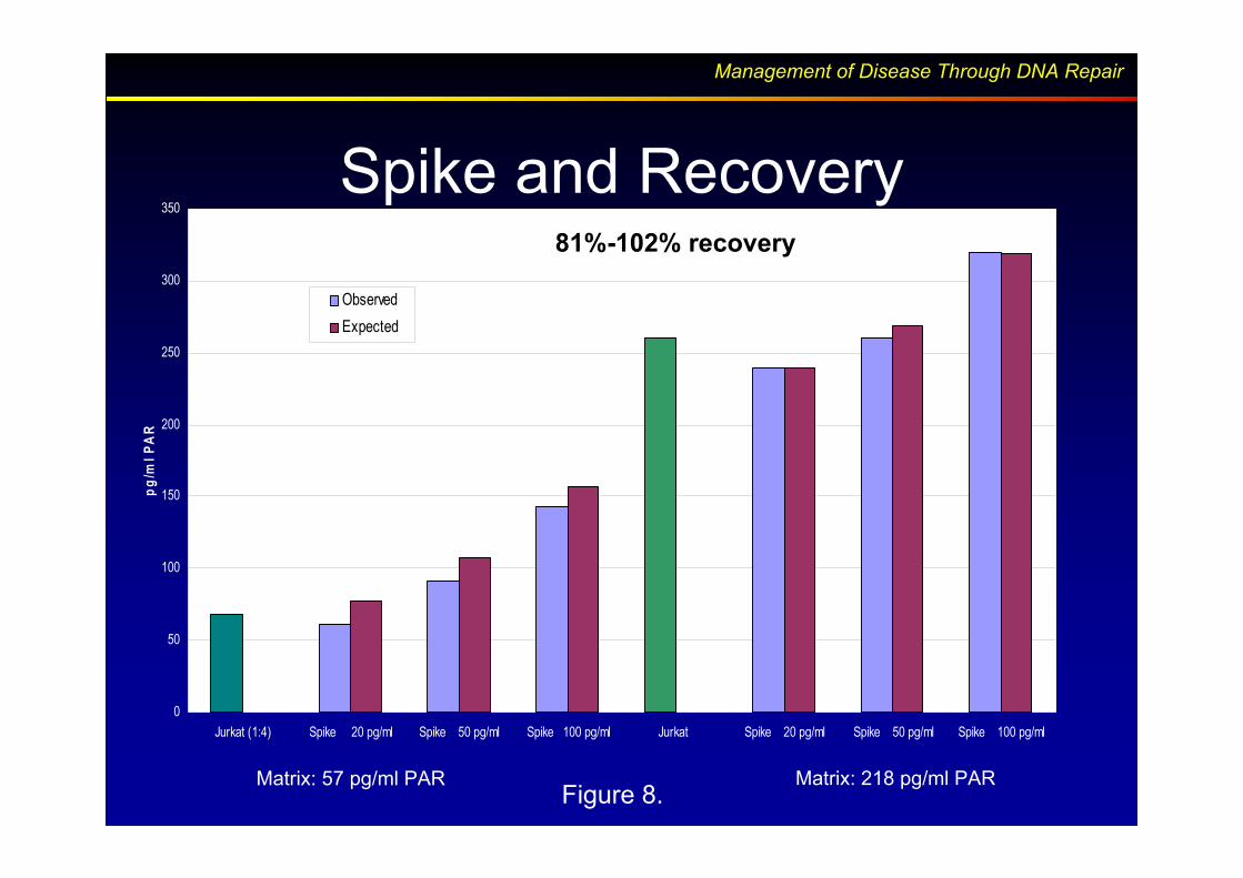
Management of Disease Through DNA RepairManagement of Disease Through DNA Repair
Spike and Recovery
0
50
100
150
200
250
300
350
Jurkat (1:4) Spike 20 pg/ml Spike 50 pg/ml Spike 100 pg/ml Jurkat Spike 20 pg/ml Spike 50 pg/ml Spike 100 pg/ml
pg
/ml
PA
R
Observed
Expected
81%-102% recovery
Matrix: 57 pg/ml PAR Matrix: 218 pg/ml PARFigure 8.

Management of Disease Through DNA RepairManagement of Disease Through DNA Repair
Samples Can be stored Prior to Assay
• It may be economical to batch samples prior
to PAR determination.
• Extracts can be prepared from frozen cell
pellets.
• Cells can be stored in liquid nitrogen prior to
lysate preparation without noticeable
changes to PAR levels.

Management of Disease Through DNA RepairManagement of Disease Through DNA Repair
Freezing has No Effect on the
Recovery of PAR from Lymphocytes
Effect of Freezing PBMCs on PAR Recovery
0.0
50.0
100.0
150.0
200.0
250.0
J Fresh J Frozen L Fresh L Frozen
pg
/ml P
AR
Figure 9
Donor L
Donor J

Management of Disease Through DNA RepairManagement of Disease Through DNA Repair
The Assay Provides Reproducible
Results
• Blood was drawn from three volunteers three times over a period of two weeks
• Reagents were tested three times over a period of two weeks (figures 6-9).
• Jurkat Cell lysates containing 22 pg/ml and 88 pg/ml PAR used as positive control
• Statistical analysis to determine performance of the assay over a two week period.

Management of Disease Through DNA RepairManagement of Disease Through DNA Repair
PARP Pharmacodynamic Assay
Validation 1
PAR Standard Curve
PAR pg/ml
Net
RL
U
Net RLU
Linear (Net RLU)
0
20000
40000
60000
80000
100000
120000
140000
0 100 200 300
PAR in PBMC and Jurkat Lysates
0
50
100
150
200
250
300
350
L10 L1
1
L12
J10
J11
J12
S10
S12
JurK
AT1:
4JU
R n
eat
pg
/ml
PA
R
R
y = 633.05x - 2377
2= 0.9964
Figure 10.

Management of Disease Through DNA RepairManagement of Disease Through DNA Repair
Validation 2
PAR in PBMCS and Jurkat lysates
Jur ne
at
0
50
100
150
200
250
300
L10 L1
1
L12
J10 J1
1
J12
S10
S12
Jur1
:4
PA
R p
g/m
l
PAR Standard Curve
Net
av
RL
U
0
20000
40000
60000
80000
100000
120000
140000
0 100 200 300
PARP Pharmacodynamic Assay
Net RLU
Linear (Net RLU)
R
y = 648.05x - 2354
2= 0.9964
PAR pg/ml
Figure 11.

Management of Disease Through DNA RepairManagement of Disease Through DNA Repair
PARP Pharmacodynamic Assay
y = 748.39x + 2043.9
R2
= 0.9899
0
20000
40000
60000
80000
100000
120000
140000
160000
0 50 100 150 200 250
Net
Av R
LU
PAR in PBMC and Jurkat lysates
0
50
100
150
200
250
300
L10
L11
L12
J10
J11
J12
S10
S12
Jur1
;4Ju
r Nea
t
PA
R p
g/m
l
Validation 3
PAR pg/ml
PAR Standard Curve
Net RLU
Linear (Net RLU)
Figure 12.

Management of Disease Through DNA RepairManagement of Disease Through DNA Repair
The Assay Provides Reproducible
Results (cont.)
• Standard curve shown in figure 12 derived from three experiments performed on three days has an R2 Value of 0.995.
• PAR levels shown in figure 9 from Jurkat cell lysates obtained from three experiments were 22 1 and 88 6 pg/ml.

Management of Disease Through DNA RepairManagement of Disease Through DNA Repair
PARP Pharmacodynamic ELISA
ASSAY
Average of 3 PAR Standard Curves
y = 661.5x + 1342.1
R2 = 0.995
0
20000
40000
60000
80000
100000
120000
140000
160000
0 50 100 150 200 250PAR pg/ml
Net
Av R
LU
Average of Validation Data
PAR in PBMC and Jurkat lysates
0
50
100
150
200
250
300
350
L10L1
1L12 J1
0J1
1J1
2S10
S12Ju
rKAT1:4
JUR
neat
Net RLU
Linear (Net RLU)
Figure 13.

Management of Disease Through DNA RepairManagement of Disease Through DNA Repair
The Assay provides Reproducible
Results
• Figure 10 shows that both PAR and the
PARP Pharmacodynamic assay are stable
for at least 3 months.

Management of Disease Through DNA RepairManagement of Disease Through DNA Repair
Assay stability evaluated over a 3
month period
0
20
40
60
80
100
120
140
1 2 3 4 5 6 7 8 9 10
Jur 1:4
Jur neat
Figure 14.

Management of Disease Through DNA RepairManagement of Disease Through DNA Repair
PARP Pharmacodynamic Assay compared
to PARP in vitro assay
• PARP Pharmacodynamic assay:
– Direct determination of the modulation of PARP activity in vivo.
• Assays that determine enzymatic activity in Lysates as opposed to actual PAR levels
– Are at best an indirect assay that depends upon retention of a PARP inhibitor on the enzyme through multiple in vitro steps.
Management of Disease Through DNA RepairManagement of Disease Through DNA Repair
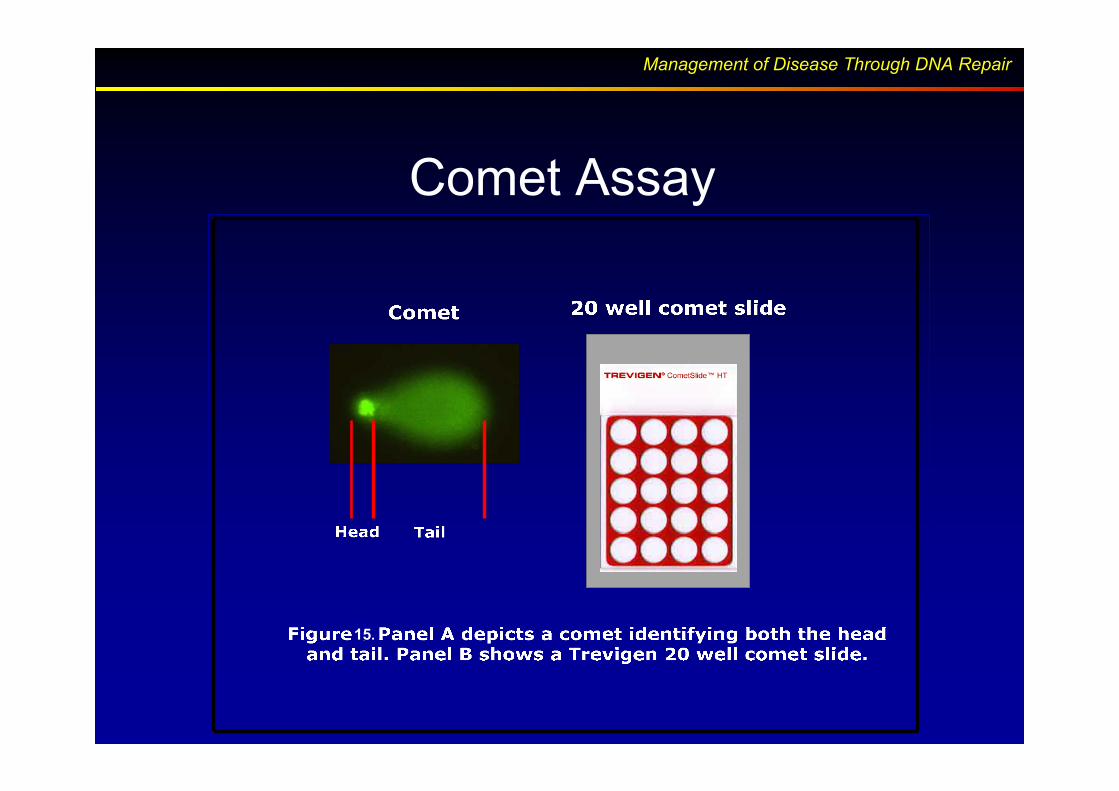
Comet Assay
15.

Management of Disease Through DNA RepairManagement of Disease Through DNA Repair
Comet Definitions
• Percent DNA in the Tail
– The integrated tail intensity x 100 divided by the total
integrated cell intensity for a normalized measure of
the percent of total cell DNA found in the tail
• Tail Moment
– The product of distance and normalized intensity
integrated over the tail length, (Lx • % DNAx)
– A damage measure combining the amount of DNA in
the tail with the distance of migration (severity of
damage)

Management of Disease Through DNA RepairManagement of Disease Through DNA Repair
Comet DNA Repair Assay
Comet Head: l l
Comet Tail: l l
1
7
2
5
8
3
6
9
4
Time
Figure 16. Cells are allowed to repair after exposure to DNA damaging agent

Management of Disease Through DNA RepairManagement of Disease Through DNA Repair
Slide Formats
• Slides are treated to promote adherence of low melting agarose
• Hydrophobic barrier allows easy application of cells directly to slide
• Formats designed to shorten assay time for rapid analysis of larger sample numbers
Figure 17. Different Comet slide formats

Management of Disease Through DNA RepairManagement of Disease Through DNA Repair
Comet Electrophoresis SystemComet Electrophoresis System
Overlay
Interlocking Safety Lid
Ceramic
2/20 Well Tray
Water Chamber Ports
•• 2/20 well Tray2/20 well Tray
•• Ten 2Ten 2--well slideswell slides
•• Five 20Five 20--well slideswell slides
•• 96 well Tray96 well Tray
•• 3 slides 3 slides -- 288 samples288 samples
96 Well
Tray
Figure. 18

Management of Disease Through DNA RepairManagement of Disease Through DNA Repair
+/- PARP inhibitor
Pre-Warm Media
30 and 60 min 37°C
5 x 10⁵ cells/ml
Jurkat and CCRF-CEM
+/- PARP inhibitor
1 hr 37°C
+/- PARP inhibitor
100 mM H202
15 min 37°C
+/- PARP inhibitor
PBS rinse
Samples
+/- PARP Inhibitor
Healthy
Treated
Recovery
Cell Treatment for PAR and Comet Analysis

Management of Disease Through DNA RepairManagement of Disease Through DNA Repair
Panel B
Jurkat-150 pg/ml PAR CCRF-CEM PAR
below detection limits
Figure 19. DNA repair measured by the comet assay
Panel A
Do PAR Levels Reflect DNA
Repair Capacity?

Management of Disease Through DNA RepairManagement of Disease Through DNA Repair
Comet
PARP inhibitor potentiates effect of
Hydrogen Peroxide
Basal PAR Level: 150 pg/ml Basal PAR Level: ND
Figure 20. DNA Repair measured by Comet Assay

Management of Disease Through DNA RepairManagement of Disease Through DNA Repair
Are Different DNA responses due to Are Different DNA responses due to
different PAR levels?different PAR levels?
Figure 21. PAR measured by Pharmacodynamic assay

Management of Disease Through DNA RepairManagement of Disease Through DNA Repair
Summary
• We have developed a validated “PDA” for PARP (Cat # 4510-096-K).
• In combination with the COMET assay (Cat # 4250-050-K), the relationship between basal levels of PAR and DNA repair can be determined.
• Preliminary experiments suggest that cell lines with low basal levels of PAR may have low repair capacity
![Flyer PARP FAmily NP V01 - biolinks k.k.1].pdf · Tomorrow’s Reagents Manufactured Today® International Edition The PARP Family T highlight PRODUCT FLYER The PARP Family](https://img.pdfslide.us/doc/110x75/5cb9946788c993f37c8c0cfc/flyer-parp-family-np-v01-biolinks-kk-1pdf-tomorrows-reagents-manufactured.jpg)