Embed Size (px)
Citation preview

Journal of Neurochemislry, 1970, Vol. 17, pp. 189 to 200. Pergamon Press. Printed in Northern Ireland
THE NATURE OF SULPHATION OF URONIC ACID- CONTAINING GLYCOSAMINOGLYCANS CATALYSED
BY BRAIN SULPHOTRANSFERASE
ELIZABETH GEORGE, MANORANJAN SINGHI and B. K. BACHHAWAT Neurochemistry Laboratory, Department of Neurological Sciences,
Christian Medical College Hospital, Vellore, S . India
(Received 24 June 1969. Accepted 6 Auguyt 1969)
Abstract-A sulphotransferase system of rat brain catalyses the transfer of sulphate from 3’-phosphoadenosine 5’-phosphosulphate to the low-sulphated glycosaminoglycans isolated from normal adult human brain. These were shown to be precursors of higher-sulphated gly- cosaminoglycans by DEAE-Sephadex column chromatography and paper electrophoresis. Xitrous acid degradation and mild acid hydrolysis of enzymically-sulphated fractions further confirmed the presence of heparan sulphate in human brain.
A partially purified sulphotransferase preparation was obtained from neonatal human brain using chondroitin-4-sulphate as sulphate acceptor. This sulphotransferase catalyses the transfer of sulphate to the various uronic acid containing glycosaminoglycans. Heparan sulphate was the best sulphate acceptor followed by dermatan sulphate, N-desulphoheparin, chondroitin-4-sulphate and chondroitin-6-sulphate in decreasing order. Sulphotransferase obtained from I-day-old rat, rabbit and guinea pig brain also had the same pattern of speci- ficity towards various sulphate acceptors.
This sulphotransferase catalyses both N-sulphation and 0-sulphation. Studies on the sulphotransferase obtained from both rat and human brain of various age groups indicate that the ratio of N-sulphation: 0-sulphation decreases as the brain matures.
PREVIOUS work from this laboratory (BALASUBRAMANIAN and BACHHAWAT, 1964) showed the transfer of sulphate from 3‘-phosphoadenosine 5’-phosphosulphate (PAPS) to various glycosaniinoglycans (GAG) such as heparan sulphate, dermatan sulphate, chondroitin-Csulphate and chondroitin-6-sulphate by a cell free extract of young rat brain. The possibility that there are specific sulphotransferase enzymes for each sulphated GAG was indicated by SUZUKI, TRENN and STROMINGER (1961), BALASUBRAMANIAN, JOUN and MARX (1968) and DAVIDSON and RILEY (1960).
A number of GAG fractions containing uronic acid were isolated from normal human brain on DEAE-Sephadex columns and some of them had a low degree of sulphation (SINGH and BACHHAWAT, 1968). These low-sulphated fractions of GAG were found to be rich in heparan sulphate. Earlier work from Meyer’s laboratory (MEYER, HOFFMAN, GRUMBACH and SAMPSON, 1959) also showed the presence of heparan sulphate in normal human brain. Recently, SINGH, CHANDRASEKARAN, CHERIAN and BACHHAWAT (1969) and CUNNINGHAM and GOLDBERG (1968) reported the presence of heparan sulphate in brains of various species. However, MARGOLIS (1967), SZABO and ROBOZ-EINSTEIN (1962) and CLAUSEN and HANSEN (1963) were unable to detect this GAG in nervous tissue. In the light of thesecontradictory reports, a study was undertaken using these low-sulphated GAG fractions from human brain
Present Address : Basic Biochemistry Unit, Veterans Administration Hospital, 4500 S . Lan- caster Road, Dallas, Texas, U.S.A.
Abbreviations used: PAPS, 3’-phosphoadenosine-5‘-phosphosulphate ; GAG, glycosaminoglycans ; CPB, cetyl pyridinium bromide; I , ionic strength; DEAE, diethylaminoethyl; ISB, IISB & IISC; low sulphated GAG isolated from adult human brain having the constituents uronic acid, hexosamine and sulphate in the following ratios: ISB, 1 :0%9:0.39; IISB, 1 :095:0.72 and IISC, 1 :0.93:0.92.
189
190 ELIZABETH GEORGE, MANORANJAN SINGH and B. K. BACHHAWAT
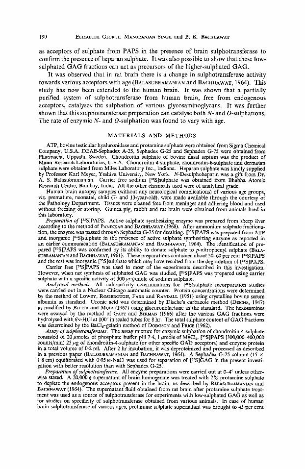
as acceptors of sulphate from PAPS in the presence of brain sulphotransferase to confirm the presence of heparan sulphate. I t was also possible to show that these low- sulphated GAG fractions can act as precursors of the higher-sulphated GAG.
I t was observed that in rat brain there is a change in sulphotransferase activity towards various acceptors with age (BALASUBRAMANIAN and BACHHAWAT, 1964). This study has now been extended to the human brain. I t was shown that a partially purified system of sulphotransferase from human brain, free from endogenous acceptors, catalyses the sulphation of various glycosaminoglycans. I t was further shown that this sulphotransferase preparation can catalyse both N- and 0-sulphations. The rate of enzymic N- and 0-sulphation was found to vary with age.
MATERIALS AND METHODS
ATP, bovine testicular hyaluronidase and protamine sulphate were obtained from Sigma Chemical Company, U.S.A. DEAE-Sephadex A-25, Sephadex G-25 and Sephadex G-75 were obtained from Pharmacia, Uppsala, Sweden. Chondroitin sulphate of bovine nasal septum was the product of Mann Research Laboratories, U.S.A. Chondroitin-4-sulphate, chondroitin-6-sulphate and dermatan sulphate were obtained from Miles Laboratory Inc., Indiana. Heparan sulphate was kindly supplied by Professor Karl Meyer, Yeshiva University, New York. N-Desulphoheparin was a gift from Dr. A. S. Balasubramanian. Carrier free sodium [35S]sulphate was obtained from Bhabha Atomic Research Centre, Bombay, India. All the other chemicals used were of analytical grade.
Human brain autopsy samples (without any neurological complications) of various age groups, viz. premature, neonatal, child (7- and 13-year-old), were made available through the courtesy of the Pathology Department. Tissues were cleaned free from meninges and adhering blood and used without freezing or storing. Guinea pig, rabbit and rat brain were obtained from animals bred in this laboratory.
Preparation of [35S]PAPS. Active sulphate synthesizing enzyme was prepared from sheep liver according to the method of PANIKKAR and BACHHAWAT (1968). After ammonium sulphate fractiona- tion, the enzyme was passed through Sephadex (3-75 for desalting. [35SlPAPS was prepared from ATP and inorganic [S5S]sulphate in the presence of active sulphate synthesizing enzyme as reported in an earlier communication (BALASUBRAMANIAN and BACHHAWAT, 1964). The identification of pre- pared [35S]PAPS was confirmed by its ability to donate sulphate to p-nitrophenyl sulphate (BALA- SUBRAMANIAN and BACHHAWAT, 1961). These preparations contained about 50-60 per cent [s5S]PAPS and the rest was inorganic [35S]sulphate which may have resulted from the degradation of [S5S]PAPS.
Carrier free [35S]PAPS was used in most of the experiments described in this investigation. However, when net synthesis of sulphated GAG was studied, [s5S]PAPS was prepared using carrier sulphate with a specific activity of 300,uc/,umole of sodium sulphate.
Analytical methods. All radioactivity determinations for [35S]sulphate incorporation studies were carried out in a Nuclear Chicago automatic counter. Protein concentrations were determined by the method of LOWRY, ROSEBROUGH, FARR and RANDALL (1951) using crystalline bovine serum albumin as standard. Uronic acid was determined by Dische's carbazole method (DISCHE, 1947) as modified by BITTER and MUIR (1962) using glucuronolactone as the standard. The hexosamines were assayed by the method of GATT and BERMAN (1966) after the various GAG fractions were hydrolysed with ~N-HCI at 100" in sealed tubes for 8 hr. The total sulphate content of GAG fractions was determined by the BaC1,-gelatin method of DODGSON and PRICE (1962).
Assay of sulphotransferase.~. The assay mixture for enzymic sulphation of chondroitin-4-sulphate consisted of 20 pmoles of phosphate buffer pH 7.4, 1 pmole of MgCIz, [s5S]PAPS [300,00&400,000 counts/min) 25 ,ug of chondroitin-4-sulphate (or other specific GAG acceptors) and enzyme protein in a total volume of 0.2 ml. After 2 hr incubation, it was deproteinized and processed as described in a previous paper (BALASUBRAMANIAN and BACHHAWAT, 1964). A Sephadex G-75 column (15 x 1.8 cm) equilibrated with 0.05 M-NaCI was used for separation of [35S]GAG in the present investi- gation with better resolution than with Sephadex G-25.
Preparation of sulphotransferase. All enzyme preparations were carried out at W unless other- wise stated. A 20,000 g supernatant of brain homogenate was treated with 2% protamine sulphate to deplete the endogenous acceptors present in the brain, as described by BALASUBRAMAMAN and BACHHAWAT (1964). The supernatant fluid obtained from rat brain after protamine sulphate treat- ment was used as a source of sulphotransferase for experiments with low-sulphated GAG as well as for studies on specificity of sulphotransferase obtained from various animals. In case of human brain sulphotransferase of various ages, protamine sulphate supernatant was brought to 45 per cent
Glycosaminoglycan sulphotransferase from brain 191
ammonium sulphate saturation by the gradual addition of solid ammonium sulphate with stirring. After stirring for 20 min, it was centrifuged at 10,OOOg for 20 min. Both the precipitates and super- natant were collected separately. Ammonium sulphate saturation of the supernatant was raised to 65 per cent by further addition of solid ammonium sulphate in the same manner. Both 0-45 per cent and 45-65 per cent ammonium sulphate precipitates were dissolved in minimal amounts of 0.02 M- tris-HC1 buffer, pH 7.4 (20-30 mg proteinlml), and they were dialysed for 8 hr against 50 vols. of the same buffer. The 45-65 per cent ammonium sulphate fraction was used for comparison of specific activity of the sulphotransferases obtained from brain of different age groups towards various GAG acceptors as well as for the study on thc site of sulphate incorporation in the case of heparan sulphate.
Isolation of low sulphated GAG. Three low-sulphated GAG fractions containing uronic acid, designated as ISB, IISB and IISC, were obtained from normal adult human brain as described by SINGH and BACHHAWAT (1968). The uronic acid:hexosamine:sulphate ratio of ISB, IISB and IISC were 1 : 0.89 : 0.39, 1 : 0.95 : 0.72 and 1 : 0.93 : 092 respectively. However, to isolate the GAG complex bound to peptide, the processing of the brain was carried out similarly except for the omission of alkali treatment, and a similar GAG pattern was obtained. The non-alkali treated fraction contained a considerably greater amount of ninhydrin positive material as determined by Rosen's method (ROSEN, 1957) after 6 N-HCI hydrolysis at 140" for 3 hr. The fractions obtained after alkali treatment are designated as peptide-free ISB and peptide-free IISB. The low-sulphated GAG fractions were separately digested with testicular hyaluronidase according to the method of MATHEW and INOYE (1961) and undigested fractions designated as hyaluronidase-resistant ISB and hyaluronidase-resistant IISB were obtained by gel filtration on Sephadex G-25 as described by SINGH and BACHHAWAT (1965). All these fractions were used for the sulphate incorporation studies.
Enzymic sulphation of low sulphated GAG. All the sulphated GAG fractions were incubated at 37" with carrier [35S]PAPS using protamine sulphate supernatant obtained from rat brain as the enzyme source.
For the characterization of enzymically sulphated [35S]GAG, 8.4 nmoles (532,000 countslmin) of [35]PAPS, 68.0 nmoles uronic acid of acceptor GAG and an enzyme preparation from 5-day-old rat brain were incubated for 3 hr, to obtain [35S]GAG with higher radioactivity. The ra5S]GAG obtained after gel filtration was dialysed against distilled water for 8 hr and then used for further identification as follows. A portion of isolated t3%]GAG was precipitated with CPB in the presence of celite, extracted with increasing concentrations of NaCI, and precipitated with alcohol (80 %) as reported previously (SINGH and BACHHAWAT, 1968). The GAG fractions were further subjected to DEAE- Sephadex column chromatography and eluted with various concentrations of NaCI. The radio- activity of each fraction was measured. ISB and IISB were designated as [35S]ISB and [S5S]IISB after enzymic sulphation in vitro.
Nitrous acid treatment. [35S]GAG obtained after enzymic sulphation was subjected to nitrous acid degradation at room temperature for SOmin, using the method of LAGUNOFF and WARREN (1962) for the determination of heparan sulphate in which the free amino group and N-sulphate of hexmamines are susceptible to the action of nitrous acid, giving rise to anhydromannose with the concomittant rupture of adjacent glycoside bonds. The reaction products resulting from nitrous acid degradation were fractionated by gel filtration on Sephadex G-25 (20 x 1.8 cm) equilibrated with 0.02 M-Nacl.
Determination of N- and 0-sulphation. [35S]GAG fractions obtained after enzymic sulphation were hydrolysed in sealed ampules at 100" in 0.04 N-HCI for 90 min for desulphation of the sulphamino groups (LAGUNOFF and WARREN, 1962; DANISHEFSKY, 1965). After hydrolysis, the incubate was neutralised with NaOH and a portion was passed through a Sephadex G-25 column (20 x 1.8 cm) equilibrated with 0.02 M-NaCl to separate the [%]GAG and inorganic [35S]sulphate. When chon- droitin [3sS]sulphate mixture was subjected to hydrolysis under the same conditions, 80 per cent of the radioactivity remained with the GAG.
Paper electrophoresis. Paper electrophoresis of the isolated fractions obtained after enzymic sulphation was carried out using 0.1 I lithium sulphate (FOSTER and PEARCE, 1961), and was followed by radioautography. The paper was stained with 05% alcian blue in 3 % acetic acid.
RESULTS Enzymic sulphation of low-sulphated-GAG. As shown in Table 1, there was a net
transfer of sulphate from PAPS to the two low-sulphated GAG acceptors. Fraction ISB which had a sulphate/uronic acid ratio of 0.39 was the best acceptor. Sulphate incorporation into IISB was comparatively low and it had a higher sulphate content (0.72 pmole sulphate/pmole of uronic acid). In the case of fraction IISC, sulphate incorporation was almost negligible and the ratio of sulphate/uronic acid was 0.92.

192 ELIZABETH GEORGE, MANORANJAN SINGH and B. K. BACHHAWAT
TABLE ENZYMIC SULPHATION OF LOW-SULPHATED GAG
[s5S]Sulphate incorporated
Counts/rnin nmoles Acceptor GAG
Experiment I ISB IISB IISC Chondroitin sulphate from bovine nasal septum
Experiment I1 ISB Peptide-free ISB IISB Peptide-free IISB
Hyaluronidase resistant ISB Peptide-free ISB IISB Peptide-free IISB
6900 2500 640
1400
18,200 15,800
8800 10,900
17,600 32,100 26,700 21,500
0.03 0.01 0,003
0.006
0.29 0.25 0.14 0.18
0.28 0.52 0.14 0.18
Incubation mixture for experiment I consisted of 40 pmoles of tris-HC1, pH 8.0, 2 pmoles of MgCI,, 0.6 m o l e (131,500 counts/ min) of [35S]PAPS, 50pg of sulphate-acceptor GAG and 0.56 mg of enzyme protein obtained from protamine sulphate supernatant of 5-day-old rat brain in a total volume of 0.3 ml. For experiment 11, it consisted of 40 pmoles of tris-HC1, pH 8.0, 2 pmoles of MgCI,, 12 nmoles (760,000 counts/min) of [35S]PAPS, 14.0 moles uronic acid of sulphate-acceptor GAG and 1.2 mg of enzyme protein obtained from protamine sulphate supernatant of 15-day- old rat brain in a total volume of 0.4 ml. The incubation was carried out for 2 hr at 37".
TABLE 2.-CPB FRACTIONATION OF LOW-SULPHATED GAG AFTER ENZYMIC SULPHATION
Counts/min Sulphated GAG
Fraction I (0.4 M-NaCI Fraction 11
extract) (1.2 M-NaCl extract)
9400 11,700 0 18,300
Hyaluronidase resistant [3JS]ISB 9600 10,700 [35S]IISB 0 35,700
Carrier hyaluronic acid and chondroitin sulphate (1 rng each) and chondroitin sulphate (1 mg) were added to [%]ISB and [35S]IISB respectively. After precipitation with CPB, the sediments were then extracted with various concentrations of NaCI. NaCl extraction was repeated until the extracts were free of radioactivity.

Glycosaminoglycan sulphotransferase from brain 193
Hyaluronidase resistant fractions also incorporated 0.28-0.52 nmole of sulphate in the presence of the sulphotransferase system. There was no significant difference in the incorporation of [35S]sulphate into the peptide free fractions and into the peptide rich fractions except in the case of hyaluronidase resistant ISB.
Characterization of [35S]ISB and r5S]IISB. The steps involved in the characteriz- ation of [35S]ISB and [35S]IISB after enzymic sulphation are summarized in the following scheme:
[S5S]ISB [35S]ILSB
Precipitation of the GAG by CPB and frac- tionation of the complex with NaCl
I
Precipitation of the GAG by CPB and frac- tionation of the complex with NaCl
I 1 1 1 1 I 0.4 M-Nacl 1.2 M-NaCI 2.1 M-NaCI 0.4 M-NaCI 1.2 M-NaCl 2.1 M-NaCl Fraction I Fraction I1 Fraction 111 Fraction I Fr
DEAE- Sephadex chromatog- raphy
I 1 1-
DEAE- Sephadex chromatography
1 4-
:ion I1 Fraction I11 (ND)
DEAE- Sephadex chromatography
1 0.5 M-NacI 0.9 M-NaCI 0.9 M-NaCl 1.5 M-NaCI 0.9 M-NaCl 1.5 M-NaCl
(ND) Peak A Peak B in 0.01 M-HCI Peak D in 0.01 M-HC1 Peak C Peak E
ND, Radioactivity not detectable.
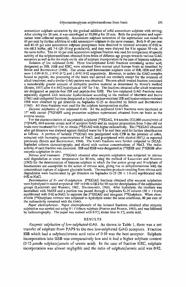
The results of the fractionation of the [35S]GAG-cetylpyridinium bromide com- plex with various concentrations of NaCl are shown in Table 2. The radioactivity of [35S]ISB and hyaluronidase-resistant r5S]ISB was equally distributed in both 0.4 M-
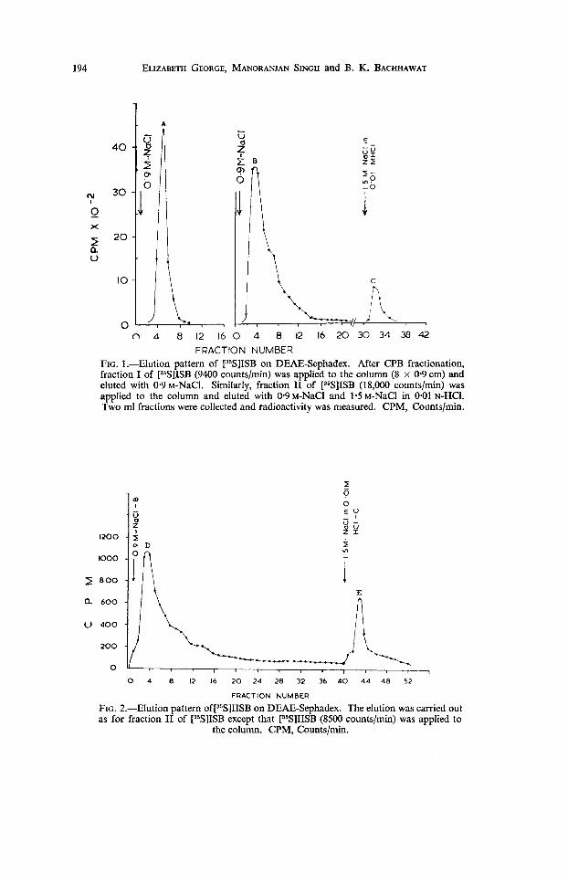
NaCl and 1.2 M-NaCI fractions, whereas the radioactivity of r5S]IISB and hyaluroni- dase-resistant [35S]IISB was obtained in the 1.2 M-NaCI fraction only. All these fractions of [35S]GAG obtained by the CPB procedure were further fractionated on DEAE-Sephadex. The 0.4 M-NaCI fraction of [35S]ISB was completely eluted in one peak, A, as shown in Fig. 1, whereas the 1-2 M-NaCI fraction of [35S]ISB was eluted in two peaks, B and C, showing that the charge density of the molecule had been in- creased. The elution pattern of peaks A, B and C were identical with the original glycosaminoglycan fractions ISB, IISB and IISC, respectively (SINGH and BACHHAWAT, 1968). Figure 2 shows that after in vitro sulphation of IISB, two radioactivity peaks, D and E, were obtained. The emergence of these peaks on sulphation of ISB and IISB indicated that ISB and IISB contained some lower-sulphated form of heparan sulphate, chondroitin-4-sulphate and chondroitin-6-sulphate.
Results of nitrous acid treatment of all the r5S]GAG obtained after sulphation in vitro is given in Table 3. A typical gel-filtration profile of one of the fractions treated with nitrous acid is given in Fig. 3. Various radioactive peaks which were retained on Sephadex G-25 may be due to 35S-sulphated oligosaccharides of varying
194
-
4 0 -
c;v 3 0 - / O
0 X
20 - z V
ELIZABETH GEORGE, MANORANJAN SINGH and B. K. BACHHAWAT
A - V
4 9 E B 0, C I S 0
f - 3 0.
1
c .- - sy 22 I- - 0 ,?
0 4 8 I2 16 2 0 24 28 32 36 4 0 4 4 4 8 5 2
FRACTION NUMBER
FIG. 2.-Elution pattern o~[~~S]I ISB on DEAE-Sephadex. The elution was carried out as for fraction I1 of [3SS]ISB except that [35S]IISB (8500 counts/min) was applied to
the column. CPM, Countslmin.

Glycosaniinoglycan sulphotransferase from brain
120
100.
8 0. I a " 60.
40.
2 0.
195
TABLE ANALYSIS OF [36S]GAG FRACTIONS OBTAINED BY GEL FILTRATION AFTER NITROUS ACID DEGRADAIJON
Radioactivity obtained (countslmin)
In void volume* In inner volumet Sulphated GAG
[35S]ISB Peptide-free [35S]ISB [35S]IISB Peptide-free [35S]IISB
Hyaluronidase resistant [35S]ISB Peptide-free [35S]ISB [35S]IISB Peptide-free [35S]11SB
580 290 430 590
650 1600 1140 750
1090 1060 620 640
1310 4860 1430 1310
* [35S]GAG which was unaffected by nitrous acid treatment
t [35S]Sulphate fractions containing oligosaccharides were was eluted in void volume.
retarded in the column.
~~~ .
10 20 30 40 50 F R A C T I O N NUMBER
FIG. 3.-Elution pattern of the gel filtration of ["SIGAG after nitrous acid degradation. CPM, counts/min. Experimental details are given in the text. One ml fractions were
collected.
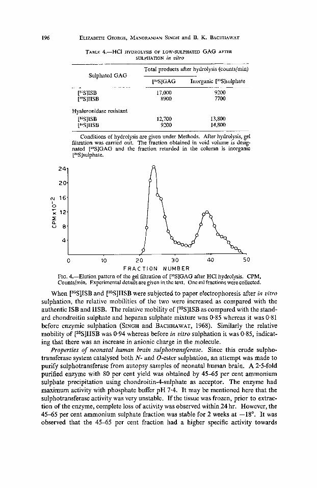
chain length. Furthermore, following in uitro sulphation, it was observed that when various GAG fractions were subjected to similar gel filtration without nitrous acid treatment, all the glycosaminoglycans were eluted in the void volume. A similar observation was made by CIFONELLI (1968) when he subjected heparan sulphate to gel filtration before and after nitrous acid treatment. All the [35S]GAG fractions were partially degraded by nitrous acid treatment indicating that they contain heparan sulphate (N-sulphated GAG). The site of [35S]sulphate incorporation into the low- sulphated GAG was further studied by 0.04 N-HCl hydrolysis. The results of HCI hydrolysis of low-sulphated GAG after enzymic sulphation is given in Table 4. A typical gel filtration pattern of [35S]GAG after HCl hydrolysis is depicted in Fig. 4. The sulphamino group was easily removed under this condition of hydrolysis whereas only 20 per cent of the [35S]sulphate of chondroitin-4-sulphate was removed. This result shows that both N- and 0-sulphation were taking place in the molecule.
196 ELIZABETH GEORGE, MANORANJAN SINGH and B. K. BACHHAWAT
TABLE 4.-HC1 HYDROLYSIS OF LOW-SULPHATED GAG AFTER SULPHATION in vitro
~~
Total products after hydrolysis (counts/min) Sulphated GAG
[35S]GAG Inorganic [35S]sulphate
17,000 8900
9200 I700
Hyaluronidase resistant [35S]ISB 12,700 13,800 [35S]IISB 9200 14,800
Conditions of hydrolysis are given under Methods. After hydrolysis, gel filtration was carried out. The fraction obtained in void volume is desig- nated [35S]GAG and the fraction retarded in the column is inorganic [35S]sulphate.
0 10 2 0 30 40 50
FIG. 4.-Elution pattern of the gel filtration of [35S]GAG after HCl hydrolysis. CPM, Countslmin. Experimental details are given in the text. One ml fractions were collected.
When [35S]ISB and [35S]IISB were subjected to paper electrophoresis after in vitro sulphation, the relative mobilities of the two were increased as compared with the authentic ISB and IISB. The relative mobility of [35S]ISB as compared with the stand- ard chondroitin sulphate and heparan sulphate mixture was 0.85 whereas it was 0-81 before enzymic sulphation (SINGH and BACHHAWAT, 1968). Similarly the relative mobility of [35S]IISB was 0.94 whereas before in vitro sulphation it was 0.85, indicat- ing that there was an increase in anionic charge in the molecule.
Properties of neonatal human brain sulphotransferase. Since this crude sulpho- transferase system catalysed both N- and 0-ester sulphation, an attempt was made to purify sulphotransferase from autopsy samples of neonatal human brain. A 2.5-fold purified enzyme with 80 per cent yield was obtained by 45-65 per cent ammonium sulphate precipitation using chondroitin-Csulphate as acceptor. The enzyme had maximum activity with phosphate buffer pH 7.4. It may be mentioned here that the sulphotransferase activity was very unstable. If the tissue was frozen, prior to extrac- tion of the enzyme, complete loss of activity was observed within 24 hr. However, the 45-65 per cent ammonium sulphate fraction was stable for 2 weeks at -18". It was observed that the 45-65 per cent fraction had a higher specific activity towards
F R A C T I O N NUMBER
Glycosaminoglycan sulphotransferase from brain 197
chondroitin-4-sulphate and dermatan sulphate as compared to the 0-45 per cent fraction, whereas the specific activity towards heparan sulphate was high in the 0-45 per cent fraction as compared to that of the 45-65 per cent ammonium sulphate fraction. Following enzymic sulphation of heparan sulphate by both these fractions, the analysis of N- and 0-sulphate by HCI hydrolysis showed that the N-sulphate to 0-sulphate ratio was same in both the fractions.
Sulphation of various GAG acceptors using sulphotransferases from human brains of various ages. Sulphotransferase activity in child brain of various age-groups was studied in the 45-65 per cent ammonium sulphate fraction. [35S]Sulphate incorpora- tion into various glycosaminologlycans using brain sulphotransferase from these four age-groups are given in Table 5. The best acceptor in all cases was heparan sulphate,
TABLE 5.-SULPHOTRANSFERASE ACTIVITY OF HUMAN BRAIN OF DIFFERENT AGES TOWARDS VARIOUS GAG ACCEPTORS
~~
Enzyme activity*
1 2 3 4 GAG Acceptors
Chondroitin-4-sulphate 4000 4490 4500 6000 Dermatan sulphate 9900 9000 7100 10,100 Chondroitin-6-sulphate 1800 N.D. 1600 840 N-Desulpho heparin 5700 7800 2300 3500 Heparan sulphate 34,800 28,000 24,000 19,300
* Enzyme activity is expressed as counts/min of [35S]GAG formed per mg of enzyme protein under the conditions of assay.
Sulphotransferase (45-65 % ammonium sulphate fraction) was prepared from : 1 , pre- mature baby brain; 2, I-day-old child brain; 3, 7-year-old child brain; 4, 13-year-old child brain. Conditions of assay were the same as described in text. N.D., not deter- mined.
followed by dermatan sulphate. N-Desulphoheparin was a poor acceptor, although 70 per cent of the amino group of N-desulphoheparin was free for sulphation (BALASUBRAMANIAN et aE., 1968). This shows that, in addition to free sites in the molecule available for sulphation, other pre-requisites are necessary for the sulpho- transferase activity. Chondroitin-4-sulphate also accepted sulphate, the amount of incorporation being 40004000 counts/min/mg enzyme protein. Chondroitin-6- sulphate was a poor acceptor as compared to other GAG. There were variations in the enzymic sulphation of various GAG with the age of the brain. In general, sulphotransferase has a very high activity in neonatal brain towards all the GAG employed as acceptors. However, the specific activity of sulphotrans- ferase towards heparan sulphate decreased as the age of tissue increased. The ratio of sulphotransferase activity towards heparan sulphate to that towards chon- droitin-4- sulphate also decreased with age, it was 8.5 in premature baby brain, and decreased to 3.2 in the brain of a 13-year-old child. This finding with sulphotrans- ferase of human brain is of interest since a similar change in sulphotransferase activity towards heparan sulphate and chondroitin-4-sulphate was made earlier in this labora- tory (BALASUBRAMANIAN and BACHHAWAT, 1964) with extracts of rat brain.
Enzyme activity from brnin preparations of various animals. The specific activities of the sulphotransferases obtained from brain of various species towards GAG is given in Table 6. The best acceptor in all cases, was heparan sulphate,
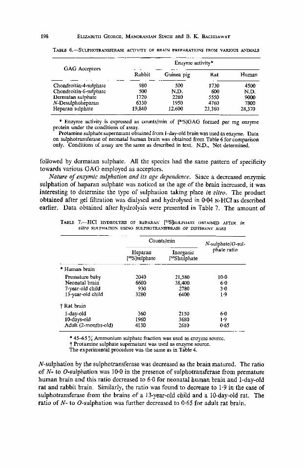
198 ELIZABETH GEORGE, MANORANJAN SINGH and B. K. BACHHAWAT
TABLE 6.--&JLPHOTRANSFERASE ACTIVITY OF BRAIN PREPARATIONS FROM VARIOUS ANIMALS ~ ~~~
Enzyme activity* GAG Acceptors
Rabbit Guinea pig Rat Human
Chondroitin-4-sulpha te 980 500 1730 4500
Dermatan sulphate 1720 2280 5550 9000 Chondroitin-6-sulphate 500 N.D. 600 N.D.
N-Desulphoheparan 6330 1950 4760 7800 Heparan sulphate 19,840 12,600 23,360 28,370
* Enzyme activity is expressed as counts/min of [36S]GAG formed per mg enzyme protein under the conditions of assay.
Protamine sulphate supernatant obtained from 1-day-old brain was used as enzyme. Data on sulphotransferase of neonatal human brain was obtained from Table 6 for comparison only. Conditions of assay are the same as described in text. N.D., Not determined.
followed by dermatan sulphate. All the species had the same pattern of specificity towards various GAG employed as acceptors.
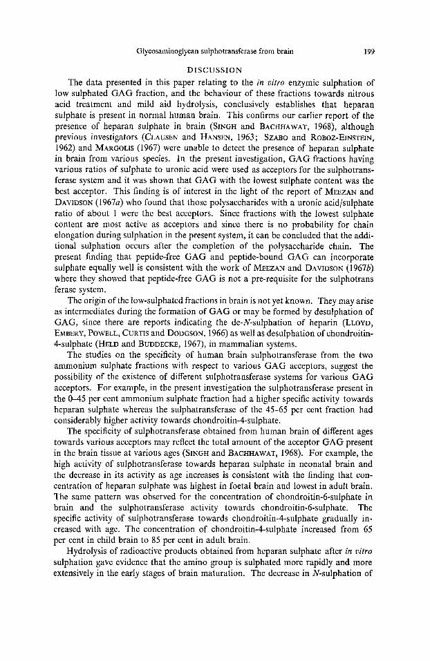
Nature of enzymic sulphation and its age dependence. Since a decreased enzymic sulphation of haparan sulphate was noticed as the age of the brain increased, it was interesting to determine the type of sulphation taking place in vifro. The product obtained after gel filtration was dialysed and hydrolysed in 0.04 N-HCI as described earlier. Data obtained after hydrolysis were presented in Table 7. The amount of
TABLE 7.-HCI HYDROLYSIS OF HEPARAN ["S]SULPHATE OBTAINED AFTER in o h 0 SULPHATION USING SULPHOTRANSFERASE OF DIFFERENT AGES
N-sulphatelU-sul- Counts/min
Heparan Inorganic phate ratio [35S]sulphate [35S]sulphate
* Human brain Premature baby 2040 21,580 10.0 Neonatal brain 6600 38,400 6.0 7-year-old child 930 2780 3.0 13-year-old child 3280 6400 1.9
t Rat brain 1-day-old 3 60 2150 6.0
Adult (2-months-old) 4130 2610 0.65 10-days-old 1960 3680 1.9
* 45-65 % Ammonium sulphate fraction was used as enzyme source. t Protamine sulphate supernatant was used as enzyme source. The experimental procedure was the same as in Table 4.
N-sulphation by the sulphotransferase was decreased as the brain matured. The ratio of N- to U-sulphation was 10.0 in the presence of sulphotransferase from premature human brain and this ratio decreased to 6.0 for neonatal human brain and 1-day-old rat and rabbit brain. Similarly, the ratio was found to decrease to 1.9 in the case of sulphotransferase from the brains of a 13-year-old child and a 10-day-old rat. The ratio of N- to U-sulphation was further decreased to 0.65 for adult rat brain.
Glycosaminoglycan sulphotransferase from brain 199
DISCUSSION The data presented in this paper relating to the in vitro enzymic sulphation of
low sulphated GAG fraction, and the behaviour of these fractions towards nitrous acid treatment and mild aid hydrolysis, conclusively establishes that heparan sulphate is present in normal human brain. This confirms our earlier report of the presence of heparan sulphate in brain (SINGH and BACHHAWAT, 1968), although previous investigators (CLAUSEN and HANSEN, 1963 ; SZABO and ROBOZ-EINSTEIN, 1962) and MARGOLIS (1967) were unable to detect the presence of heparan sulphate in brain from various species. In the present investigation, GAG fractions having various ratios of sulphate to uronic acid were used as acceptors for the sulphotrans- ferase system and it was shown that GAG with the lowest sulphate content was the best acceptor. This finding is of interest in the light of the report of MEEZAN and DAVIDSON (19674 who found that those polysaccharides with a uronic acid/sulphate ratio of about 1 were the best acceptors. Since fractions with the lowest sulphate content are most active as acceptors and since there is no probability for chain elongation during sulphation in the present system, it can be concluded that the addi- tional sulphation occurs after the completion of the polysaccharide chain. The present finding that peptide-free GAG and peptide-bound GAG can incorporate sulphate equally well is consistent with the work of MEEZAN and DAVIDSON (1967b) where they showed that peptide-free GAG is not a pre-requisite for the sulphotrans ferase system.
The origin of the low-sulphated fractions in brain is not yet known. They may arise as intermediates during the formation of GAG or may be formed by desulphation of GAG, since there are reports indicating the de-N-sulphation of heparin (LLOYD, EMJIERY, POWELL, CURTIS and DODGSON, 1966) as well as desulphation of chondroitin- 4-sulphate (HELD and BUDDECKE, 1967), in mammalian systems.
The studies on the specificity of human brain sulphotransferase from the two ammonium sulphate fractions with respect to various GAG acceptors, suggest the possibility of the existence of different sulphotransferase systems for various GAG acceptors. For example, in the present investigation the sulphotransferase present in the 0-45 per cent ammonium sulphate fraction had a higher specific activity towards heparan sulphate whereas the sulphatransferase of the 45-65 per cent fraction had considerably higher activity towards chondroitin-4-sulphate.
The specificity of sulphotransferase obtained from human brain of different ages towards various acceptors may reflect the total amount of the acceptor GAG present in the brain tissue at various ages (SINGH and BACHHAWAT, 1968). For example, the high activity of sulphotransferase towards heparan sulphate in neonatal brain and the decrease in its activity as age increases is consistent with the finding that con- centration of heparan sulphate was highest in foetal brain and lowest in adult brain. The same pattern was observed for the concentration of chondroitin-6-sulphate in brain and the sulphotransferase activity towards chondroitin-6-sulphate. The specific activity of sulphotransferase towards chondroitin-4-sulphate gradually in- creased with age. The concentration of chondroitin-4-sulphate increased from 65 per cent in child brain to 85 per cent in adult brain.
Hydrolysis of radioactive products obtained from heparan sulphate after in uitro sulphation gave evidence that the amino group is sulphated more rapidly and more extensively in the early stages of brain maturation. The decrease in N-sulphation of

200 ELIZABETH GEORGE. MANORANJAN SINGH and B. K. BACHHAWAT
heparan sulphate as the brain matures in an interesting finding. It agrees with our previous work on the sulphotransferase system, when lower specific activity towards heparan sulphate was found in adult rat brain as compared to young rat brain (BALASUBRAMANIAN and BACHHAWAT, 1964). Although the significance of the change in specific activity of sulphotransferase towards heparan sulphate in relation to brain function and maturation is not known, it may be mentioned here that abnormal metabolism of heparan sulphate leads to a marked abnormality in brain function, as in the case of a mucopolysaccharidosis, namely San Fillip0 Syndrome (MCKUSICK, 1966). Acknowledgements-The work was supported by Grant No. NIH-IND 6x4324 from the National Institute of Health, U.S. Public Health Service and a grant from the Council of Scientific and Indus- trial Research, India.
REFERENCES BALASUBRAMANIAN A. S. and BACHHAWAT B. K. (1961) J. Sci. Industr. Res. 20C, 202. BALASUBRAMANIAN A. S. and BACHHAWAT B. K. (1964) J. Neurochem. 11, 877, BALASUBRAMANIAN A. S., JOUN N. S. and MARX W. (1968) Arch. Biochem. 128, 623. BITTER T . and MUIR H. (1962) Analyt. Biochem. 4, 330. CIFONELLI J. A. (1968) Carbohydrate Res. 8,233. CLAUSEN J . and HANSEN A. (1963) J. Neurochem. 10, 165. CUNNINGHAM W. L. and GOLDBERG J. M. (1968) Biochem. J. 110, 35P. DANISHEFSKY I. (1965) In Methods in Carbohydrate Chemistry (Edited by WHISTLER R. L.) Vol. 5,
DAVIDSON E. A. and RILEY J. G. (1960) J. bid. Chem. 235, 3367. DISCHE Z. (1947) J. bid. Chem. 167, 189. DODGSON K. S. and PRICE R. G. (1962) Biochem. J. 84, 106. FOSTER T. S. and PEARCE R. H. (1961) Canad. J. Biochem. 39, 1771. GATT R. and BERMAN E. R. (1966) Analyt. Biochem. 15, 167. HELD L. and BUDDECKE E. (1967) Hoppe Seylers Z. Physiol. Chem. 348, 104, LANGUNOFF D. and WARREN G . (1962) Arch. Biochern. 99, 396. LLOYD A. G., EMBERY G., POWELL G. M., CURTIS C. G. and DODCSON K. S. (1966) Biochem. J. 98,
LOWRY 0. H., ROSEBROUGH N. J., FARR A. L. and RANDALL R. J. (1951) J. biol. Chem. 193, 265. MCKUSICK V. A. (1966) Heritable Heriditary Disorders of Connective Tissue. 3rd Ed., p. 325.
MARGOLIS R. U. (1967) Biochim. biophys. Acta (Amst.) 141, 91. MATHEW M. B. and INOYE M. (1961) Biochim. biophys. Acta (Amst.) 53, 509. MEEZAN E. and DAWSON E. A. (1967~) J. bid. Chem. 242, 1685. MEEZAN E. and DAVJDSON E. A. (19676) J. biol. Chem. 242,4956. MEYER K., HOFFMAN P., GRUMBACH M. M. and SAMPSON P. (1959) Proc. SOC. exp. Biol. (N.Y.)
PANIKKAR K. R. and BACHHAWAT B. K. (1968) Biochim. biophys. Acta (Amst.) 151, 725. ROSEN H. (1957) Arch. Biochem. 67, 10. SINGH M. and BACHHAWAT B. K. (1965) J. Neurochem. 12, 519. SINGH M. and BACHHAWAT B. K. (1968) J. Neurochem. 15, 249. SINGH M., CHANDRASEKARAN E. V., CHERIAN R. and BACHHAWAT B. K. (1969) J. Neurochem. 16,
SUZUKI S., TRENN R. and STROMINGER J. L. (1961) Biochim. biophys. Acta (Amst.) 50, 169. SZABO M. M. and ROBOZ-EINSTEIN E. (1962) Arch. Biochem. 98, 406.
p. 407. Academic Press, London.
34P.
St. Louis, C.V. Mosby Comp.
102, 587.
11 57.