Embed Size (px)
Citation preview

Research ArticleThe NADPH Oxidase Nox4 Controls MacrophagePolarization in an NFκB-Dependent Manner
V. Helfinger,1 K. Palfi,1 A. Weigert ,2 and K. Schröder 1
1Institute for Cardiovascular Physiology, Goethe-University, Frankfurt, Germany2Institute for Biochemistry I, Goethe-University, Frankfurt, Germany
Correspondence should be addressed to K. Schröder; [email protected]
Received 13 December 2018; Revised 4 March 2019; Accepted 21 March 2019; Published 18 April 2019
Academic Editor: Mithun Sinha
Copyright © 2019 V. Helfinger et al. This is an open access article distributed under the Creative Commons Attribution License,which permits unrestricted use, distribution, and reproduction in any medium, provided the original work is properly cited.
The family of NADPH oxidases represents an important source of reactive oxygen species (ROS) within the cell. Nox4 is a specialmember of this family as it constitutively produces H2O2 and its loss promotes inflammation. A major cellular component ofinflammation is the macrophage population, which can be divided into several subpopulations depending on their phenotype,with proinflammatory M(LPS+IFNγ) and wound-healing M(IL4+IL13) macrophages being extremes of the functional spectrum.Whether Nox4 is expressed in macrophages is discussed controversially. Here, we show that macrophages besides a high level ofNox2 indeed express Nox4. As Nox4 contributes to differentiation of many cells, we hypothesize that Nox4 plays a role indetermining the polarization and the phenotype of macrophages. In bone marrow-derived monocytes, ex vivo treatment withLPS/IFNγ or IL4/IL13 results in polarization of the cells into M(LPS+IFNγ) or M(IL4+IL13) macrophages, respectively. In thisex vivo setting, Nox4 deficiency reduces M(IL4+IL13) polarization and forces M(LPS+IFNγ). Nox4-/- M(LPS+IFNγ)-polarizedmacrophages express more Nox2 and produce more superoxide anions than wild type M(LPS+IFNγ)-polarized macrophages.Mechanistically, Nox4 deficiency reduces STAT6 activation and promotes NFκB activity, with the latter being responsible forthe higher level of Nox2 in Nox4-deficient M(LPS+IFNγ)-polarized macrophages. According to those findings, in vivo, in amurine inflammation-driven fibrosarcoma model, Nox4 deficiency forces the expression of proinflammatory genes andcytokines, accompanied by an increase in the number of proinflammatory Ly6C+ macrophages in the tumors. Collectively, thedata obtained in this study suggest an anti-inflammatory role for Nox4 in macrophages. Nox4 deficiency results in less M(IL4+IL13) polarization and suppression of NFκB activity in monocytes.
1. Introduction
Reactive oxygen species (ROS) regulate a variety of complexcellular processes including angiogenesis, inflammation, dif-ferentiation, and proliferation. The family of NADPH oxi-dases (Nox) consists of 7 members with tissue- and celltype-specific expression profiles. The main function of allfamily members is a controlled ROS production [1]. Impor-tantly, the NADPH oxidases differ in the type of ROS pro-duced. While Nox2 upon activation produces ⋅O2
−, Nox4 isconstitutively active and predominantly produces H2O2 [2, 3].
Inflammation and wound healing are processes thatstrongly depend on the function of macrophages. Macro-phages are quite heterogeneous and represent a group ofdiversely polarized cells from the same monocyte origin
[4]. The nomenclature of polarized macrophages has beenchanged recently. In particular, the M1 and M2 pheno-types have now been replaced by M(LPS+IFNγ) and M(IL4+IL13), respectively, according to the stimulation by cyto-kines forcing in vitro polarization to one or the other pheno-type [5]. We followed this new nomenclature throughoutthe manuscript.
Nox2 and its product ⋅O2− promote an M(LPS+IFNγ)
phenotype with phagocytic activity and proinflammatoryproperties [6, 7]. In contrast, in tissue remodeling and woundhealing, M(IL4+IL13) polarization of macrophages is char-acterized by both reduced Nox2 activity and reducedsuperoxide anion production [8]. H2O2 is a second mes-senger that enforces the polarization of monocytes to theM(IL4+IL13) phenotype (despite a lower Nox2-dependent
HindawiOxidative Medicine and Cellular LongevityVolume 2019, Article ID 3264858, 11 pageshttps://doi.org/10.1155/2019/3264858

ROS production observed in other studies [9]). Although,there is evidence that Nox4 is expressed in macrophages[10], this is rather inconsistent throughout the literature,leading to the conclusion that Nox4 expression is dynamicover the course of a macrophage life. Nox4 is a major deter-minant of differentiation of a number of cells, including adi-pocytes [11] and osteoclasts [12]. Therefore, we hypothesizethat Nox4 plays a role in macrophage polarization. With theaid of an in vivo model of tumorigenesis, as well as isolatedmurine bone marrow and human blood monocytes, we ana-lyzed the contribution of Nox4 in macrophage polarization.
2. Material and Methods
2.1. Material. The following chemicals were used: 3-methylcholanthrene (MCA), NaCl, NH4Cl, NaHCO3Hank’s BSS without Ca2+ and Mg2+, Trypsin-EDTA solution(T3924) and LPS from Sigma-Aldrich (Munich, Germany),Dulbecco’s PBS (Gibco, Life Technologies, Carlsbad, CA,USA), Hank’s buffer, Sybr Green from Bio-Rad (California,USA), Tris (Carl Roth) NFκB inhibitor #sc-3060 fromSanta Cruz (Texas, USA), and GKT 137928 from Genkyotex(Switzerland). IL4, IL13, and IFNγ were purchased fromPeproTech (NJ, USA). The following antibodies wereused: anti-β-actin (AC-15) from Sigma-Aldrich (Munich,Germany), pSTAT6, STAT6, pSTAT1, and STAT1 from CellSignaling (Danvers, MA, USA), and p65, β-tubulin, andtopoisomerase from Santa Cruz (Texas, USA). YM1 wasfrom Chemicon-Millipore (Darmstadt, Germany), and YY1was from Bethyl Laboratories (Texas, USA).
2.2. Animals and Animal Procedures. All animal experi-ments were approved by the local governmental authorities(approval numbers: F28/27 and F28/46) and were per-formed in accordance with the animal protection guidelines.C57Bl/6J and Nox2y/- mice were purchased from JacksonLaboratories (Bar Harbor, Maine). Nox4-/- mice were gener-ated by targeted deletion of the translation initiation site andof exons 1 and 2 of the Nox4 gene [13] and backcrossed intoC57Bl/6J for more than 10 generations. Nox1y/- mice,kindly provided by Karl-Heinz Krause and previously char-acterized, were used for the same experiments [14]. Micewere housed in a specified pathogen-free facility with12/12 hours day and night cycle and free access to waterand chow. All experiments were performed with male miceat the age of 10-12 weeks. To induce fibrosarcomas, thechemical carcinogen MCA was injected subcutaneously intothe right flank of the mice. In response to this, tumors wereformed within the next three to four months. Once thetumors reached a diameter of 1.5 cm (around 100 days),mice were sacrificed by isoflurane anesthesia and subsequentdecapitation. Subsequently, the tumor tissue was processedfor biochemical analysis.
2.3. Cell Culture. Cell populations were isolated using thetumor dissociation kit for the mouse and the gentleMACSDissociator (Miltenyi Biotec, Bergisch Gladbach, Germany),following the manufacturer instructions. Briefly, tumor tissuewas homogenized enzymatically, erythrocytes were lysed,
and only fibrosarcoma cells were cultured whereas the restof the cell suspension was only used for FACS analysis.Murine monocytes were isolated from bone marrow byflushing the bones with PBS containing 1% of PenStrep. Cellswere filtered (Falcon; #340605, BD) and centrifuged, anderythrocytes were lysed. Erythrocyte depletion buffer con-tained 155mM NH4Cl, 10 nM NaHCO3, and 100nM EDTAin double distilled water,pH = 7 4. For isolation of humanmonocytes, whole blood samples were centrifuged (400×gfor 30 minutes) on a Ficoll gradient (Bicoll separation solu-tion #L6115, Millipore) without brake. In order to force mac-rophage development, human peripheral blood mononuclearcells (PBMCs) and murine bone marrow-derived mono-cytes were cultured in Dulbecco’s modified Eagle’s medium(DMEM+glutaMAX) (Gibco, Life Technologies, Carlsbad,CA, USA), supplemented with 10% fetal calf serum (FCS),1% penicillin (50U/ml), and streptomycin (50μg/ml), as wellas 20% conditioned medium of L929 cells (contains M-CSF)for one week. Media were changed every 4 days. Beforepolarization, medium was exchanged to an unsupplementedDMEM/FCS. Polarization to M(LPS+IFNγ) was inducedby 1μg/ml LPS and 100U/ml IFNγ; and M(IL4+IL13) polar-ization by IL4 and IL13 100 ng/ml each. After 4 hours, cellswere used for nuclear extraction, Western Blot, PCR, orROS measurements.
2.4. mRNA Isolation and RT-qPCR. Total mRNA from frozenhomogenized tissue and isolated cells was obtained withan RNA-Mini-kit (Bio&Sell, Feucht, Germany) accordingto the manufacturers’ protocol. Random hexamer primers(Promega, Madison, WI, USA) and SuperScript III ReverseTranscriptase (Invitrogen, Darmstadt, Germany) were usedfor cDNA synthesis. Semi-quantitative real-time PCR wasperformed with the Mx3500P qPCR cycler (Agilent Tech-nologies, Santa Clara, CA, USA) using the PCR Sybr GreenqPCR Mix with ROX (Bio&Sell, Feucht, Germany) andappropriate primers. Relative expression of target genes wasnormalized to eukaryotic translation elongation factor 2(EF2), analyzed by the delta-delta-ct method. Primersequences are listed in Table 1.
2.5. Protein and Western Blot Analysis. For protein isolation,cells were lysed in a buffer containing 20mM Tris/cl pH 7.5,150mM NaCl, 10mM NaPPi, 20 nM NaF, 1% Triton, 10nMokadaic acid (OA), 2mM orthovanadate (OV), proteininhibitor mix (PIM), and 40μg/ml phenylmethylsulfonyl-fluorid (PMSF). Separation of nucleus and cytosol wasachieved by lysing the cells in hypotonic buffer (10 nMHEPES pH 7.9, 10 nM KCl, 0.1mM EDTA, 0.1mM EGTA,1% Nonidet, 10mM DTT, protein inhibitor mix (PIM), and40μg/ml phenylmethylsulfonylfluorid (PMSF)). Cells werecentrifuged at 17000 g, and the supernatant was collectedas the cytosolic fraction. The pellet was further lysed witha hypertonic buffer (20mM HEPES pH 7.9, 0.4M NaCl,1mM EDTA, 1mM EGTA, 10mM DTT, protein inhibi-tor mix (PIM), and 40μg/ml phenylmethylsulfonylfluorid(PMSF)). After centrifugation at 17000 g, the supernatantcontained most soluble nuclear proteins, while membranes,organelles, and DNA were collected in the pellet. Protein
2 Oxidative Medicine and Cellular Longevity
content was determined with the Bradford assay [15].Samples were boiled in reducing the Laemmli sample bufferand were subjected to SDS-PAGE followed by WesternBlotting. After incubation with first antibodies, membraneswere analyzed with an infrared-based detection system, usingfluorescent dye-conjugated secondary antibodies from LI-COR Biosciences.
2.6. Electrophoretic Mobility Shift Assay. The electropho-retic mobility shift assay (EMSA) was performed accordingto the manufacturer protocol (LI-COR). Shortly, cells werelysed, and nuclear extract was gained as described above.5 μg nuclear extract (14 μl including water and sample)was incubated with 2 μl 10x binding buffer (100mM Tris,500mM KCl, and 10mM DTT; pH 7.5), 1 μl poly(dI·dC)(1 μg/μl in 10mM Tris and 1mM EDTA; pH 7.5), 2 μl25mM DTT/2.5% Tween® 20 (all components of the Odys-sey® EMSA Buffer Kit #829-07910), and 1 μl IRDye® NFκBOligonucleotide for 30min in the dark. After that, 10xOrange loading buffer was added, and the total mixturewas loaded onto a 4% native polyacrylamide gel. Detectionwas performed with an Odyssey® Infrared Imaging Systemat 700nm.
2.7. ROS Measurements with Chemiluminescence. Afterpolarization, macrophages were dissociated from the platewith Ca2+-free EDTA/EGTA (Versene). ROS levels wereassessed in intact cells with either L-012 (200μmol/l) or lumi-nol (100μmol/l)/horseradish peroxidase (HRP at 1U/ml)in a Berthold 6-channel luminometer (LB9505, Berthold,Wildbad, Germany). All measurements were performedin the HEPES-Tyrode (HT) buffer containing (in mmol/l)137 NaCl, 2.7 KCl, 0.5 MgCl2, 1.8 CaCl2, 5 glucose, 0.36
NaH2PO4, and 10 HEPES. Substances added during theexperiment were used as follows: PMA 100nM, DPI 10μM,L-NAME 300μM, PEG-catalase 250U/ml, and PEG-SOD50U/ml.
2.8. Flow Cytometry. Tumor tissue was lysed with the aid ofthe tumor dissociation kit, mouse (Miltenyi) according tothe manufacturer protocol. 3∗10E6 cells were used for flowcytometry. Cells were pelleted by centrifugation at 500 gand resuspended in 100μl PBS+0.5% BSA. CD16/32 block-ing antibody was added to the cells for 15 minutes sub-sequently followed by a 15-minute incubation with theprepared mastermix of all antibodies indicated in Table 2.After staining, FACS flow was added; cells were centri-fuged and resuspended in FACS flow for measurement.Samples were acquired with a LSRII/Fortessa flow cyt-ometer (BD Biosciences) and analyzed using FlowJo soft-ware Vx (Treestar).
2.9. Statistics. All values are displayed as mean ± SEM. Sta-tistical analysis was performed by ANOVA followed by LSDpost hoc testing or by the t test if appropriate. Densitometrywas performed with the aid of the Odyssey software. Ap value of less than 0.05 was considered statistically significant.
3. Results
3.1. Nox4 Deficiency Promotes Inflammation in MurineFibrosarcomas. In a murine fibrosarcoma model, the absenceof Nox4 forces tumor growth [16]. Simultaneously, mRNAabundance of proinflammatory cytokines such as IL1β andTNFα and other markers of inflammation, such as ICAM-1, was elevated (Figure 1(a)). Accordingly, IL1β and TNFα
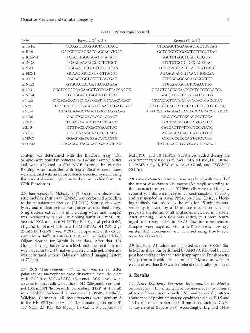
Table 1: Primer sequences used.
Gene Forward (5′ to 3′) Reverse (5′ to 3′)m TNFα CCCGACTACGTGCTCCTCACC CTCCAGCTGGAAGACTCCTCCCAG
m IL1β GACCTTCCAGGATGAGGACATGAG GGTGGGTGTGCCGTCTTTCATTAC
m ICAM-1 TGGCCTGGGGGATGCACACT GGCTGTAGGTGGGTCCGGGT
m iNOS TGAAGAAAACCCCTTGTGCT TTCTGTGCTGTCCCAGTGAG
m YM1 CTGGAATTGGTGCCCCTACAA TCATAACCAACCCACTCATTACC
m FIZZ1 GCAACTGCCTGTGCTTACTC AGAAGCAGGGTAAATGGGCAA
m ARG1 GACAGGGCTCCTTTCAGGAC CTTGGGAGGAGAAGGCGTTT
m Nox2 GTGCACCATGATGAGGAGAA TTGCAATGGTCTTGAACTCG
m Nox1 CGCTCCCAGCAGAAGGTCGTGATTACCAAGG GGAGTGACCCCAATCCCTGCCCCAACCA
m Nox4 TGTTGGGCCTAGGATTGTGTT AGGGACCTTCTGTGATCCTCG
h Nox2 GTCACACCCTTCGCATCCATTCTCAAGTCAGT CTGAGACTCATCCCAGCCAGTGAGGTAG
h Nox1 TTCACCAATTCCCAGGATTGAAGTGGATGGTC GACCTGTCACGATGTCAGTGGCCTTGTCAA
h Nox4 CTGGAGGAGCTGGCTCGCCAACGAAG GTGATCATGAGGAATAGCACCACCACCATGCAG
h iNOS GACCTGGGACCCGCACCACT AGGATGGTGGCACGGCTGGA
h TNFα TGGAGAAGGGTGACCGACTC TCCTCACAGGGCAATGATCC
h IL1β CTGTACGATCACTGAACTGC CACCACTTGTTGCTCCATATC
h ARG1 TTCTCAAAGGGACAGCCACG AGCACCAGGCTGATTCTTCC
h MRC1 GGAGTGATGGAACCCCAGTG CTGTCCGCCCAGTATCCATC
h TGM2 TTCAGGGTACAAACTGAGGCTGCT TATTCAAGTTCACCCACTGGCCCT
3Oxidative Medicine and Cellular Longevity

were elevated, when measured with an ELISA or a cytometricbead assay, respectively. In contrast, the anti-inflammatorycytokine IL10 was strongly reduced in tumors of Nox4-deficient mice (Figure 1(b)). These data point towards amore severe inflammation in tumors of Nox4-/- mice. How-ever, the total number of immune cells per tissue was sim-ilar in wild type and Nox4-/- mice (Supplemental Figure 1)as measured by flow cytometry. Therefore, we analyzed thenumber of proinflammatory macrophages, identified byhigh expression of the surface marker Ly6C [17], and founda substantial increase in Ly6Chi monocytes in the tumorsof Nox4-/- mice (Figure 1(c)). When we further analyzedthe tumor tissue for pro- and anti-inflammatory markers,we observed a trend towards more inflammation, togetherwith lower expression of markers typical for M(IL4+IL13)-polarized macrophages (Supplemental Figure 2). Accordingly,we conclude that the absence of Nox4 favors the polarizationof macrophages towards a proinflammatory phenotype,which was further investigated.
3.2. Loss of Nox4 Promotes M(LPS+IFNγ) Polarization ofMacrophages. Human and murine macrophages were gener-ated and analyzed for the expression of individual NADPHoxidases. As expected, Nox2 expression was the highest inboth macrophage populations, followed by Nox4 and Nox1(Supplemental Figure 3). In order to analyze if Nox4influences macrophage polarization, we isolated monocytesfrom bone marrow of wild type and Nox4-deficient mice,challenged them (with M-CSF) to become macrophages,and eventually polarized them to either M(LPS+IFNγ) orM(IL4+IL13) phenotype. Nox4 knockout promoted theexpression of M(LPS+IFNγ) macrophage markers includingTNFα and IL1β (Figure 2(a)), whereas typical M(IL4+IL13)markers were significantly downregulated (Figure 2(b)).This effect was mediated by H2O2, the product of Nox4:external H2O2 or increased intracellular H2O2 formationvia PMA-induced activation of Nox2 and conversion of theresulting ⋅O2
− into H2O2 by SOD induced M(IL4+IL13)polarization. Depletion of H2O2 by catalase forces the
expression of M(LPS+IFNγ) markers, both without anyfurther treatment with cytokines (Supplemental Figure 4).Exemplary verification of the PCR results on the proteinlevel revealed the same effect for the M(IL4+IL13) markerYM1 (Figure 2(c)). STAT6 is one of the main transcriptionfactors involved in the expression of M(IL4+IL13) markers.In line with the decreased level of M(IL4+IL13) markers inNox4-/- cells, phosphorylation of STAT6 was attenuated(Figure 2(d)). In order to analyze whether or not theeffects seen are specific for Nox4, macrophage polarizationwas determined in Nox2- and Nox1-deficient macrophagesas well. In contrast to Nox4-/- macrophages, loss of Nox2induced a small but significant reduction in M(LPS+IFNγ)polarization with no effect on M(IL4+IL13) polarization orSTAT6 phosphorylation (Supplemental Figure 5). Knockoutof Nox1 had no effect on macrophage polarization,compared to wild type littermates (Supplemental Figure 6).
3.3. Formation of Reactive Oxygen Species upon M(LPS+IFNγ) Polarization Is Elevated in the Absence of Nox4. Severalpublications indicate that polarization of macrophages isdependent on ROS production and simultaneously forcesROS formation [18]. Polarization of macrophages towardsthe proinflammatory M(LPS+IFNγ) phenotype resulted inan increase in superoxide anion as well as in hydrogen perox-ide production compared to M(IL4+IL13)-polarized macro-phages (Figures 3(a) and 3(b)). Surprisingly, the absence ofNox4 further increased ROS formation in M(LPS+IFNγ)-polarized macrophages (Figures 3(a) and 3(b)). A majorsource of ROS in M(LPS+IFNγ)-polarized macrophages isNox2, whose expression was elevated in Nox4-deficientM(LPS+IFNγ)-polarized macrophages (Figure 3(c)). Accord-ingly, when measuring ⋅O2
− in a more specific way withthe aid of L-012 in intact cells, we found that both LPSand IFNγ separately increase the level of ⋅O2
− production inmacrophages as well as the combination of both(Figure 3(d)). Knockout of Nox2 in macrophages completelyabolished L-012 detectable ⋅O2
− formation (Figure 3(e)). Inconclusion, the increase in Nox2 expression, which pre-dominantly produces ⋅O2
− over H2O2, indicates thatNox2 is the major source of ROS in M(LPS+IFNγ)-polar-ized macrophages.
3.4. Nox4 Mediates the Proinflammatory MacrophagePolarization via Activation of NFκB. Inflammation is oftenassociated with an increased activity of NFκB [19].Indeed, TNFα and IL1β as well as ICAM-1 and Nox2 aretarget genes of NFκB. Therefore, we analyzed the potentialrole of Nox4 in NFκB activation in the course of macro-phage polarization.
M(LPS+IFNγ) polarization was accompanied by anincreased translocation of p65 from the cytosol to thenucleus in the Nox4-deficient macrophages when comparedto wild type cells (Figures 4(a) and 4(b)). However, nucleartranslocation alone is not sufficient as the indicator of a tran-scription factor activity. In order to test for both, NFκBnuclear translocation and DNA binding activity, an electromobility shift assay (EMSA) was utilized. Activity of NFκBwas enhanced in M(LPS+IFNγ) macrophages in the absence
Table 2: Antibodies used.
Antigen Dye
CD3 PE-CF594
CD4 V500
CD8 BV650
CD11b eFluor 605
CD11c Alexa Fluor 700
CD19 APC-H7
CD45 VioBLue
CD49b PE-CF594
F4/80 PE-Cy7
HLA-DR (MHCII) APC-H7
Ly-6C PerCP-Cy5.5
Ly-6G APC-Cy7
Siglec H FITC
4 Oxidative Medicine and Cellular Longevity

of Nox4 (Figure 4(c)). In M(IL4+IL13)-polarized macro-phages, no such effect of Nox4 was observed.
3.5. Activated NFκB Promotes Nox2 Expression in the Absenceof Nox4. NFκB is one of the transcription factors that controlNox2 expression. We therefore hypothesized that elevatedactivation of NFκB in the absence of Nox4 promotes Nox2expression during macrophage M(LPS+IFNγ) polarization.The upregulation of Nox2 however is not accompanied byan elevated expression of its cytosolic subunits or antioxi-dative enzymes such as SOD1 or 3 in wild type vs. Nox4-/-cells (Supplemental Figure 7). Treatment of the cells withan NFκB inhibitor prevented the increase in p65 nucleartranslocation (Supplemental Figure 8), and Nox2 expressionwas reduced in Nox4-/- macrophages to the level similar tothat of the wild type, when cells were pretreated with theNFκB inhibitor prior to M(LPS+IFNγ) polarization(Figure 4(d)). NFκB acts in concert with othertranscription factors to regulate the expression of Nox2[20]. One of which is the redox-sensitive zinc-fingertranscription factor Yin Yang 1 (YY1), which directlycontrols the activity of NFκB [21]. As such, YY1represents a potential target of Nox4-derived ROS, whichis upstream of NFκB and controls Nox2 expression. Asignificant increase in the YY1 protein level was observedin M(LPS+IFNγ)-polarized Nox4-/- macrophages; which
was not the case for M(IL4+IL13)-polarized macro-phages (Supplemental Figure 9A). Inhibition of Nox4with GKT137928 in Nox2-deficient macrophages resultsin a small but significant increase in LPS and IFNγ-induced M(LPS+IFNγ) polarization. Under basalconditions, treatment with GKT only increased iNOS,compared to DMSO-treated samples. Those resultsindicate that inhibition of Nox4 favors M(LPS+IFNγ)polarization even in the absence of Nox2 (SupplementalFigure 10). The interpretation of this result could be thatNFκB even in the absence of Nox2 promotes M(LPS+IFNγ) polarization in macrophages. Although manystudies provide evidence for the involvement of NFκB inmacrophage polarization, the exact role of NFκB and itseffects besides induction of Nox2 are unclear. Therefore,investigation of how NFκB triggers M(LPS+IFNγ)polarization in the absence of Nox2 would be worth asecond study. Another transcription factor involved inM(LPS+IFNγ) polarization is STAT1 [22]. We thereforechecked for a potential effect of Nox4 on phosphorylation ofSTAT1 in M(LPS+IFNγ) polarization without observing anyeffect of Nox4 (Supplemental Figure 9B). Thus, Nox4appears to selectively regulate the activity of NFκB andpotentially YY1. In conclusion, the absence of Nox4promotes Nox2 expression and subsequently M(LPS+IFNγ)polarization of macrophages.
WT0
1
2
3
4
Nox4-/- WT Nox4-/- WT Nox4-/-
IL1�훽
rel.
IL1�훽
mRN
A ex
pres
sion
0
1
2
3
4
rel.
TNF�훼
mRN
A ex
pres
sion
0.0
0.5
1.0
1.5
2.0
rel.
ICA
M-1
mRN
A ex
pres
sion
TNF�훼 ICAM-1⁎ ⁎ ⁎
(a)
WT Nox4-/- WT Nox4-/- WT Nox4-/-0
50
100
150
200
pmol
/ml
0
2
4
6
pg/m
l
0
1
2
3
4
pg/m
l
ELISA IL1�훽 CBA TNF�훼 CBA IL10⁎
⁎
⁎
(b)
WT Nox4-/-0
40000
20000
60000
80000
100000
Cell
s/g
tum
or
Ly6C+ monocytes⁎
(c)
Figure 1: Nox4 deficiency favors inflammation in a murine tumor model. (a) Proinflammatory markers including the cytokines IL1β andTNFα and the adhesion molecule ICAM-1 were quantified by RT-qPCR in tumor tissue of WT and Nox4-/- mice. (b) Proinflammatorymarkers IL1β (ELISA) and TNFα as well as IL10 (CBA: cytometric bead assay) as anti-inflammatory markers were measured in tumortissue, n = 3; ∗p < 0 05. (c) Single-cell suspension of tumor tissue was analyzed by FACS for Ly6C+ monocytes; ∗p < 0 05, n = 5-10.
5Oxidative Medicine and Cellular Longevity

3.6. Pharmacological Inhibition of Nox4 Promotes M(LPS+IFNγ) Polarization of Human Macrophages. In order todetermine whether our findings in a mouse can be trans-lated to human cells, human macrophages generated fromperipheral blood of healthy donors were analyzed. Inhibi-tion of Nox4 was achieved by treatment of the cells withthe Nox1/Nox4 inhibitor GKT137928. Upon treatment ofthe macrophages with GKT137928, an increased M(LPS+IFNγ) polarization was observed. This was accompaniedby a decrease in M(IL4+IL13) polarization (Figure 5). Asshown above in the murine system, knockout of Nox1 has
no influence on macrophage polarization. Therefore, weassume that usage of the inhibitor will mainly affect Nox4-mediated signaling in the process of polarization. We con-clude that the findings in the murine system also apply tothe human system.
4. Discussion
Macrophages are a heterogeneous population of cells. Gen-erally, they can be categorized into two discrete subsets aseither classically activated M1 or alternatively activated
WT0.0
0.5
1.0
1.5
Nox4-/-
WT Nox4-/-
IL1�훽
rel.
IL1�훽
mRN
A ex
pres
sion
0.0
0.5
1.0
1.5
2.0re
l. TN
F�훼 m
RNA
expr
essio
n TNF�훼
M(LPS+IFN�훾) marker
⁎
WT0.0
0.5
1.0
1.5
Nox4-/-
iNOS
rel.
iNO
S m
RNA
expr
essio
n ⁎
(a)
WT Nox4-/-0.0
0.5
1.0
rel.
ARG
1 m
RNA
expr
essio
n Arginase 1
M(IL4+IL13) marker
⁎
WT Nox4-/-
0.5
1.0
rel.
YM1
mRN
A ex
pres
sion
YM1
WT Nox4-/-0.0
0.5
1.0
rel.
FIZZ
1 m
RNA
expr
essio
n
FIZZ1
⁎
0.0
(b)
WTNox4-/-
WT Nox4-/- WT Nox4-/-
0
5
10
15
20
YM1/�훽
-act
in
YM1
�훽-Actin
45
42
YM1 protein expression
M(LPS+IFN�훾) M(IL4+IL13)
⁎
(c)
WTNox4-/-
WT Nox4-/- WT Nox4-/-
0
5
10
15
20pS
TAT6
/STA
T6
pSTAT6
STAT6
�훽-Actin
100
100
42
p-STAT6/STAT6 protein expression
M(LPS+IFN�훾) M(IL4+IL13)
⁎#
#
(d)
Figure 2: Nox4 knockout leads to a decreased M(IL4+IL13) polarization of macrophages. The specific M(LPS+IFNγ) markers IL1β, TNFα,and iNOS (a) and specific M(IL4+IL13) markers arginase 1, YM1, and FIZZ1 (b) were quantified by RT-qPCR after stimulation withcytokines polarizing the bone marrow-derived macrophages from WT and Nox4-/- mice to M(LPS+IFNγ) or M(IL4+IL13) phenotype.Protein expression of the M(IL4+IL13) marker YM1 (c) and the ratio of pSTAT6 to STAT6 (d) as determined by Western Blot; ∗p < 0 05WT vs. Nox4-/- and #p < 0 05 WT/Nox4-/- M(LPS+IFNγ) vs. WT/Nox4-/- M(IL4+IL13), n = 5-6.
6 Oxidative Medicine and Cellular Longevity

M2 macrophages, herein referred to as M(LPS+IFNγ) orM(IL4+IL13). In this context, M(LPS+IFNγ) macrophagesrepresent proinflammatory “killers,” while M(IL4+IL13)
macrophages serve as “builders” in inflammatory woundrepair. This polarization of the macrophage populationresults from interactions with other cells or molecules within
WTNox4-/-
⁎
# #0
50
100
Aver
age l
umin
esce
nce (
%)
150
200�㴆O2
− level in WT/Nox4-deficient macrophages
WT Nox4-/-M(LPS+IFN�훾) M(IL4+IL13)
WT Nox4-/-
(a)
0
50
100
Aver
age l
umin
esce
nce (
%)
Buffer
Luminol/HRP H2O2 measurement
WT M(IL4+IL13)
Nox4-/- M(IL4+IL13)
WT M(LPS+IFN�훾)
Nox4-/- M(LPS+IFN�훾)
Cells PMA DPIL-NAME
(b)
WTNox4-/-
⁎
# #
0.0
1.0
0.5
1.5
rel.
Nox
2 m
RNA
expr
essio
n
2.0
2.5 Nox2 mRNA expression
WT Nox4-/-M(LPS+IFN�훾) M(IL4+IL13)
WT Nox4-/-
(c)
⁎
⁎⁎
⁎
012345
100
150
200
50
Aver
age l
umin
esce
nce (
%)
L-012 �㴆O2− measurement
IFN�훾 IFN�훾LPS LPS
Acute 4 hours
LPS/IFN�훾CTL
(d)
0
10000
5000
Buffer Cells PMA DPI SOD
L-012 �㴆O2− measurement
Nox2y/- M(IL4+IL13)
WT M(LPS+IFN�훾)Nox2y/- M(LPS+IFN�훾)
15000
Aver
age l
umin
esce
nce (
RLU
)
20000
25000
(e)
Figure 3: ROS measurements reveal increased ROS production in Nox4-deficient cells due to an increase in Nox2. Superoxide anionproduction measured with L-012 (a) and hydrogen peroxide levels measured with luminol and HRP (b) in polarized macrophages of wildtype and Nox4 knockout mice. (c) RT-qPCR for Nox2 mRNA expression in polarized macrophages of WT and Nox4-deficient animals;∗p < 0 05 (n = 3 − 8). (d) Superoxide anion production, as measured with L-012 in WT macrophages with or without LPS (10 μg/ml) andIFNγ (100U/ml) directly after stimulation or 4 hours after addition. (e) Superoxide anion production in polarized WT and Nox2-deficient macrophages; ∗p < 0 05 WT vs. Nox4-/- or treated vs. CTL and #p < 0 05 WT/Nox4-/- M(LPS+IFNγ) vs. WT/Nox4-/- M(IL4+IL13) (n = 3-5).
7Oxidative Medicine and Cellular Longevity

the host tissues [23]. In previous work, we found that knock-out of the NADPH oxidase Nox4 enhances inflammationand tumorigenesis [16, 24]. In an angiotensin II-inducedmodel of vascular dysfunction, loss of Nox4 promotedthe expression of the proinflammatory cytokines IL6 andIL1β [12]. The present study underlines the protectivepotential of Nox4 in inflammation, as it promotes M(IL4+IL13) polarization of macrophages. Our results were con-firmed in a very recent study in a myocardial infarctionmodel, where overexpression of Nox4 promoted M(IL4+IL13) polarization of cardiac macrophages and protectsfrom postinfarction remodeling [25].
The balance between activation of STAT1 and STAT3/-STAT6 plays a crucial role in macrophage polarization: a pre-dominance of STAT1 activation promotes M(LPS+IFNγ),
while STAT3/STAT6 activation increases M(IL4+IL13)macrophage polarization [26]. In fact, STAT6 is the mostimportant transcription factor regulating M(IL4+IL13)polarization of macrophages [27], and phosphorylationof STAT6 can be regulated by redox-sensitive phosphatases[28]. Therefore, it is likely that Nox4-derived H2O2 at leastcontributes to STAT6 phosphorylation and thereby toM(IL4+IL13) polarization. Importantly, STAT6 suppressesNFκB activation via Klf4. Here, we provide evidence thatNox4 deficiency prevents STAT6 phosphorylation and sup-ports NFκB activation. NFκB has been shown to promoteM(LPS+IFNγ) polarization of phagocytes [29]. Besides regu-lation by STAT6/Klf4, the activity of NFκB is redox sensitiveand potentially regulated by NADPH oxidases [30]. Both,increased NFκB and reduced phosphorylation of STAT6,
WT Nox4-/- WT Nox4-/-M(LPS+IFN�훾) M(IL4+IL13)
WTNox4-/-
0.0
65
55
0.5
1.0
p65/�훽
-tubu
lin1.5
p65
�훽-Tubulin
⁎
Cytosolic fraction p65
(a)
WT Nox4-/- WT Nox4-/-M(LPS+IFN�훾) M(IL4+IL13)
WTNox4-/-
p65/
TOPO
p65
5
4
3
2
1
0
65
100 TOPO1
⁎
Nuclear extract p65
(b)
WT Nox4-/- WT Nox4-/-
2
1
0
⁎
EMSA for NF�휅B
(c)
WT Nox4-/- WT Nox4-/-CTL NF�휅B inhibitor
2.0
1.0
0.0
0.5
1.5
⁎
Nox2 mRNAre
l. N
ox2
mRN
A ex
pres
sion
#
(d)
Figure 4: Increased NFκB activation in M(LPS+IFNγ)-polarized macrophages of Nox4-/- is responsible for elevated Nox2 expression.Translocation of p65 was analyzed by Western Blot in the cytosol (a) and nuclear fraction (b) of M(LPS+IFNγ)- and M(IL4+IL13)-polarized macrophages of WT and Nox4-/- mice. (c) Electrophoretic mobility shift assay for NFκB was performed in M(LPS+IFNγ)-polarized macrophages of WT and Nox4-/- animals. The left bar graph shows quantification, and the right bar graph representative shift.(d) Nox2 mRNA expression was quantified by RT-qPCR after M(LPS+IFNγ) polarization with and without an NFκB inhibitor (30 ng/ml,1 h pretreatment before polarization); ∗p < 0 05 WT vs. Nox4-/- and #p < 0 05 CTL vs. NFκB inhibitor (n = 3-8). TOPO: topoisomerase I.
8 Oxidative Medicine and Cellular Longevity

inhibit the polarization of macrophages towards the M(IL4+IL13) phenotype. Consequently, since there exists an intrin-sic balance, less force in the direction of M(IL4+IL13) willlead to more M(LPS+IFNγ) polarization, as observed in thecurrent study. Since Nox4 produces H2O2, a Nox4 knockoutwould naturally lead to a reduced production of H2O2.Therefore, our data can be supported by a finding concerningCuZn-SOD, an enzyme catalyzing the conversion of super-oxide anion to hydrogen peroxide. Consequently, less H2O2is formed in the absence of CuZn-SOD. Knockout of thisenzyme promotes M(IL4+IL13) polarization of macrophages[8], favoring the hypothesis that indeed H2O2 mediates theeffect of Nox4.
Different to Nox4, Nox2 produces superoxide anions(⋅O2
−), and knockout of Nox2 results in a decreased M(LPS+
IFNγ) polarization. In contrast, Nox2, via production ofsuperoxide anions, contributes to M(LPS+IFNγ) polariza-tion [6]. We observed not only a reduced M(LPS+IFNγ)polarization in Nox2-deficient macrophages but also anincrease in Nox2 expression and subsequently ⋅O2
− forma-tion, in Nox4 knockout macrophages. This is potentially aconsequence of the abovementioned enhanced NFκB activa-tion in the absence of Nox4, as Nox2 expression is enhancedby NFκB [31].
We conclude that the specific types of ROS, such asH2O2 or ⋅O2
−, differentially contribute to M(LPS+IFNγ) orM(IL4+IL13) macrophage polarization. Importantly, knock-out of Nox4 not only favors M(LPS+IFNγ) polarizationbut also results in an increased expression of Nox2 inM(LPS+IFNγ)-polarized macrophages.
Data Availability
All data used to support the findings of this study areincluded within the article or the supplements.
Conflicts of Interest
The authors declare no conflict of interest.
Acknowledgments
This research was supported by the Else KrÖner-Fresenius-Stiftung Foundation (EKFS), Research Training GroupTranslational Research Innovation–Pharma (TRIP), andCardio-Pulmonary Institute (CPI), EXC 2026, Project ID:390649896 and by grants from the Deutsche Forschungsge-meinschaft (DFG) (to KS; SCHR1241/1-1, SFB815/TP1,and SFB834/TPA2).
Supplementary Materials
Supplemental Figure 1: FACS analysis of tumor tissuerevealed only a tendency for differences in B cells withinNox4-deficient tumors. (A) Fibrosarcoma tissues of wild typeand Nox4-/- mice were analyzed for cell composition withFACS using specific antibodies for cells indicated. The tablecontains the different T cell populations in cells/g tumor tis-sue, no statistical differences (n = 6-10). Supplemental Figure2: tumor tissue was analyzed for different inflammatory andanti-inflammatory markers. (A) Fibrosarcoma tissues ofwildtype and Nox4-/- mice were analyzed for proinflamma-tory markers iNOS and CD163, anti-inflammatory markersFIZZ1, arginase 1, and YM1, and tissue remodeling markersMMP9 and collagens I and III with RT-qPCR; ∗p < 0 05(n = 6-10). Supplemental Figure 3: NADPH oxidase expres-sion in isolated murine and human macrophages. Nox1,Nox2, and Nox4 expressions were determined by RT-qPCRin isolated murine (A) and human (B) macrophages, andcorresponding CT values were included (n = 3). Supplemen-tal Figure 4: H2O2 mediates polarization of macrophageswithout cytokine stimulation. (A) WT macrophages weretreated with basal medium or IL4 and IL13 to polarize. Forpolarization without cytokines, cells were treated with 5μMH2O2 or PEG-SOD (50U/ml) and PMA (100nM) for 4 h,
M(LPS+IFN�훾) markeriNOS
TNF�훼
IL1�훽
0.0DMSO GKT
0.5
1.0
rel.
iNO
S m
RNA
expr
essio
nre
l. TN
F�훼 m
RNA
expr
essio
n
0.0
DMSO GKT
DMSO GKT
1.5
2.0
0.5
1.0
0.0
1.5
2.0
0.5
1.0
rel.
IL1�훽
mRN
A ex
pres
sion
⁎
(a)
M(IL4+IL13) markerArginase 1
MRC1
Transglutaminase
DMSO GKT0.0
0.5
1.0
rel.
ARG
1 m
RNA
expr
essio
n
DMSO GKT0.0
0.5
1.0
rel.
MRC
1 m
RNA
expr
essio
n
DMSO GKT0.0
0.5
1.0
rel.
TGM
2 m
RNA
expr
essio
n
⁎
⁎
⁎
(b)
Figure 5: Treatment of human macrophages with the Nox4inhibitor GKT137928 forces M(LPS+IFNγ) polarization. Thespecific M(LPS+IFNγ) markers iNOS, TNFα, and IL1β (a) andM(IL4+IL13) markers arginase 1, MRC1, and transglutaminase 2(b) were quantified by RT-qPCR. Cells were preincubated withNox4 inhibitor GKT (10 μM, 2 h) followed by stimulation withcytokines polarizing human macrophages; ∗p < 0 05 (n = 6).
9Oxidative Medicine and Cellular Longevity

and polarization markers ARG1, FIZZ1, and YM1 werequantified with RT-qPCR; ∗p < 0 05 (n = 6). (B) WT macro-phages were treated with basal medium or LPS and IFNγ orPEG-catalase (500U/ml) for 4 h to polarize, followed by sub-sequent analysis of polarization markers TNFα, IL1β, andiNOS; ∗p < 0 05 (n = 3). Supplemental Figure 5: Nox2 knock-out decreases M(LPS+IFNγ) polarization of macrophages.The specific M(LPS+IFNγ) markers IL1β, TNFα, and iNOS(A) and specific M(IL4+IL13) markers arginase 1, YM1,and FIZZ1 (B) were quantified by RT-qPCR after stimulationwith cytokines polarizing the bone marrow-derived macro-phages from Nox2KO/C57Bl6J mice to M(LPS+IFNγ) orM2(IL4+IL13) phenotype. Protein expression of the M(IL4+IL13) marker YM1 (C) and the ratio of pSTAT6 to STAT6(D) as determined by Western Blot; ∗p < 0 05 and #p < 0 05WT/Nox2y/- M(LPS+IFNγ) vs. WT/Nox2y/- M(IL4+IL13)(n = 4 − 8). Supplemental Figure 6: deficiency in Nox1 doesnot affect polarization of macrophages. The specific M(LPS+IFNγ) markers IL1β, TNFα, and iNOS (A) and specificM(IL4+IL13) markers arginase 1, YM1, and FIZZ1 (B) werequantified by RT-qPCR after stimulation with cytokinespolarizing the bone marrow-derived macrophages fromNox1KO/C57Bl6J mice to M(LPS+IFNγ) or M2(IL4+IL13)phenotype. Protein expression of the M(IL4+IL13) markerYM1 (C) and the ratio of pSTAT6 to STAT6 (D) as deter-mined byWestern Blot; ∗p < 0 05 and #p < 0 05WT/Nox1y/-M(LPS+IFNγ) vs. WT/Nox1y/- M(IL4+IL13) (n = 3 − 6).Supplemental Figure 7: SOD and Nox2 cytosolic subunitexpressions in WT and Nox4-deficient macrophages. SOD1(A) and SOD3 (B) expressions were analyzed in WT andNox4-/- M(LPS+IFNγ)- and M(IL4+IL13)-polarized macro-phages by RT-qPCR. Expressions of cytosolic Nox2 subunits(C: p40phox, D: p47phox, and E: p67phox) and Nox1 (F)were analyzed in WT and Nox4-/- M(LPS+IFNγ)- andM(IL4+IL13)-polarized macrophages using RT-qPCR; ∗p <0 05 WT/Nox4-/- M(LPS+IFNγ) vs. WT/Nox4-/- M(IL4+IL13) (n = 5-8). Supplemental Figure 8: NFκB inhibition pre-vents p65 translocation into the nucleus in M(LPS+IFNγ)-polarized macrophages of Nox4-/-. P65 expression in cyto-sol (A) and nucleus (B) was assessed with Western Blotafter M(LPS+IFNγ) polarization with and without treat-ment of NFκB inhibitor (30 ng/ml, 1 h pretreament beforeM(LPS+IFNγ) polarization); ∗p < 0 05 WT vs. Nox4-/- and#p < 0 05 CTL vs. NFκB inhibitor (n = 3-8). TOPO: topo-isomerase I. Supplemental Figure 9: YY1 is increased inNox4-deficient macrophages after M(LPS+IFNγ) polariza-tion. (A) YY1 expression was determined by Western Blotafter polarization of WT and Nox4-/- macrophages. (B)Phosphorylation of pSTAT1 and total STAT1 quantified byWestern Blot in M(LPS+IFNγ)- and M(IL4+IL13)-polarizedmacrophages of WT and Nox4-deficient animals; ∗p < 0 05WT vs. Nox4-/- and #p < 0 05 WT/Nox4-/- M(LPS+IFNγ)vs. WT/Nox4-/- M(IL4+IL13) (n = 3-5). Supplemental Fig-ure 10: inhibition of Nox4 in Nox2-deficient macrophageselevates M(LPS+IFNγ) polarization in M(LPS+IFNγ)-polar-ized macrophages. Nox2-deficient macrophages were treatedwith Nox4 inhibitor GKT (10μM) 2h prior to cell polari-zation to M(LPS+IFNγ) or only control medium (CTL).M(LPS+IFNγ) markers TNFγ, IL1β, and iNOS were
evaluated using RT-qPCR; ∗p < 0 05 DMSO vs. GKT and#p < 0 05 DMSO/GKT CTL vs. DMSO/GKT M(LPS+IFNγ)(n = 3). (Supplementary Materials)
References
[1] M. Ushio-Fukai and Y. Nakamura, “Reactive oxygen speciesand angiogenesis: NADPH oxidase as target for cancer ther-apy,” Cancer Letters, vol. 266, no. 1, pp. 37–52, 2008.
[2] L. Serrander, L. Cartier, K. Bedard et al., “NOX4 activity isdetermined by mRNA levels and reveals a unique pattern ofROS generation,” The Biochemical Journal, vol. 406, no. 1, Part1, pp. 105–114, 2007.
[3] K. Bedard and K.-H. Krause, “The NOX family of ROS-generating NADPH oxidases: physiology and pathophysiol-ogy,” Physiological Reviews, vol. 87, no. 1, pp. 245–313,2007.
[4] A. Weigert, A. von Knethen, D. Fuhrmann, N. Dehne, andB. Brüne, “Redox-signals and macrophage biology,”MolecularAspects of Medicine, vol. 63, pp. 70–87, 2018.
[5] P. J. Murray, J. E. Allen, S. K. Biswas et al., “Macrophage acti-vation and polarization: nomenclature and experimentalguidelines,” Immunity, vol. 41, no. 1, pp. 14–20, 2014.
[6] A. Kumar, J. P. Barrett, D.-M. Alvarez-Croda, B. A. Stoica,A. I. Faden, and D. J. Loane, “NOX2 drives M1-like micro-glial/macrophage activation and neurodegeneration followingexperimental traumatic brain injury,” Brain, Behavior, andImmunity, vol. 58, pp. 291–309, 2016.
[7] D. Sanmun, E.Witasp, S. Jitkaew et al., “Involvement of a func-tional NADPH oxidase in neutrophils and macrophages dur-ing programmed cell clearance: implications for chronicgranulomatous disease,” American Journal of Physiology-CellPhysiology, vol. 297, no. 3, pp. C621–C631, 2009.
[8] D. R. Balce, B. Li, E. R. O. Allan, J. M. Rybicka, R. M. Krohn,and R. M. Yates, “Alternative activation of macrophages byIL-4 enhances the proteolytic capacity of their phagosomesthrough synergistic mechanisms,” Blood, vol. 118, no. 15,pp. 4199–4208, 2011.
[9] C. He, A. J. Ryan, S. Murthy, and A. B. Carter, “Accelerateddevelopment of pulmonary fibrosis via Cu,Zn-superoxidedismutase-induced alternative activation of macrophages,”The Journal of Biological Chemistry, vol. 288, no. 28,pp. 20745–20757, 2013.
[10] C. F. Lee, M. Qiao, K. Schröder, Q. Zhao, and R. Asmis, “Nox4is a novel inducible source of reactive oxygen species in mono-cytes and macrophages and mediates oxidized low densitylipoprotein-induced macrophage death,” Circulation Research,vol. 106, no. 9, pp. 1489–1497, 2010.
[11] K. Schroder, K. Wandzioch, I. Helmcke, and R. P. Brandes,“Nox4 acts as a switch between differentiation and prolifera-tion in preadipocytes,” Arteriosclerosis, Thrombosis, and Vas-cular Biology, vol. 29, no. 2, pp. 239–245, 2009.
[12] C. Goettsch, A. Babelova, O. Trummer et al., “NADPH oxidase4 limits bone mass by promoting osteoclastogenesis,” TheJournal of Clinical Investigation, vol. 123, no. 11, pp. 4731–4738, 2013.
[13] K. Schröder, M. Zhang, S. Benkhoff et al., “Nox4 is a protectivereactive oxygen species generating vascular NADPH oxidase,”Circulation Research, vol. 110, no. 9, pp. 1217–1225, 2012.
[14] G. Gavazzi, C. Deffert, C. Trocme, M. Schäppi, F.̧. R.Herrmann, and K. H. Krause, “NOX1 deficiency protects
10 Oxidative Medicine and Cellular Longevity

from aortic dissection in response to angiotensin II,” Hyper-tension, vol. 50, no. 1, pp. 189–196, 2007.
[15] M. M. Bradford, “A rapid and sensitive method for thequantitation of microgram quantities of protein utilizingthe principle of protein-dye binding,” Analytical Biochemis-try, vol. 72, no. 1-2, pp. 248–254, 1976.
[16] V. Helfinger, F. F. von Gall, N. Henke et al., “Hydrogenperoxide formation by Nox4 limits malignant transforma-tion,” http://arxiv.org/abs/2017:177055. https://www.biorxiv.org/content/early/2017/08/16/177055.full.pdf.
[17] F. Geissmann, M. G. Manz, S. Jung, M. H. Sieweke,M. Merad, and K. Ley, “Development of monocytes, macro-phages, and dendritic cells,” Science, vol. 327, no. 5966,pp. 656–661, 2010.
[18] H.-Y. Tan, N. Wang, S. Li, M. Hong, X. Wang, and Y. Feng,“The reactive oxygen species in macrophage polarization:reflecting its dual role in progression and treatment of humandiseases,” Oxidative Medicine and Cellular Longevity,vol. 2016, Article ID 2795090, 16 pages, 2016.
[19] Y. Zhou, T. Zhang, X. Wang et al., “Curcumin modulatesmacrophage polarization through the inhibition of theToll-like receptor 4 expression and its signaling pathways,”Cellular Physiology and Biochemistry, vol. 36, no. 2,pp. 631–641, 2015.
[20] J. D. Lambeth, “Nox enzymes, ROS, and chronic disease: anexample of antagonistic pleiotropy,” Free Radical Biology &Medicine, vol. 43, no. 3, pp. 332–347, 2007.
[21] W. Zhu, S. Y. Olson, and H. J. Garbán, “Transcription regula-tor Yin-Yang 1: from silence to cancer,” Critical Reviews inOncogenesis, vol. 16, no. 3-4, pp. 227–238, 2011.
[22] Y.-B. Liang, H. Tang, Z.-B. Chen et al., “DownregulatedSOCS1 expression activates the JAK1/STAT1 pathway andpromotes polarization of macrophages into M1 type,”Molecu-lar Medicine Reports, vol. 16, no. 5, pp. 6405–6411, 2017.
[23] L. Parisi, E. Gini, D. Baci et al., “Macrophage polarizationin chronic inflammatory diseases: killers or builders?,” Journalof Immunology Research, vol. 2018, Article ID 8917804,25 pages, 2018.
[24] C. Schürmann, F. Rezende, C. Kruse et al., “The NADPH oxi-dase Nox4 has anti-atherosclerotic functions,” European HeartJournal, vol. 36, no. 48, pp. 3447–3456, 2015.
[25] H. Mongue-Din, A. S. Patel, Y. H. Looi et al., “NADPHoxidase-4 driven cardiac macrophage polarization protectsagainst myocardial infarction–induced remodeling,” JACC:Basic to Translational Science, vol. 2, no. 6, pp. 688–698,2017.
[26] A. Sica and A. Mantovani, “Macrophage plasticity and polari-zation: in vivo veritas,” Journal of Clinical Investigation,vol. 122, no. 3, pp. 787–795, 2012.
[27] T. Lawrence and G. Natoli, “Transcriptional regulation ofmacrophage polarization: enabling diversity with identity,”Nature Reviews. Immunology, vol. 11, no. 11, pp. 750–761,2011.
[28] S. Hirakawa, R. Saito, H. Ohara, R. Okuyama, and S. Aiba,“Dual oxidase 1 induced by Th2 cytokines promotes STAT6phosphorylation via oxidative inactivation of protein tyrosinephosphatase 1B in human epidermal keratinocytes,” Journalof Immunology, vol. 186, no. 8, pp. 4762–4770, 2011.
[29] N. Wang, H. Liang, and K. Zen, “Molecular mechanisms thatinfluence the macrophage M1–M2 polarization balance,”Frontiers in Immunology, vol. 5, p. 614, 2014.
[30] G. Bonizzi, J. Piette, S. Schoonbroodt et al., “Reactive oxygenintermediate-dependent NF-kappaB activation by interleukin-1beta requires 5-lipoxygenase or NADPH oxidase activity,”Molecular and Cellular Biology, vol. 19, no. 3, pp. 1950–1960,1999.
[31] J. Anrather, G. Racchumi, and C. Iadecola, “NF-kappaB regu-lates phagocytic NADPH oxidase by inducing the expressionof gp91phox,” The Journal of Biological Chemistry, vol. 281,no. 9, pp. 5657–5667, 2006.
11Oxidative Medicine and Cellular Longevity

Stem Cells International
Hindawiwww.hindawi.com Volume 2018
Hindawiwww.hindawi.com Volume 2018
MEDIATORSINFLAMMATION
of
EndocrinologyInternational Journal of
Hindawiwww.hindawi.com Volume 2018
Hindawiwww.hindawi.com Volume 2018
Disease Markers
Hindawiwww.hindawi.com Volume 2018
BioMed Research International
OncologyJournal of
Hindawiwww.hindawi.com Volume 2013
Hindawiwww.hindawi.com Volume 2018
Oxidative Medicine and Cellular Longevity
Hindawiwww.hindawi.com Volume 2018
PPAR Research
Hindawi Publishing Corporation http://www.hindawi.com Volume 2013Hindawiwww.hindawi.com
The Scientific World Journal
Volume 2018
Immunology ResearchHindawiwww.hindawi.com Volume 2018
Journal of
ObesityJournal of
Hindawiwww.hindawi.com Volume 2018
Hindawiwww.hindawi.com Volume 2018
Computational and Mathematical Methods in Medicine
Hindawiwww.hindawi.com Volume 2018
Behavioural Neurology
OphthalmologyJournal of
Hindawiwww.hindawi.com Volume 2018
Diabetes ResearchJournal of
Hindawiwww.hindawi.com Volume 2018
Hindawiwww.hindawi.com Volume 2018
Research and TreatmentAIDS
Hindawiwww.hindawi.com Volume 2018
Gastroenterology Research and Practice
Hindawiwww.hindawi.com Volume 2018
Parkinson’s Disease
Evidence-Based Complementary andAlternative Medicine
Volume 2018Hindawiwww.hindawi.com
Submit your manuscripts atwww.hindawi.com