Embed Size (px)
Citation preview

The Muscular SystemTypes of Muscles

Skeletal Muscles
There are close to 700 muscles in the body You will learn about 60 of them and this is a
monumental task The job of learning all of these muscles will still
require a great effort on your part Memorization will be easier if you apply what you
have learned so far Start with the name of the muscle and identifying it on
a figure Then figure out its points of attachments To learn their functions act it out on your own body!

Skeletal Muscles
Muscles can’t push; they can only pull (contraction) Body movements are the result of two or more
muscles acting together Insertion (attachment on a movable bone) moves
towards origin (immovable point of attachment) The fleshy part of the muscle between the two
tendons of the origin and insertion is the belly

Functional Groups
Movements are usually the result of several skeletal muscles acting as a group rather than acting alone
Most skeletal muscles are arranged in opposing pairs at joints
Muscles can be classified into four functional groups
Prime mover (agonist) Muscle with the major responsibility for a certain movement
Ex: The biceps brachii is the prime mover of elbow flexion
Antagonist Muscle that opposes or reverses a movement
Ex: The triceps act as antagonists for the biceps in elbow flexion
If the triceps is also considered a prime mover of the elbow extension, then what muscle would be the antagonist?

Functional Groups
Synergist Muscle that helps a prime mover by adding a little extra force to
the same movement or reducing undesirable movements that might occur
Ex: Finger-flexor muscles cross both the wrist and the finger joints. You can make a fist without bending your wrist because synergist muscles stabilize the wrist joints.
Fixator Synergists that immobilize a bone or a muscle’s origin so that the
prime mover has a stable base on which to act
Ex: fixator muscles that run from the axial skeleton to the scapula can immobilize the scapula so that only desirable movements occur at the shoulder joint; erector spinae group

Naming Skeletal Muscles
Skeletal muscles are named for structural or functional characteristics
Direction of muscle fibers
Some muscles reveal the direction in which their fibers run in reference to some imaginary line in the body (midline)
Ex: rectus (parallel); oblique (diagonal); transverse (perpendicular)
Relative size of the muscle
Ex: maximus (largest), minimus (smallest), longus (long), brevis (short), latissimus (widest), longissimus (longest), magnus (large), major (larger), minor (smaller), vastus (huge)

Naming Skeletal Muscles
Location of the muscle
Some muscle names indicate the bone or body region with which they are associated
Ex: temporalis (temporal), frontalis (frontal), intercostals (costal = ribs)
Number of origins
Some muscles have more than one origin or head
Ex: biceps (two), triceps (three), quadriceps (four)

Location of the attachments (insertion and origin)
Some muscles are named according to their points of origin and insertion
The origin is always named first
Ex: sterno (on the sternum), cleido (clavicle). mastoid (mastoid process); sternocleidomastoid
Shape of the muscle
Some muscles are named for their distinctive shapes
Ex: deltoid (triangular); trapezius (trapezoid), orbicularis (circular), platys (flat), quadratus (square, four-sided), gracilis (slender)
Naming Skeletal Muscles

Action of the muscle
Some muscles are named for the movement that they produce
Ex: flexor (decreases angle), extensor (increases an angle), abductor (away from midline), adductor (towards the midline)
Often several criteria are combined in naming a muscle
Ex: Extensor carpi radialis longus
Muscle’s action = extensor
Location = carpi (wrist); radialis (over radius)
Size = longus
Naming Muscles Video
Naming Skeletal Muscles

Gross Anatomy of Skeletal Muscles
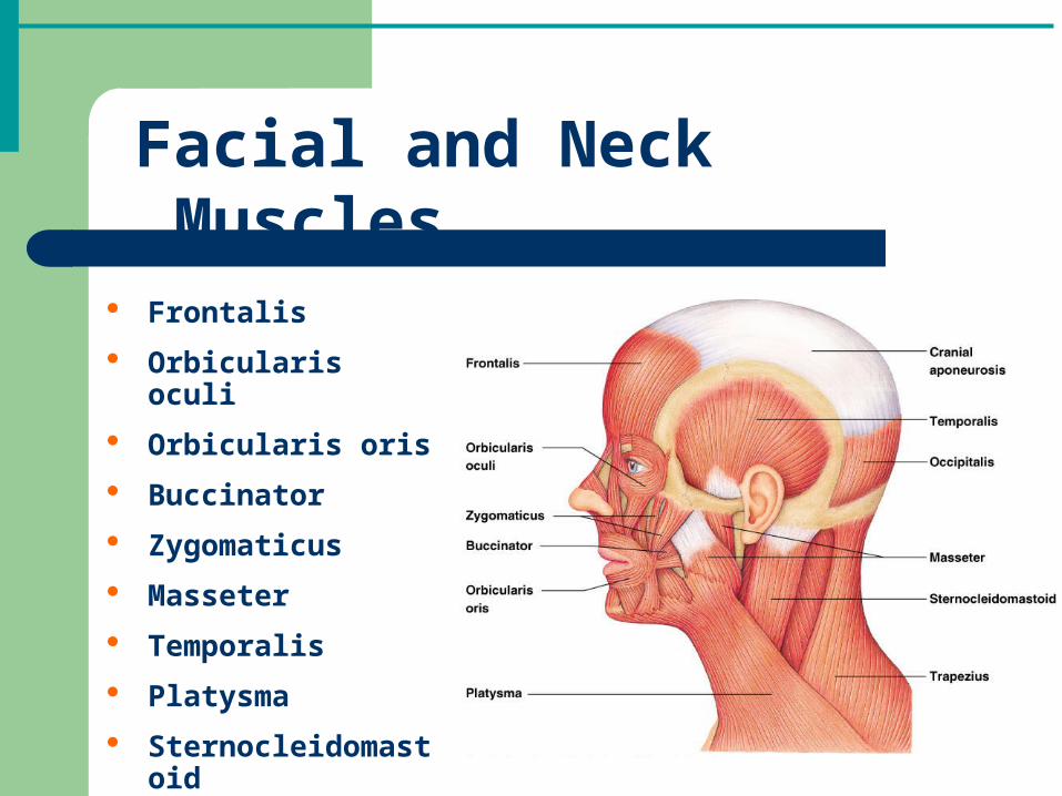
Facial and Neck Muscles
Frontalis
Orbicularis oculi
Orbicularis oris
Buccinator
Zygomaticus
Masseter
Temporalis
Platysma
Sternocleidomastoid
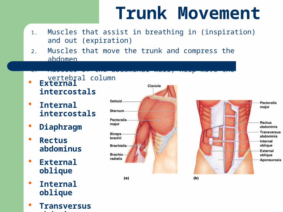
Trunk Movement1. Muscles that assist in breathing in (inspiration) and out (expiration)
2. Muscles that move the trunk and compress the abdomen
3. Muscles of the abdominal wall; help move the vertebral column
External intercostals
Internal intercostals
Diaphragm
Rectus abdominus
External oblique
Internal oblique
Transversus abdominus

Back Extension
Erector Spinae Group
Iliocostalis
Longissimus
Spinalis

Scapula Movement
Trapezius

Movement of Humerus Pectoralis major
Deltoid
Latissimus Dorsi

Movement of Elbow
Biceps brachii
Brachialis
Brachioradialis
Triceps brachii

Movement of Wrist
Flexion
Flexor carpi radialis
Flexor carpi ulnaris
Extension
Extensor carpi ulnaris
Extensor carpi radialis longus

Movement of the Hip
Iliopsoas
Iliacus
Psoas major
Adductor group(3)
Adductor magnus
Adductor longus
Adductor brevis
Gluteus maximus
Gluteus medius
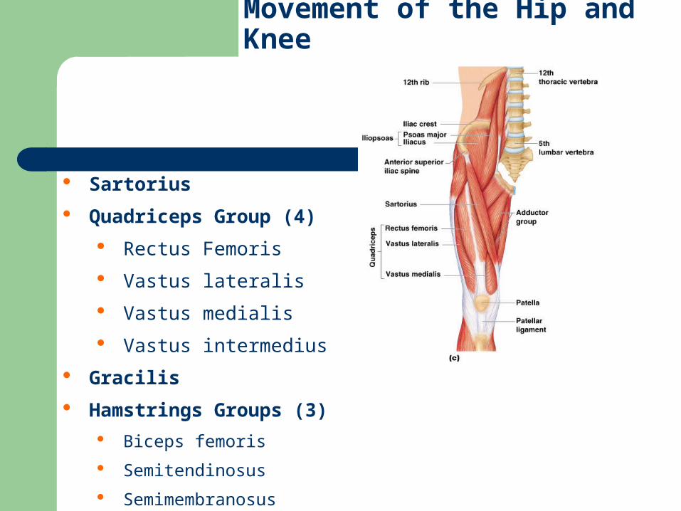
Movement of the Hip and Knee
Sartorius
Quadriceps Group (4)
Rectus Femoris
Vastus lateralis
Vastus medialis
Vastus intermedius
Gracilis
Hamstrings Groups (3)
Biceps femoris
Semitendinosus
Semimembranosus

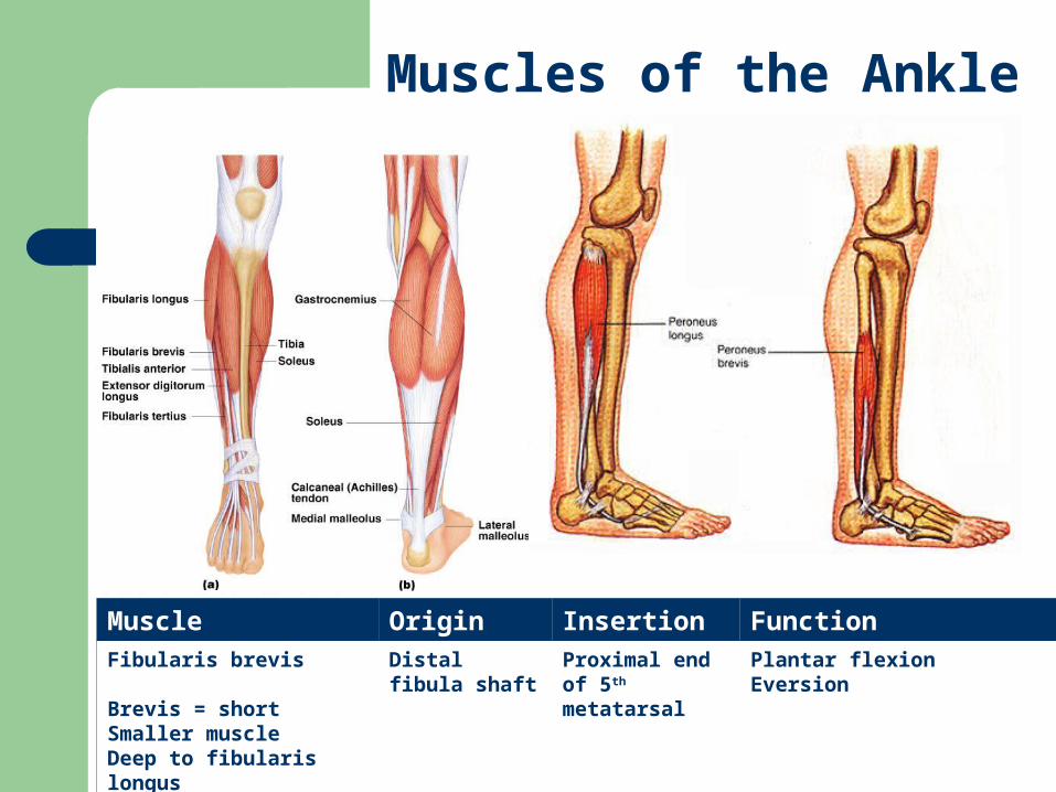
Muscles of the Ankle
Gastrocnemius (knee also)
Tibialis anterior Fibularis longus Fibularis brevis Soleus Tibialis posterior Extensor digitorum
longus (toes also) Flexor digitorum
longus (toes also)

Anterior Muscles

Posterior Muscles
Figure 6.21


Facial and Neck Muscles
Muscles that provide the ability to express emotions
Muscles usually insert directly into skin; therefore the skin is what moves

Facial and Neck Muscles
Frontalis
Orbicularis oculi
Orbicularis oris
Buccinator
Zygomaticus
Masseter
Temporalis
Platysma
Sternocleidomastoid

Facial Muscles
Muscle Origin Insertion FunctionFrontalis
Covers the frontal boneNo bony attachments
Cranial aponeurosis
Skin of eyebrows
Raises eyebrowsWrinkles skin of foreheadPulls scalp anteriorly

Facial Muscles
Muscle Origin Insertion FunctionOrbicularis oculi
Orb = circularOculi = eye
Frontal bone and maxilla bone
Tissue of eyelid Closes eye, squint, blink and wink

Facial Muscles
Muscle Origin Insertion FunctionOrbicularis oris
“The Kissing Muscle”Oris = mouth
Muscles encircling mouth
Skin of at corner of mouth
Closes and protrudes lipsCompresses lips against teeth

Facial Muscles
Muscle Origin Insertion FunctionBuccinator
Bucc = cheek
Maxilla and mandible
Orbicularis oris
Draws corners of mouth laterallyHolds food between teeth when chewingCompresses cheeks against teeth Used in whistling, blowing and suckingWell developed in infants

Facial Muscles
Muscle Origin Insertion FunctionZygomaticus
“Smiling Muscle”
Zygomatic bone Skin and muscle at corner of lips
Raises corners of lips

Facial Muscles
Muscle Origin Insertion Function Type
Masseter
Masseter = chewer
Maxilla and zygomatic arch
Ramus of mandible
Elevates mandibleCloses jaw
Prime mover

Facial Muscles
Muscle Origin Insertion Function TypeTemporalis Temporal
boneMandible Closes jaw
Elevates mandibleSynergist to masseter
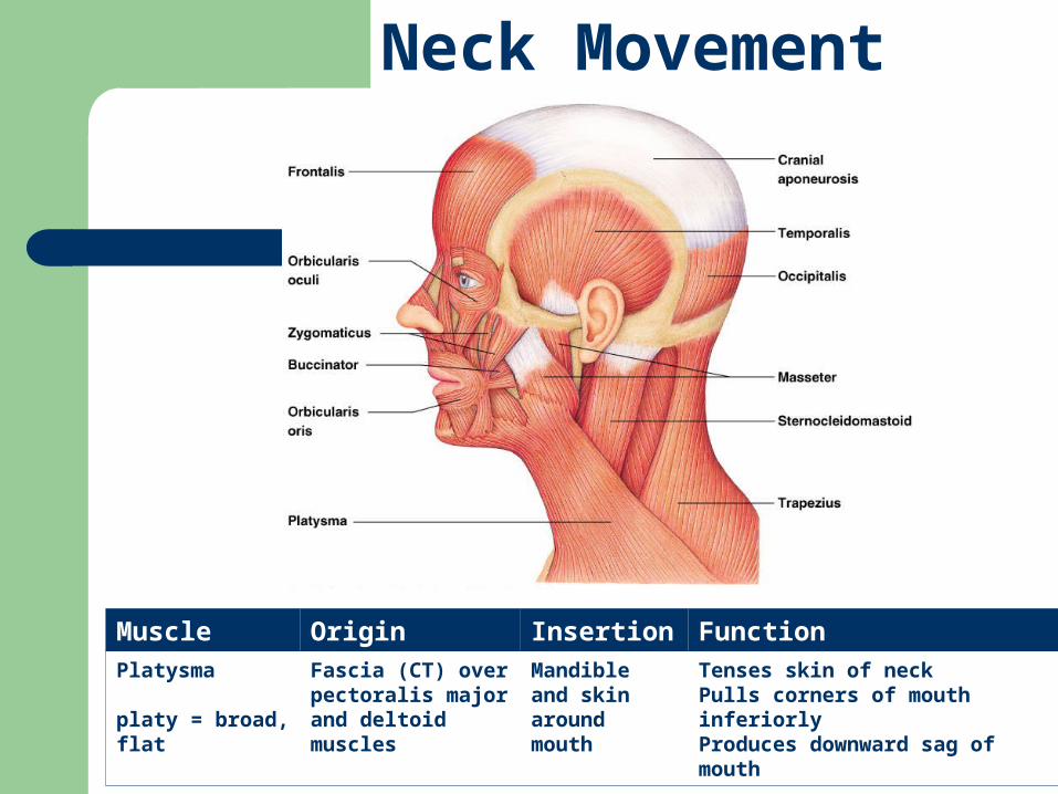
Neck Movement
Muscle Origin Insertion FunctionPlatysma
platy = broad, flat
Fascia (CT) over pectoralis major and deltoid muscles
Mandible and skin around mouth
Tenses skin of neckPulls corners of mouth inferiorlyProduces downward sag of mouth

Neck Movement
Muscle Origin Insertion Function TypeSternocleidomastoid
Anterior surface of neck“Two-headed”Sterno = sternumCleido = clavicleMastoid = mastoid process
1. Manubrium of sternum
2. Clavicle
Mastoid process of temporal bone
Flexes neckRotates head“The Prayer Muscle”
Prime mover

Trunk Movement
External intercostalis
Internal intercostalis
Diaphragm
Rectus abdominus
External oblique
Internal oblique
Transversus abdominus
1. Muscles that assist in breathing in (inspiration) and out (expiration)
2. Muscles that move the trunk and compress the abdomen
3. Muscles of the abdominal wall; help move the vertebral column

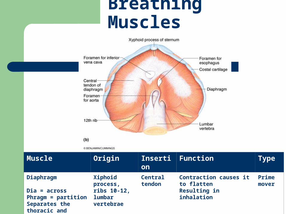
Breathing Muscles
Muscle Origin Insertion Function TypeExternal Intercostalis
External = outsideInter = betweenCostals = ribs
Inferior border of rib above (1-11)
Superior border of rib below (2-12)
InhalationElevates ribs Increases thoracic volume
Synergists to diaphragm

Breathing Muscles
Muscle Origin Insertion Function TypeInternal Intercostalis
Internal = insideInter = betweenCostals = ribs
Superior border of rib below (1-11)
Inferior border of rib above (2-12)
ExhalationDepresses rib cage Decreases thoracic volume
Antagonist to external intercostals
Breathing Muscles
Muscle Origin Insertion Function TypeDiaphragm
Dia = acrossPhragm = partitionSeparates the thoracic and abdominal cavities
Xiphoid process, ribs 10-12, lumbar vertebrae
Central tendon
Contraction causes it to flatten Resulting in inhalation
Prime mover

Abdominal Compression
Four paired muscles that protect the abdominal cavity
All muscles meet at the midline to form the linea alba, a tough, fibrous band that extends from the xiphoid process to the pubic symphysis
Abdominal compression of these muscles assists in urination, defecation, childbirth, vomiting, coughing, sneezing, screaming, burping, and nose blowing

Muscle Origin Insertion FunctionRectus abdominus
Rectus= straightAbdom = abdomenLong, medial superficial muscle
Pubic crest and pubic symphysis
Xiphoid process and ribs 5-7
Flexes vertebral columnCompresses abdomen Stabilizes pelvis Used in sit-ups“Six pack”
Abdominal Compression
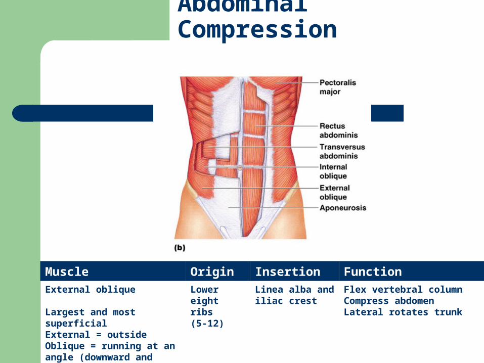
Muscle Origin Insertion FunctionExternal oblique
Largest and most superficialExternal = outsideOblique = running at an angle (downward and medially)
Lower eight ribs (5-12)
Linea alba and iliac crest
Flex vertebral columnCompress abdomenLateral rotates trunk
Abdominal Compression

Muscle Origin Insertion FunctionInternal oblique
Internal = deepOblique = running at an angle (upward and medially)
Inguinal ligament and iliac crest
Linea alba and last four ribs
Flex vertebral columnCompress abdomen Laterally rotates trunk
Abdominal Compression

Muscle Origin Insertion FunctionTransversus abdominus
Transversus = running straight acrossDeepest muscle
Inguinal ligament, ribs 7-12, iliac crest
Xiphoid process, linea alba and pubis
Compress abdomen
Abdominal Compression

Back Extension
Erector Spinae Group
Iliocostalis
Longissimus
Spinalis
Three columns of paired, deep muscles of back
Prime mover of back extension
Provide resistance to control bending forward
Extensors that promote return to erect position

Back Extension
Muscle Origin Insertion Function TypeIliocostalis
Ilio = iliumCostal = ribsMost lateral of the group
Iliac crestRibs 3 - 12Vertebrae
Ribs, thoracic and cervical vertebrae
Rotates and extends vertebral column and headMaintain posture
Prime mover of back extension
Erector Spinae Group
Iliocostalis
Longissimus
Spinalis

Back Extension
Muscle Origin Insertion Function TypeLongissimus
Middle of the groupLongissimus = longest
Transverse processes of lumbar through cervical
Transverse processes of thoracic through cervical, ribs
Rotates and extends vertebral column and headExtends head and turns the face toward the same side
Prime mover of back extension
Erector Spinae Group
Iliocostalis
Longissimus
Spinalis

Back Extension
Muscle Origin Insertion Function TypeSpinalis
Spin = vertebral columnMost medial of the group
Spinous process of upper and lower thoracic vertebrae
Occipital boneSpinous process of upper thoracic and cervical vertebrae
Rotates and extends vertebral column
Prime Mover
Erector Spinae Group
Iliocostalis
Longissimus
Spinalis

Scapula Movement
Muscle Origin Insertion FunctionTrapezius
Trapezoid = irregular four-sided figureLarge muscle in upper back
Occipital bone, spinous process of C7-T12
Acromion and spine of scapula; clavicle
Stabilizes, elevates,rotates and adducts scapula (shrugging shoulders)Extends neck
Trapezius

Movement of Humerus Pectoralis major
Deltoid
Latissimus Dorsi

Muscle Origin Insertion Function TypePectoralis Major
Pectus = chestMajor = largerSuperior portion of chest
Clavicle, sternum and ribs 1-7
Greater tubercle of humerus
Flexion, adduction and medial rotation of armPulls rib cage upwardUsed in movements for climbing, throwing, etc
Prime mover of arm flexion and adduction
Movement of Humerus

Muscle Origin Insertion Function TypeDeltoid
Delta = triangularRounded shoulder muscleCommonly used for IM injection
Clavicle, acromion and spine of scapula
Deltoid tuberosity of humerus
Flexion and medial rotation of arm (anterior)Extension and lateral rotation of the arm (posterior) Arm abduction (lateral)
Prime mover of arm flexion (anterior), extension (posterior), and abduction
Movement of Humerus

Muscle Origin Insertion Function TypeLatissimus dorsi
Latissimus = widestDorsi = backBroad, flat muscle of lower back“The Lats”
T7-T12 , L1-L5, sacrum, ribs 10-12, iliac crest
Proximal humerus
Extension and adduction of armMedially rotates arm at shoulderDraws arm inferiorly and posteriorly
Prime mover of arm extension and adduction
Movement of Humerus
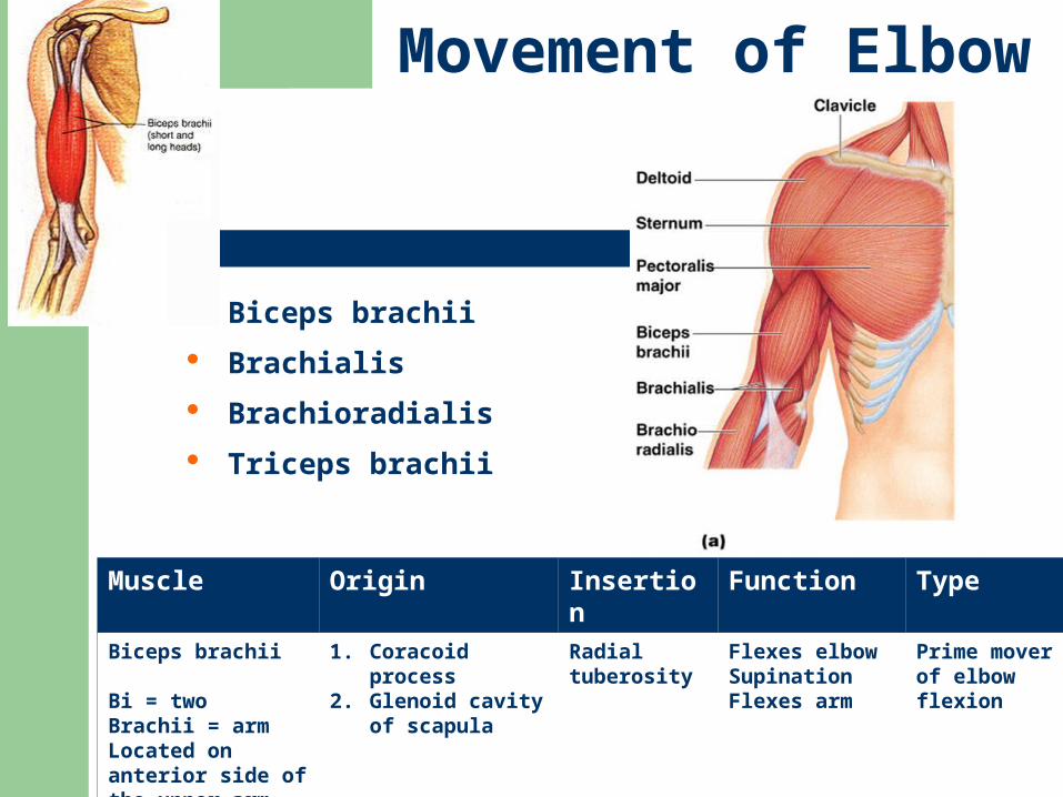
Movement of Elbow
Muscle Origin Insertion Function TypeBiceps brachii
Bi = twoBrachii = armLocated on anterior side of the upper arm
1. Coracoid process2. Glenoid cavity of
scapula
Radial tuberosity
Flexes elbowSupinationFlexes arm
Prime mover of elbow flexion
Biceps brachii
Brachialis
Brachioradialis
Triceps brachii

Movement of Elbow
Muscle Origin Insertion Function Type
Brachialis
Deep to biceps
Anterior, distal humerus
Coronoid process of ulna
Flexes forearm (lifts the ulna as biceps lift the radius)
Prime mover
Brachioradialis
Superficial muscle of lateral forearmRadi = radius
Lateral, distal end of humerus
Styloid process of radius
Flexes forearm (elbow) Synergist to biceps and brachialis

Movement of Elbow
Muscle Origin Insertion Function TypeTriceps brachii
Tri – threePosterior side of upper arm
1. Scapula2. & 3. proximal
humerus
Olecranon process of ulna
Elbow extensionArm extensionAdducts arm
Prime mover of elbow extensionAntagonist of forearm flexion

Movement of Wrist
Flexion
Flexor carpi radialis
Flexor carpi ulnaris
Extension
Extensor carpi ulnaris
Extensor carpi radialis longus
Flexors are usually on anterior side of the body
Extensors are usually on the posterior side of the body
Exception: Knee flexion and extension

Movement of Wrist
Muscle Origin Insertion Function TypeFlexor carpi radialis
Flex = decrease angleCarpi = wristRadi = radiusRuns diagonally across forearm (anterior)
Medial epicondyle of humerus
Metacarpal 2 and 3
Wrist flexion and abduction
Prime mover for wrist flexion
Flexion
Flexor carpi radialis
Flexor carpi ulnaris

Movement of Wrist
Muscle Origin Insertion Function TypeFlexor carpi ulnaris
Ulnar = ulna(anterior)
Medial epicondyle of humerus and proximal ulna
Metacarpal 5 Wrist flexionWrist adduction
Prime mover of wrist flexion
Flexion
Flexor carpi radialis
Flexor carpi ulnaris

Movement of Wrist
Muscle Origin Insertion FunctionExtensor carpi ulnaris
Most medial of superficial muscles (posterior)Extend = increase angle between two bones
Lateral epicondyle of humerus and proximal ulna
Metacarpal 5 Wrist extensionWrist adduction
Extension
Extensor carpi ulnaris
Extensor carpi radialis longus

Movement of Wrist
Muscle Origin Insertion FunctionExtensor carpi radialis longus
Humerus Metacarpals 2 & 3 Wrist extension Wrist abduction
Extension
Extensor carpi ulnaris
Extensor carpi radialis longus

Movement of the Hip
Most originate on pelvis and insert on femur
Iliopsoas
Iliacus
Psoas major
Adductor group (3)
Adductor magnus
Adductor longus
Adductor brevis
Gluteus maximus
Gluteus medius

Muscle Origin Insertion Function Type
Iliacus
Iliac = iliumLarge, fan-shaped lateral muscle
Iliac fossa and sacrum
Lesser trochanter of femur
Flexion of thighLateral rotation of thighFlexion of trunk
Prime Mover hip flexion
Psoas major
Psoa = loin muscleTenderloin in beefLonger, thicker and more medial muscle
Transverse processes of L1-L5 and T12
Lesser trochanter of femur
Flexes thighLateral rotation of thighFlexes lumbar vertebrae
Prime mover hip flexion
Iliopsoas
Iliacus
Psoas major
Movement of the Hip

Muscle Origin Insertion FunctionAdductor Magnus
Magnus = largeTriangular muscle with broad insertion
Ischium and pubis
Femur Adducts thighMedially rotates thighExtends thigh (posterior)
Adductor Muscles
Magnus
Longus
Brevis
Form medial aspect of thigh
Adduct = movement towards midline
“Pulled groin”
Movement of the Hip

Muscle Origin Insertion FunctionAdductor Longus
Longus = long
Pubis crest and pubic symphysis
Femur Adducts thighFlexes thighMedially rotates thigh
Adductor Muscles
Magnus
Longus
Brevis
Movement of the Hip

Movement of the Hip
Muscle Origin Insertion FunctionAdductor brevis
Brevis = short
Inferior ramus of pubis
Femur Adducts thighsFlexes thighMedially rotates thigh
Adductor Muscles
Magnus
Longus
Brevis

Muscle Origin Insertion Function TypeGluteus maximus
Glutos = buttockMaximus = largestLarge, superficial posterior muscle at the top of each legSite of IM injection
Iliac crest, sacrum, and coccyx
Tuberosity of femur
Thigh extensionLateral rotation of thighAbducts thigh
Prime mover of thigh extension
Movement of the Hip
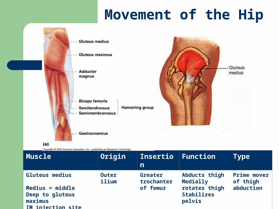
Muscle Origin Insertion Function TypeGluteus medius
Medius = middleDeep to gluteus maximusIM injection site
Outer ilium Greater trochanter of femur
Abducts thighMedially rotates thighStabilizes pelvis
Prime mover of thigh abduction
Movement of the Hip

Movement of the Hip and Knee
Sartorius
Quadriceps Group (4)
Rectus femoris
Vastus lateralis
Vastus medialis
Vastus intermedius
Gracilis
Hamstrings Groups (3)
Biceps femoris
Semimembranosus
Semitendinosus

Muscle Origin Insertion FunctionSartorius
Sartor = tailor “Tailor’s muscle” – crossing the legLongest muscle in the bodyRuns obliquely across anterior surface of thighCrosses both hip and knee joints
Anterior iliac spine
Medial side of proximal tibia
Thigh flexionThigh abductionLaterally rotation thighKnee flexion (weak)
Movement of the Hip and Knee
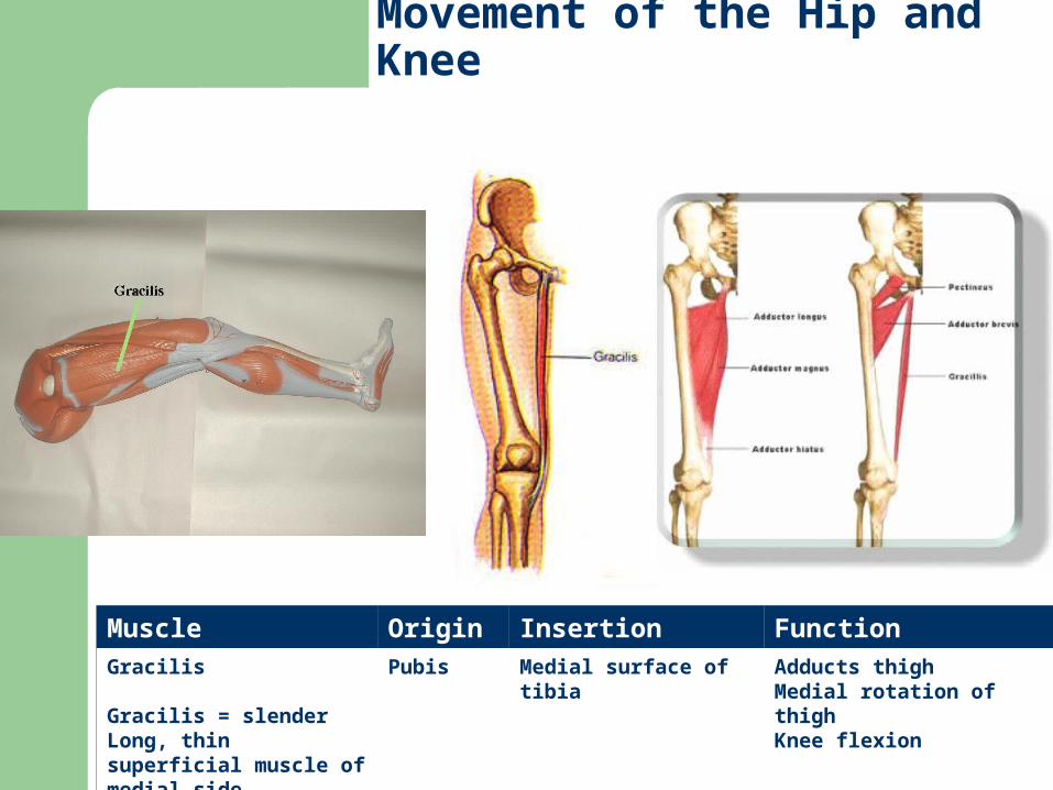
Muscle Origin Insertion FunctionGracilis
Gracilis = slenderLong, thin superficial muscle of medial side
Pubis Medial surface of tibia Adducts thighMedial rotation of thighKnee flexion
Movement of the Hip and Knee

Movement of the Hip and Knee
Muscle Origin Insertion Function TypeRectus femoris
Rectus = straightFemoris = femurSuperficial muscle of anterior thigh
Anterior iliac spine (pelvis)
Tibial tuberosity via patellar ligament
Knee extensionThigh flexion
Prime mover of knee extension
Quadriceps Femoris = 4 heads
Rectus femoris
Vastus lateralis
Vastus medialis
Vastus intermedius
4 heads share insertion at quadriceps tendon which inserts into the patella which continues as the patellar ligament and into the tibial tuberosity

Muscle Origin Insertion Function TypeVastus lateralis
Vastus = largeLateralis = lateralCommon IM injection site
Greater trochanter (femur)
Patella and tibial tuberosity
Extend kneeStabilizes knee
Prime mover of knee extension
Quadriceps Group = 4 heads
Rectus femoris
Vastus lateralis
Vastus medialis
Vastus intermedius
Movement of the Hip and Knee

Muscle Origin Insertion Function TypeVastus medialis
Medialis = medial
Femur Patella and tibial tuberosity
Extend knee Prime mover of knee extension
Quadriceps Group = 4 heads
Rectus femoris
Vastus lateralis
Vastus medialis
Vastus intermedius
Movement of the Hip and Knee

Muscle Origin Insertion Function TypeVastus intermedius
Intermedius = intermediate
Proximal femur
Patella tendon and tibia
Extend knee Prime mover of knee extension
Quadriceps Group = 4 heads
Rectus femoris
Vastus lateralis
Vastus medialis
Vastus intermedius
Movement of the Hip and Knee

Movement of the Hip and Knee
Hamstring Group (posterior)
Biceps femoris
Semimembranosus
Semitendinosus
Posterior thigh muscles
Cross both hip and knee joints
Name comes from butchers using their tendons to hang hams
Muscle Origin Insertion Function TypeBiceps femoris
Bi = twoLateral muscle of group
1. Ischial tuberosity2. Distal femur
Proximal fibula Thigh extensionKnee flexionLaterally rotates leg when knee is flexed
Prime mover of thigh extension and knee flexion

Muscle Origin Insertion Function TypeSemimembranosus
Membranosus = membraneDeep to semitendinosus
Ischial tuberosity
Medial condyle of tibia
Extends thighFlexes knee
Prime mover of knee flexion
Movement of the Hip and Knee
Hamstring Group (posterior)
Biceps femoris
Semimembranosus
Semitendinosus

Muscle Origin Insertion Function TypeSemitendinosus
Semi = halfTendinosus = tendonMedial to biceps femoris
Ischial tuberosity
Medial tibia Extends thighFlexes knee
Prime mover of knee flexion
Movement of the Hip and Knee
Hamstring Group (posterior)
Biceps femoris
Semimembranosus
Semitendinosus

Muscles of the Ankle
Gastrocnemius (knee also)
Soleus
Tibialis posterior
Tibialis anterior
Fibularis longus
Fibularis brevis
Muscles of Ankle and Toes
Extensor digitorum longus (anterior)
Flexor digitorum longus (posterior)

Muscles of the Ankle
Muscle Origin Insertion Function TypeGastrocnemius
Gastro = bellycneme = leg Two bellied muscle“Dancer’s Toe”
Medial and lateral condyles of femur
Calcaneus via Achilles tendon
Plantar flexionKnee flexion
Prime mover of plantar flexion

Muscles of the Ankle
Muscle Origin Insertion Function TypeSoleus
Soleus = a type of flat fishBroad, flat muscleDeep to gastrocnemius on posterior surface
Posterior tibia and fibula
Calcaneus via Achilles tendon
Plantar flexion Prime mover of plantar flexion

Muscles of the Ankle
Muscle Origin Insertion Function TypeTibialis posterior
Posterior = backThick, flat muscle deep to soleus
Posterior tibia and fibula
Metatarsals 2-4 and tarsals
Foot inversion Plantar flexion
Prime mover of inversion

Muscles of the Ankle
Muscle Origin Insertion Function TypeTibialis anterior
Anterior = frontSuperficial muscle of anterior leg
Lateral tibia
1st metatarsal and tarsal
DorsiflexionInversion
Prime mover of dorsiflexion

Muscles of the Ankle
Muscle Origin Insertion FunctionFibularis longus
Superficial lateral muscle over fibula
Proximal fibula and lateral condyle of tibia
1st metatarsal and tarsal
Plantar flexionEversion
Muscles of the Ankle
Muscle Origin Insertion FunctionFibularis brevis
Brevis = shortSmaller muscleDeep to fibularis longus
Distal fibula shaft
Proximal end of 5th metatarsal
Plantar flexion Eversion

Muscle Origin Insertion Function TypeExtensor digitorum longus
Digit = toesLateral to tibialis anterior muscle
Lateral condyle of tibia and fibula
Distal phalanges 2-5
Extends toes 2-5Dorsiflexion
Prime mover of toe extension 2-5
Muscles of the Ankle and Toes

Muscles of the Ankle and Toes
Muscle Origin Insertion Function TypeFlexor digitorum longus
Long, narrow muscleRuns medial to tibialis posterior
Posterior tibia
Distal phalanges of toes 2-5
Toe flexionPlantar flexionInversionHelps foot grip ground
Prime mover of toe flexion 2-5

Anterior Muscles

Posterior Muscles