Embed Size (px)
Citation preview

www.aromaflex.co.nz Telephone: 03 545 6218
UNIT FOUR
__________________________
ANATOMY & PHYSIOLOGY
THE MUSCULAR SYSTEM

Aromaflex Academy, 1st Floor, Development House, 280 – 282 Trafalgar Street, Nelson. NZ. [email protected]
Unit 4 – The Muscular System
2
Certificate of Anatomy & Physiology Course Notes:
1998 First Revised Edition
2000 Second Revised Edition
2003 Third Revised Edition
2008 Fourth Revised Edition
2013 Fifth Revised Edition
Published by Aromaflex Academy,
Development House,
First Floor
280-282 Trafalgar Street,
Nelson,
New Zealand
References to updating material in Fourth Revised Edition:
A Textbook of Anatomy and Physiology, 2nd
Edition; William Arnold-Taylor MSc PhD
Ross & Wilson Anatomy and Physiology in Health and Illness, 7th
Edition; Kathleen J. W. Wilson OBE
Human Anatomy and Physiology, 3rd
Edition.; Elaine N. Marieb
Principles of Anatomy and Physiology, 8th
Edition; Gerald J. Tortora and Sandra Reynolds Grabowski
The Human Body in Health and Disease, 9th
Edition; Cohen and Woods.
A massage Therapists Guide to PathologWerner and Benjamin.
An Introductory Guide to Anatomy & Physiology; Louise Tucker
NCEA Level 1 Chemistry; Dorothy Kane
Year 10 Science: Paul King,Tania Lineham, Marea Goode, Jenny Pollock, Cara Scott, Mark Standley
All rights reserved. No part of the publication may be reproduced, stored in a retrieval system, or transmitted, in any
form or by any means, electronic, mechanical, photocopying, recording or otherwise, without the prior permission of the
copyright holder.
Aromaflex Academy asserts the moral right to be identified as author of this work.

Aromaflex Academy, 1st Floor, Development House, 280 – 282 Trafalgar Street, Nelson. NZ. [email protected]
Unit 4 – The Muscular System
3
UNIT FOUR.
THE MUSCULAR SYSTEM
The main framework of the skeleton of the body is covered in muscles. They are responsible for 50% of
our body weight. It’s the alternating contraction and relaxation of the muscle tissue which allows
movement to occur. Our bones provide leverage but skeletal muscles have to work cooperatively with
them for movement to occur.
Your muscular strength reflects the prime function of muscle – changing chemical energy into
mechanical energy to generate force, perform work and produce movement.
In addition, muscle tissue stabilises the body’s position, regulates organ volume, generates heat and
propels fluids and food matter through various body systems
The muscle system is under the control of the nervous system. There are nerve endings in the
muscles and their actions are dependent on the vitality of the nervous system and the connecting
nerves.
Muscle Functions.
1) To permit movement. Every part of the body capable of motion, performs its movement by means
of it’s own special set of muscles. Skeletal muscles enable us to move and express emotions while
smooth muscle inside the body works to move substances such as fluids and food, or a baby through
internal body channels.
2) To maintain posture. We are rarely aware of this yet these muscles function continually, making tiny
adjustments to keep us upright against the pull of gravity.
3) They generate heat. As they contract heat is generated which is vital in maintaining normal body
temperature. It’s mostly the voluntary (skeletal) muscles that are responsible for this. When there is
a danger of our body temperature getting too low, we shiver. ( this is an involuntary action ) About
30% of the energy produced in muscular activity results in work, the remaining 70% being released
as heat which warms the body.
4) They stabilise joints such as the knees or the shoulders where there is poor reinforcement.
5) Regulating Organ Volume sustained contractions of ring like bands of smooth muscles called
sphincters may prevent the outflow of contents of hollow organ e.g. urine in the urinary bladder
6) Moving substances in the body Cardiac muscle contractions move blood through the circulatory
system. Smooth muscle contractions also move food substances e.g. bile through the gastro
intestinal tract.
Skeletal muscle contractions promote the flow of lymph and aid the return of blood to the heart.
7) They also give our body shape.

Aromaflex Academy, 1st Floor, Development House, 280 – 282 Trafalgar Street, Nelson. NZ. [email protected]
Unit 4 – The Muscular System
4
Muscle Tissue.
Firstly all muscle cells are elongated and for this reason called muscle fibres. These fibres are elastic,
able to stretch and relax. In its relaxed state a muscle is soft, but within a few milliseconds it can become
a hard elastic structure.
There are three types of muscle tissue in the body;
Voluntary (skeletal)
Cardiac,
Involuntary (smooth).
Voluntary or Skeletal muscle tissue.
This is the muscle tissue that is packaged together to form our skeletal muscles that attach to the body’s
skeleton.
This can be described as skeletal, striated, striped or voluntary muscle. It is called voluntary because
contraction is under control of the will. We must however remember that skeletal muscles can be
activated by reflexes not under conscious control. e.g. shivering.
Each skeletal muscle is made up of hundreds to thousands of muscle fibres. Like all body cells, skeletal
muscle fibres are soft and fragile and these fibres are bundled together for strength and support. The
plasma membrane of a muscle cell is called the sarcolemma and the cytoplasm content is called
sarcoplasm. Microscopically this sarcoplasm appears to have alternating light and dark bands hence it’s
said the cells have a striated appearance.
The connective tissue coverings support each cell, reinforce the muscle as a whole and provide muscle
tissue with its natural elasticity.
The fibres are long and thread like have this striped appearance and are multinucleate, that is they have
more than one nucleus per cell.
Each muscle fibre is surrounded by a fine sheath of connective tissue called endomycium.
Several sheathed muscle fibres are then gathered together, side by side, into bundles of muscle cells
called fascicles. Each fascicle is bound together by a collagenic sheath called a perimysium.
Bundles of fascicles are then bound together by even coarser dense fibrous connective tissue called
epimysium.
The epimysium surrounds the entire muscle. This sheath is extended at the end to form strong fibrous
bands known as tendons, by means of which the muscle is attached to the bones.
External to the epimysium is the deep fascia, an even coarser sheet of fibrous connective tissue, which
binds muscles into functional groups.
N.B. Tendons are cords of dense connective tissue extending beyond the muscle that attaches a muscle to
the periosteum of a bone. When the connective tissue extends in a broad flat layer it’s called an
aponeuroses.
The microscopic structure of skeletal muscle:
Skeletal muscle fibres are huge cells and when examined microscopically are found to be roughly
cylindrical or cigar shaped. They may be as long as 35cms. and some fibres are so big and coarse they
can be seen with the naked eye. Their diameter is typically up to 10 times larger than that of an average
body cell.
As we have said they have several nuclei and these are situated just under the cell membrane of each cell.
The muscle fibres lie parallel to one another and when viewed under the microscope they show well these
marked transverse dark and light bands, the striations or stripes. At high magnification, each muscle cell is
seen to contain a large number of rod like myofibrils that run in parallel fashion and extend the entire
length of the cell. There are hundreds to thousands of myofibrils in a single muscle fibre, and they account
for about 80% of cellular volume. It’s these myofibrils that are the contractile elements of skeletal muscle.

Aromaflex Academy, 1st Floor, Development House, 280 – 282 Trafalgar Street, Nelson. NZ. [email protected]
Unit 4 – The Muscular System
5
The cytoplasm in muscle fibres contains unusually large amounts of stored glycogen and a unique oxygen
binding protein called myoglobin, a red pigment that stores oxygen in the muscle cells. Skeletal muscle
contraction can be fast and strong but it tires easily and needs frequent rest periods.
Diagram showing organization of skeletal muscle and its connective tissue coverings.

Aromaflex Academy, 1st Floor, Development House, 280 – 282 Trafalgar Street, Nelson. NZ. [email protected]
Unit 4 – The Muscular System
6
Diagram showing microscopic organization of skeletal muscle.
Aromaflex Academy, 1st Floor, Development House, 280 – 282 Trafalgar Street, Nelson. NZ. [email protected]
Unit 4 – The Muscular System
7
Involuntary or Smooth Muscle Tissue
Smooth muscle has no striations and is involuntary.
Except for the heart, which is made up of cardiac muscle, smooth muscle is found in the walls of hollow
visceral organs such as blood and lymph vessels, the alimentary tract, the respiratory tract, the urinary
bladder, the biliary tract and the uterus.
Although contraction is essentially the same in all muscle tissue, smooth muscle is different in several
ways:
Smooth muscle fibres are small, spindle - shaped cells, each with only one central nucleus.
Skeletal muscle fibres are 20 times wider and thousands of times longer than the smooth muscle
fibres.
Between the smooth muscle fibres is a small amount of fine connective tissue, (endomycium,)
containing blood vessels and nerves. Smooth muscle lacks the courser connective tissue sheaths seen in
skeletal muscle.
Bundles of fibres are organised into sheets or layers of muscle. Most often there are two layers, one
running circularly and the other longitudinally. As the two layers alternately contract and relax they
change the size and shape of the organ.
No striations are visible, hence the name smooth muscle.
As we have already said these muscles are involuntary, that is they are not under the control of the
will. The nerve fibres are part of the autonomic nervous system.
Contraction of smooth muscle is slow, sustained and resistant to fatigue. It takes 30 times longer to
contract and relax than a skeletal muscle.
Cardiac Muscle Tissue
Cardiac muscle is only found in the walls of the heart.
It is involuntary muscle and is therefore not under the control of the will.
It is striated in appearance, resembling voluntary muscle.
It has the greatest supply of blood vessels of all the muscles because cardiac muscle is constantly
working and requires much energy.
Cardiac muscle cells are short, fat, branched and interconnected. The cells fit together tightly at
unique junctions called intercalated discs. A wave of contraction spreads from cell to cell across these
discs which means that the cells do not need to be stimulated individually.
The fibres are in a spiral or a figure 8 arrangement and cushioned by small amounts of connective
tissue. The loose connective tissue of the intercellular spaces contains numerous blood vessels.
Each cell contains only one, or occasionally two, pale centrally located nucleus.
Contractions usually occur at a steady pace under the control of the inbuilt pacemaker but can speed
up when necessary as a result of nervous system stimulation.

Aromaflex Academy, 1st Floor, Development House, 280 – 282 Trafalgar Street, Nelson. NZ. [email protected]
Unit 4 – The Muscular System
8
PROPERTIES OF MUSCLE TISSUES
Muscle tissue has four special properties that enable it to function and contribute to
homeostasis
1) Electrical Exatability
A property of both muscle fibres (cells) and neurons is the ability to respond to certain
stimuli by producing electrical signals.
2) Contractility
Is the ability to generate tension to do work
3) Extensibility
Is the ability of the muscle to stretch without being damaged
4) Elasticity
Is the ability of muscle tissue to return to its original length and shape after contraction or
extension
NERVE AND BLOOD SUPPLY
Skeletal muscles are well supplied with nerves & blood vessels. Generally an artery and one or two
veins go with each nerve that penetrates a skeletal muscle. Blood capillaries bring in oxygen &
nutrients and remove heat and waste products of muscle metabolism. The neurons that stimulate
skeletal muscle to contract are the somatic motor neurons
Energy Source for Muscle Contraction
Skeletal muscles have a good supply of blood and nerves.
The blood supplies the oxygen and nutrients for muscle contractions and removes the waste products.
Muscle contraction requires energy in the form of ATP and the muscle cells need a supply of oxygen and
glucose or other usable nutrient to produce this. This production process is called cellular respiration.
Glucose is also stored in the muscle fibres in the form of glycogen so that it is immediately available to
produce energy for movements.
For a skeletal muscle fibre to contract it must be stimulated by a motor nerve ending, a motor neurone.
The motor neurone plus the skeletal muscle fibres it stimulates is called a motor unit.
A motor unit can consist of 2 to 2000 fibres and these fibres all contract at the same time under the
stimulus of the neurone. The junction where the axon of the neurone reaches the muscle cell membrane is
called a neuromuscular junction.
The nerve stimulus sets up chemical changes in the muscle cells. These changes include the breaking
down of glucose, glycogen, fat, and oxygen to form the ATP which in turn liberates the energy required
for contraction. Protein molecules inside the fibres are used to provide energy when supplies of
carbohydrate and fat are deficient. As we said, for the complete breakdown of all these various molecules
an adequate supply of oxygen is required and ‘mother nature’ has taken care of this by storing extra
oxygen in the muscle cells in the form of myoglobin. Vigorous exercise then increases the need for
oxygen.
Muscle Fatigue
If we exercise our muscles for a long period of time fatigue occurs as a result of insufficient oxygen and
ATP. When muscles lack oxygen lactic acid begins to accumulate in the muscle fibres resulting in a
build up of waste products, more than the venous and lymph systems can cope with. When waste
products remain between the muscle fibres, they contract less efficiently and there is a feeling of
soreness. The accumulating lactic acid is a sticky substance preventing the muscle fibres sliding

Aromaflex Academy, 1st Floor, Development House, 280 – 282 Trafalgar Street, Nelson. NZ. [email protected]
Unit 4 – The Muscular System
9
smoothly over each other. Friction then occurs leading to the soreness. Massage can ease the pain by
increasing local circulation
True muscle fatigue is when the muscle stops functioning altogether but we usually feel tired before this
occurs and slow down or stop the activity. Marathon runners sometimes experience true muscle fatigue
and collapse when their muscles can no longer work.
.
Factors which influence contraction of muscle.
Satisfactory metabolic conditions. Making sure there is an adequate supply of food and oxygen,
oxidation is complete, and waste products are removed.
Strength of the stimulus, so disorders of the nervous system affect muscle contraction
Exercise increases the flow of blood and therefore increases the supply of oxygen and nutrients to
muscle tissue. Waste products can then be removed, thus reducing fatigue.
Prolonged immobility can cause reduced muscle tone.
Temperature. Warmth enables muscle fibres to react to stimuli more rapidly and contract more
efficiently, this is why athletes warm up before exercise. Exposure of the skin to cold will stimulate
muscle contraction as a means of maintaining body temperature.
Types of Muscle Contractions
Until now we have been looking at contraction in terms of shortening behaviour. Sometimes
muscles do not shorten during contraction but remain unchanged in length or actually lengthen.
There are two main categories of contractions, isotonic and isometric:
Isotonic contractions
These occur when the muscle shortens or lengthens and movement occurs.
Smiling, bending the knee moving the arms are all examples of isotonic contractions.
Isometric contractions
Shortening of the muscle may be prevented because a load is applied. The force of contraction then
results in a great increase in tension within the muscle but its length remains the same.
Muscles that act primarily to maintain upright posture, or trying to lift a 400 pound object alone
would be examples.
Most body movements involve both types of contraction.
Muscle Tone
The word tone is used to describe the state of slight tension in the muscles in which they are kept all
the time. It also refers to the efficiency with which the muscles can switch from the resting to the
active state.
This minimum tension keeps the bones to which the muscles are attached in their normal positions
but it is not sufficient to cause fatigue.
Bad posture can cause fatigue and pain because more than the usual amount of tension is needed to
maintain the body in balance.
Tension
If the amount of tension being maintained in the muscles, even when not moving, is excessive pain
and fatigue and other complications can arise. The constant contraction of the muscles will cause
local pain due simply to the fatigue of the muscle itself. The muscle may even go into spasm.
If a state of excessive tension exists over a prolonged period actual displacement of bones may
occur because the muscles attached to those bones are continually pulling them. Any muscle or
group of muscles in the body may be affected but this kind of stress- induced pain often appears in
the neck, shoulder and back muscles.

Aromaflex Academy, 1st Floor, Development House, 280 – 282 Trafalgar Street, Nelson. NZ. [email protected]
Unit 4 – The Muscular System
10
Muscle Spasm
When a muscle becomes locked in a contracted state, instead of releasing after a movement is
completed, we say it is in spasm.
Spasm can occur following injury or sudden excessive demands on the muscle such as lifting an
unaccustomed weight. Some of the muscle fibres do not return to their longer, relaxed state as they
ought to.
The shortened fibres can be felt during massage as a ‘knot’ or nodule. The nodule may be as small
as a pea but the whole muscle will be affected by pain.
You may find several nodules clustered together in high tension areas such as the lower back or the
shoulders.
In some areas of the body spasm can be felt as long hard strips. They are sometimes so hard that
they may be mistaken for bones. These hard areas or nodules are made up of contracted muscle
filaments and accumulated acid wastes.
Posture
The muscular system plays a vital role in maintaining correct body posture by means of good
muscle tone and co-ordinated activity.
Maintenance of good posture when standing, sitting, lying down or moving about has the following
advantages:
It improves ones general appearance.
It prevents deformities in bones, joints and muscles.
It prevents the fatigue, discomfort and or pain induced by some forms of abnormal posture.
It encourages free circulation of blood.
It lessens the risk of injury to joints and muscles during strenuous activity.
It allows internal organs to function without interference, e.g. full expansion of the lungs is
possible when the body posture is correct.
The Mechanics of Muscle Movement
To be able to produce movement at a joint, a muscle or a tendon must stretch across the joint. When a
muscle contracts, its fibres shorten and it pulls one bone towards another. e.g bending the elbow.
Skeletal muscles have no less than two points of attachment to bone.
The point of origin is the bone to which they are attached and which they do not move. The point
of insertion is the bone to which they are attached and which they do move.
When the muscle contracts the insertion moves towards the origin.
Because muscles pull but cannot push they need to work in pairs. Whatever one muscle or muscle group
can do there is always another muscle or muscle group that can “undo” the action.
A muscle that causes the movement is the agonist.
As the agonist contracts, the other muscle in the pair relaxes and lengthens. This muscle is called the
antagonist.
An example would be when the biceps (agonist) contracts the triceps (antagonist) relaxes, the fore-arm
bends. When the triceps contracts (agonist) and the biceps relaxes (antagonist) the fore-arm straightens.

Aromaflex Academy, 1st Floor, Development House, 280 – 282 Trafalgar Street, Nelson. NZ. [email protected]
Unit 4 – The Muscular System
11
A diagram showing Antagonist and Agonist pair of muscles ~~~~~
Biceps contracts - (agonist) Biceps relaxes - (antagonist)
Triceps relaxes - (antagonist) Triceps contracts - (agonist)

Aromaflex Academy, 1st Floor, Development House, 280 – 282 Trafalgar Street, Nelson. NZ. [email protected]
Unit 4 – The Muscular System
12
Diagrams illustrating Types of Body Movement
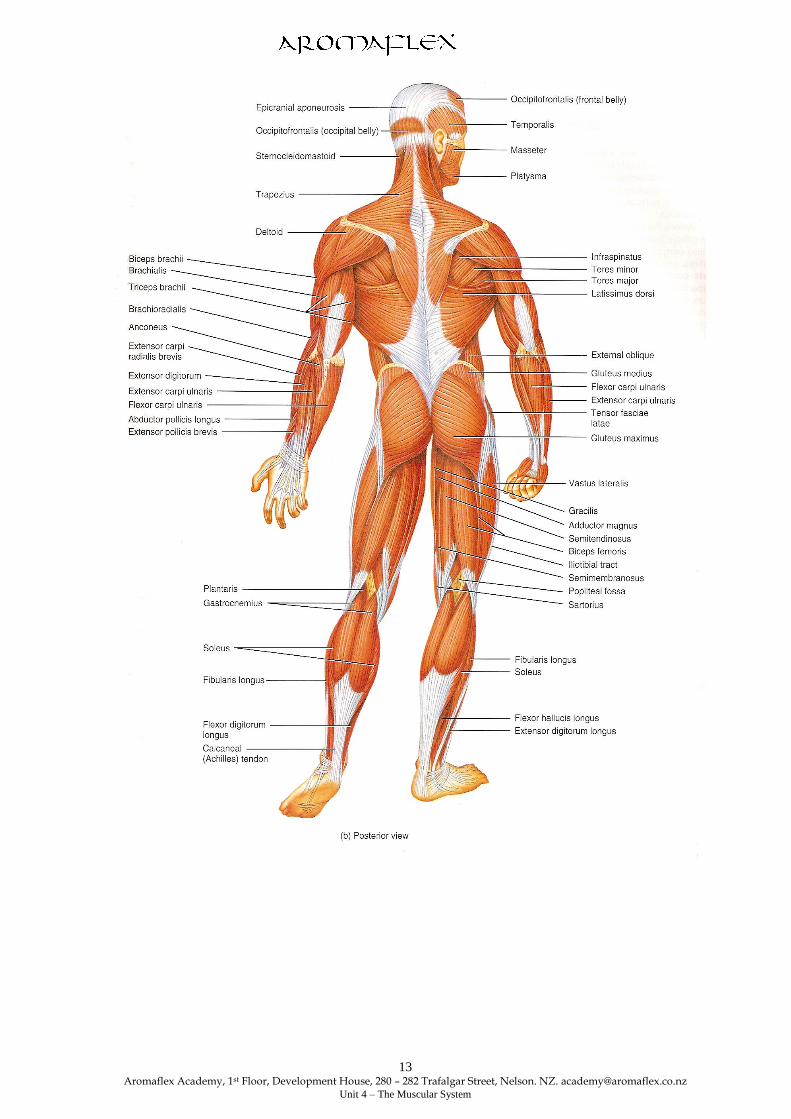
Major Skeletal Muscles Of The Body
Aromaflex Academy, 1st Floor, Development House, 280 – 282 Trafalgar Street, Nelson. NZ. [email protected]
Unit 4 – The Muscular System
13

Aromaflex Academy, 1st Floor, Development House, 280 – 282 Trafalgar Street, Nelson. NZ. [email protected]
Unit 4 – The Muscular System
14
Muscles of the Face and Neck.
There are many muscles involved in changing facial expression and with movement of the lower jaw
during chewing and speaking. Only the main muscles are described here.
Except where indicated the muscles are present in pairs, one on each side.
There are the muscles of expression and the muscles of mastication.
Occipitofrontalis - (unpaired) consists of a posterior muscular part over the occipital bone, an anterior
part over the frontal bone and an extensive flat tendon or aponeurosis that stretches over the dome of the
skull and joins the two muscular parts. It raises the eyebrows.
N.B. An aponeurosis is a flat wide band of fibrous connective tissue that connects muscle to muscle
and muscle to bone.
Levator Palpebrae Superioris - extends from the posterior part of the orbital cavity to the upper
eyelid. It raises the eyelid.
Orbicularis Oculi - surrounds the eye, eyelid and orbital cavity. It closes the eye and when strongly
contracted screws up the eyes.
Buccinator - This flat muscle of the cheek draws the cheek muscles in towards the teeth in chewing
and in forcible expulsion of air from the mouth (trumpeters muscle.)
Orbicularis Oris (unpaired) surrounds the mouth and blends with the muscles of the cheeks. It
closes the lips and, when strongly contracted, shapes the mouth for whistling.
Masseter - is a broad muscle extending from the zygomatic arch to the angle of the jaw. In chewing
it draws the mandible up to the maxilla and exerts pressure on the food.
Temporalis - covers the squamous part of the temporal bone. It passes behind the zygomatic arch to
be inserted into the coronoid process of the mandible.
It closes the mouth and assists with chewing.
Pterygoid Muscle - extends from the sphenoid bone to the mandible.
It closes the mouth and pulls the lower jaw forward.

Aromaflex Academy, 1st Floor, Development House, 280 – 282 Trafalgar Street, Nelson. NZ. [email protected]
Unit 4 – The Muscular System
15
Muscles that Move the Head
Sternocleidomastoid
Origin - Sternum and clavicle
Insertion - Mastoid process of temporal bone.
Action - Acting together – flex cervical portion of vertebral column and extend head.
- Acting singly – laterally flex and rotate head to side opposite contracting muscle.
Semi Spinalis Capitus
Origin - Articular processes of 4th
, 5th
, 6th
cervical vertebrae. Transverse process of 7th
cervical and
6th
or 7th
thoracic vertebrae.
Insertion - Occipital bone between superior and inferior nuchal lines.
Action - Acting together – extended head
- Acting singly – rotate head side to side opposite contracting muscle.
Splenius Capitus
Origin - Ligamentum nuchae & spinous processes of 7th
cervical and first 3 or 4 thoracic
vertebrae.
Insertion - Occipital bone and mastoid process fo temporal bone.
Action - Acting together - extend head
- Acting singly - Laterally flex and rotate head to same side as contracting muscle.
Longissimus Capitis
Origin - Transverse processes of upper four thoracic vertebrae and articular processes of last four
cervical vertebrae.
Insertion - Mastoid process of temporal bone.
Action - Acting together - extend head
- Acting singly – Laterally flex and rotate head to same side as contracting muscle.
Muscles that move the Pectoral (shoulder girdle)
Muscles that move the pectoral girdle can be divided into two groups based on their location in the
thorax: Anterior and Superior
Anterior Thoracic Muscles
Subclavius (sub-KLA-vē-us; sub = under; clavius = clavicle)
Origin - First rib
Insertion - Clavicle
Action - Depresses and moves clavicle anteriorly and helps stabilze pectoral girdle.
Pectoralis minor (pek’-tor-A-lis; pectus = breast, chest, thorax; minor = lesser)
Origin - Second through fifth, third through fifth, or second through fourth ribs.
Insertion - Corocoid process of scapula.
Action - Depresses and abducts scapula and rotates it downwards; elevates third through fifth ribs
during forced inspiration when scapula is fixed.
Serratus anterior ser-Ā-tus; serratus = saw toothed: anterior = before)
Origin - Superior eight or nine ribs.
Insertion - Vertebral border and inferior angle of scapula
Action - Abducts scapula and rotates it upwards; elevates ribs when scapula is fixed; known as
“boxer’s muscle.”

Aromaflex Academy, 1st Floor, Development House, 280 – 282 Trafalgar Street, Nelson. NZ. [email protected]
Unit 4 – The Muscular System
16
Posterior Thoracic Muscles
Trapezius (tra-PĒ-zē-us; trapezoids = trapezoid-shaped). The trapezius is a large flat triangular
sheet of muscle. It is the most superficial back muscle.
Origin - Superior nuchal line of occipital bone, ligamentum nuchae, and spines of seventh cervical
and all thoracic vertebrae.
Insertion - Clavicle and acromion and spine scapula.
Action - Superior fibres elevate scapula and can help extend head; middle fibres adduct scapular;
inferior fibres depress scapular; superior and inferior fibres together rotate scapula upwards;
stabilizes scapular.
Levator scapulae (le –VĀ-tor SKA-pyoo-lē; levare = to raise; scapulae = scapula
Origin - Superior four or five cervical vertebrae.
Insertion - Superior vertebtral border of scapular
Action - Elevates scapula and rotates it downwards
Rhomboideus major (rom-BOID-ē-us; rhomboids=rhomboid or diamond-shaped)
Origin - Spines of second to fifth thoracic vertebrae.
Insertion – Vertebral border of scapula inferior to spine.
Action – Elevates and adducts scapula and rotates it downwards; stabilizes scapula.
Rhomboideus minor (rom-BOID-ē-us)
Origin - Spines of seventh cervical and first thoracic vertebrae.
Insertion - Vertebral border of scapular superior to spine.
Action - Elevates and adducts scapula and rotates it downwards; stabilizes scapula.
Muscles of the Arm:
Of the muscles that cross the shoulder joint, only two of them do not have their origin on the
scapula. They are Pectoralis Major and Latissmun Dorsi. These two muscles are called Axial
muscles as their origins are on the Axial Skeleton.
The Pectoralis Major
Origin – Clavicle (clavivular head), sternum, and costal cartilages of second to sixth ribs
(sometimes first to seventh ribs).
Insertion – Greater tubercle and intertubercular sulcus of humerus.
Action – As a whole, adducts and medially rotates arm at shoulder joint; clavicular head alone
flexes arm and sternocostal head alone extends arm at shoulder joint.
The Latissimus Dorsi
Origin – Spines of inferior six thoracic vertebrae, lumbar vertebrae, crests of sacrum and ilium,
inferior four ribs.
Insertion – Intertubercular sulcus of humerus.
Action – Extends, adducts, and medially rotates arm at shoulder joint; draws arm inferiorly and
posteriorly.

Aromaflex Academy, 1st Floor, Development House, 280 – 282 Trafalgar Street, Nelson. NZ. [email protected]
Unit 4 – The Muscular System
17
The Deltoid
Origin - Is a triangular muscle lying over the point of the shoulder.
Insertion -
Action - Is to abduct (raise) the arm.
Biceps Braehii (BĪ-ceps BRĀ-kē-ī; biceps= two heads of origin; brachion= arm
Origin – Long head originates from tubercle above glenoid cavity of scapula (supraglenoid
tubercle); short head originates from coracoids process of scapula.
Insertion – Radial tuberosity and bicipital aponeurosis.
Action – Flexes forearm at elbow joint, supinates forearm at radioulnar joints, and flexes arm at
shoulder joint.
The Brachialis (brā-kē-A-lis) Lies below the biceps on the anterior aspect of the upper arm. It helps
with flexion of the elbow. It is a single joint muscle and an important flexor of the elbow joint.
Origin – Distal, anterior surface of the humerus.
Insertion – Ulna tuberosity and coronoid process of ulna.
Action – Flexes forearm at elbow joint..

Aromaflex Academy, 1st Floor, Development House, 280 – 282 Trafalgar Street, Nelson. NZ. [email protected]
Unit 4 – The Muscular System
18
Brachioradialis (bra’-kē-ō-rā-dē-A-lis; radialis-radius)
Origin – Medial and lateral borders of distal end of humerus.
Insertion – Superior to styloid process of radius.
Action – Fleses forearm at elbow joint and supinates and pronates forearm at radioulnar joints to
neutral position.
Coracobrachialis (kor’-a-kō-BRĀ-kē-a’-lis; coraco=coracoids process)
Origin – Coracoid process of scapula.
Insertion – Middle of medial surface of shaft of humerus.
Action – Flexes and adducts arm at shoulder joint.
Teres major – (TE-rēz; teres= long and round)
Origin – Inferior angle of scapula.
Insertion – Intertubercular sulcus of humerus.
Action – Extends arm at shoulder joint and assists in adduction and medial rotation of arm at
shoulder joint.
Teres minor (TE-rēz)
Origin - Inferior lateral border of scapula.
Insertion – Greater tubercle of humerus.
Action – Laterally rotates, extends, and adducts arm at shoulder joint.
The strength and stability of the shoulder is provided by four muscles:
- Subscapularis
- Suprs spinatus
- Infra spinatus
- Teres minor
These muscles join the shoulder to the humerus. Their flat tendons fuse together to form a nearly
complete circle around the shoulder joint. This arrangement is referred to as the musculoteninous
cuff or rotator cuff. The supraspinatus muscle because of its position can suffer a great deal of
wear and tear.
Main Muscles of the Abdominal Wall.
Rectus Abdominis – a broad flat muscle forming the front abdominal wall. Origin in the symphysis
pubis and insertion into lower ribs and sternum. Action – flexes trunk, assists in forced expiration,
childbirth and defecation.
External Oblique – forms outer coat of side wall. Flexes and rotates vertebral column.
Internal oblique – Flexes and rotates vertebral column.
Transverse Abdominis - compresses abdomen.
Muscles that move the chest wall.
External Intercostals – lie between the ribs and draws them upwards and outwards, thus
increasing the size of the thoracic cavity.
Internal Intercostals – also lie between the ribs and are antagonists to the external intercostals.
Action to draw the ribs downwards and inwards.

Aromaflex Academy, 1st Floor, Development House, 280 – 282 Trafalgar Street, Nelson. NZ. [email protected]
Unit 4 – The Muscular System
19
Muscles of the Neck and Back.
Sternocleidomastoid – flexes neck and rotates head.
Trapezius – pulls the head backward and elevates, stabilizes and lowers shoulders.
Latissimus Dorsi – rotates and adducts the humerus and assists in extension of the arm at the
shoulder joint.
Deltoid – prime movers of arm abduction.
Teres major – extends the arm and draws humerus down and back.
Erector Spinae – extends the back. A common source of lower back pain after injury.
Psoas – flexes and rotates thigh laterally and flexes vertebral column.
Sacrospinalis – backward flexion or extension of vertebral column.
Quadratus Lamborum – extends vertebral column and extends it sideways.
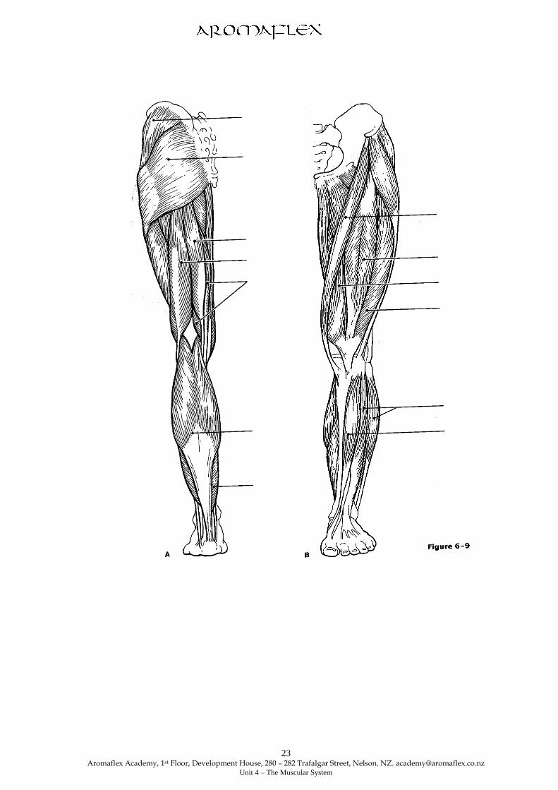
Muscles of The Back.

Aromaflex Academy, 1st Floor, Development House, 280 – 282 Trafalgar Street, Nelson. NZ. [email protected]
Unit 4 – The Muscular System
20
Muscles Which move the Leg.
The Sartorius – the longest muscle in the body extending from the pelvic girdle to the upper part of
the tibia. Action flexes thigh on hip; helps with the cross legged position.
Quadriceps group – all extend the knee; rectus femoris also flexes hip on thigh.
Adductor muscles – Adduct thigh.
Gracilis – adducts thigh and flexes leg.
Tibialis Anterior – dorsiflexes and inverts the foot.
The Hamstrings – flex knee and extend hip.
Gastrocnemius – plantar flexes foot and flexes knee.
The Soleus – plantar flexes foot.
The Gluteus maximus – forms the buttocks; extends the hip when power is needed e.g. jumping
and climbing stairs.
The Gluteus Medius – a hip abductor; important in steadying the hip when walking.
Tensor Fasciae latae – flexes and abducts thigh.

Aromaflex Academy, 1st Floor, Development House, 280 – 282 Trafalgar Street, Nelson. NZ. [email protected]
Unit 4 – The Muscular System
21
ANATOMY & PHYSIOLOGY
ASSIGNMENT COVER SHEET
Name:
Address:
Telephone Number:
E-Mail Address:
Unit Name and Number:
Date Assignment Submitted:
Questions/Comments from student:
Requirements:
Send in the above cover sheet along with the question sheet.
Enclose a stamped addressed envelope to have your assignment returned.
Answer each question on a separate sheet of paper, with your name at the top of every page.
All resubmissions need to be sent in with original marked unit.
Make sure your resubmission work is clearly in a different colour, or highlighted as a
“resubmission, attempt 2”, “resubmission, attempt 3” etc..
Answer the questions in black or blue pen, not pencil. Drawings may be in pencil.
Submit clearly drawn and clearly labelled diagrams
Answer the questions clearly and in order they are set on the question page.
All questions need to be answered correctly for the unit to pass.
If you don’t understand any questions contact the A&P tutor.
You have as many attempts as you like to get the question right, and to pass the unit.
Keep a copy of your work

Aromaflex Academy, 1st Floor, Development House, 280 – 282 Trafalgar Street, Nelson. NZ. [email protected]
Unit 4 – The Muscular System
22
Unit Four Questions on the Muscular System.
1. State the functions of skeletal muscle.
2. Create a table to compare the 3 types of muscle tissue with respect to function, appearance,
control, nature of contraction, nuclei, location, and
special characteristics.
3. Produce a flow chart to describe the process by which the energy requirements for skeletal
muscle contraction are typically met, starting with the Stimulus.
4. Compare isotonic and isometric contractions under the following headings: muscle length
changes, movement occurs, tension changes, example other than in workbook.
5. A client/person comes to you for advice on how to relieve muscle pain and stiffness after exertion and
asks you why his/her muscles feel this way. In your own words explain current thoughts on this topic
and the relief advice you would give.
6. List 4 advantages of good posture.
7. Name an antagonistic pair of muscles and explain how they work. Choose an example other than
the one given in the unit.
8. Using your own words define what is meant by muscle tone.
9. Label the diagram of the back clearly.
10. Clearly label the diagrams of the arm and leg.
11. State the function of the following muscles;
The Latissimus Dorsi.
The Orbicularis Oculi.
The Orbicularis Oris.
The Gracilis.
The sternocleidomastoid.
12. a) What proportion of body weight are muscles?
b) What percentage of the energy is released as heat when muscles contract?
Aromaflex Academy, 1st Floor, Development House, 280 – 282 Trafalgar Street, Nelson. NZ. [email protected]
Unit 4 – The Muscular System
23

Aromaflex Academy, 1st Floor, Development House, 280 – 282 Trafalgar Street, Nelson. NZ. [email protected]
Unit 4 – The Muscular System
24

Aromaflex Academy, 1st Floor, Development House, 280 – 282 Trafalgar Street, Nelson. NZ. [email protected]
Unit 4 – The Muscular System
25


![· 2005-07-25 · is part of the visceral peritoneum [Tortora and Grabowski, 1996]. The stomach wall is impermeable to the passage of most materials; so most substances are not absorbed](https://img.pdfslide.us/doc/110x75/5e5475c099fb4215f9503beb/2005-07-25-is-part-of-the-visceral-peritoneum-tortora-and-grabowski-1996-the.jpg)