Embed Size (px)
Citation preview
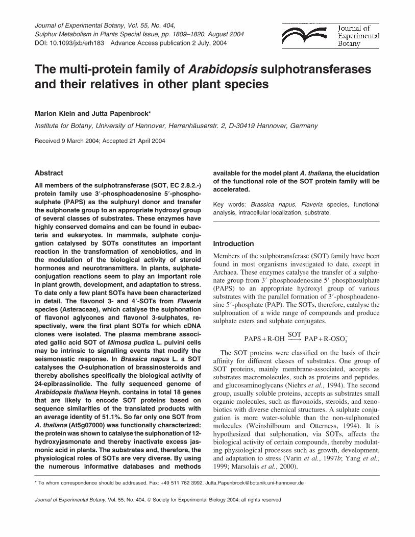
The multi-protein family of Arabidopsis sulphotransferasesand their relatives in other plant species
Marion Klein and Jutta Papenbrock*
Institute for Botany, University of Hannover, Herrenhauserstr. 2, D-30419 Hannover, Germany
Received 9 March 2004; Accepted 21 April 2004
Abstract
All members of the sulphotransferase (SOT, EC 2.8.2.-)
protein family use 39-phosphoadenosine 59-phospho-
sulphate (PAPS) as the sulphuryl donor and transfer
the sulphonate group to an appropriate hydroxyl group
of several classes of substrates. These enzymes have
highly conserved domains and can be found in eubac-
teria and eukaryotes. In mammals, sulphate conju-
gation catalysed by SOTs constitutes an important
reaction in the transformation of xenobiotics, and in
the modulation of the biological activity of steroid
hormones and neurotransmitters. In plants, sulphate-
conjugation reactions seem to play an important role
in plant growth, development, and adaptation to stress.
To date only a few plant SOTs have been characterized
in detail. The flavonol 3- and 49-SOTs from Flaveria
species (Asteraceae), which catalyse the sulphonation
of flavonol aglycones and flavonol 3-sulphates, re-
spectively, were the first plant SOTs for which cDNA
clones were isolated. The plasma membrane associ-
ated gallic acid SOT of Mimosa pudica L. pulvini cells
may be intrinsic to signalling events that modify the
seismonastic response. In Brassica napus L. a SOT
catalyses the O-sulphonation of brassinosteroids and
thereby abolishes specifically the biological activity of
24-epibrassinolide. The fully sequenced genome of
Arabidopsis thaliana Heynh. contains in total 18 genes
that are likely to encode SOT proteins based on
sequence similarities of the translated products with
an average identity of 51.1%. So far only one SOT from
A. thaliana (At5g07000) was functionally characterized:
the proteinwas shown to catalyse the sulphonationof 12-
hydroxyjasmonate and thereby inactivate excess jas-
monic acid in plants. The substrates and, therefore, the
physiological roles of SOTs are very diverse. By using
the numerous informative databases and methods
available for the model plant A. thaliana, the elucidation
of the functional role of the SOT protein family will be
accelerated.
Key words: Brassica napus, Flaveria species, functional
analysis, intracellular localization, substrate.
Introduction
Members of the sulphotransferase (SOT) family have beenfound in most organisms investigated to date, except inArchaea. These enzymes catalyse the transfer of a sulpho-nate group from 39-phosphoadenosine 59-phosphosulphate(PAPS) to an appropriate hydroxyl group of varioussubstrates with the parallel formation of 39-phosphoadeno-sine 59-phosphate (PAP). The SOTs, therefore, catalyse thesulphonation of a wide range of compounds and producesulphate esters and sulphate conjugates.
PAPS+R-OH ��!��!SOTPAP+R-OSO
�3
The SOT proteins were classified on the basis of theiraffinity for different classes of substrates. One group ofSOT proteins, mainly membrane-associated, accepts assubstrates macromolecules, such as proteins and peptides,and glucosaminoglycans (Niehrs et al., 1994). The secondgroup, usually soluble proteins, accepts as substrates smallorganic molecules, such as flavonoids, steroids, and xeno-biotics with diverse chemical structures. A sulphate conju-gation is more water-soluble than the non-sulphonatedmolecules (Weinshilboum and Otterness, 1994). It ishypothesized that sulphonation, via SOTs, affects thebiological activity of certain compounds, thereby modulat-ing physiological processes such as growth, development,and adaptation to stress (Varin et al., 1997b; Yang et al.,1999; Marsolais et al., 2000).
* To whom correspondence should be addressed. Fax: +49 511 762 3992. [email protected]
Journal of Experimental Botany, Vol. 55, No. 404,
Sulphur Metabolism in Plants Special Issue, pp. 1809–1820, August 2004
DOI: 10.1093/jxb/erh183 Advance Access publication 2 July, 2004
Journal of Experimental Botany, Vol. 55, No. 404, ª Society for Experimental Biology 2004; all rights reserved

In Arabidopsis thaliana Heyn. 18 SOT genes have beenidentified through alignment search tools (described in thisarticle), that may encode SOT proteins based on sequenceidentities, but little is known about their functions. The firstSOT gene identified in A. thaliana (RaR047, At2g03760)was only expressed in shoots and in A. thaliana cellcultures, and its expression was enhanced by challengingplants with isolates of Xanthomonas campestris pathovarcampestris 147 and of Pseudomonas syringae pathovarmaculicola M2 undergoing an incompatible interaction(Lacomme and Roby, 1996).Structural similarities are present among SOTs from
eubacteria, plants, and mammals (Yamazoe et al., 1994).SOTs comprise a very important and interesting group ofenzymes because they arewidely distributed and are involvedin a broad spectrum of physiological functions (Weinshil-boum and Otterness, 1994; Klaassen and Boles, 1997; Varinet al., 1997b; Hanai et al., 2000; Marsolais et al., 2000).
What are sulphotransferases not?
Sulphotransferases are different from sulphurtransferases.The names of both groups of enzymes, sulphotransferasesand sulphurtransferases, are very similar; both groups ofproteins were often mixed up in the past. To avoid con-fusion about the catalytic activity of members of eachprotein family, the difference will be defined.Sulphurtransferases/rhodaneses are a group of enzymes
widely distributed in all three phyla that catalyse the transferof reduced sulphur from a donor, such as thiosulphate or 3-mercaptopyruvate, to a thiophilic acceptor substrate (West-ley, 1973). In the reaction catalysed by sulphurtransferaseproteins the transferred sulphur is highly reduced (oxidationstate –II), whereas the SOT proteins catalyse the transferof sulphur that is highly oxidized (oxidation state VI). Thesequences of both protein families are not related. InA. thaliana the family of sulphurtransferases also consistsof 18 members (Bauer and Papenbrock, 2002).In the past the enzyme in the sulphur-assimilation
pathway which reduces adenosine-59-phosphosulphate(APS) to sulphite was misleadingly called APS sulpho-transferase. Only recently, after the reaction mechanism hadbeen elucidated in detail, was the protein renamed APSreductase (Suter et al., 2000). In the older literature theenzymes catalysing the APS reduction are always referredto as sulphotransferase. However, they do not belong to theSOT protein family described in this paper and do notpossess any sequences similarities with SOT proteins.The nomenclature for protein families often has a histor-
ical background. To minimize confusion between differentprotein families, it is proposed to abbreviate the sulpho-transferases ‘SOT’ instead of the formerly used abbrevia-tion ‘ST’, which is now broadly used as abbreviation forsulphate transporters. The abbreviation for sulphurtrans-ferases has been changed from the formerly used ‘ST’
into ‘STR’ (The Arabidopsis information resource, http://arabidopsis.org/info/genefamily/STR_genefamily.html).
Biosynthesis of APS and PAPS
PAPS is an obligate co-substrate for sulphonation reactionscatalysed by SOTs. PAPS is synthesized from ATP andendogenous sulphate in a two-step reaction. In the first stepATP sulphurylase (EC 2.7.7.4) catalyses sulphate activa-tion. The enzyme hydrolyses the bond between the b- andthe c-phosphates of ATP and then adds sulphate to the c-phosphate. The activation step is necessary because sul-phate is metabolically inert. The energy is stored in thephosphoric acid-sulphuric anhydride bond of the reactionproduct, 59-adenylylsulphate (APS), allowing sulphate toundergo further reactions.
ATP +SO2�4 ������������! ������������
ATP sulphurylaseAPS+PPi
The energetics of the sulphate adenylylation reactionfavours ATP formation. Therefore, the reaction products,APS and pyrophosphate (PPi), must be maintained at a lowconcentration by the enzymes, inorganic pyrophosphatasethat hydrolyses PPi, and APS reductase (EC 1.8.4.9), andAPS kinase (EC 2.7.1.25) that metabolize APS. APSreductase catalyses the first step of sulphate reduction.APS kinase catalyses the ATP-dependent phosphorylationon the 39-position of APS. The product PAPS is thesubstrate for the SOT proteins.
ATP+APS �������! �������
APS kinasePAPS+ADP
In general the availability of PAPS for sulphonation invivo depends on its synthesis, transport, degradation, andutilization. As the SOT proteins from mammals have beeninvestigated in more detail, the situation in mammals will beoutlined initially. In mammals, PAPS synthesis dependsdirectly on sulphate availability which is the limiting factorin the first step. On the other hand, steady-state PAPSconcentration cannot be increased by increasing endogenoussulphate concentration. The second part of PAPS biosyn-thesis depends on both PAPS and APS. PAPS inhibits itsown synthesis through negative product feedback. PAPSutilization or transport out of cytoplasm promotes PAPSsynthesis (Klaassen and Boles, 1997). In vitro tests haveshown that excess APS (substrate) also inhibits APS kinase.PAPS is formed in the cytoplasm. Sulphonation of macro-molecules takes place in the lumen of the Golgi apparatus(Capasso andHirschberg, 1984;Mandon et al., 1994). Theirsulphonation depends on PAPS synthesis and transport. APAPS transporter was purified fromGolgi vesicles (Mandonet al., 1994); the transporter is inhibited by 39,59-adenosinediphosphate (Zaruba et al., 1988).
In mammals, PAPS is degraded by two differentpathways leading to the same end product, 59-adenosine
1810 Klein and Papenbrock
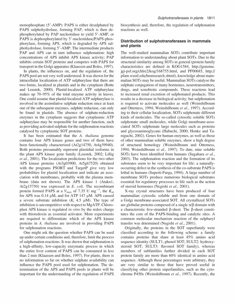
monophosphate (59-AMP): PAPS is either desulphated byPAPS sulphohydrolase, forming PAP, which is then de-phosphorylated by PAP nucleotidase to yield 59-AMP, orPAPS is dephosphorylated by 39-nucleotidase/PAPS-phos-phorylase, forming APS, which is degraded by APS sul-phohydrolase, forming 59-AMP. The intermediate productsPAP and APS can in turn influence sulphonation: highconcentrations of APS inhibit APS kinase activity; PAPinhibits certain SOT proteins and competes with PAPS fortransport in the Golgi apparatus (Klaassen and Boles, 1997).
In plants, the influences on, and the regulation of, thePAPS pool are not very well understood. It was shown for theintracellular localization of ATP sulphurylase that there aretwo forms, localized in plastids and in the cytoplasm (Rotteand Leustek, 2000). Plastid-localized ATP sulphurylasemakes up 70–95% of the total enzyme activity in leaves.One could assume that plastid-localized ATP sulphurylase isinvolved in the assimilative sulphate reduction since at leastone of the subsequent enzymes, sulphite reductase, can onlybe found in plastids. The absence of sulphate-reductionenzymes in the cytoplasm suggests that cytoplasmic ATPsulphurylase may be responsible for another function, suchas providing activated sulphate for the sulphonation reactionscatalysed by cytoplasmic SOT proteins.
It has been estimated that the A. thaliana genomecontains four APS kinase genes and two of them havebeen functionally characterized (At2g14750, At4g39940).Both proteins presumably represent plastidial isoforms inthe plant APS kinase gene family (Leustek, 2002; Lilliget al., 2001). The localization predictions for the two otherAPS kinase proteins (At3g03900, At5g67520) obtainedwith the programs PSORT and TargetP give very lowprobabilities for plastid localization and indicate an asso-ciation with membranes, probably with the plasma mem-brane (data not shown). The APS kinase 1 (Atakn1,At2g15750) was expressed in E. coli. The recombinantprotein formed PAPS at a Vmax of 7.35 U mg�1, the Km
for APS was 0.14 lM, and for ATP 147 lM. APS causeda severe substrate inhibition (Ki 4.5 lM). The type ofinhibition is uncompetitive with respect to MgATP. Chloro-plast APS kinase is regulated in vitro by the redox chargewith thioredoxin as essential activator. More experimentsare required to differentiate which of the APS kinaseproteins in A. thaliana are involved in providing PAPSfor sulphonation reactions.
One might ask the question whether PAPS can be usedup under certain conditions and, therefore, limit the processof sulphonation reactions. It was shown that sulphonation isa high-affinity, low-capacity enzymatic process in whichthe entire liver content of PAPS can be consumed in lessthan 2 min (Klaassen and Boles, 1997). For plants, there isno information so far on whether sulphate availability caninfluence the PAPS pool used for sulphonation. The de-termination of the APS and PAPS pools in plants will beimportant for the understanding of the regulation of PAPS
biosynthesis and, therefore, the regulation of sulphonationreactions as well.
Distribution of sulphotransferases in mammalsand plants
The well-studied mammalian SOTs contribute importantinformation to understanding about plant SOTs. Due to thestructural similarity among SOTs in general (protein familycharacteristics are defined in KOG1584, http://genome.jgi-psf.org/chlre2/kog/168755.html, and PF00685, http://pfam.wustl.edu/hmmsearch.shtml), knowledge about mam-malian SOTs may be useful. Mammalian SOTs catalyse thesulphate conjugation of many hormones, neurotransmitters,drugs, and xenobiotic compounds. These reactions leadto increased renal excretion of sulphonated products. Thisresults in a decrease in biological activity; but sulphonationis required to activate molecules as well (Weinshilboumand Otterness, 1994; Weinshilboum et al., 1997). Accord-ing to their cellular localization, SOTs sulphonate differentkinds of molecules. The so-called cytosolic soluble SOTssulphonate small molecules, while Golgi membrane-asso-ciated SOTs sulphonate large molecules such as proteinsand glycosaminoglycans (Habuchi, 2000; Honke and Ta-niguchi, 2002). Genes for human enzymes, as well as thoseof other mammalian soluble enzymes, show a high degreeof structural homology (Weinshilboum and Otterness,1994; Weinshilboum et al., 1997). To date, nine solubleSOTs have been identified from humans (Sugahara et al.,2003). The sulphonation reaction and the formation of itssubstrates seem to be very important for life: a naturally-occurring defect in the synthesis of the co-substrate PAPS islethal in humans (Superti-Furga, 1994). A large number ofmembrane SOTs produce numerous biological substratesessential for regulatory processes in life such as the actionof steroid hormones (Negishi et al., 2001).
X-ray crystal structures have been produced of fourmammalian cytosolic soluble and from one domain ofa Golgi membrane-associated SOT. All crystallized SOTsare globular proteins composed of a single a/b domain witha characteristic five-stranded b-sheet. The b-sheet consti-tutes the core of the PAPS-binding and catalytic sites. Acommon molecular mechanism reaction of the sulphuryltransfer was determined (Negishi et al., 2001).
Originally, the proteins in the SOT superfamily wereclassified according to the following scheme: a familycontains proteins that share at least 45% amino acidsequence identity (SULT1, phenol SOT; SULT2: hydroxy-steroid SOT; SULT3: flavonol SOT family), whereasmembers of subfamilies further divided in each SOTprotein family are more than 60% identical in amino acidsequence. Although these percentages were arbitrary, theyare very similar to those that have proved useful inclassifying other protein superfamilies, such as the cyto-chrome P450s (Weinshilboum et al., 1997). Recently, the
Sulphotransferases in plants 1811

human SOT families were further subdivided in thefollowing way: the SULT1 phenol SOT family containsfour subfamilies, the phenol SOT (SULT1A, EC 2.8.2.-),the Dopa/tyrosine SOT (SULT1B, EC 2.8.2.9), the hydrox-yarylamine SOT (SULT1C, EC 2.8.2.3), and the estrogenSOT (SULT1E, EC 2.8.2.15). The SULT2 hydroxysteroidSOT family consists of two subfamilies, the dehydroepian-drosterone SOT (SULT2A) and the cholesterol SOT(SULT2B) (Sugahara et al., 2003; Yoshinari et al.,2001). However, based on the above-mentioned criteriaa number of other eukaryotic SOT proteins, whose sub-strate specificity was elucidated, could not be classified inthe existing scheme. Either the proteins showed lesssequence identity, although they possessed the same sub-strate specificity as other members of the subfamily, or theyused a substrate chemically very different from the sub-strate typical for a particular subfamily, but showed highsequence identities (Sugahara et al., 2003). Thus, theclassification scheme has to be adapted to the latest resultswith respect to substrate specificity.Little is known about plant SOTs compared with mam-
malian SOTs. SOTs of Flaveria species and Brassica napusL. are well characterized bymeans of molecular biology andbiochemistry. Thus, the SOTs from Flaveria species rep-resent a general model for plant SOTs. The flavonol 3- and49-SOTs from Flaveria chloraefolia were the first plantSOTs for which cDNA clones were isolated and character-ized (Varin et al., 1992). Additional SOTs from Flaveriabidentis (L.) Kuntze have been characterized. This group ofSOTs accept different flavonols as sulphate acceptors(Varin et al., 1997b). These enzymes exhibit strict speci-ficity for the substrate and the position of the hydroxylgroup to be sulphonated. The biological function of flavo-nols and their derivatives are not fully understood. Flavo-nols might be involved in adaptation to stress, for example,microbial attack (Hahlbrock and Scheel, 1989). Flavonolsmay also act as a regulator of polar auxin transport(Faulkner and Rubery, 1992). The common characteristicsof flavonol SOTs are as follows: they all do not use divalentcations for sulphonation, they have a similar mass of 35kDa, and the active form of these enzymes is a monomer.Km values range between 0.2 lM and 0.4 lM for PAPS andvarious flavonols (Varin et al., 1997b).A very different substrate type for SOT proteins was
described in halophytic plants. Choline-O-sulphate isa compatible osmolyte accumulated under saline conditionsby members of the halophytic genus Limonium and otherPlumbaginaceae. A choline SOT (EC 2.8.2.6) responsiblefor the formation of choline-O-sulphate was characterizedin Limonium species. The choline SOT activity wascatalysed by a soluble protein and required PAPS as thesulphate donor. Apparent Km values were 25 lM forcholine and 5.5 lM for PAPS. In roots and leaves ofLimonium perezii (Stapf) F.T. Hubb., the activity wasincreased at least 4-fold by salinization with 40% (v/v)
artificial sea water. Here the sulphonated choline has a rolein tolerance against salt stress as a beneficial osmoprotec-tant. Among the non-accumulators such as barley, maize,sunflower, and Brassica species, none had significantcholine SOT activity (Rivoal and Hanson, 1994). The typeand the sequence of the SOT protein catalysing this reactionhas not been identified so far.
In Mimosa pudica L. a SOT activity was characterizedfrom plasma membrane preparations. The SOT proteincatalysed the transfer of sulphate from PAPS to gallic acidglucoside; the reaction product was identical with gallic acid,b-D-gluco-pyranosyl-69-sulphate, the periodic leafmovementfactor. Therefore the 42 kDa SOT protein analysed in M.pudicamight be involved in the induction of the seismonasticresponse movement (Varin et al., 1997a).
Evidence exists that plants, like mammals, use steroids toregulate their growth and development. In B. napus a SOTprotein was characterized that catalysed the sulphonation ofbrassinosteroids and mammalian estrogenic steroids. Thesulphonation abolishes specifically the biological activityof 24-epibrassinolide. Treatment with salicylic acid, a signalmolecule in plant defence, leads to increased expression ofthe B. napus SOT gene. This suggests an involvement of atleast one SOT protein in plant responses to pathogeninfection (Rouleau et al., 1999).
The first SOT encoding cDNA clone from A. thaliana,RaR047 (At2g03760), was isolated by Lacomme and Roby(1996). However, the gene product was not functionallyanalysed. The first A. thaliana SOT protein (At5g07000)was functionally analysed only recently. A different groupof chemical compounds was shown to be sulphonated bythis SOT protein. In vitro the recombinant A. thaliana SOTprotein exhibited strong substrate specificity for 11- and 12-hydroxyjasmonate. The Km value for PAPS was found to be1 lM. In vivo the naturally occurring 12-hydroxyjasmonatewas sulphonated in A. thaliana (Gidda et al., 2003).Initially, 12-hydroxyjasmonate was isolated as a tuber-inducing compound from Solanum tuberosum (Yoshiharaet al., 1989). Hydroxylation and subsequent sulphonationmight be components of a pathway that controls thebiological activity of 12-hydroxyjasmonate or inactivatesexcess jasmonic acid in plants (Gidda et al., 2003).
This short summary demonstrates the diversity of sub-strates used by SOT proteins in addition to the identical co-substrate PAPS, and the broad spectrum of physiologicalprocesses where sulphonated compounds are involved. Thedifferent Km values for PAPS (from 0.1–5 lM) provide theplant with a regulatory system for the use of the PAPS poolfor different sulphonation reactions.
The multi-protein family of A. thalianasulphotransferases
In recent years the scientific community was provided withvaluable information about the model plant A. thaliana
1812 Klein and Papenbrock

(The Arabidopsis genome initiative, 2000). Extensive useof all sources available will help to analyse and differentiatebetween the members of protein families. Therefore, theaim of this study was to identify all genes and gene productswhich might be classified as SOT in A. thaliana. The fullysequenced genome of A. thaliana was searched for SOTsequences applying the BLAST program with the alreadyisolated SOT RaR047 protein sequence from A. thaliana(Lacomme and Roby, 1996). 18 SOT protein sequencesshowing high similarity to already known SOT proteinsequences and to each other were identified. The phylo-genetic tree of the family of A. thaliana SOTs showsthe relationships among these 18 sequences (Fig. 1). Theprotein sequences were divided into seven groups accord-ing to their sequence similarities; the results are displayed
in Table 1. The table shows an overview of the completeSOT family with additional information including geneidentification, numbers of amino acids, number of ESTsidentified, and intracellular localization predictions (asexplained later). Apart from two proteins, all members ofthis family consist of an approximately equal number ofamino acids of at least 310 residues. Computer analysisof A. thaliana amino acid sequences indicates that all 18SOTs might be soluble and none of the SOT proteinscontains a transmembrane region as indicated by hydrop-athy plots in SOSUI (http://sosui.proteome.bio.tuat.ac.jp).
In human and mouse SOT sequences, a dimerizationmotif near the C-terminus was identified, designated as theKTVE motif (Negishi et al., 2001). In mammals, cytosolicsoluble SOTs are predominantly dimers, both homo- andheterodimers (Weinshilboum et al., 1997). In plants, theenzymes characterized so far exist as catalytically activemonomers (Varin and Ibrahim, 1989). A. thaliana SOTprotein sequences do not contain a KTVE motif; thereforethey also might occur as monomers.
Interestingly, nearly all of theA. thalianaSOTgenes donotcontain introns (exceptions: AtSOT3, 4, and 10), in contrasttomammalian SOT genes. Genes for human SOTs, as well asfor other mammalian SOTs, show a high degree of struc-tural homology with conservation of the locations of mostintron/exon splice junctions (Weinshilboum et al., 1997).
Remarkably, the numbers of EST clones available arevery low. For the A. thaliana genes in group IV, VI, andVII, the number of EST clones identified are in the samerange as in other gene families (Bauer and Papenbrock,2002) whereas, in the other groups, the numbers arerelatively low. For seven out of the 18 putative SOT genesno EST clone has been identified so far. There are severalhypothetical explanations: several of the SOT genes mightbe pseudogenes which are not expressed; the abundanceof the SOT mRNA molecules is very low, or the mRNAsare not very stable; the genes are only expressed in veryspecific conditions not included in the EST projects done sofar. Fortunately, the coding sequence for most of theAtSOTs could be amplified from genomic DNA becausethe genes do not contain introns. It will be a challenge tofind conditions for the expression of these seven SOT genes.
Sequence/function analysis of A. thaliana SOTproteins
A comparison of amino acid sequences of A. thaliana SOTswas done with other plant SOTs with known substratespecificities (Fig. 2). The aim of this comparison was to getindications about substrate specificities of A. thalianaSOTs. RaRO47 (At2g03760) was the first cDNA cloneisolated encoding a SOT from A. thaliana and has not yetbeen characterized biochemically. The clone shows a highsimilarity of 87% with a SOT of B. napus (steroidST-3).Another SOT in the same group (At2g03770, group V) also
Fig. 1. Phylodendrogram of Arabidopsis thaliana sulphotransferases(SOTs). The fully sequenced genome of A. thaliana was searched forSOT sequences applying the BLAST program with the already isolatedSOT RaR047 protein sequence from A. thaliana (At2g03760) (http://www.ncbi.nlm.nih.gov/BLAST/). 18 SOT sequences were identifiedin A. thaliana. The respective proteins were grouped according to theiramino acid sequence similarities using the Clustal W program (http://www.ebi.ac.uk/clustalw). The phylogenetic tree shows the relationshipamong the 18 SOT proteins.
Sulphotransferases in plants 1813

shows a good correspondence of 57% with steroidST-3. B.napus SOTs catalyse the O-sulphonation of brassinoste-roids and mammalian estrogenic steroids (Rouleau et al.,1999). Because of the mentioned close similarity, RaR047should be tested for catalysis of these substrates as well.AtSOT14 in group VI has been characterized biochemi-
cally. The recombinant AtSOT14 protein sulphonated 11-and 12-hydroxyjasmonate, whereas for the closely relatedAtSOT15 protein (87% identity with AtSOT14) no activitywas observed with these substrates (Gidda et al., 2003).Both SOTs in group VI show a smaller correspondence ofonly 42–43% compared with the flavonol SOT of F.chloraefolia (F4-ST) and B. napus SOTs (steroidST1-3,41–43%). Therefore, other substrates related in structure tojasmonates have to be tested to determine the biochemicalfunction of AtSOT15.The comparison of sequence similarities to proteins with
known substrate specificities was also applied for group I toIV and VII, however, the differences are probably notsignificant. Group VII shows a better correspondence with
the flavonol SOT of F. chloraefolia (F4-ST, 46–48%) thanthe comparison with B. napus SOTs (steroidST1-3, about41%). Thus, the substrate for this group of A. thalianaSOTs may be a flavonol or a similar substrate. If sucha substrate could be identified, the SOT group VII wouldrepresent an A. thaliana flavonol protein family.
Group II shows a slightly better correspondence with theB. napus SOTs (steroidST1-3, 43–49%) than with the F.chloraefolia SOT (F4-ST, 39–43%). Despite these results,At3g45070 has been shown to accept a number of flavonolsand flavone aglycones as well as their sulphonated derivates(Marsolais et al., 2000). The remaining groups I, III, and IVshow an average similarity of about 44% with B. napusSOTs (steroidST1-3). This value is only slightly higherthan the respective F. chloraefolia SOT (F4-ST) value (40–41%). In summary, the sequence/function analysis forthe SOT protein family did not reveal clear results. Therelatively weak similarities and the small differences amongthe groups make a biochemical analysis to identify thenatural substrate for each SOT protein essential.
Table 1. Features of the SOT family from A. thaliana and localization prediction for the SOT proteins
Protein name, gene identification, number of amino acids, and EST clones for 18 SOTs are summarized. The program PSORT was used for thelocalization prediction (http://psort.ims.u-tokyo.ac.jp). Numbers in brackets give the certainty of prediction. The number of EST clones was determinedon 14 February 2004 (http://mips.gsf.de, http://arabidopsis.org). Abbreviations: aa, amino acids; ER, endoplasmic reticulum; ID, identification; No.,number.
Group Name Gene ID No. aa EST Localization prediction
I AtSOT1 At5g43690 331 2 Microbody (peroxisome) (0.748)Chloroplast stroma (0.200)
AtSOT2 At3g51210 67 - ER (membrane) (0.550)Microbody (peroxisome) (0.320)
II AtSOT3 At4g26280 314 - Cytoplasm (0.450)Microbody (peroxisome) (0.313)
AtSOT4 At2g27570 273 - Nucleus (0.980)Microbody (peroxisome) (0.429)
AtSOT5 At3g45070 323 4 Microbody (peroxisome) (0.575)Cytoplasm (0.450)
AtSOT6 At3g45080 329 - Microbody (peroxisome) (0.522)Cytoplasm (0.450)
III AtSOT7 At1g28170 326 - Microbody (peroxisome) (0.572)Mitochondrial matrix (0.100)
AtSOT8 At1g13420 331 2 Cytoplasm (0.650)Mitochondrial matrix (0.100)
AtSOT9 At1g13430 351 2 Cytoplasm (0.450)Microbody (peroxisome) (0.405)
AtSOT10 At2g14920 333 - Cytoplasm (0.450)Microbody (peroxisome) (0.392)
IV AtSOT11 At2g03750 351 9 ER (membrane) (0.550)Microbody (peroxisome) (0.291)
V AtSOT12 At2g03760 326 5 Microbody (peroxisome) (0.622)(RaR047) Mitochondrial matrix (0.100)
AtSOT13 At2g03770 324 - Microbody (peroxisome) (0.705)Mitochondrial matrix (0.100)
VI AtSOT14 At5g07000 347 6 Microbody (peroxisome) (0.602)Chloroplast stroma (0.200)
AtSOT15 At5g07010 359 15 Microbody (peroxisome) (0.640)Mitochondrial matrix (0.484)
VII AtSOT16 At1g74100 338 25 Microbody (peroxisome) (0.640)Cytoplasm (0.450)
AtSOT17 At1g18590 346 8 Microbody (peroxisome) (0.640)Chloroplast stroma (0.566)
AtSOT18 At1g74090 350 6 Microbody (peroxisome) (0.640)Cytoplasm (0.450)
1814 Klein and Papenbrock

The rules for subdivision into families and subfamiliesaccording to the percentage of their sequence identity(Weinshilboum et al., 1997) are not very useful for theplant SOT families. The detailed comparison above showsthat high sequence similarity alone does not necessarilyindicate specificity for the same chemical group of sub-strates. Even from high sequence identity of more than 85%among two SOT proteins, one cannot conclude the samesubstrate specificity. Probably SOT proteins specific fora group of substrates evolved independently on more thanone occasion. Thus, for each SOT protein the in vitro andfinally the in vivo substrate specificity has to be detected.The crystal structures of a number of mammalian SOTproteins are available which might be used for three-dimensional modelling of the active site and putativesubstrates from ligand libraries.
Alignment of the highly conserved regions
Cytosolic soluble SOTs from mammalian species and plantSOT proteins have high structural similarities. All SOTshave conserved amino acid motives which are involved inPAPS binding (regions I and IV) (Marsolais and Varin,1995; Weinshilboum et al., 1997). Figure 3 shows a partialamino acid alignment of the putative PAPS-binding regionsof the A. thaliana SOT protein family. Region I is localizednear the N-terminus and region IV at the C-terminus. Thisalignment indicates that the typical binding site for PAPSexists in all 18 SOTs. However, a comparison of theconsensus sequences for SOT proteins from a broad spec-
trum of species described previously (region I,TYPKSGTxW; region IV, RKGxxGDWKxxFT) (Wein-shilboum et al., 1997) are different from the consensussequences in the PAPS-binding regions of A. thaliana SOTproteins (region I, PKxGTTWLKALTFA; region IV,FRKGxVGDWxxxLT). In at least 14 A. thaliana SOTs,the amino acids of these consensus sequences are identical;there are not more than three different amino acids at oneposition among all 18 SOT proteins. The amino acids in theA. thaliana consensus sequence printed in bold are identicalwith the overall consensus sequence. In the first publishedA. thaliana RaR047 sequence (Lacomme and Roby, 1996),there are a number of sequence deviations compared withthe sequence published later (The Arabidopsis genomeinitiative, 2000), including in the consensus sequence ofregion IV.
Intracellular localization of A. thaliana SOT
In mammalian species one group of SOT proteins isassociated with membranes and accepts as substratesmacromolecules such as proteins and glycosylaminogly-cans, and a second group of SOT proteins is soluble andaccepts small organic molecules as substrates (Habuchi,2000; Niehrs et al., 1994; Sugahara et al., 2003). In plants,membrane-associated SOT proteins might be involved inthe biosynthesis of phytosulphokines (Hanai et al., 2000).A SOT protein characterized in more detail was shown tobe localized in the plasma membrane and to be involved inthe seismonastic response in M. pudica. The size of this
Fig. 2. Comparison of amino acid sequences of Arabidopsis thaliana SOTs with well-known plant SOTs from Flaveria chloraefolia and Brassicanapus (Jotun Hein method in MegAlign/DNASTAR, Madison, WI, USA). Gene identifications, with subdivision into groups and abbreviations ofknown plant SOTs, are listed in the right columns. F49-ST represents flavonol-49-SOT from F. chloraefolia (Accession no. M84135), steroidST1-3represent the steroid SOT 1–3 from B. napus (Accession no. AF000305–307). Shadowed areas are as mentioned in the text. The values indicate theidentity in percent at the amino acid level.
Sulphotransferases in plants 1815

membrane SOT protein was 42 kDa, whereas most solubleplant SOT proteins have only a molecular mass of about 35kDa. The difference might reflect the addition of a trans-membrane domain (Varin et al., 1997a).For the localization prediction of nuclear-encoded
proteins in the cell, several programs have been devel-oped. PSORT, TargetP, and further programs in http://www.expasy.ch/tools were applied. The prediction pro-grams use different algorithms. PSORT is based on anexpert system with a knowledge-base and is a collection of‘if-then’-type rules (Nakai and Kanehisa, 1992). TargetP isa neural network-based tool using N-terminal sequenceinformation only. It discriminates between proteins destinedfor the mitochondrion, the chloroplast, the secretory path-way, and ‘other’ localizations with a calculated success rateof 85% (plant) and 90% (non-plant) in redundancy-reducedtest sets (Emanuelsson et al., 2000). The results are sum-marized in Table 1. None of the A. thaliana SOT proteins
contains an N-terminal transit peptide or a mitochondrialpre-sequence in TargetP, apart from AtSOT11 (At2g03750)which possess a 17 amino acid extension indicating a labelfor the secretory pathway (probability 0.794). The programspecific for the prediction of peroxisomal proteins (http://mendel.imp.univie.ac.at/mendeljsp/sat/pts1/PTS1predictor.jsp) did not recognize and classify any of the A. thalianaSOT proteins as peroxisomal, although the PSORT programsuggested transport into peroxisomes with probabilitieshigher than 0.5 for AtSOT1, AtSOT5 to 7, and AtSOT12to 18. In summary, the computer predictions for the in-tracellular localization of the A. thaliana SOT proteins donot reveal clear and reliable results. Therefore, the intra-cellular localization has to be investigated experimentally.One method to demonstrate the intracellular localizationof nuclear-encoded proteins is the transient expression offusion constructs with the green fluorescent protein (GFP)(Bauer et al., 2004; Nowak et al., 2004).
Fig. 3. Partial amino acid alignment of Arabidopsis thaliana SOTs. The 18 protein sequences of the identified SOTs were aligned (Jotun Hein method inMegAlign/DNASTAR, Madison, WI, USA). The consensus sequences are listed at the top (regions I and IV). ‘x’ represents any amino acid. Numbers onthe right refer to amino acid position within the protein. Majority values are shadowed, including residues critical for 39-phosphoadenosine 59-phosphosulphate (PAPS) binding. (A) Shows the highly conserved region I, and (B) the highly conserved region IV. Both conserved regions, critical forPAPS binding, are present among all 18 SOT proteins.
1816 Klein and Papenbrock

One example of this approach is shown in Fig. 4.According to the intracellular localization prediction, At-SOT18 is either localized in the peroxisomes (0.640) or inthe cytoplasm (0.450). The full-length cDNA sequenceencoding AtSOT18 was cloned in a frame downstream(pGFP-C) or upstream (pGFP-N) of the GFP reading frame.A. thaliana protoplasts were transiently transformed withthe GFP constructs according to standard procedures.Bright-field images are shown to visualize the protoplast’scell borders and the chloroplasts. Fluorescent images of thesame protoplasts were done using a fluorescence micro-scope. As a control, the protoplasts were transformed withthe pGFP-C and pGFP-N vector without additional in-sertion. According to the results, the AtSOT18 protein is
cytoplasmic. These studies will be extended to obtaina complete set of data of the intracellular localization ofall A. thaliana SOT proteins.
Expression studies
So far the results concerning gene expression and proteinsteady-state levels of A. thaliana SOTs are very limited.Only for RaR047 (AtSOT12, At2g03760) was mRNAaccumulation determined in different conditions. ThemRNA coding for RaR047/AtSOT12 was expressed duringactive growth of A. thaliana cell cultures and in the aerialparts of young seedlings, but not in roots. Treatment ofA. thaliana seedlings with hormonal or stress-related com-pounds showed that mRNA for RaR047/AtSOT12 wasinduced in response to salicylic acid and methyl jasmonate.Infection with avirulent bacterial pathogens causing anhypersensitive response increased the RaR047/AtSOT12mRNA levels (Lacomme and Roby, 1996). The expressionof AtSOT14 mRNA (At5g07000) was induced followingtreatment of A. thaliana rosette leaves with methyljas-monate and 12-hydroxyjasmonate, the substrate for theAtSOT14 protein. However, the expression of the geneencoding the thionin Thi2.1 protein, specifically inducedby wounding, pathogen infection, and methyljasmonatetreatment, was not induced by 12-hydroxyjasmonate, in-dicating two independent response pathways mediated bymethyljasmonate and 12-hydroxyjasmonate (Gidda et al.,2003). So far the experiments described above are the onlyresults on expression levels of SOTs in A. thaliana.
There are numerous results of microarray analyses avail-able in the databases performed under a broad varietyof conditions. The authors’ own microarray data of anexperiment with A. thaliana seedlings treated with meth-yljasmonate for several hours indicated differential expres-sion of the A. thaliana SOT genes included on the chip.Some genes were up-regulated, others were down-regulatedwith expression maxima at different time points (R Jostand J Papenbrock, unpublished results). A detailed searchof microarray analyses using the 18 A. thaliana SOT genesmight help to characterize the expression pattern and toform groups of similarly expressed SOT genes.
How to identify the respective substrates andtheir function?
It is a challenging task to analyse the respective substrates foreach SOT. The study of the structure/function relationship ofSOT proteins in order to elucidate the mechanism of sulph-onate transfer, and to define the amino acids responsible forsubstrate binding and catalysis will help to identify putativesubstrates. Using site-directed mutagenesis of the flavonol3-SOT, several amino acids required for catalysis and co-substrate binding were mapped, while the construction of
Fig. 4. Intracellular localization of AtSOT18. The full length cDNAencoding AtSOT18 (At1g74090) was amplified from a phage librarycontaining genomic DNA. The SOT gene for AtSOT18 does contain anintron. The cDNA sequence was cloned in frame downstream (pGFP-C)or upstream (pGFP-N) of the green fluorescent protein (GFP) readingframe. The gene-cassettes were driven by the CaMV-35S promoter witha double enhancer and the polyA-tail from CaMV-35S. A. thalianaprotoplasts were transiently transformed with the GFP constructs asdescribed in Bauer and co-workers (2004). Bright field images, shown in(A, C, E, G), were made to visualize the protoplast’s cell borders and thechloroplasts. Fluorescence images of the protoplasts, shown in (B, D, F,H), were taken using an Axioskop microscope with filter sets optimal forGFP fluorescence (BP 450–490/LP 520). As a control, the protoplasts,shown in (A, B, E, F), were transformed with the pGFP-C and the pGFP-N vector, respectively, without an insert. All scale bars represent 10 lm.
Sulphotransferases in plants 1817

chimeric proteins allowed definition of the domain responsi-ble for substrate specificity (Varin et al., 1995).A domainwasfound which is involved in substrate binding, designated asdomain II. Within this domain, two subdomains of highdivergence were identified, probably participating in therecognition and binding of different acceptor substrates(Marsolais and Varin, 1997, 1998; Varin et al., 1995). Thesedomains of high divergence could be used for modellingligands which might serve as substrates using dockingprograms.To characterize the biochemical function of SOT proteins
one could think about all putative substrate candidates whichmight be found in plants in the desulpho- and in thesulphonated form, such as coumarins, desulphoglucosino-lates, flavonoids, gibberellic acids, hydroxyjasmonates,phenolic acids, steroids, sulphate esters, such as choline-O-sulphate, and terpenoids, and test them by using recombinantSOT proteins. This idea was followed by building up a sub-strate library comprising more than 100 desulpho-derivativesofmost of the knownplant-sulphonatedmetabolites aswell asa collection of metabolites for which no sulphonated metab-olites have been reported (Gidda et al., 2003).In the review written by Marsolais et al. (2000), the
putative substrates and functions of two more A. thalianaSOT proteins are mentioned as unpublished results of thesame group. The substrates already identified for other SOTproteins were tested using two A. thaliana SOT proteins.The purified recombinant AtSOT5 (At3g45070) was foundto exhibit strict specificity for position 7 of flavonoids. Thenatural occurrence of a SOT protein exhibiting highspecificity for flavonoids in A. thaliana was surprising,considering the absence of reports on the presence offlavonoid sulphates in this plant. It was hypothesized thatflavonoid sulphates may act as regulators of polar auxintransport (Faulkner and Rubery, 1992). In the same reviewAtSOT10 (At2g14920) was shown to exhibit strict speci-ficity for brassinosteroids having 22R-, 23R-hydroxyls, anda 24S-methyl or ethyl group on the steroid side chain. Dueto the high sequence identity to the B. napus SOTs,AtSOT12 was suggested to be involved in stereospecificinactivation of brassinosteroid by sulphonation.The rare expression of the A. thaliana SOT genes might
suggest that plants use the sulphonation reaction to modulatethe biological activity of hormones andmessengersmoleculesunder special conditions where only low amounts of pro-tein are necessary. The presence of the sulphate group mightsuggest a role in signalling processes, as shown for othersulphate metabolites such as the Nod factors in the interactionbetweenRhizobiummeliloti andMedicago sativaL. (Truchetet al., 1991). However, so far no sulphatase-like sequencescould be identified in plant genomes. In the enzyme databases(http://au.expasy.org/enzyme) there are already 29 differentSOT protein reactions described giving more ideas forsubstrates. Thus, there are a number of strategies to followfor the elucidation of the SOT’s physiological role.
Experimental evidence for an involvement ofa SOT in phytosulphokine biosynthesis
Phytosulphokine-a (PSK-a) is a sulphonated pentapeptide(Tyr(SO3H)-Ile-Tyr(SO3H)-Thr-Gln) and was shown to actas a plant growth factor (Matsubayashi and Sakagami,1996). The biosynthetic pathway of a preprophytosulpho-kine was partially elucidated in Oryza sativa L. and A.thaliana (Yang et al., 1999, 2001). The PSK-a is onlyactive in its sulphonated form, thus the identification ofa tyrosyl peptide SOT (EC 2.8.2.20) is of high importancefor the understanding of PSK-a and related peptides in theplant organism. Protein tyrosyl O-sulphation is one ofthe post-translational modifications that occurs with manysecretory and membrane proteins in animal cells. Inmammals, a tyrosyl protein SOT was localized specificallyin the trans-Golgi-network (Lee and Huttner, 1983). In cellcultures of a number of plant species the existence ofa tyrosyl protein SOT activity was demonstrated also in theGolgi-network (Hanai et al., 2000). However, the sequenceof the respective protein has not been determined so far(Youji Sakagami, Nagoya, Japan, personal communica-tion). The sequence of the mammalian tyrosyl protein SOTdoes not show any similarity to a DNA or protein sequencefrom a plant species. Therefore, it might be possible thatone of the 18 A. thaliana SOT acts as tyrosyl protein SOT.
Involvement of sulphotransferases inglucosinolate biosynthesis
Glucosinolates are found in vegetative and reproductivetissues of 16 plant families, but are most well known as themajor secondary metabolites in the Brassicaceae (Mithen,2001; Mikkelsen et al., 2002). In A. thaliana more than 20different glucosinolates have been identified (Mithen, 2001;Reichelt et al., 2002). Glucosinolates and their degradationproducts have a wide range of biological activities, forexample, in plant defence as deterrents against insect andfungi. Their biosynthetic pathways are partially identified(Mikkelsen et al., 2002; Reichelt et al., 2002). Interest-ingly, glucosinolates contain two forms of sulphur in dif-ferent oxidation states. The reduced form is a thioetherand is derived from cysteine (Mikkelsen et al., 2002;Reichelt et al., 2002), whereas the oxidized form is asulphate ester and is derived from the sulphonation path-way. The last step from the different aliphatic, aromatic,and indole desulphoglucosinolates to the active glucosino-lates might be catalysed by members of the SOT family.Glendening and Poulton (1990) partially purified a proteinfrom Lepidium sativum L. that had PAPS-dependentdesulphoglucosinolate SOT activity, however, no molecu-lar data are available to date. AtSOT16, AtSOT17, andAtSOT18 were suggested as being involved in sulphona-tion of desulphoglucosinolates, and might be used tomodulate the glucosinolate pattern of plants (patent WO
1818 Klein and Papenbrock

03010318-A, L. Varin and D. Spertini, 06/02/2003, Con-cordia University, Canada). However, at the moment theresults of the substrate specificity of the three SOT proteinsmentioned are not publicly available. The approach hasgreat potential for design of metabolically engineeredplants with improved pest resistance and increased nutri-tional value.
A. thaliana as a model plant: suited for theelucidation of all SOT functions?
In the almost fully sequenced genome of A. thaliana, 18SOT encoding genes have been identified while, in theclose relative B. napus, at least 12 genes were detected(Marsolais et al., 2000), and the genome of the mono-cotyledonous plant O. sativa, about 3.5-times larger insize than the A. thaliana genome, contains 13 SOT genes(M Klein and J Papenbrock, unpublished results). Thus, theSOT gene number is about 1.5-times higher in A. thaliana.However, similar genes might have evolved from geneduplications and might be functionally redundant or silent.Evaluation of already available microarray analyses willhelp to characterize expression patterns. Metabolic pro-filing of desulpho and sulphonated compounds in T-DNAinsertion mutants available for almost all A. thaliana SOTgenes and in mutants obtained with the RNAi techniquesin combination with exact observation of the physiologicalphenotype should be very successful for analysing the func-tion of the members of this protein family in A. thaliana.On the other hand, detection of the sulphonation reaction ofcholine in halophytes and of gallic acid glucoside involvedin the seismonastic response shows the limitations of themodel plant A. thaliana. Results of expression and meta-bolic profiling will be required for the analysis of thefunctional role of SOT proteins in non-A. thaliana species.
Acknowledgements
We would like to thank P von Trzebiatowski for her excellenttechnical assistance and M Bauer for performing most of thetransient expression experiments with GFP.
References
Bauer M, Dietrich C, Nowak K, Sierralta WD, Papenbrock J.2004. Intracellular localization of sulphurtransferases from Arabi-dopsis thaliana. Plant Physiology 135, (in press).
Bauer M, Papenbrock J. 2002. Identification and characterizationof single-domain thiosulphate sulphurtransferases from Arabidop-sis thaliana. FEBS Letters 532, 427–431.
Capasso JM, Hirschberg CB. 1984. Effect of atractylosides,palmitoyl coenzym A, and anion transport inhibitors on trans-location of nucleotide sugars and nucleotide sulphate into Golgivesicles. Journal of Biological Chemistry 259, 4263–4266.
Emanuelsson O, Nielsen H, Brunak S, von Heijne G. 2000.Predicting subcellular localization of proteins based on their N-
terminal amino acid sequence. Journal of Molecular Biology 300,1005–1016.
Faulkner IJ, Rubery PH. 1992. Flavonoids and flavonoid sulphatesas probes of auxin transport regulation in Cucurbita pepo hypo-cotyl segments and vesicles. Planta 186, 618–625.
Gidda SK, Miersch O, Levitin A, Schmidt J, Wasternak C, VarinL. 2003. Biochemical and molecular characterization of a hydroxy-jasmonate sulphotransferase from Arabidopsis thaliana. Journal ofBiological Chemistry 278, 17895–17900.
Glendening TM, Poulton JE. 1990. Partial purification and char-acterization of a 39-phosphoadenosine 59-phosphosulphate: desul-phoglucosinolate sulphotransferase from cress (Lepidium sativum).Plant Physiology 94, 811–818.
Habuchi O. 2000. Diversity and functions of glycosaminoglycansulphotransferases. Biochimica et Biophysica Acta 1474, 115–127.
Hahlbrock K, Scheel D. 1989. Physiology and molecular biologyof phenylpropanoid metabolism. Annual Review Plant Physiologyand Plant Molecular Biology 41, 339–367.
Hanai H, Nakayama D, Yang HP, Matsubayashi Y, Hirota Y,Sakagami Y. 2000. Existence of a plant tyrosylprotein sulpho-transferase: novel plant enzyme catalysing tyrosine O-sulphationof preprophytosulphokine variants in vitro. FEBS Letters 470,97–101.
Honke K, Taniguchi N. 2002. Sulphotransferases and sulphatedoligosaccarides. Medicinal Research Reviews 22, 637–654.
Klaassen CD, Boles JW. 1997. The importance of 39-phosphoade-nosine 59-phosphosulphate (PAPS) in the regulation of sulphation.FASEB Journal 11, 404–418.
Lacomme C, Roby D. 1996. Molecular cloning of a sulphotransfer-ase in Arabidopsis thaliana and regulation during developmentand in response to infection with pathogenic bacteria. PlantMolecular Biology 30, 995–1008.
Lee RW, Huttner WB. 1983. Tyrosine-O-sulphated proteins ofPC12 pheochromocytoma cells and their sulphation by a tyrosyl-protein sulphotransferase. Journal of Biological Chemistry 258,11326–11334.
Leustek T. 2002. Sulphate metabolism. In: Somerville CR, Meyer-owitz EM, eds. The Arabidopsis Book. Rockville, MD, USA:American Society of Plant Biologists, Vol. doi/10.1199/tab.0017,http://www.aspb.org/publications/arabidopsis/.
Lillig CH, Schiffmann S, Berndt C, Berken A, Tischka R,Schwenn JD. 2001. Molecular and catalytic properties of Arabi-dopsis thaliana adenylyl sulphate (APS)-kinase. Archives of Bio-chemistry and Biophysics 392, 303–310.
Mandon EC, Milla ME, Kempner E, Hirschberg CB. 1994.Purification of the Golgi adenosine 39-phosphate 59-phosphosul-phate transporter, a homodimer within the membrane. Proceedingsof the National Academy of Sciences, USA 91, 10707–10711.
Marsolais F, Gidda SK, Boyd J, Varin L. 2000. Plant solublesulphotransferases: Structural and functional similarity withmammalian enzymes. Recent Advances in Phytochemistry 34,433–456.
Marsolais F, Varin L. 1995. Identification of amino acid residuescritical for catalysis and cosubstrate binding in the flavonol3-sulphotransferase. Journal of Biological Chemistry 270,30458–30463.
Marsolais F, Varin L. 1997. Mutational analysis of domain II offlavonol 3-sulphotransferase. European Journal of Biochemistry247, 1056–1062.
Marsolais F, Varin L. 1998. Recent developments in the study ofthe structure-function relationship of flavonol sulphotransferases.Chemico-Biological Interactions 109, 117–122.
Matsubayashi Y, Sakagami Y. 1996. Phytosulphokine, sulphatedpeptides that induce the proliferation of single mesophyll cells of
Sulphotransferases in plants 1819

Asparagus officinalis L. Proceedings of the National Academy ofSciences, USA 93, 7623–7627.
Mikkelsen MD, Petersen BL, Olsen CE, Halkier BA. 2002.Biosynthesis and metabolic engineering of glucosinolates. AminoAcids 22, 279–295.
Mithen R. 2001. Glucosinolates – biochemistry, genetics and bio-logical activity. Plant Growth Regulation 34, 91–103.
Nakai K, Kanehisa M. 1992. A knowledge base for predictingprotein localization sites in eukaryotic cells. Genomics 14, 897–911.
Negishi M, Pedersen LG, Petrotchenko E, Shevtsov S, GorokhovA, Kakuta Y, Pedersen LC. 2001. Structure and function ofsulphotransferases. Archives of Biochemistry and Biophysics 390,149–157.
Niehrs C, Beisswanger R, Huttner WB. 1994. Protein tyrosinesulphation, 1993—an update. Chemico-Biological Interactions92, 257–271.
Nowak K, Luniak N, Meyer S, Mendel RR, Hansch R. 2004.Fluorescent proteins in poplar: a useful tool to study promotorfunction and protein localization. Plant Biology 6, 65–73.
Reichelt M, Brown PD, Schneider B, Oldham NJ, Stauber E,Tokuhisa J, Kliebenstein DJ, Mitchell-Olds T, Gershenzon J.2002. Benzoic acid glucosinolate esters and other glucosinolatesfrom Arabidopsis thaliana. Phytochemistry 59, 663–671.
Rivoal J, Hanson AD. 1994. Choline-O-Sulphate biosynthesis inplants (identification and partial characterization of a salinity-inducible choline sulphotransferase from species of Limonium(Plumbaginaceae). Plant Physiology 106, 1187–1193.
Rotte C, Leustek T. 2000. Differential subcellular localization andexpression of ATP sulphurylase and 59-adenylylsulphate reductaseduring ontogenesis of Arabidopsis leaves indicates that cytosolicand plastid forms of ATP sulphurylase may have specializedfunctions. Plant Physiology 124, 715–724.
RouleauM,Marsolais F, RichardM, Nicolle L, Voigt B, AdamG,Varin L. 1999. Inactivation of brassinosteroid biological activityby a salicylate-inducible steroid sulphotransferase from Brassicanapus. Journal of Biological Chemistry 274, 20925–20930.
Sugahara T, Liu CC, Pai G, Collodi P, Suiko M, Sakakibara Y,Nishiyama K, Liu MC. 2003. Sulphation of hydroxychlorobi-phenyl. Molecular cloning, expression, and functional character-ization of zebrafish SULT1 sulphotransferases. European Journalof Biochemistry 270, 2404–2411.
Superti-Furga A. 1994. A defect in the metabolic activation ofsulphate in a patient with achondrogenesis type IB. AmericanJournal of Human Genetics 55, 1137–1145.
Suter M, von Ballmoos P, Kopriva S, den Camp RO, Schaller J,Kuhlemeier C, Schurmann P, Brunold C. 2000. Adenosine 59-phosphosulphate sulphotransferase and adenosine 59-phosphosul-phate reductase are identical enzymes. Journal of BiologicalChemistry 275, 930–936.
The Arabidopsis Genome Initiative. 2000. Analysis of the genomesequence of the flowering plant Arabidopsis thaliana. Nature408, 796–815.
Truchet G, Roche P, Lerouge P, Vasse J, Camut S, DeBilly F,Prome JC, Denarie J. 1991. Sulphated lipo-oligosaccharidesignals of Rhizobium meliloti elicit root nodule organogenesis inalfalfa. Nature 351, 670–673.
Varin L, Chamberland H, Lafontaine JG, Richard M. 1997a.The enzyme involved in sulphation of the turgorin, gallic acid4-O-(beta-D-glucopyranosyl-69-sulphate) is pulvini-localized inMimosa pudica. The Plant Journal 12, 831–837.
Varin L, DeLuca V, Ibrahim RK, Brisson N. 1992. Molecularcharacterization of two plant flavonol sulphotransferases. Proceed-ings of the National Academy of Sciences, USA 89, 1286–1290.
Varin L, Ibrahim RK. 1989. Partial purification and characteriza-tion of three flavonol-specific sulphotransferases from Flaveriachloraefolia. Plant Physiology 90, 977–981.
Varin L, Marsolais F, Brisson N. 1995. Chimeric flavonol sulpho-transferases define a domain responsible for substrate and positionspecificities. Journal of Biological Chemistry 270, 12498–12502.
Varin L, Marsolais F, Richard M, Rouleau M. 1997b. Biochem-istry and molecular biology of plant sulphotransferases. FASEBJournal 11, 517–525.
Weinshilboum RM, Otterness DM. 1994. Sulphotransferase En-zymes. In: Kauffman FC, ed. Handbook of Experimental Phar-macology. Berlin: Springer-Verlag, 45–78.
Weinshilboum RM, Otterness DM, Aksoy IA, Wood TC, Her C,Raftogianis RB. 1997. Sulphotransferase molecular biology:cDNAs and genes. FASEB Journal 11, 3–14.
Westley J. 1973. Rhodanese. Advances in Enzymology 39, 327–368.Yamazoe Y, Nagata K, Ozawa S, Kato R. 1994. Structuralsimilarity and diversity of sulphotransferases. Chemico-BiologicalInteractions 92, 107–117.
Yang HP, Matsubayashi Y, Nakamura K, Sakagami Y. 1999.Oryza sativa PSK gene encodes a precursor of phytosulphokine-a,a sulphated peptide growth factor found in plants. Proceedings ofthe National Academy of Science USA 96, 13560–13565.
Yang HP, Matsubayashi Y, Nakamura K, Sakagami Y. 2001.Diversity of Arabidopsis genes encoding precursors forphytosulphokine, a peptide growth factor. Plant Physiology 127,842–851.
Yoshihara T, Omer EA, Koshino H, Sakamura S, Kikuta Y, KodaY. 1989. Structure of tuber inducing stimulus from potato leaves.Agricultural and Biological Chemistry 53, 2835–2837.
Yoshinari K, Petrotchenko EV, Pedersen LC, Negishi M. 2001.Crystal structure-based studies of cytosolic sulphotransferase.Journal of Biochemical Molecular Toxicology 15, 67–75.
Zaruba ME, Schwartz NB, Tennekoon GI. 1988. Reconstitutionof adenosine 39-phosphosulphate transporter from rat brain.Biochemical and Biophysical Research Communications 155,1271–1277.
1820 Klein and Papenbrock