Embed Size (px)
Citation preview
Prec. Indian Acad Sci., Vol. 84 B, No. 3, 1976, pp. 109-115.
The m o r p h o l o g y of two n e w flagellates o f Monocercomonoides travis, 1932 f rom
in Ind i a
the genus insects
R. KRISHNAMURTHY AND TAYYABA SULTANA
Department of Zoology, Marathwada University, Aurangabad 431002
MS received 21 January 1976
ABSTRACT
The morphology of two new species of flagellates, namely Mono. cercomonoides marathwadensis n.sp., from Periplaneta amerlcana and M. polyphagae n.sp., from Polyphaga indica is described. The former species is charaeterised by the presence of four basal granules arranged in two groups of two each, a moderately long funis and a flexible axe- style of uneven thickness. The latter has two blepharoplasts, a short, thick and rod-like funis and a filamentous axostyle. The organisms measure 6"%14"9 X 4"6-14"4tz(9"5 X 8"2tz) and 6"7-12'3 × 4" 6-10" 8/z (8" 6 × 7" 2/0 respectively.
1. INTRODUCTION Tr~ genus Monoeercomonoides (Order Oxymonadida Grass6, 1952 emend. Honigberg, 1963: Family Monocorcomonoididae Honigberg, 1963)was established by Travis 1 and has at present many species described from insects among the invertebrates and amphibians, reptiles and mammals among the vertebrates. Several species of this genus were encountered during a study of the flagellates of the gut of various arthropods in Maharashtra. Two of these are described in detail in this communication, which also gives detailed comments on their specific identity.
2. MATERIALS AND METHODS During the course of study, 995 numbers of Periplaneta americana and
249 numbers of Polyphaga indiea were examined for flagellates. About 80~o of the cockroaches and 27~o of Polyphaga indiea revealed the presence of flagellates of the genus Monocercomonoides in their middle and hind gut. The flagellates were examined in the living condition with the help of vital stains like methylene blue, toluidine blue, etc. Permanent prepaxations were stained with either phospho-tungstic haematoxylin after fixation in Schau- dinn's fluid or with Giemsa stain after exposur0 to osmic vapours and fixation
109 B5--Sept. 76
II0 R. KRISI-INAMURTHY AND TAYYABA SULTANA
in methanol. The drawings wore made with a Leitz Camera lueida at a magnification of 2000 x .
3. Monocercomonoides marathwadensis N.SP. MORPHOLOGY
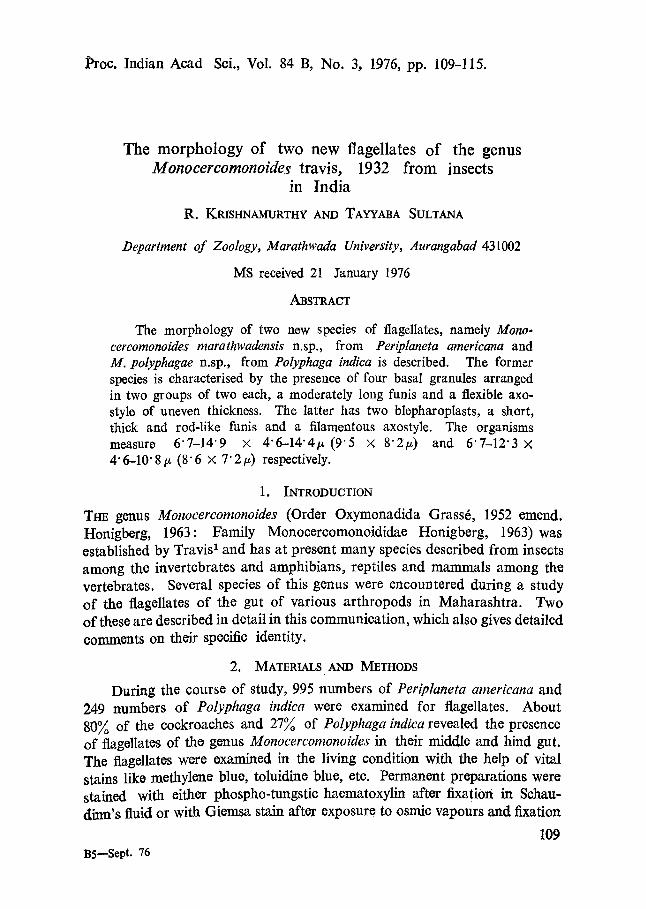
The body of the organism is rounded (figure 1 : 4, 6) or often irregular (figure 1: 1, 2, 7, 8) ha outline. The periplast is very thin.
/
9
,) t0.,a I
Figure 1 : 1-9. Monocercomonoides marathwadensis n.sp. (1-7 from smears exposed to osmic vapours, fixed in methanol and stained with Giemsa's stain. 8-9. from smears fixed in Sehaudirn's .fluid and stained with phospho-tungstic haematoxytin), (1) Showing three basal granules, rhizo- pl~t and aeronematie flagella. (2 and 3) Showing a long axostyle of variable thickness. (.4) Showing funis and aeronematie flagella. (5) Showing four basal granules ano origin 6fHagdla: (6) Showing funis, three basal granules and origin of axostyle, (7) Sh0wfng ~l'~izoplast and axostyle of variable thickness. (8, 9) Showing endosome in nucleus

MORPHOLOGrf OF NEW FLAGELLATES 111
The flagella arise in two groups from two sots of basal granules, about 2/z apart and lying at slightly different levels just above the nucleus (figure 1 : 1, 4-7). The two groups are connected by a thin rhizoplast which forms an arch-qke loop over the nucleus (figure 1 : 1, 7). In well stained preparations, either one (figure 1 : 1, 6, 7) or both (figure 1 : 5, 8) appear to be bilobed or composed of two closely placed granules. Thus there are four granules placed in two groups. One of the groups gives origin to two anterior flagella, one from each granule. Of the other group, one granule gives rise to the third anterior flagellum while the other is the point of origin of the trailing flagellum as well as the funis and axostyle (figure 1 : 1-3, 7, 9). The anterior flagella are slightly unequal and are of the same length as the body or slightly more. The trailing flagellum is relatively longer and measures about twice the body length. The funis arises from the same basal granule and runs along the surface of the body, more or less parallel to the base of the trailing flagellum. It is about the same thickness as the flagellum and measures 2" 6-6" 2/z (average 4" 8 tz) in length. Either all or at least some of the flagella terminate in distinct acronemes (figure 1: 1, 4).
The axostyle originates from the same granule as the trailing flagellum and passes backwards through the body. It is almost as long as the body (figure 1: 1, 5-9) or slightly longer and projecting outside the body (figure 1: 2, 3). It is apparently quite flexible and somewhat uneven in its thickness (figure 1: 2, 4, 5, 7).
Thonuelous is fairly large and situated anteriorly. It is spherical (figure 1 : 7), ovoid (figure 1 : 2) or somewhat irregular in outline (figure 1 : 4-6). It has a well defined endosome in the centre, surrounded by a halo (figure 1 : 8, 9).
The cytoplasm is granular and vacuolated. organism are as follows:
(All dimensions in microns)
Length of the body 6.7-14 Maximum width of the body L~gth of anterior flagellum I Length of anterior flagellum II Le-agth of anterior flagellum III Length of trailing flagellum Size of rtudous
The dimensions of the
• 9 (9.5) 4.6-14.4 (8.2) 5.7-18.0 (12.6) 5.1-20.6 (•2.5) 5.7-20.6 (14.1)
12;9-26.7 (19.0) 1-0 × 1'5-7-7 × 6.2 (3.2 × 3.4)
112 R. KRISHNAMURTHY AND TAYYABA SULTANA
DISCUSSION
Seven species of this genus are so far described from insect hosts. These are as follows: M. melolonthae Grassi, 1879; M. orthopterorum Parisi, 1910; M. garnhami Bhaskar Rao, i969; M. qadrii Bhaskar Rao, •969; M. ganapatii Bhaskar Rao, 1969; M. krishnamurthff Sultana (In press) and M. chakra- vartii Krishnamurthy and Sultana, 1976. The organism described above is easily distinguished from all these species by the presence of four basal granules arranged in two groups of two each, a pattern not so far described in any of the species.
It comes c/ose to M. garnhami and M. chakravartii, in the presottce of a funis. However, the funis is long in the former (average 7"5 tz) and rela- tively short in the latter (average 3"6 t*)while it is moderately long in the present organism (4..8/z). Sultana, 1975 described a short funis ,(3"1 ~) in M. ganapatii. This is also much shorter than the funis in the present organism. The parasite is ,further distinguished from M. garnhami by the nature of.the axostyle and the absence,of the pelta. It is marked off from M. chakramrtii by its larger size and better developed axostyle. It is contrasted from M, ganapatff hy its larger size and the nature of the axostyle.
The organism under discussion is marked off from the other four species namely M. tnelolonthae, M. orthopterorum, M. qadrii and M. krishnamurthii by the presence of a distinct funts, which is absent in all these species. It is further distinguished from M. melolonthae by the nature of the axostyle and the presence of a long trailing flagellum. It differs markedly from M. orthopterorum in size measuring 6" ~/-14" 9 tz × 4" 6--14.4 ~ (9" 5 × 8" 2 t~) as against 3" 7-7" 0/, × 2" 1-3' 8/z (6" 2 x 3" 1/z) and in possessing a well defined axostyle. It is slightly smaller in size than M. qadrii and has a longer axostyle of lmeven thickness. The species differs considerably from M. krishnamurthff in the nature of its nuclear structure as well as the axostyle.
Thus the o:rganism is distinct from all the species described so far and hence is designg, ted as M. marathwadensis n.sp., after the iegion where it was found.
Species Host Habitat Locality
: Monocercomonoides marathwadensis n.sp. : Periplaneta americana : Hind gut : Aurangabad, Maharashtra, India,
(The type slides are deposited in the Department of Zoology, Marathwada University, Auranga, bad).
MORPHOLOGY OF NEW' FLAGELLATES 113
2 >3 , -,
4
7
5
8
6:
x
I IO7~ i
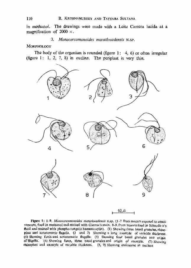
Figure 2: 1-8. Monocercomonoides polyphagae n.sp. (1-=7 from smears exposed to osmic vapours, fixed in methanol and stained with Giemsa's stain. 8. from a smear fixed in Schaudinn's fluid and stained with phospho-tungstic haematoxylin). (1) Showing a straight and slightly projecting axostyle. (2) Showing two blepharoplasts and origin of flagella. (3) Show- ing origin of flagella and axostyle with a thicker posterior end. (4) Showing general structure. (5, 6) Showing funis and acronematic flagella. (7) Showing blepharoplasts, origin of flagella and funis. (8) Showing the uniformly distributed chromatin in nucleus.
4. Monocercomonoides polyphagae, N.SP. MORPHOLOGY
The organism appoars spherical (figur0 2 : 2-4), pyriform. (figuro: 2: 5, 6, 8) or somowhat irrogular in shape, in tho stained preparations. Tho poriplast is thin.

114 R. KRISHNAMURTHY AND TAYYABA, SULTANA
Two blepharoplasts are situated at the anterior end of the body just above the nucleus (figure 2: 2, 3, 7). The two granules are about 1"5/z apart and lie at slightly different levels. The one placed somewhat anteriorly gives rise to two anterior flagella, while from the other arises the third anterior flagellum, the trailing flagellum and the axostyle (figure 2: 2, 3, 7).
The three anterior flagella are almost equal in length and longer than the body. The trailing flagellum is about twice the length of the body. Arising along with the trailing flagellum and runDing along the body surface is a thick, slightly curved and rod-like tunis (figure 2: 5-7). It is 3.1-7.2tt in length (average 5" 1/0. In some organisms the flagella end in aeronemes (figure 2: 5, 6).
The axostyle is filamentous. It is as long as the body (figure 2 : 6-8) or slightly longer (figure 2: 1), following a straight or somewhat curved path. It is uniform in its thickness in most cases (figure 2: 1, 2, 4, 6, 7) while in some (figure 2: 3) the terminal part appears to be slightly thicker.
The anteriorly situated nucleus is fairly large in size and spherical or ovoid in shape (figure 2: 3, 5-8). It is flattened or somewhat bilobed anteriorly in some cases (figure 2: 2, 4). The chromatin is distributed uniformly (figure 2: 8).
The cytoplasm is vacuolated but does not show any granules or bacteria except in rare cases (figure 2: 3). The dimensions of the organism are as follows:
(All measurements in microns)
Length of Maximum Length of Length of
the body width of the body anterior flagellum I anterior flagellum II
Length of anterior flagellvm III Length of trailing flagellum Size of nucleus
6'7-12-3 (8"6) 4"6-10"8 (7"2) 6"2-16"5 (11"0) 6"2-17"5 (11"0) 7"2-18"5 01:3) 9.3-24.2 (16-4) 2 . 1 × 2 ' 1 - 3 . 6 × 4 . 1 (2"9 × 2"9)
D~SCUSS~ON
In possessing a funis this organism resembles M. garnhami, M. ganapatii, M. chakravartii and M. marathwadensis n.sp. However, its funis is relatively thicker and rod-like contrasted with the thin and filamentous funis in all the. other species. In addition, there, are also other features distinguish'ing it fiom them. It differs from M. garnhami in possessing a shorter furiis
MORPHOLOGY OF NEW FLAGELLATES 115
and a shorter axostyle and in lacking a pelta. The organism has a much longer funis than M. ganapatii and differs from it in the nature of the axostyle as well as the body dimensions. It is marked off from M. ehakravartii and M. marathwadensis in having only two blepharoplasts, as against 3 and 4 respectively in the latter speeies.
This parasite differs from all the other species namely, M. melolonthae, M. orthopterorum, M. qadrii and M. krishnamurthii in the presence of the funis and in body dimensions, besides other differences. It is marked off from M. melolonthae by its trailing flagellum, from M. qadrii and M. ortho- perorum by the different type of axostyle and from M. krishnamurthii by the absence of the axostylar thickening at the point of emergence and the surround- ing vacuole.
In view of its distinctness, this organism is designated as Monocerco- monoides polyphagae n.sp., after the generic name cf the host in which it was found.
Species : Monoeercomonoides polyphagae n.sp. Host : Polyphaga indica. Habitat : Hind gut. Locality : Aurangabad, Maharashtra, India.
(The type slides are deposited in the Department of Zoology, Marath- wada University, Aurangabad).
ACKNOWLEDGEMENTS
The authors are grateful to Dr. R. Nagabhushanam, Head of the Depart- ment of Zoology, Marathwada University, Aurangabad, for facilities. T.S. thanks the authorities of the Marathwada University for the award of a junior research fellowship.
REFERENCES
1. Bhaskar Rao, T., Prec. Indian Acad. Sci. B70 208 (1969).
2. GrassY, P. P., Trall6 de Zoologie Tome 1, Masson et Cie., Paris (1952), 3. Grassi, B., Gaz. Med. ltal. Lambardi, 39 445 (1879). 4. Honigberg, B. M., in Progress in Protozoology, Academic Press, p. 68 (•963). 5. Krishnatrturthy, R and Sultana, T., Curr. Sci. 45 (5), 184 (1e76).
6. Parisi, B., Arch. Protistenk. 19 232 (1910). 7. Sultana, T., Marathwada Univ. J. Sci. 14 (7), 229 (1975). 8. Sultana, T., (In Press). Marathwada Univ. J. Sci. 9. Travis, B. V., Iowa St. Coll. J. Sci. 6 317 (1932).
![THE PARTNERSHIP ACT, 1932 (IX OF 1932) - Punjab PARTNERSHIP ACT, 1… · TEXT THE PARTNERSHIP ACT, 1932 (IX OF 1932) [8th April 1932] An Act to define and amend the law relating to](https://img.pdfslide.us/doc/110x75/5a797b9f7f8b9ade698c0b32/the-partnership-act-1932-ix-of-1932-partnership-act-1text-the-partnership.jpg)