Embed Size (px)
Citation preview
Jour
nal o
f Cel
l Sci
ence
RESEARCH ARTICLE
The Mon1–Ccz1 GEF activates the Rab7 GTPase Ypt7 via alongin-fold–Rab interface and association with PI3P-positive membranes
Margarita Cabrera1, Mirjana Nordmann1,*, Angela Perz1, David Schmedt2, Andreas Gerondopoulos3,Francis Barr3, Jacob Piehler2, Siegfried Engelbrecht-Vandre1 and Christian Ungermann1,{
ABSTRACT
To function in fusion and signaling, Rab GTPases need to be
converted into their active GTP form. We previously identified
the conserved Mon1–Ccz1 complex as the guanine nucleotide
exchange factor (GEF) of the yeast Rab7 GTPase Ypt7. To address
the possible GEF mechanism, we generated a homology model of
the predicted longin domains of Mon1 and Ccz1 using the Rab-
binding surface of the TRAPP complex as a template. On the basis
of this, we identified mutations in both yeast Mon1 and Ccz1 that
block Ypt7 activation, without affecting heterodimer formation and
intracellular localization of Mon1 and Ccz1 at endosomes. Strikingly,
the activity of the isolated Mon1–Ccz1 complex for Ypt7 is highly
stimulated on membranes, and is promoted by the same anionic
phospholipids such as phosphatidylinositol-3-phosphate (PI3P),
which also support membrane association of the GEF complex.
Our data imply that the GEF activity of the Mon1–Ccz1 complex
towards Rab7/Ypt7 requires the interface formed by their longin
domains and profits strongly from its association with the organelle
surface.
KEY WORDS: Guanine nucleotide exchange factor, Mon1–Ccz1,
Rab GTPase, Endosome, Membrane fusion
INTRODUCTIONWithin the endomembrane system, vesicles transport cargo between
organelles. These organelles need to maintain their identity despite
the fact that they continuously fuse with incoming vesicles and losemembrane as a result of vesicle generation. Rab GTPases, tethering
factors and the SNARE machinery localize to specific organelles and
cooperate to promote fusion of membranes. Rab proteins are smallGTP/GDP-binding proteins with C-terminal prenyl anchors, which
function as molecular switches. They bind in their GTP form to
tethering factors to bring membranes into proximity before SNAREspresent in each membrane form four-helix bundles and thus drive
bilayer mixing.
As a result of the slow rate of dissociation of GDP, Rabs
require guanine nucleotide exchange factors (GEFs) for their
conversion into the active GTP-bound form, which can interact
with effectors on membranes such as tethering factors or lipid
kinases. In addition, Rabs depend on a GTPase activating protein
(GAP) to hydrolyze the GTP to GDP, and then become substrates
of the GDI chaperone, which can extract the Rab-GDP and keep it
soluble in the cytoplasm (Lachmann et al., 2011; Hutagalung and
Novick 2011; Itzen and Goody 2011; Barr, 2013).
Among the Rab interactors, GEFs are most crucial because
they provide the activated Rab at a specific location in the
cell. In this context, structures of Rab–GEF complexes and
the subsequent analyses have been very informative. For the
exocytic Sec4 Rab, the GEF Sec2 promotes nucleotide exchange
via its a-helical domain (Dong et al., 2007; Sato et al., 2007).
Rab5-like proteins that operate in the endosomal pathway
depend on Vps9-domain-containing GEFs (Delprato et al.,
2004; Delprato and Lambright 2007). DENN domains were
identified as a signature domain for multiple GEFs (Yoshimura
et al., 2010; Allaire et al., 2010; Marat et al., 2011) and molecular
details of their function were revealed for DENND1-BS and
Rab35 (Wu et al., 2011).
GEFs might also act as part of a multiprotein complex. The
best-characterized example is the heptameric TRAPPI, which
interacts with the Rab1/Ypt1 protein through a surface consisting
of four subunits: Bet5, Trs23 and the two flanking copies of Bet3
(Kummel et al., 2006; Kim et al., 2006; Cai et al., 2008). GEF
dimers are required to activate Rab6/Ypt6 (Ric1–Rgp1) (Pusapati
et al., 2012; Siniossoglou et al., 2000) and Rab7/Ypt7 (Mon1–
Ccz1) (Nordmann et al., 2010; Gerondopoulos et al., 2012),
although structural information on these complexes is lacking. In
agreement with the function of the Mon1–Ccz1 dimer in Ypt7
activation, endosomal biogenesis is strongly impaired if either of
the two proteins is defective (Wang et al., 2002; Kinchen and
Ravichandran 2010; Poteryaev et al., 2010; Nordmann et al.,
2010; Yousefian et al., 2013).
TRAPP subunits Bet5 and Trs23 interact both with each other
and with Ypt1 through their longin domains (Kim et al., 2006;
Cai et al., 2008). These are arranged into a continuous ten-strand
b-sheet supporting two parallel a-helices, which provide a
significant portion of the interacting residues. Interestingly, the
formation of the Mon1–Ccz1 heterodimer also requires the
predicted longin domains of both subunits (Nordmann et al.,
2010). This suggests that the architecture and mode of GEF action
of Mon1 and Ccz1 on Ypt7 might resemble those of the TRAPPI
complex. Here, we present evidence that Mon1 and Ccz1, which
are linked to Rab7/Ypt7 both in mammals and yeast, cooperate
1University of Osnabruck, Department of Biology/Chemistry, Biochemistry section,Barbarastrasse 13, 49076 Osnabruck, Germany. 2University of Osnabruck,Department of Biology/Chemistry, Biophysics section, Barbarastrasse 13, 49076Osnabruck, Germany. 3Department of Biochemistry, University of Oxford, South ParksRoad, Oxford OX1 3QU, UK.
*Present address: University of Osnabruck, Department of Biology/Chemistry,Section of Molecular Cell Biology, Barbarastrasse 13, 49076 Osnabruck,Germany.
{Author for correspondence ([email protected])
Received 16 August 2013; Accepted 9 December 2013
� 2014. Published by The Company of Biologists Ltd | Journal of Cell Science (2014) 127, 1043–1051 doi:10.1242/jcs.140921
1043
Jour
nal o
f Cel
l Sci
ence
via their longin-domain surface to activate the Rab GTPase. Thisactivity is strongly promoted upon membrane recruitment of the
GEF, suggesting that previously determined activities might havebeen significantly underestimated.
RESULTSMon1 and Ccz1 require a common interface for Ypt7 activationSecondary structure prediction using PredictProtein (http://predictprotein.org) and PsiPred (http://bioinf.cs.ucl.ac.uk/
psipred/) identified possible longin domains in Ccz1 (aa 1–156)and Mon1 (aa 180–287), which are required for the assembly ofthe Mon1–Ccz1 heterodimer (Nordmann et al., 2010). This
suggests a similar structure–function relationship for Mon1–Ccz1and the TRAPPI GEF complex, because the two TRAPP subunitsTrs23 and Bet5 also contain longin domains that contact the Rab
GTPase Ypt1 directly (Kim et al., 2006; Cai et al., 2008). Wetherefore built a homology model for the predicted longindomains of Mon1–Ccz1 using Trs23 and Bet5 as templates.
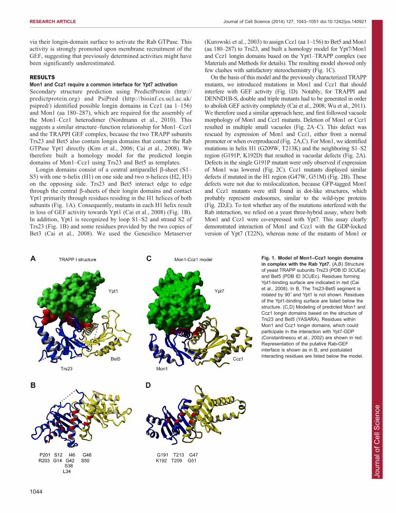
Longin domains consist of a central antiparallel b-sheet (S1–
S5) with one a-helix (H1) on one side and two a-helices (H2, H3)on the opposing side. Trs23 and Bet5 interact edge to edgethrough the central b-sheets of their longin domains and contact
Ypt1 primarily through residues residing in the H1 helices of bothsubunits (Fig. 1A). Consequently, mutants in each H1 helix resultin loss of GEF activity towards Ypt1 (Cai et al., 2008) (Fig. 1B).
In addition, Ypt1 is recognized by loop S1–S2 and strand S2 ofTrs23 (Fig. 1B) and some residues provided by the two copies ofBet3 (Cai et al., 2008). We used the Genesilico Metaserver
(Kurowski et al., 2003) to assign Ccz1 (aa 1–156) to Bet5 and Mon1(aa 180–287) to Trs23, and built a homology model for Ypt7/Mon1
and Ccz1 longin domains based on the Ypt1–TRAPP complex (seeMaterials and Methods for details). The resulting model showed onlyfew clashes with satisfactory stereochemistry (Fig. 1C).
On the basis of this model and the previously characterized TRAPP
mutants, we introduced mutations in Mon1 and Ccz1 that shouldinterfere with GEF activity (Fig. 1D). Notably, for TRAPPI andDENND1B-S, double and triple mutants had to be generated in order
to abolish GEF activity completely (Cai et al., 2008; Wu et al., 2011).We therefore used a similar approach here, and first followed vacuolemorphology of Mon1 and Ccz1 mutants. Deletion of Mon1 or Ccz1
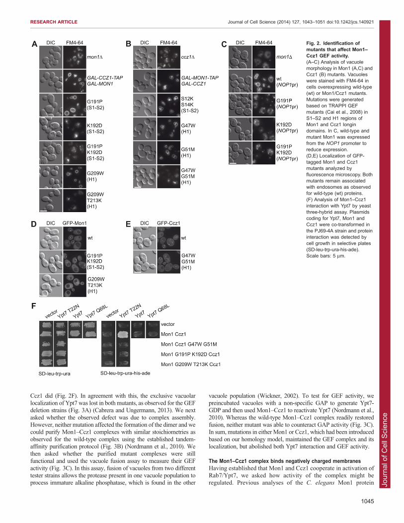
resulted in multiple small vacuoles (Fig. 2A–C). This defect wasrescued by expression of Mon1 and Ccz1, either from a normalpromoter or when overproduced (Fig. 2A,C). For Mon1, we identified
mutations in helix H1 (G209W, T213K) and the neighboring S1–S2region (G191P, K192D) that resulted in vacuolar defects (Fig. 2A).Defects in the single G191P mutant were only observed if expressionof Mon1 was lowered (Fig. 2C). Ccz1 mutants displayed similar
defects if mutated in the H1 region (G47W, G51M) (Fig. 2B). Thesedefects were not due to mislocalization, because GFP-tagged Mon1and Ccz1 mutants were still found in dot-like structures, which
probably represent endosomes, similar to the wild-type proteins(Fig. 2D,E). To test whether any of the mutations interfered with theRab interaction, we relied on a yeast three-hybrid assay, where both
Mon1 and Ccz1 were co-expressed with Ypt7. This assay clearlydemonstrated interaction of Mon1 and Ccz1 with the GDP-lockedversion of Ypt7 (T22N), whereas none of the mutants of Mon1 or
Fig. 1. Model of Mon1–Ccz1 longin domainsin complex with the Rab Ypt7. (A,B) Structureof yeast TRAPP subunits Trs23 (PDB ID 3CUEa)and Bet5 (PDB ID 3CUEc). Residues formingYpt1-binding surface are indicated in red (Caiet al., 2008). In B, The Trs23-Bet5 segment isrotated by 90˚ and Ypt1 is not shown. Residuesof the Ypt1-binding surface are listed below thestructure. (C,D) Modeling of predicted Mon1 andCcz1 longin domains based on the structure ofTrs23 and Bet5 (YASARA). Residues withinMon1 and Ccz1 longin domains, which couldparticipate in the interaction with Ypt7-GDP(Constantinescu et al., 2002) are shown in red.Representation of the putative Rab-GEFinterface is shown as in B, and postulatedinteracting residues are listed below the model.
RESEARCH ARTICLE Journal of Cell Science (2014) 127, 1043–1051 doi:10.1242/jcs.140921
1044
Jour
nal o
f Cel
l Sci
ence
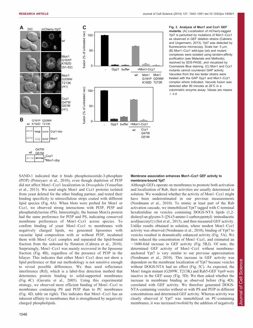
Ccz1 did (Fig. 2F). In agreement with this, the exclusive vacuolarlocalization of Ypt7 was lost in both mutants, as observed for the GEFdeletion strains (Fig. 3A) (Cabrera and Ungermann, 2013). We next
asked whether the observed defect was due to complex assembly.However, neither mutation affected the formation of the dimer and wecould purify Mon1–Ccz1 complexes with similar stoichiometries as
observed for the wild-type complex using the established tandem-affinity purification protocol (Fig. 3B) (Nordmann et al., 2010). Wethen asked whether the purified mutant complexes were still
functional and used the vacuole fusion assay to measure their GEFactivity (Fig. 3C). In this assay, fusion of vacuoles from two differenttester strains allows the protease present in one vacuole population toprocess immature alkaline phosphatase, which is found in the other
vacuole population (Wickner, 2002). To test for GEF activity, wepreincubated vacuoles with a non-specific GAP to generate Ypt7-GDP and then used Mon1–Ccz1 to reactivate Ypt7 (Nordmann et al.,
2010). Whereas the wild-type Mon1–Ccz1 complex readily restoredfusion, neither mutant was able to counteract GAP activity (Fig. 3C).In sum, mutations in either Mon1 or Ccz1, which had been introduced
based on our homology model, maintained the GEF complex and itslocalization, but abolished both Ypt7 interaction and GEF activity.
The Mon1–Ccz1 complex binds negatively charged membranesHaving established that Mon1 and Ccz1 cooperate in activation ofRab7/Ypt7, we asked how activity of the complex might beregulated. Previous analyses of the C. elegans Mon1 protein
Fig. 2. Identification ofmutants that affect Mon1–Ccz1 GEF activity.(A–C) Analysis of vacuolemorphology in Mon1 (A,C) andCcz1 (B) mutants. Vacuoleswere stained with FM4-64 incells overexpressing wild-type(wt) or Mon1/Ccz1 mutants.Mutations were generatedbased on TRAPPI GEFmutants (Cai et al., 2008) inS1–S2 and H1 regions ofMon1 and Ccz1 longindomains. In C, wild-type andmutant Mon1 was expressedfrom the NOP1 promoter toreduce expression.(D,E) Localization of GFP-tagged Mon1 and Ccz1mutants analyzed byfluorescence microscopy. Bothmutants remain associatedwith endosomes as observedfor wild-type (wt) proteins.(F) Analysis of Mon1–Ccz1interaction with Ypt7 by yeastthree-hybrid assay. Plasmidscoding for Ypt7, Mon1 andCcz1 were co-transformed inthe PJ69-4A strain and proteininteraction was detected bycell growth in selective plates(SD-leu-trp-ura-his-ade).Scale bars: 5 mm.
RESEARCH ARTICLE Journal of Cell Science (2014) 127, 1043–1051 doi:10.1242/jcs.140921
1045
Jour
nal o
f Cel
l Sci
ence
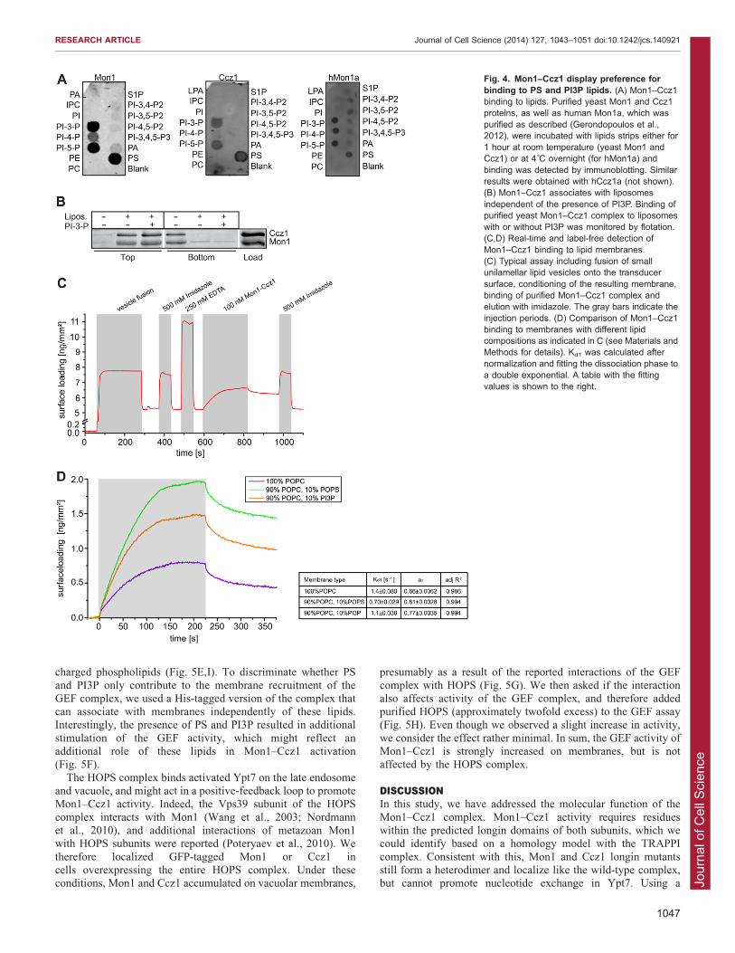
SAND-1 indicated that it binds phosphoinositide-3-phosphate(PI3P) (Poteryaev et al., 2010), even though depletion of PI3Pdid not affect Mon1–Ccz1 localization in Drosophila (Yousefian
et al., 2013). We used single Mon1 and Ccz1 proteins isolatedfrom yeast deleted for the other binding partner, and tested theirbinding specificity to nitrocellulose strips coated with different
lipid species (Fig. 4A). When blots were probed for Mon1 orCcz1, we observed strong interactions with PI3P, PI5P andphosphatidylserine (PS). Interestingly, the human Mon1a protein
had the same preference for PI3P and PS, indicating conservedmembrane preferences of Mon1–Ccz1 across species. Toconfirm binding of yeast Mon1–Ccz1 to membranes withnegatively charged lipids, we generated liposomes with
vacuolar lipid composition with or without PI3P, incubatedthem with Mon1–Ccz1 complex and separated the lipid-boundfraction from the unbound by flotation (Cabrera et al., 2010).
Surprisingly, Mon1–Ccz1 was mainly recovered in the liposomefraction (Fig. 4B), regardless of the presence of PI3P in thebilayer. This indicates that either Mon1–Ccz1 does not show a
lipid preference or that our methodology is not sensitive enoughto reveal possible differences. We thus used reflectanceinterference (Rif), which is a label-free detection method that
determines protein binding to solid-supported membranes(Fig. 4C) (Gavutis et al., 2005). Using this experimentalstrategy, we observed more efficient binding of Mon1–Ccz1 tomembranes containing PS and PI3P than to PC membranes
(Fig. 4D, table on right). This indicates that Mon1–Ccz1 has aninherent affinity to membranes that is strengthened by negativelycharged phospholipids.
Membrane association enhances Mon1–Ccz1 GEF activity tomembrane-bound Ypt7Although GEFs operate on membranes to promote both activation
and localization of Rab, their activities are usually determined insolution. We wondered whether the activity of Mon1–Ccz1 mighthave been underestimated in our previous measurements
(Nordmann et al., 2010). To mimic at least part of the Rabactivation cascade, we immobilized Ypt7 tagged with C-terminalhexahistidine on vesicles containing DOGS-NTA lipids (1,2-
dioleoyl-sn-glycero-3-{[N-(5-amino-1-carboxypentyl) iminodiaceticacid]succinyl}) (Sot et al., 2013), and then measured GEF activity.Unlike results obtained in solution, where modest Mon1–Ccz1activity was observed (Nordmann et al., 2010), binding of Ypt7 to
vesicles resulted in dramatically enhanced activity (Fig. 5A). Wethen reduced the concentration of Mon1–Ccz1, and estimated an,1600-fold increase in GEF activity (Fig. 5B,I). Of note, the
determined GEF activity of Mon1–Ccz1 without membrane-anchored Ypt7 is very similar to our previous approximation(Nordmann et al., 2010). This increase in GEF activity was
dependent on the membrane localization of Ypt7 because vesicleswithout DOGS-NTA had no effect (Fig. 5C). As expected, theMon1 longin mutant (G209W, T213K) and Rab5-GEF Vps9 were
inactive in the GEF assay (Fig. 5D). We then asked whether theincrease in membrane binding as observed before (Fig. 4D),correlated with GEF activity. We therefore generated DOGS-NTA-containing vesicles without or with PS and PI3P in different
concentrations and determined GEF activity. Whereas activity wasclearly observed if Ypt7 was immobilized on PC-containingmembranes, it was increased twofold by the addition of negatively
Fig. 3. Analysis of Mon1 and Ccz1 GEFmutants. (A) Localization of mCherry-taggedYpt7 is perturbed by mutations of Mon1–Ccz1as observed in GEF deletion strains (Cabreraand Ungermann, 2013). Ypt7 was detected byfluorescence microscopy. Scale bar: 5 mm.(B) Mon1–Ccz1 wild-type (wt) and mutantcomplexes were isolated using tandem-affinitypurification (see Materials and Methods),resolved by SDS-PAGE, and visualized byCoomassie Blue staining. (C) Mon1 and Ccz1mutants cannot counteract GAP activity.Vacuoles from the two tester strains weretreated with the GAP Gyp1 and Mon1–Ccz1complex where indicated. Vacuole fusion wasdetected after 90 minutes at 26˚C in acolorimetric enzyme assay. Values are means6 s.d.
RESEARCH ARTICLE Journal of Cell Science (2014) 127, 1043–1051 doi:10.1242/jcs.140921
1046
Jour
nal o
f Cel
l Sci
ence
charged phospholipids (Fig. 5E,I). To discriminate whether PS
and PI3P only contribute to the membrane recruitment of theGEF complex, we used a His-tagged version of the complex thatcan associate with membranes independently of these lipids.Interestingly, the presence of PS and PI3P resulted in additional
stimulation of the GEF activity, which might reflect anadditional role of these lipids in Mon1–Ccz1 activation(Fig. 5F).
The HOPS complex binds activated Ypt7 on the late endosomeand vacuole, and might act in a positive-feedback loop to promoteMon1–Ccz1 activity. Indeed, the Vps39 subunit of the HOPS
complex interacts with Mon1 (Wang et al., 2003; Nordmannet al., 2010), and additional interactions of metazoan Mon1with HOPS subunits were reported (Poteryaev et al., 2010). We
therefore localized GFP-tagged Mon1 or Ccz1 incells overexpressing the entire HOPS complex. Under theseconditions, Mon1 and Ccz1 accumulated on vacuolar membranes,
presumably as a result of the reported interactions of the GEF
complex with HOPS (Fig. 5G). We then asked if the interactionalso affects activity of the GEF complex, and therefore addedpurified HOPS (approximately twofold excess) to the GEF assay(Fig. 5H). Even though we observed a slight increase in activity,
we consider the effect rather minimal. In sum, the GEF activity ofMon1–Ccz1 is strongly increased on membranes, but is notaffected by the HOPS complex.
DISCUSSIONIn this study, we have addressed the molecular function of the
Mon1–Ccz1 complex. Mon1–Ccz1 activity requires residueswithin the predicted longin domains of both subunits, which wecould identify based on a homology model with the TRAPPI
complex. Consistent with this, Mon1 and Ccz1 longin mutantsstill form a heterodimer and localize like the wild-type complex,but cannot promote nucleotide exchange in Ypt7. Using a
Fig. 4. Mon1–Ccz1 display preference forbinding to PS and PI3P lipids. (A) Mon1–Ccz1binding to lipids. Purified yeast Mon1 and Ccz1proteins, as well as human Mon1a, which waspurified as described (Gerondopoulos et al.,2012), were incubated with lipids strips either for1 hour at room temperature (yeast Mon1 andCcz1) or at 4˚C overnight (for hMon1a) andbinding was detected by immunoblotting. Similarresults were obtained with hCcz1a (not shown).(B) Mon1–Ccz1 associates with liposomesindependent of the presence of PI3P. Binding ofpurified yeast Mon1–Ccz1 complex to liposomeswith or without PI3P was monitored by flotation.(C,D) Real-time and label-free detection ofMon1–Ccz1 binding to lipid membranes.(C) Typical assay including fusion of smallunilamellar lipid vesicles onto the transducersurface, conditioning of the resulting membrane,binding of purified Mon1–Ccz1 complex andelution with imidazole. The gray bars indicate theinjection periods. (D) Comparison of Mon1–Ccz1binding to membranes with different lipidcompositions as indicated in C (see Materials andMethods for details). Kd1 was calculated afternormalization and fitting the dissociation phase toa double exponential. A table with the fittingvalues is shown to the right.
RESEARCH ARTICLE Journal of Cell Science (2014) 127, 1043–1051 doi:10.1242/jcs.140921
1047

Jour
nal o
f Cel
l Sci
ence
modified GEF assay, we further show that Mon1–Ccz1 is stronglyactive once it is recruited to the membranes, suggesting that GEF
activities might have been underestimated previously.The presence of longin domains in Mon1–Ccz1 and TRAPPI,
and the similar phenotype of the mutants suggest that both GEFs
might share a related Rab-binding surface and nucleotide
exchange mechanism. Also, the recently resolved DENND1GEF domain contains a longin fold, although it binds Rab35 via a
different surface compared with the interaction between TRAPPIand Ypt1 (Wu et al., 2011). We are aware that within the TRAPPIcomplex, the two Bet3 subunits contribute to the binding of Ypt1
and the C-terminal residues of Bet3A have a crucial role to
Fig. 5. Mon1–Ccz1 GEF activity is stimulated by PS and PI3P lipids. 500 pmoles of Ypt7-His loaded with MANT-GDP were used in all experiments. Lipidconcentration is indicated in mol% in brackets. Mon1–Ccz1 concentrations are indicated in each figure legend. (A) GEF activity of Mon1–Ccz1 towards Ypt7 isincreased in the presence of POPC (52.5):DOGS-NTA(30):PS(10):PI3P(7.5) vesicles. 500 pmoles of Mon1–Ccz1 complex was used. (B) Titration of Mon1–Ccz1 complex in the GEF assay containing POPC (67.5):DOGS-NTA (15):PS(10):PI3P(7.5) vesicles. Mon1–Ccz1 complex was used at the indicatedconcentration. (C) Vesicles stimulate Mon1–Ccz1 GEF activity only if they contain bound Ypt7. POPC (67.5): DOGS-NTA(15):PS(10):PI3P(7.5) and POPC(82.5) PS(10):PI3P(7.5) vesicles were used in the presence of 25 pmoles of Mon1–Ccz1 complex. (D) Rab5-GEF Vps9 and Mon1 G209W, T213K-Ccz1 mutantcomplex are inactive towards membrane-bound Ypt7. 25 pmoles or Vps9 or Mon1 G209W T213K-Ccz1 complex were added. POPC (67.5):DOGS-NTA(15):PS(10):PI3P(7.5) vesicles were used in this assay. (E) Complete GEF stimulation requires PS and PI3P lipids. GEF activity of Mon1–Ccz1 wascompared in vesicles of different composition, POPC(85):DOGS-NTA(15), POPC (67.5):DOGS-NTA(15):PS(10):PI3P(7.5) and POPC(77):DOGS-NTA(15):PS(5):PI3P(3). Mon1–Ccz1 concentration was 25 pmoles. (F) Contribution of PS and PI3P to recruitment and activation of Mon1–Ccz1. Whereindicated, 25 pmoles of Mon1–Ccz1 complex carrying a His tag were added. POPC(85):DOGS-NTA(15) and POPC (67.5):DOGS-NTA(15):PS(10):PI3P(7.5)vesicles were used in the GEF assay. (G) Excess of HOPS relocalizes Mon1 and Ccz1 to the vacuole membranes. Distribution of Mon1 and Ccz1 was analyzedby fluorescence microscopy in wild-type (wt) and cells overexpressing all HOPS subunits. Scale bar: 5 mm. (H) Mon1–Ccz1 GEF activity was slightly enhancedby the HOPS complex. GEF assay included 500 pmoles of Ypt7-His loaded with MANT-GDP, 25 pmoles Mon1–Ccz1 complex, HOPS complex at the indicatedconcentration and POPC(85):DOGS-NTA(15) vesicles. (I) Estimation of Mon1–Ccz1 GEF activity. kcat/Km was calculated after fitting the nucleotide releasecurves to a double exponential curve.
RESEARCH ARTICLE Journal of Cell Science (2014) 127, 1043–1051 doi:10.1242/jcs.140921
1048
Jour
nal o
f Cel
l Sci
ence
promote GEF activity (Cai et al., 2008). We thus consider it likelythat additional segments proximal to the longin domains of
Mon1–Ccz1 will be required for full activity. We also note thatour model is limited by the structural differences between Ypt1and Ypt7, especially in their N-terminal regions. However, at thispoint, our working model allows us to predict essential residues
for GEF activity (Figs 1,2).In this context, we realized that roadblock domains that are
found in subunits of the EGO/ragulator complex have been
predicted as GEFs for the Rag GTPases (BarPeled et al., 2013).Even though the order of secondary structure elements differsbetween roadblock and longin domains, their overall structure is
quite similar (Kurzbauer et al., 2004; Qian et al., 2005; Zhanget al., 2012). These domains have probably evolved as generalinteractors of small GTPases (Levine et al., 2013), and a
subfamily might act as dimeric GEFs. Importantly, the dimericBLOC-3 complex also harbors a longin domain in each of its twosubunits Hps1 and Hps4 and might function in a very similarmanner to Mon1–Ccz1 (Gerondopoulos et al., 2012). It thus
is conceivable that these domains have evolved as generalinteractors of small GTPases. Intriguingly, the dimeric Ric1–Rgp1 complex, which activates Ypt6 (Siniossoglou et al., 2000),
also depends on both subunits for activity, and might also use ashared surface to interact with its Rab Ypt6/Rab6.
Even though we have shown GEF activity of Mon1–Ccz1
previously in vitro, we did not expect a 1600-fold increase inactivity (kcat/KM) upon immobilization of Ypt7 on membranes.This might be due in part to the membrane recruitment of both
proteins. Although the assay does not recapitulate the entirecycle, it revealed that Mon1–Ccz1 activity is far higher thanpreviously anticipated. GEFs such as the Legionella DrrA proteinhave strong activity in vitro, which is sufficient to counteract Rab
extraction by GDI (Schobel et al., 2009; Zhu et al., 2010; Suhet al., 2010). It is thus likely that other GEFs use similarmechanisms and will explain how GEFs that are mistargeted to
the mitochondria can drive the recruitment of Rabs to thisorganelle (Gerondopoulos et al., 2012; Blumer et al., 2013), eventhough some targeting information might reside in additional
proteins and the Rab itself (Cabrera and Ungermann, 2013).Our adjustment in the GEF assay now provides insight intothe activity of Mon1–Ccz1. We demonstrated that acidicphospholipids such as PS and PI3P clearly enhance the inherent
ability of the yeast and human complex to bind membranes, inagreement with earlier findings on C. elegans Mon1 (Poteryaevet al., 2010). Interestingly, in Drosophila, the Mon1–Ccz1
complex localizes even in the absence of PI3P to membranes(Yousefian et al., 2013). We consider it likely that acidicphospholipids also facilitate the Rab-GEF interaction by
optimally positioning the GEF complex relative to the Rab. Webelieve that similar assays with other GEFs will also uncoverstronger GEF activities. A more extreme case was observed for
Rasal, a RasGAP that displays activity only when it is recruitedwith its Ras substrate to the same membrane (Sot et al., 2013).Because GEFs function only on membranes in vivo, we considerour approach more suitable to mimic the in vivo situation on
organelles.One aspect that we could not clarify was the timing of Mon1–
Ccz1 recruitment. It is possible that the complex is always active
on endosomal membranes or it might be activated only onceendosomes have matured, and thus can contribute to the exchangeof Rab5 for Rab7 (Rink et al., 2005; Poteryaev et al., 2010). With
respect to yeast, it is puzzling that the amounts of Ypt7 on
endosomes are rather low, even though we observe Mon1–Ccz1mostly on endosomes (Nordmann et al., 2010). This suggests that
the complex might be further regulated in vivo. At this point, ourdata indicate that Mon1–Ccz1 acts primarily at endosomes andits interaction with HOPS might create an additional GEFmicrodomain on vacuoles. Within our experimental set-up,
Mon1–Ccz1 activity was not strongly affected by HOPS, andYpt7 localization was not disturbed in the absence of Vps39,which binds to Mon1 in vitro (Nordmann et al., 2010). We expect
that the further dissection of the role of other endosomal factorsthat might function in a Rab cascade will help us to reveal howMon1–Ccz1 function is controlled in vivo. In summary, our data
reveal that the Mon1–Ccz1 GEF complex activates Ypt7 via acommon interface, has highest activity on membranes and actsindependently of the HOPS complex.
MATERIALS AND METHODSYeast strains and molecular biologyStrains and plasmids used in this study are listed in supplementary
material Tables S1 and S2, respectively. Deletions and tagging of genes
were done using homologous recombination of PCR fragments (Janke
et al., 2004; Puig et al., 1998). mCherry-tagged Ypt7 was expressed from
plasmid pRS414-TPIpr-mCherry-V5, which was generated from plasmid
pRS415-TPIpr-mCherry-V5-ATG8 (a gift from Fulvio Reggiori,
University Medical Center Utrecht, The Netherlands). Mon1 and Ccz1
mutants were generated using the QuikChange site-directed mutagenesis
kit from Stratagene (La Jolla, CA). The catalytic domain of Gyp1 (249–
637) was purified from a BL21 strain containing plasmid pET22-Gyp1-
46 (Nordmann et al., 2010). Protein expression was induced by addition
of 0.5 mM isopropyl-d-thiogalactoside overnight at 20 C. Purification
was performed using the nickel nitrilotriacetic acid resin (Qiagen, Hilden,
Germany) and elution with 300 mM imidazole.
Yeast three-hybrid assayYEP352-Mon1, pACT2-Ccz1 and pFBT9-Ypt7 plasmids containing
wild-type and mutant versions of Mon1, Ccz1 and Ypt7 were co-
transformed into PJ69-4A strain and plated on minimal medium lacking
leucine, tryptophan and uracil. Transformants were patched first on plates
lacking leucine, tryptophan and uracil and afterwards on plates lacking
leucine, tryptophan, uracil, histidine and adenine. Four clones were
analyzed for each combination and one is shown.
Modeling of Mon1 and Ccz1The Genesilico metaserver (https://genesilico.pl/meta2) (Kuroski and
Bujnicki 2003) was used to identify remotely related proteins of known
structure (by allowing for much more freedom in sequence alignments).
Secondary structure predictions were carried out with either
PredictProtein or PsiPred (http://predictprotein.org, http://bioinf.cs.ucl.
ac.uk/psipred/). Both published structures of the TRAPP complex were
identified (PDB IDs 3CUE 2J3T; protein data bank codes from www.
pdb.org). 3CUEc and 2J3Tc (chain c of yeast and mouse Bet5
respectively) were identified for the first 150 residues of Ccz1. The
server also reports Bet5 structures for the Mon1 fragment (180–287) but
with a lower score. The template for modeling consisted of Ypt7-GDP
(PDB ID 1KY3) (Constantinescu et al., 2002) and mammalian Bet5 and
Trs23 (PDB ID 2J3T) and was obtained by 3D alignment with the
respective chains of Ypt1/TRAPP structure (PDB ID 3CUE). A PDZL
domain, which is absent in yeast Trs23 was removed from the template.
The complex formed by Ypt7 GDP and the longin domains of Mon1 and
Ccz1 was modeled using YASARA 13.4.21 (Krieger et al., 2002). The
resulting model resembled the Ypt1–Trs23–Bet5 structure and showed
only few clashes in the contacting residues. The model scored as optimal
or satisfactory considering the correctness of backbone (Ramachandran
plot) and side-chain dihedrals, as well as packing interactions. Note that
the model does not include Ypt7 residues (38–40 and 67–76), which are
missing in the reported structure.
RESEARCH ARTICLE Journal of Cell Science (2014) 127, 1043–1051 doi:10.1242/jcs.140921
1049
Jour
nal o
f Cel
l Sci
ence
MicroscopyYeast cells were grown to mid-log phase in YPD, YPG or synthetic
complete (SDC) medium lacking selected amino acids or nucleotides,
collected by centrifugation, washed once with SDC or SGC medium
supplemented with all amino acids, and immediately analyzed by
fluorescence microscopy. For FM4-64 staining of vacuoles, cells were
incubated with 30 mM FM4-64 for 30 minutes, washed twice with YPD
medium, and incubated in the same medium without dye for 1 hour.
Images were acquired with a Leica DM5500 B microscope equipped with
a SPOT Pursuit camera equipped with an internal filter wheel (D460sp,
BP460-515 and D580lp; Leica Microsystems GmbH), fluorescence filters
[49002 ET-GFP (FITC/Cy2): Exc. ET470/40x, Em. ET525/50m; Wide
Green: Exc. D535/50, Em. E590lp; 49008 ET-mCherry, Texas Red:
Exc. ET560/40x, Em. ET630/75m Chroma Technology Corp.], and
Metamorph 7 software (Visitron Systems, Munich, Germany). Images
were processed using ImageJ 1.42 (National Institutes of Health) and
Autoquant x v1.3.3 (Media Cybernetics, Inc.).
Tandem affinity purification (TAP)Tandem affinity purification was performed as described (Nordmann
et al., 2010; Ostrowicz et al., 2010; Brocker et al., 2012). Three liters of
culture were grown at 30 C to OD600 ,4 and cells were harvested by
centrifugation. Cells were lysed in buffer containing 50 mM HEPES-
NaOH, pH 7.4, 300 mM NaCl, 1.5 mM MgCl2, 16FY protease inhibitor
mix (Serva, Heidelberg, Germany), 0.5 mM PMSF and 1 mM DTT.
Lysates were centrifuged for 90 minutes at 100,000 g, and supernatants
were incubated with IgG Sepharose beads for 2 hours at 4 C. Beads were
isolated by centrifugation at 800 g for 5 minutes, and washed with 15 ml
lysis buffer containing 0.5 mM DTT. Bound proteins were eluted by
TEV cleavage, and analyzed on SDS-PAGE. For Mon1–Ccz1, buffer
exchange via NAP5 columns to the fusion reaction buffer was performed.
For HOPS, buffer exchange was omitted.
Vacuole fusionVacuoles were isolated from yeast strains BJ3505 (pep4D) and DKY6281
(pho8D) via ficoll gradient centrifugation as described (Cabrera and
Ungermann 2008). Fusion reactions containing 3 mg of each vacuole type
were incubated in fusion reaction buffer (1 mM PIPES-KOH, pH 6.8,
20 mM sorbitol, 5 mM MgCl2, 125 mM KCl), 10 mM CoA, 0.01 mg His-
Sec18, 1 mM GTP, with or without ATP-regenerating system (0.5 mM
ATP, 40 mM creatine phosphate, 0.1 mg/ml creatine kinase) for
90 minutes at 27 C and developed for 5 minutes. Where indicated,
Mon1–Ccz1 or recombinant Gyp1-46 was added. Fusion values
correspond to the average of 2–4 samples.
Interaction of Mon1–Ccz1 with lipidsSingle Mon1 and Ccz1 molecules were purified from strains lacking the
respective binding partner. Proteins were incubated with the lipid strips
(Invitrogen) that had been blocked with TBS-Tween and 3% fatty acid
free BSA for 1 hour at room temperature and protein binding detected by
immunoblotting. Purification of human Mon1a was done as described
(Gerondopoulos et al., 2012). Blots were blocked with TBS-Tween and
3% fatty acid free BSA as described by the manufacturer (Invitrogen).
Incubation with human Mon1–Ccz1 was carried out at 4 C overnight.
Vesicle isolationLipids were purchased from Avanti Polar Lipids, except ergosterol
(Sigma) and phosphatidylinositol-3-phosphate (PI3P) (Mobitech/
Echelon). A vacuolar lipid mixture (Zinser et al., 1991; Mima et al.,
2008) containing (mol %) di-oleoyl-phosphatidylcholine (DOPC; 47%),
di-oleoyl-phosphatidyl-ethanolamine (DOPE; 18%), soy phosphatidyl-
inositol (PI; 18%), di-oleoyl-phosphatidyl-serine (DOPS; 4.4%), di-
oleoyl-phosphatidic acid (DOPA; 2%), cardiolipin (1.6%), ergosterol
(8%), diacylglycerol (DAG; 1%) and NBD-PE (1%) was prepared by
evaporation, and resuspended in HEPES-KOAc (HK) buffer (50 mM
HEPES-KOH, pH 7.2 and 120 mM KOAc). Where indicated,
phosphatidylinositol-3-phosphate (PI3P, 1%) was added. After five
steps of thawing and freezing in liquid nitrogen, the liposome
suspension (2 mM) was extruded through polycarbonate filters of 400,
200, 100 nm pore size using a hand extruder (Avanti). The liposome size
was determined by dynamic light scattering (DynaPro Titan, Wyatt
Technology Europe GmbH, Dernbach, Germany). To obtain the vesicles
used in the GEF assays, the lipid mixture was prepared by evaporation of
the desired lipids, resuspended in HEPES-NaCl buffer containing 20 mM
HEPES-NaOH, pH 7.4, 150 mM NaCl, and 1 mM MgCl2 to a final
concentration of 3 mM, and subjected to nine steps of thawing and
freezing in liquid nitrogen.
Liposome flotation assayThe assay was performed as described (Cabrera et al., 2010). In brief,
purified Mon1–Ccz1 complex (200 nM) was incubated with liposomes
(0.75 mM) in 150 ml HKM buffer (HK buffer with 1 mM MgCl2) at
room temperature for 5 minutes. 100 ml of a 75% sucrose solution in
HKM buffer was added to adjust the sucrose concentration to 30%. The
suspension was overlaid with two layers (200 ml of 25% sucrose solution
in HKM and 50 ml HKM buffer). Gradients were centrifuged at 220,000
g for 1 hour at 20 C. Proteins from the bottom (250 ml) and top (50 ml)
fractions were trichloroacetic acid (TCA)-precipitated, analyzed by SDS-
PAGE and Sypro Orange staining (Invitrogen). Protein binding was
detected using VersaDoc imaging system (Bio-Rad GmbH, Munich).
Real-time membrane-binding studiesBinding of Mon1–Ccz1 to artificial membranes was monitored in real
time by reflectance interference (RIf) detection in a flow system as in
principle described previously (Gavutis et al., 2005). Briefly, RIf allows
label-free detection of protein binding to the surface of a glass substrate
coated with a thin silica layer. All experiments were carried with HEPES-
NaCl buffer (20 mM HEPES, pH 7.5, 150 mM NaCl) as running buffer.
Continuous membranes were obtained by fusing small unilamellar lipid
vesicles composed of POPC only, 90% POPC and 10% POPS or 90%
POPC and 10% PI3P, onto clean transducer slides. Prior to each
measurement the system and membrane were washed by successive
injections of imidazole (500 mM, 250 ml, 86 seconds) and EDTA
(250 mM, 250 ml, 86 seconds). Subsequently, 100 nM of the Mon1–
Ccz1 complex was injected in a volume of 250 ml for 185 seconds, and
its dissociation was monitored for 150 seconds by rinsing with HBS. A
final washing step with imidazole removed all the bound protein from the
membrane to recover it for the next measurement and served as proof of
an intact bilayer.
GEF assay on vesiclesGEF assays were performed as described (Nordmann et al., 2010).
Briefly, 500 pmoles of the Rab Ypt7-His were preloaded with MANT-
GDP, and incubated with 160 ml vesicles for 10 minutes at 25 C before
incubation with different amounts of Mon1–Ccz1 complex. MANT
fluorescence was recorded for the indicated time in a fluorimeter with a
temperature-controlled cuvette and a stirring device (Jasco, Gross-
Umstadt, Germany). Samples were excited at 366 nm and fluorescence
was detected at 443 nm. After 200 seconds, GTP was added to final
concentrations of 0.1 mM to trigger the exchange reaction. The decrease
of MANT-GDP fluorescence is used as read-out of nucleotide release.
AcknowledgementsWe would like to thank Daniel Kummel for feedback on the manuscript, and allmembers of the Ungermann lab for critical suggestions.
Competing interestsThe authors declare no competing financial interests.
Author contributionsM.C., M.N, F.B, S.E.V. and C.U. designed research; M.C., M.N., A.P., D.S., A.G.and S.E.V. performed research; M.C., M.N., D.S., A.G., F.B., J.P., S.E.V. and C.U.analyzed data; M.C., S.E.V. and C.U. wrote the paper.
FundingThis work was supported by the SFB (Sonderforschungsbereich) 944 (projectP11); and the Hans-Muhlenhoff foundation (to C.U.). J.P. is funded by the SFB
RESEARCH ARTICLE Journal of Cell Science (2014) 127, 1043–1051 doi:10.1242/jcs.140921
1050
Jour
nal o
f Cel
l Sci
ence
944 (project P9); F.B. received support from the Wellcome Trust [grant number082467/Z/07/Z]. Deposited in PMC for release after 6 months.
Supplementary materialSupplementary material available online athttp://jcs.biologists.org/lookup/suppl/doi:10.1242/jcs.140921/-/DC1
ReferencesAllaire, P. D., Marat, A. L., Dall’Armi, C., Di Paolo, G., McPherson, P. S. andRitter, B. (2010). The Connecdenn DENN domain: a GEF for Rab35 mediatingcargo-specific exit from early endosomes. Mol. Cell 37, 370-382.
Bar-Peled, L., Chantranupong, L., Cherniack, A. D., Chen, W. W., Ottina, K. A.,Grabiner, B. C., Spear, E. D., Carter, S. L., Meyerson, M. and Sabatini, D. M.(2013). A Tumor suppressor complex with GAP activity for the Rag GTPasesthat signal amino acid sufficiency to mTORC1. Science 340, 1100-1106.
Barr, F. A. (2013). Review series: Rab GTPases and membrane identity: causal orinconsequential? J. Cell Biol. 202, 191-199.
Blumer, J., Rey, J., Dehmelt, L., Mazel, T., Wu, Y. W., Bastiaens, P., Goody,R. S. and Itzen, A. (2013). RabGEFs are a major determinant for specific Rabmembrane targeting. J. Cell Biol. 200, 287-300.
Boriack-Sjodin, P. A., Margarit, S. M., Bar-Sagi, D. and Kuriyan, J. (1998). Thestructural basis of the activation of Ras by Sos. Nature 394, 337-343.
Brocker, C., Kuhlee, A., Gatsogiannis, C., Balderhaar, H. J., Honscher, C.,Engelbrecht-Vandre, S., Ungermann, C. and Raunser, S. (2012). Moleculararchitecture of the multisubunit homotypic fusion and vacuole protein sorting(HOPS) tethering complex. Proc. Natl. Acad. Sci. USA 109, 1991-1996.
Cabrera, M. and Ungermann, C. (2008). Purification and in vitro analysis of yeastvacuoles. Methods Enzymol. 451, 177-196.
Cabrera, M. and Ungermann, C. (2013). Guanine nucleotide exchange factors(GEFs) have a critical but not exclusive role in organelle localization of RabGTPases. J. Biol. Chem. 288, 28704-28712.
Cabrera, M., Langemeyer, L., Mari, M., Rethmeier, R., Orban, I., Perz, A.,Brocker, C., Griffith, J., Klose, D., Steinhoff, H.-J. et al. (2010).Phosphorylation of a membrane curvature-sensing motif switches function ofthe HOPS subunit Vps41 in membrane tethering. J. Cell Biol. 191, 845-859.
Cai, Y., Chin, H. F., Lazarova, D., Menon, S., Fu, C., Cai, H., Sclafani, A.,Rodgers, D. W., De La Cruz, E. M., Ferro-Novick, S. et al. (2008). Thestructural basis for activation of the Rab Ypt1p by the TRAPP membrane-tethering complexes. Cell 133, 1202-1213.
Constantinescu, A. T., Rak, A., Alexandrov, K., Esters, H., Goody, R. S. andScheidig, A. J. (2002). Rab-subfamily-specific regions of Ypt7p are structurallydifferent from other RabGTPases. Structure (Camb) 10, 569-579.
Delprato, A. and Lambright, D. G. (2007). Structural basis for Rab GTPase activationby VPS9 domain exchange factors. Nat. Struct. Mol. Biol. 14, 406-412.
Delprato, A., Merithew, E. and Lambright, D. G. (2004). Structure, exchangedeterminants, and family-wide rab specificity of the tandem helical bundle andVps9 domains of Rabex-5. Cell 118, 607-617.
Dong, G., Medkova, M., Novick, P. and Reinisch, K. M. (2007). A catalytic coiledcoil: structural insights into the activation of the Rab GTPase Sec4p by Sec2p.Mol. Cell 25, 455-462.
Gavutis, M., Lata, S., Lamken, P., Muller, P. and Piehler, J. (2005). Lateral ligand-receptor interactions on membranes probed by simultaneous fluorescence-interference detection. Biophys. J. 88, 4289-4302.
Gerondopoulos, A., Langemeyer, L., Liang, J.-R., Linford, A. and Barr, F. A.(2012). BLOC-3 mutated in Hermansky-Pudlak syndrome is a Rab32/38guanine nucleotide exchange factor. Curr. Biol. 22, 2135-2139.
Hutagalung, A. H. and Novick, P. J. (2011). Role of Rab GTPases in membranetraffic and cell physiology. Physiol. Rev. 91, 119-149.
Itzen, A. and Goody, R. S. (2011). GTPases involved in vesicular trafficking:structures and mechanisms. Semin. Cell Dev. Biol. 22, 48-56.
Janke, C., Magiera, M. M., Rathfelder, N., Taxis, C., Reber, S., Maekawa, H.,Moreno-Borchart, A., Doenges, G., Schwob, E., Schiebel, E. et al. (2004). Aversatile toolbox for PCR-based tagging of yeast genes: new fluorescentproteins, more markers and promoter substitution cassettes. Yeast 21, 947-962.
Kim, Y. G., Raunser, S., Munger, C., Wagner, J., Song, Y. L., Cygler, M., Walz,T., Oh, B. H. and Sacher, M. (2006). The architecture of the multisubunitTRAPP I complex suggests a model for vesicle tethering. Cell 127, 817-830.
Kinchen, J. M. and Ravichandran, K. S. (2010). Identification of twoevolutionarily conserved genes regulating processing of engulfed apoptoticcells. Nature 464, 778-782.
Krieger, E., Koraimann, G. and Vriend, G. (2002). Increasing the precision ofcomparativemodels with YASARANOVA– a self-parameterizing force field.Proteins47, 393-402.
Kummel, D., Muller, J., Roske, Y., Henke, N. and Heinemann, U. (2006).Structure of the Bet3-Tpc6B core of TRAPP: two Tpc6 paralogs form trimericcomplexes with Bet3 and Mum2. J. Mol. Biol. 361, 22-32.
Kurowski, M. A. and Bujnicki, J. M. (2003). GeneSilico protein structureprediction meta-server. Nucleic Acids Res. 31, 3305-3307.
Kurzbauer, R., Teis, D., de Araujo, M. E., Maurer-Stroh, S., Eisenhaber, F.,Bourenkov, G. P., Bartunik, H. D., Hekman, M., Rapp, U. R., Huber, L. A.
et al. (2004). Crystal structure of the p14/MP1 scaffolding complex: how a twincouple attaches mitogen-activated protein kinase signaling to late endosomes.Proc. Natl. Acad. Sci. USA 101, 10984-10989.
Lachmann, J., Ungermann, C. and Engelbrecht-Vandre, S. (2011). RabGTPases and tethering in the yeast endocytic pathway. Small GTPases 2,182-186.
Levine, T. P., Daniels, R. D., Wong, L. H., Gatta, A. T., Gerondopoulos, A. andBarr, F. A. (2013). Discovery of new Longin and Roadblock domains that formplatforms for small GTPases in Ragulator and TRAPP-II. Small GTPases 4, 62-69.
Marat, A. L., Dokainish, H. and McPherson, P. S. (2011). DENN domainproteins: regulators of Rab GTPases. J. Biol. Chem. 286, 13791-13800.
Mima, J., Hickey, C. M., Xu, H., Jun, Y. and Wickner, W. (2008). Reconstitutedmembrane fusion requires regulatory lipids, SNAREs and synergistic SNAREchaperones. EMBO J. 27, 2031-2042.
Nordmann, M., Cabrera, M., Perz, A., Brocker, C., Ostrowicz, C., Engelbrecht-Vandre, S. and Ungermann, C. (2010). The Mon1-Ccz1 complex is the GEF ofthe late endosomal Rab7 homolog Ypt7. Curr. Biol. 20, 1654-1659.
Ostrowicz, C. W., Brocker, C., Ahnert, F., Nordmann, M., Lachmann, J.,Peplowska, K., Perz, A., Auffarth, K., Engelbrecht-Vandre, S. andUngermann, C. (2010). Defined subunit arrangement and rab interactions arerequired for functionality of the HOPS tethering complex. Traffic 11, 1334-1346.
Poteryaev, D., Datta, S., Ackema, K., Zerial, M. and Spang, A. (2010).Identification of the switch in early-to-late endosome transition. Cell 141, 497-508.
Puig, O., Rutz, B., Luukkonen, B. G., Kandels-Lewis, S., Bragado-Nilsson,E. and Seraphin, B. (1998). New constructs and strategies for efficient PCR-based gene manipulations in yeast. Yeast 14, 1139-1146.
Pusapati, G. V., Luchetti, G. and Pfeffer, S. R. (2012). Ric1-Rgp1 complex is aguanine nucleotide exchange factor for the late Golgi Rab6A GTPase and aneffector of the medial Golgi Rab33B GTPase. J. Biol. Chem. 287, 42129-42137.
Qian, C., Zhang, Q., Wang, X., Zeng, L., Farooq, A. and Zhou, M. M. (2005).Structure of the adaptor protein p14 reveals a profilin-like fold with distinctfunction. J. Mol. Biol. 347, 309-321.
Rink, J., Ghigo, E., Kalaidzidis, Y. and Zerial, M. (2005). Rab conversion as amechanism of progression from early to late endosomes. Cell 122, 735-749.
Sato, Y., Fukai, S., Ishitani, R. and Nureki, O. (2007). Crystal structure of theSec4p.Sec2p complex in the nucleotide exchanging intermediate state. Proc.Natl. Acad. Sci. USA 104, 8305-8310.
Schobel, S., Oesterlin, L. K., Blankenfeldt, W., Goody, R. S. and Itzen,A. (2009). RabGDI displacement by DrrA from Legionella is a consequence ofits guanine nucleotide exchange activity. Mol. Cell 36, 1060-1072.
Siniossoglou, S., Peak-Chew, S. Y. and Pelham, H. R. (2000). Ric1p and Rgp1pform a complex that catalyses nucleotide exchange on Ypt6p. EMBO J. 19,4885-4894.
Sot, B., Behrmann, E., Raunser, S. and Wittinghofer, A. (2013). Ras GTPaseactivating (RasGAP) activity of the dual specificity GAP protein Rasal requirescolocalization and C2 domain binding to lipid membranes. Proc. Natl. Acad. Sci.USA 110, 111-116.
Suh, H.-Y., Lee, D.-W., Lee, K.-H., Ku, B., Choi, S.-J., Woo, J.-S., Kim, Y.-G. andOh, B.-H. (2010). Structural insights into the dual nucleotide exchange and GDIdisplacement activity of SidM/DrrA. EMBO J. 29, 496-504.
Wang, C.-W., Stromhaug, P. E., Shima, J. and Klionsky, D. J. (2002). The Ccz1-Mon1 protein complex is required for the late step of multiple vacuole deliverypathways. J. Biol. Chem. 277, 47917-47927.
Wang, C.-W., Stromhaug, P. E., Kauffman, E. J., Weisman, L. S. and Klionsky,D. J. (2003). Yeast homotypic vacuole fusion requires the Ccz1-Mon1 complexduring the tethering/docking stage. J. Cell Biol. 163, 973-985.
Wickner, W. (2002). Yeast vacuoles and membrane fusion pathways. EMBO J.21, 1241-1247.
Wu, X., Bradley, M. J., Cai, Y. and Kummel, D., De La Cruz, E. M., Barr, F. A.and Reinisch, K. M. (2011). Insights regarding guanine nucleotide exchangefrom the structure of a DENN-domain protein complexed with its Rab GTPasesubstrate. Proc. Natl. Acad. Sci. USA 108, 18672-18677.
Yoshimura, S., Gerondopoulos, A., Linford, A., Rigden, D. J. and Barr, F. A.(2010). Family-wide characterization of the DENN domain Rab GDP-GTPexchange factors. J. Cell Biol. 191, 367-381.
Yousefian, J., Troost, T., Grawe, F., Sasamura, T., Fortini, M. and Klein,T. (2013). Dmon1 controls recruitment of Rab7 to maturing endosomes inDrosophila. J. Cell Sci. 126, 1583-1594.
Zhang, T., Peli-Gulli, M.-P., Yang, H., De Virgilio, C. and Ding, J. (2012).Ego3 functions as a homodimer to mediate the interaction between Gtr1-Gtr2 and Ego1 in the ego complex to activate TORC1. Structure 20, 2151-2160.
Zhu, Y., Hu, L., Zhou, Y., Yao, Q., Liu, L. and Shao, F. (2010). Structuralmechanism of host Rab1 activation by the bifunctional Legionella type IVeffector SidM/DrrA. Proc. Natl. Acad. Sci. USA 107, 4699-4704.
Zinser, E., Sperka-Gottlieb, C. D., Fasch, E. V., Kohlwein, S. D. and PaltaufFand Daum, G. (1991). Phospholipid synthesis and lipid composition ofsubcellular membranes in the unicellular eukaryote Saccharomyces cerevisiae.J. Bacteriol. 173, 2026-2034.
RESEARCH ARTICLE Journal of Cell Science (2014) 127, 1043–1051 doi:10.1242/jcs.140921
1051