Embed Size (px)
Citation preview
cells
Review
The Molecular Function and Clinical Role of ThyroidStimulating Hormone Receptor in Cancer Cells
Yu-De Chu 1 and Chau-Ting Yeh 2,*1 Liver Research Center, Linkou Chang Gung Memorial Hospital, Taoyuan 333, Taiwan;
[email protected] Liver Research Center, Chang Gung Memorial Hospital and Molecular Medicine Research Center,
Chang Gung University, Taoyuan 333, Taiwan* Correspondence: [email protected]; Tel.: +88633281200 (ext. 8129)
Received: 2 July 2020; Accepted: 17 July 2020; Published: 20 July 2020�����������������
Abstract: The thyroid stimulating hormone (TSH) and its cognate receptor (TSHR) are of crucialimportance for thyrocytes to proliferate and exert their functions. Although TSHR is predominantlyexpressed in thyrocytes, several studies have revealed that functional TSHR can also be detected inmany extra-thyroid tissues, such as primary ovarian and hepatic tissues as well as their correspondingmalignancies. Recent advances in cancer biology further raise the possibility of utilizing TSH and/orTSHR as a therapeutic target or as an informative index to predict treatment responses in cancerpatients. The TSH/TSHR cascade has been considered a pivotal modulator for carcinogenesis and/ortumor progression in these cancers. TSHR belongs to a sub-group of family A G-protein-coupledreceptors (GPCRs), which activate a bundle of well-defined signaling transduction pathways toenhance cell renewal in response to external stimuli. In this review, recent findings regarding themolecular basis of TSH/TSHR functions in either thyroid or extra-thyroid tissues and the potential ofdirectly targeting TSHR as an anticancer strategy are summarized and discussed.
Keywords: thyroid stimulating hormone receptor; cancer cells; extra-thyroid; G protein
1. Introduction
Thyroid stimulating hormone receptor (TSHR) is a receptor for thyrostimulin and thyroidstimulating hormone (TSH, also called thyrotropin) [1]. TSHR is a cell surface glycoprotein receptorand belongs to the leucine-rich repeat (LGR) subfamily of G-protein-coupled receptors (GPCRs).There are two groups of receptors for gonadotrophic hormones: follitropin (FSH) and leutropin(LH)/choriogonadotropin (CG), that are structurally closely related to TSHR in humans and mammals [2].Physiologically, upon binding of the blood circulating TSH on the surface of thyrocytes, TSHR isactivated, switching on the coupled signaling pathways and thereby promoting the expression ofdownstream effector genes to control thyroid growth; thyrocyte differentiation; thyroid hormonesynthesis, which includes thyroxine (T4) and triiodothyronine (T3); and hormone secretion [3]. Thefeedback loop composed of TSH–thyroid hormone has also been well characterized during the pastdecades [4]. Persistently low levels of thyroid hormone in the blood stream, or hypothyroidism,enhances TSH production and secretion, whereas over-production, or hyperthyroidism, repressesthis process in the hypothalamus. Notably, a similar regulatory loop does not exist between theexpression levels of TSH and TSHR [5–7]. TSHR also serves as an autoantigen in Graves’ disease,where autoantibody binding leads to activation of the downstream signaling cascades, mimicking theeffect of consistent stimulation of TSH and thus resulting in hyperthyroidism.
Expression of TSHR has been recognized in either benign or malignant thyrocytes, serving as acognate receptor for TSH. Activation of the coupled signaling cascades through TSHR is considered the
Cells 2020, 9, 1730; doi:10.3390/cells9071730 www.mdpi.com/journal/cells

Cells 2020, 9, 1730 2 of 17
pivotal pathway for de novo carcinogenesis and/or tumor growth promoter for thyroid cancer. Clinicallyuseful strategies have been proposed to treat well-differentiated thyroid cancer, which harbors a higherdensity of TSHR. For example, radio-iodine uptake into cancerous thyrocytes could be enhancedthrough transient elevation of the sodium-iodide symporter (NIS), a downstream gene of TSHR, byTSH stimulation [6]. This can also be achieved by either manipulating the thyroid hormone levelsor exogenously supplementing the recombinant human TSH (rhTSH) [8]. Alternatively, activationof TSHR-mediated proliferative cues in malignant thyrocytes can be minimized therapeuticallyby inducing pharmacologic hyperthyroidism, leading to suppression of endogenous TSH. Recentinvestigation has unraveled novel possibilities to directly target TSHR in thyroid cancers by usingselective small molecule antagonists of TSHR. Such strategies preclude the requirement of inducingsystemic hyperthyroidism or the use of TSHR agonists to enhance the therapeutic index for drugdelivery, such as radio-iodine uptake.
Although the roles remain elusive, the presence of TSHR in a bundle of extra-thyroid tissues [9],including some malignant tumors, such as ovarian cancer and hepatocellular carcinoma (HCC), hasbeen found by researchers. Aroused interest in the roles of TSHR demands an inclusive review ofevidences for its expression in distinct types of cancers. Herein, we review the reported evidence forthe existence of TSHR in thyroid and non-thyroid tissues and its downstream coupled signaling asa mitogenic pathway in cancer cells. Additionally, we discuss potential new therapeutic strategiesdevised according to these findings.
2. Genomic Feature, Protein Structure and Distribution of TSHR
The existence of TSHR was initially proposed and then confirmed during 1960–1980 by usingthyroid tissue slices to demonstrate a bovine TSH (bTSH) binding surface protein [10,11]. However, itwas not until the end of 1980s that the gene location of TSHR, together with its coding sequences, wereidentified and cloned from human chromosome 14q31 [12–14]. There are 10 exons in the TSHR gene.The former nine constitute the ectodomain (extracellular region) starting from the amino-terminus,whereas the tenth exon encodes seven transmembrane segments as well as a carboxyl-terminal regioncontaining the intracytoplasmic domain (Figure 1A). The relationships have been established betweengenetic variations in TSHR gene and thyroid diseases, such as autoantibody-mediated and geneticvariant-induced hyperactivation or repression of TSHR, causing hyper- or hypo-thyroidism. Forinstance, in patients with Graves’ disease or autoimmune-related hypothyroidism, such correlationshave been extensively investigated and comprehensively reviewed during the past decades [15–25].These topics are not included in this review.
The TSHR gene encodes a full-length protein of 764 amino acid residues, harboring a molecularweight of 87 kDa. Although it might exist as a single polypeptide chain under some specific situationsin thyroid cells or in extra-thyroidal cells [26–29], most of the TSHR in thyroid cells are cleaved anddivided into two subunits, A and B (or α and β)—A for an extracellular and B for a large intracellularportion, crosslinked by disulfide bonds, with exclusion of a 50-amino acid region (also called the hingeregion), subjected to post-translational proteolysis [30–35] (Figure 1B). To achieve full functionality,TSHR also undergoes N-linked glycosylation, palmitoylation and other types of post-translationalmodifications [22,36,37]. The extracellular A-subunit possesses the TSH binding sites, composed of theleucine rich repeat domain (LRRD). Conformational change upon binding with TSH or stimulatoryauto-antibodies leads to activation of TSHR and thereby switches on the intracellular B-subunit-coupleddownstream signaling pathways [34,35].
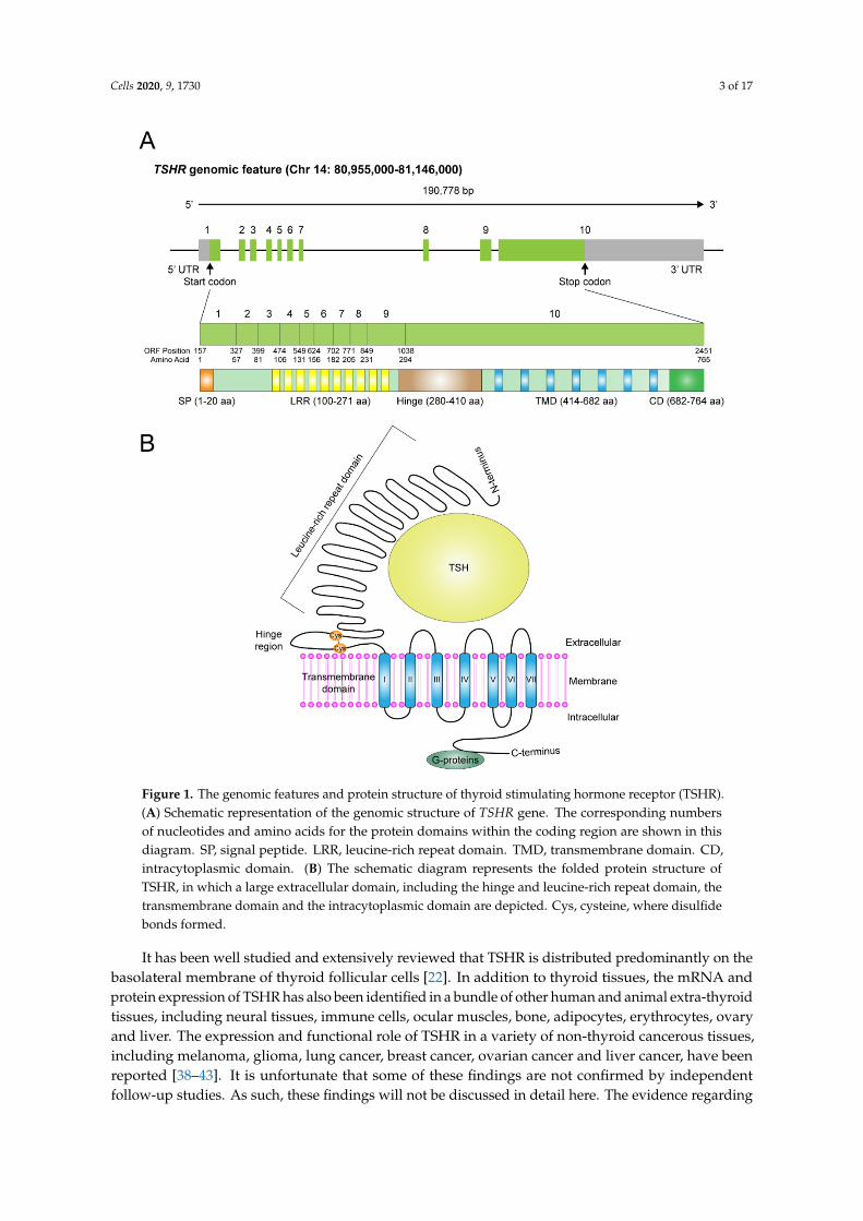
Cells 2020, 9, 1730 3 of 17Cells 2020, 9, x FOR PEER REVIEW 3 of 16
Figure 1. The genomic features and protein structure of thyroid stimulating hormone receptor (TSHR). (A) Schematic representation of the genomic structure of TSHR gene. The corresponding numbers of nucleotides and amino acids for the protein domains within the coding region are shown in this diagram. SP, signal peptide. LRR, leucine-rich repeat domain. TMD, transmembrane domain. CD, intracytoplasmic domain. (B) The schematic diagram represents the folded protein structure of TSHR, in which a large extracellular domain, including the hinge and leucine-rich repeat domain, the transmembrane domain and the intracytoplasmic domain are depicted. Cys, cysteine, where disulfide bonds formed.
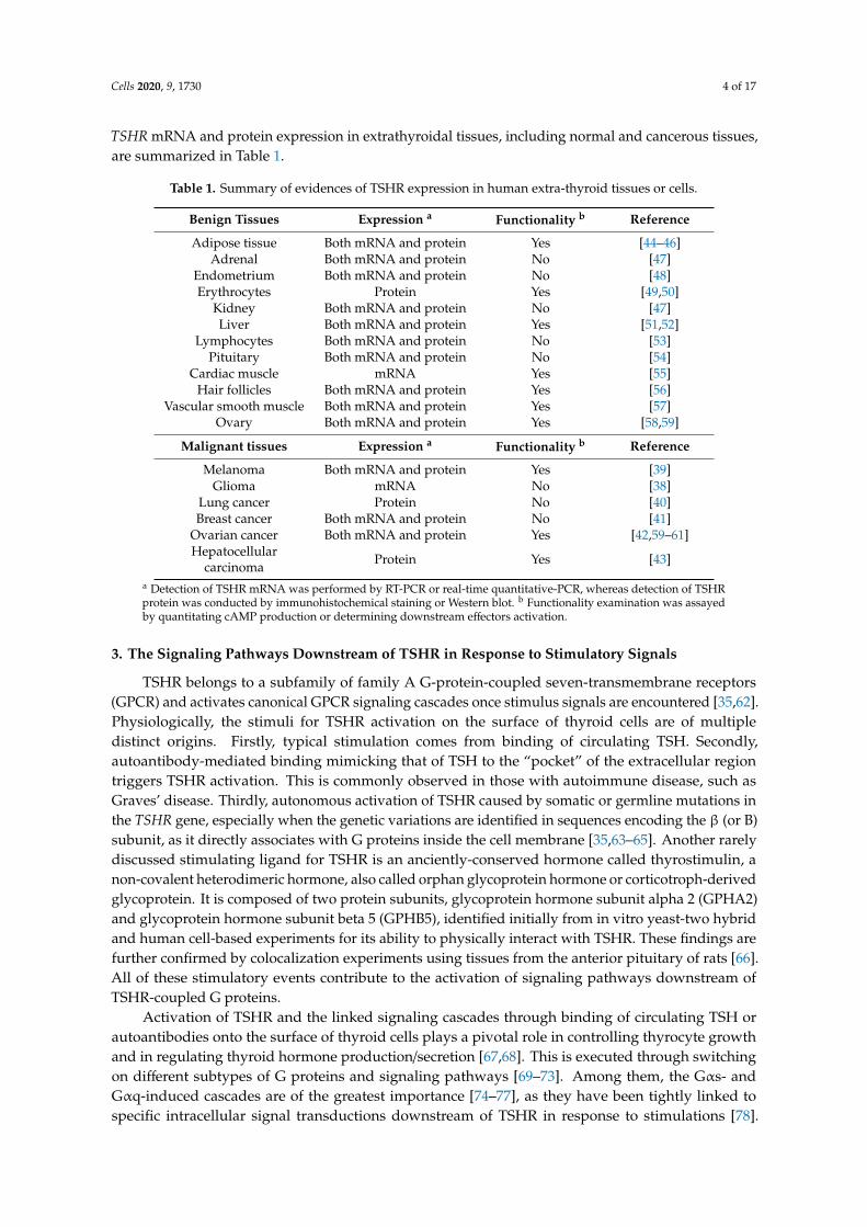
It has been well studied and extensively reviewed that TSHR is distributed predominantly on the basolateral membrane of thyroid follicular cells [22]. In addition to thyroid tissues, the mRNA and protein expression of TSHR has also been identified in a bundle of other human and animal extra-thyroid tissues, including neural tissues, immune cells, ocular muscles, bone, adipocytes, erythrocytes, ovary and liver. The expression and functional role of TSHR in a variety of non-thyroid cancerous tissues, including melanoma, glioma, lung cancer, breast cancer, ovarian cancer and liver cancer, have been reported [38–43]. It is unfortunate that some of these findings are not confirmed by independent follow-up studies. As such, these findings will not be discussed in detail here. The evidence regarding TSHR mRNA and protein expression in extrathyroidal tissues, including normal and cancerous tissues, are summarized in Table 1.
Figure 1. The genomic features and protein structure of thyroid stimulating hormone receptor (TSHR).(A) Schematic representation of the genomic structure of TSHR gene. The corresponding numbersof nucleotides and amino acids for the protein domains within the coding region are shown in thisdiagram. SP, signal peptide. LRR, leucine-rich repeat domain. TMD, transmembrane domain. CD,intracytoplasmic domain. (B) The schematic diagram represents the folded protein structure ofTSHR, in which a large extracellular domain, including the hinge and leucine-rich repeat domain, thetransmembrane domain and the intracytoplasmic domain are depicted. Cys, cysteine, where disulfidebonds formed.
It has been well studied and extensively reviewed that TSHR is distributed predominantly on thebasolateral membrane of thyroid follicular cells [22]. In addition to thyroid tissues, the mRNA andprotein expression of TSHR has also been identified in a bundle of other human and animal extra-thyroidtissues, including neural tissues, immune cells, ocular muscles, bone, adipocytes, erythrocytes, ovaryand liver. The expression and functional role of TSHR in a variety of non-thyroid cancerous tissues,including melanoma, glioma, lung cancer, breast cancer, ovarian cancer and liver cancer, have beenreported [38–43]. It is unfortunate that some of these findings are not confirmed by independentfollow-up studies. As such, these findings will not be discussed in detail here. The evidence regarding
Cells 2020, 9, 1730 4 of 17
TSHR mRNA and protein expression in extrathyroidal tissues, including normal and cancerous tissues,are summarized in Table 1.
Table 1. Summary of evidences of TSHR expression in human extra-thyroid tissues or cells.
Benign Tissues Expression a Functionality b Reference
Adipose tissue Both mRNA and protein Yes [44–46]Adrenal Both mRNA and protein No [47]
Endometrium Both mRNA and protein No [48]Erythrocytes Protein Yes [49,50]
Kidney Both mRNA and protein No [47]Liver Both mRNA and protein Yes [51,52]
Lymphocytes Both mRNA and protein No [53]Pituitary Both mRNA and protein No [54]
Cardiac muscle mRNA Yes [55]Hair follicles Both mRNA and protein Yes [56]
Vascular smooth muscle Both mRNA and protein Yes [57]Ovary Both mRNA and protein Yes [58,59]
Malignant tissues Expression a Functionality b Reference
Melanoma Both mRNA and protein Yes [39]Glioma mRNA No [38]
Lung cancer Protein No [40]Breast cancer Both mRNA and protein No [41]
Ovarian cancer Both mRNA and protein Yes [42,59–61]Hepatocellular
carcinoma Protein Yes [43]
a Detection of TSHR mRNA was performed by RT-PCR or real-time quantitative-PCR, whereas detection of TSHRprotein was conducted by immunohistochemical staining or Western blot. b Functionality examination was assayedby quantitating cAMP production or determining downstream effectors activation.
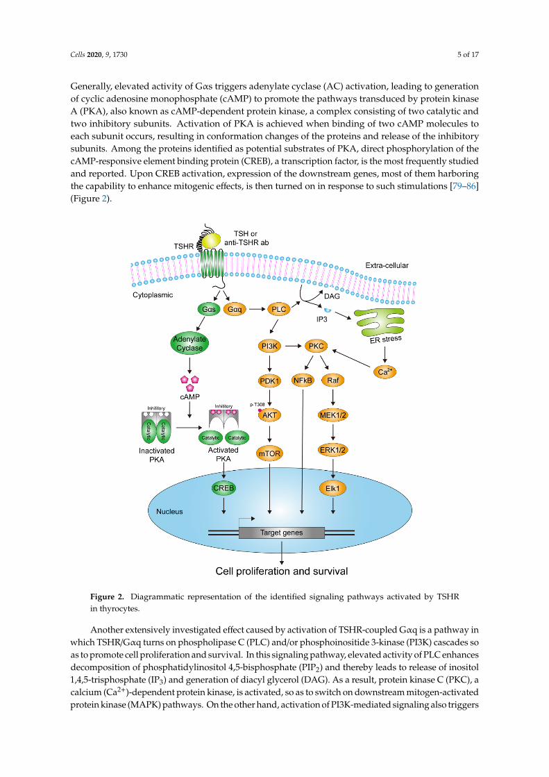
3. The Signaling Pathways Downstream of TSHR in Response to Stimulatory Signals
TSHR belongs to a subfamily of family A G-protein-coupled seven-transmembrane receptors(GPCR) and activates canonical GPCR signaling cascades once stimulus signals are encountered [35,62].Physiologically, the stimuli for TSHR activation on the surface of thyroid cells are of multipledistinct origins. Firstly, typical stimulation comes from binding of circulating TSH. Secondly,autoantibody-mediated binding mimicking that of TSH to the “pocket” of the extracellular regiontriggers TSHR activation. This is commonly observed in those with autoimmune disease, such asGraves’ disease. Thirdly, autonomous activation of TSHR caused by somatic or germline mutations inthe TSHR gene, especially when the genetic variations are identified in sequences encoding the β (or B)subunit, as it directly associates with G proteins inside the cell membrane [35,63–65]. Another rarelydiscussed stimulating ligand for TSHR is an anciently-conserved hormone called thyrostimulin, anon-covalent heterodimeric hormone, also called orphan glycoprotein hormone or corticotroph-derivedglycoprotein. It is composed of two protein subunits, glycoprotein hormone subunit alpha 2 (GPHA2)and glycoprotein hormone subunit beta 5 (GPHB5), identified initially from in vitro yeast-two hybridand human cell-based experiments for its ability to physically interact with TSHR. These findings arefurther confirmed by colocalization experiments using tissues from the anterior pituitary of rats [66].All of these stimulatory events contribute to the activation of signaling pathways downstream ofTSHR-coupled G proteins.
Activation of TSHR and the linked signaling cascades through binding of circulating TSH orautoantibodies onto the surface of thyroid cells plays a pivotal role in controlling thyrocyte growthand in regulating thyroid hormone production/secretion [67,68]. This is executed through switchingon different subtypes of G proteins and signaling pathways [69–73]. Among them, the Gαs- andGαq-induced cascades are of the greatest importance [74–77], as they have been tightly linked tospecific intracellular signal transductions downstream of TSHR in response to stimulations [78].
Cells 2020, 9, 1730 5 of 17
Generally, elevated activity of Gαs triggers adenylate cyclase (AC) activation, leading to generationof cyclic adenosine monophosphate (cAMP) to promote the pathways transduced by protein kinaseA (PKA), also known as cAMP-dependent protein kinase, a complex consisting of two catalytic andtwo inhibitory subunits. Activation of PKA is achieved when binding of two cAMP molecules toeach subunit occurs, resulting in conformation changes of the proteins and release of the inhibitorysubunits. Among the proteins identified as potential substrates of PKA, direct phosphorylation of thecAMP-responsive element binding protein (CREB), a transcription factor, is the most frequently studiedand reported. Upon CREB activation, expression of the downstream genes, most of them harboringthe capability to enhance mitogenic effects, is then turned on in response to such stimulations [79–86](Figure 2).
Cells 2020, 9, x FOR PEER REVIEW 5 of 16
adenosine monophosphate (cAMP) to promote the pathways transduced by protein kinase A (PKA), also known as cAMP-dependent protein kinase, a complex consisting of two catalytic and two inhibitory subunits. Activation of PKA is achieved when binding of two cAMP molecules to each subunit occurs, resulting in conformation changes of the proteins and release of the inhibitory subunits. Among the proteins identified as potential substrates of PKA, direct phosphorylation of the cAMP-responsive element binding protein (CREB), a transcription factor, is the most frequently studied and reported. Upon CREB activation, expression of the downstream genes, most of them harboring the capability to enhance mitogenic effects, is then turned on in response to such stimulations [79–86] (Figure 2).
Figure 2. Diagrammatic representation of the identified signaling pathways activated by TSHR in thyrocytes.
Another extensively investigated effect caused by activation of TSHR-coupled Gαq is a pathway in which TSHR/Gαq turns on phospholipase C (PLC) and/or phosphoinositide 3-kinase (PI3K) cascades so as to promote cell proliferation and survival. In this signaling pathway, elevated activity of PLC enhances decomposition of phosphatidylinositol 4,5-bisphosphate (PIP2) and thereby leads to release of inositol 1,4,5-trisphosphate (IP3) and generation of diacyl glycerol (DAG). As a result, protein kinase C (PKC), a calcium (Ca2+)-dependent protein kinase, is activated, so as to switch on downstream mitogen-activated protein kinase (MAPK) pathways. On the other hand, activation of PI3K-mediated signaling also triggers production of various 3-phosphorylated phosphoinositides, phosphatidylinositol (3,4,5)-trisphosphate (PI(3,4,5)P3), phosphatidylinositol 3-phosphate (PI3P) and
Figure 2. Diagrammatic representation of the identified signaling pathways activated by TSHRin thyrocytes.
Another extensively investigated effect caused by activation of TSHR-coupled Gαq is a pathway inwhich TSHR/Gαq turns on phospholipase C (PLC) and/or phosphoinositide 3-kinase (PI3K) cascades soas to promote cell proliferation and survival. In this signaling pathway, elevated activity of PLC enhancesdecomposition of phosphatidylinositol 4,5-bisphosphate (PIP2) and thereby leads to release of inositol1,4,5-trisphosphate (IP3) and generation of diacyl glycerol (DAG). As a result, protein kinase C (PKC), acalcium (Ca2+)-dependent protein kinase, is activated, so as to switch on downstream mitogen-activatedprotein kinase (MAPK) pathways. On the other hand, activation of PI3K-mediated signaling also triggers
Cells 2020, 9, 1730 6 of 17
production of various 3-phosphorylated phosphoinositides, phosphatidylinositol (3,4,5)-trisphosphate(PI(3,4,5)P3), phosphatidylinositol 3-phosphate (PI3P) and phosphatidylinositol (3,4)-bisphosphate(PI(3,4)P2) for instance, to recruit and activate an assorted group of signaling proteins containingspecific phosphoinositide-binding domains, such as the phosphoinositide-dependent kinase-1 (PDK1)and pleckstrin homology domains (PH domains) harboring protein AKT. The activated PDK1 thenphosphorylates AKT at the T308 residue to partially elevate its function, although exercising the fullfunctionality of AKT needs additional phosphorylation at S473 [69,70,87–89] (Figure 2).
A similar mechanism seems to be employed by the ancestrally-conserved thyrostimulin, which isestimated to have 30-fold stronger binding affinity to TSHR than TSH, acting as a circulatory hormonereleased from the anterior pituitary or a paracrine hormone secreted from neighboring tissues [59,90,91].Thyrostimulin binds to the binding sites on TSHR, where GPHA2 binds to the transmembrane domainand GPHB5 associates with the extracellular domain, leading to activation of TSHR-mediated signalingcascades. The binding between GPHA2 and TSHR has been found to be similar to that between TSHand TSHR, wherein the N-linked glycosylation at specific residues either on GPHA2 or TSH is pivotalfor activation of TSHR [92,93]. Although the existence of thyrostimulin in humans remains doubtfulcurrently, its roles and functions have been extensively investigated in a bundle of animal models (fora comprehensive review of thyrostimulin, see [90]).
4. TSHR in Thyroid Cancer Cells
Expression of TSHR is predominantly identified in thyroidal tissues, either in benign and malignantthyroid tissues, serving as a target mainly for TSH [9]. The TSHR-mediated growth of thyroid wasinitially observed in TSH-secreting pituitary adenoma and Graves’ disease [94,95]. Subsequently,multiple large cohort studies have proposed that higher serum TSH levels are linked to an increasedrisk of thyroid cancer [96]. The activated TSHR-induced signaling cascades, as mentioned above, havebeen shown to serve as oncogenic pathways in thyroid cancer, especially in tumors carrying mutationsat V600E in B-Raf proto-oncogene. Furthermore, it has been demonstrated by subsequent studiesthat TSH signaling overcomes senescence induced by BRAFV600E mutations in vitro in cell culture andin vivo in mice [97–100]. Moreover, by using a TSHR-knockout mouse model (TSHR(-/-)) to study therole of TSH/TSHR-mediated growth signaling, it was discovered that existing additional oncogenicmutation, such as that in TRbeta(PV/PV) mice, is indispensable for follicular thyroid cancer (FTC) cellsto metastasize [101], indicating that although the signaling transduction downstream of TSH/TSHRplays an important role in modulating thyroidal cell proliferation, other coexisting mutations capableof turning on other signaling cascades are required to establish an invasive phenotype. Except forthe TSH-induced activation of TSHR, there are numerous spontaneously occurring mutations locatedwithin the TSHR gene, which are identified in thyroid cancer patients. These can be either germlineor tumor-specific mutations, carrying the ability to automatously turn on downstream cascades andthereby promoting cell proliferation, such as V509A, C672Y and M453T [65,102]. Taken together,TSHR has a growth-promoting or oncogene-like function in thyroid cancer, although other co-existingmutations are required to establish a fully malignant phenotype. Nevertheless, clinically, the expressionlevels of TSHR seem to be downregulated in patients with more advanced stages of thyroid cancer,especially for those with poorly differentiated cell types, suggesting the existence of an uncharacterizedselection/adaption mechanism [103].
5. TSHR in Extra-Thyroid Cancer Cells
5.1. Ovarian Cancer
Existence of both TSHR and thyroid hormone receptor (TRs) in human ovarian tissue was initiallyreported in 2009 by Aghajanova et al., using immunohistochemical staining and reverse transcriptionpolymerase chain reaction (RT-PCR). Functionality of TSHR and TRs in primary human ovarian tissuesunder TSH or thyroid hormone stimulation was also demonstrated through assessing downstream
Cells 2020, 9, 1730 7 of 17
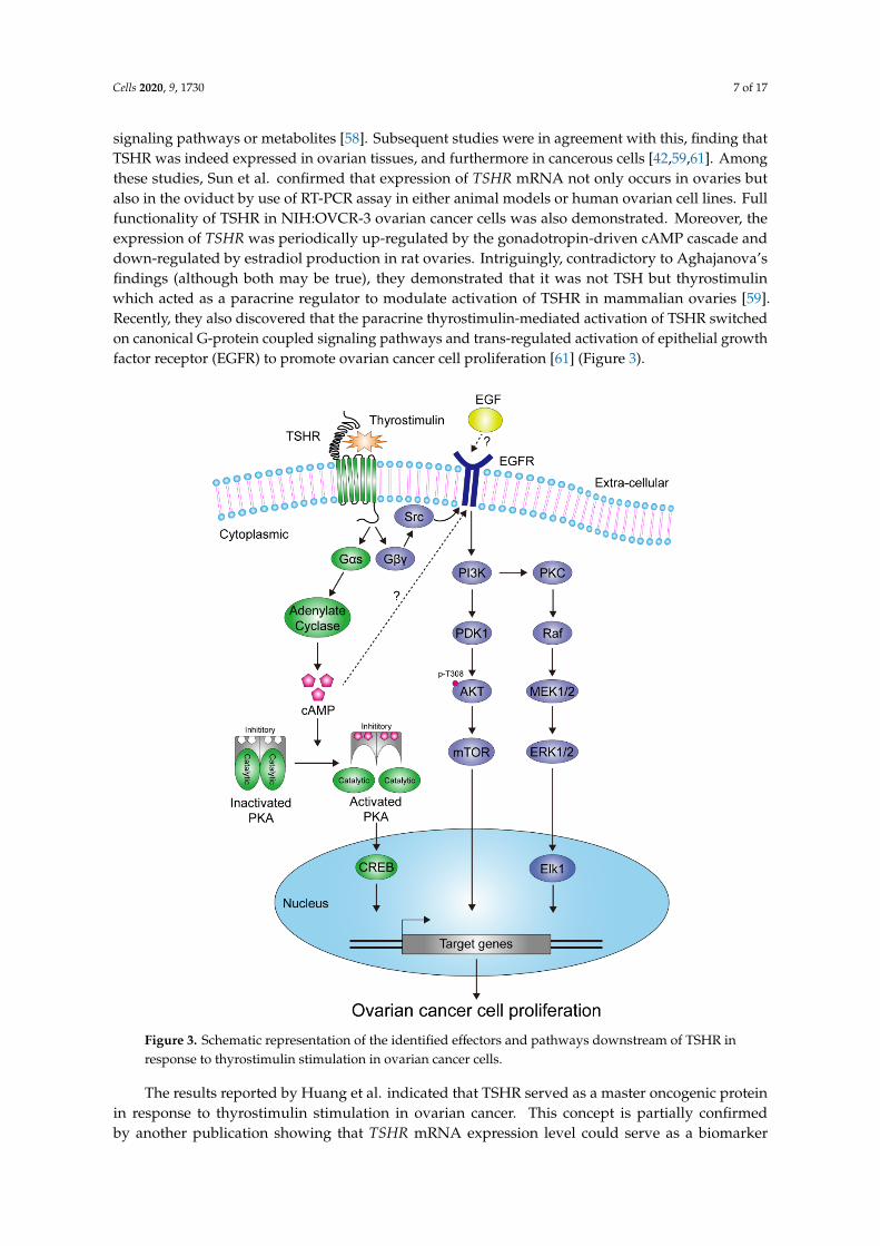
signaling pathways or metabolites [58]. Subsequent studies were in agreement with this, finding thatTSHR was indeed expressed in ovarian tissues, and furthermore in cancerous cells [42,59,61]. Amongthese studies, Sun et al. confirmed that expression of TSHR mRNA not only occurs in ovaries butalso in the oviduct by use of RT-PCR assay in either animal models or human ovarian cell lines. Fullfunctionality of TSHR in NIH:OVCR-3 ovarian cancer cells was also demonstrated. Moreover, theexpression of TSHR was periodically up-regulated by the gonadotropin-driven cAMP cascade anddown-regulated by estradiol production in rat ovaries. Intriguingly, contradictory to Aghajanova’sfindings (although both may be true), they demonstrated that it was not TSH but thyrostimulinwhich acted as a paracrine regulator to modulate activation of TSHR in mammalian ovaries [59].Recently, they also discovered that the paracrine thyrostimulin-mediated activation of TSHR switchedon canonical G-protein coupled signaling pathways and trans-regulated activation of epithelial growthfactor receptor (EGFR) to promote ovarian cancer cell proliferation [61] (Figure 3).
Cells 2020, 9, x FOR PEER REVIEW 7 of 16
in ovaries but also in the oviduct by use of RT-PCR assay in either animal models or human ovarian cell lines. Full functionality of TSHR in NIH:OVCR-3 ovarian cancer cells was also demonstrated. Moreover, the expression of TSHR was periodically up-regulated by the gonadotropin-driven cAMP cascade and down-regulated by estradiol production in rat ovaries. Intriguingly, contradictory to Aghajanova’s findings (although both may be true), they demonstrated that it was not TSH but thyrostimulin which acted as a paracrine regulator to modulate activation of TSHR in mammalian ovaries [59]. Recently, they also discovered that the paracrine thyrostimulin-mediated activation of TSHR switched on canonical G-protein coupled signaling pathways and trans-regulated activation of epithelial growth factor receptor (EGFR) to promote ovarian cancer cell proliferation [61] (Figure 3).
Figure 3. Schematic representation of the identified effectors and pathways downstream of TSHR in response to thyrostimulin stimulation in ovarian cancer cells.
The results reported by Huang et al. indicated that TSHR served as a master oncogenic protein in response to thyrostimulin stimulation in ovarian cancer. This concept is partially confirmed by another publication showing that TSHR mRNA expression level could serve as a biomarker to predict the response of adjuvant intravenous platinum-taxane chemotherapy in patients with ovarian cancer, wherein those with increased TSHR mRNA level are associated with poorer clinical outcomes [60]. These lines of evidence partially uncover the growth-promoting role of TSH/TSHR and/or thyrostimulin/TSHR in ovary and ovarian cancer. Nevertheless, the details of underlying
Figure 3. Schematic representation of the identified effectors and pathways downstream of TSHR inresponse to thyrostimulin stimulation in ovarian cancer cells.
The results reported by Huang et al. indicated that TSHR served as a master oncogenic proteinin response to thyrostimulin stimulation in ovarian cancer. This concept is partially confirmedby another publication showing that TSHR mRNA expression level could serve as a biomarker
Cells 2020, 9, 1730 8 of 17
to predict the response of adjuvant intravenous platinum-taxane chemotherapy in patients withovarian cancer, wherein those with increased TSHR mRNA level are associated with poorer clinicaloutcomes [60]. These lines of evidence partially uncover the growth-promoting role of TSH/TSHRand/or thyrostimulin/TSHR in ovary and ovarian cancer. Nevertheless, the details of underlyingmechanisms by which TSHR modulates oncogenesis or tumor growth of ovarian cancer, and newstrategies designed accordingly to intervene these processes, still await further exploration.
5.2. Liver Cancer
The presence of TSHR on the cell surface of human liver tissues as well as a hepatocyte cell line(L-02) was first demonstrated by Zhang et al. via RT-PCR and immuno-fluorescence assay (IFA),respectively [51]. The functionality of TSHR, driving cAMP accumulation in response to the stimulationof bovine TSH (bTSH) and immunoglobulins isolated from patients with Graves’ disease, was alsodemonstrated in this study. Subsequently, these findings were confirmed in an animal model byshowing that TSH/TSHR played a role in maintaining hepatic blood glucose, triacylglycerol and bile acidhomeostasis [104–106]. Lines of evidence from other studies also indicated that TSH/TSHR modulatedprogression of liver-associated metabolic diseases in human and in mice model [52,106–110]. Recently,it has been unveiled that abnormality in metabolic processes can lead to enhanced carcinogenesisand/or metastasis of several cancers, including liver cancer [111]. However, it was not until 2018 thatShih and coworkers demonstrated that the levels of TSHR predicted clinical outcomes of patientswith hepatocellular carcinoma (HCC), of which poorer survival rates were observed in those withhigher TSHR expression, and the presence of TSH altered sensitivity of HCC cells to cisplatin, achemotherapeutic drug for HCC, in vitro [43]. Moreover, several mutations within exon 10 of theTSHR gene were identified in HCC tissues, despite the fact that the roles of these mutants in promotingtumorigenesis or cancer progression in HCC remained uncharacterized.
In liver cancer, another frequently debated issue is the roles of the thyroid hormones T3 andT4 in tumorigenesis, cell growth and metastasis. Thyroid hormones might impact HCC growththrough various pathways. According to current understandings in this field, most of the literaturehas considered T3/T4 a potential tumor suppressor, while others have considered them to be oncogenicpromoters in HCC (for a comprehensive review of the effects of thyroid hormones in HCC, see [112]).Some of these studies provided a possible explanation for the finding that higher TSH and freeT4 levels in blood were associated with poorer prognoses and larger tumor size in unresectableHCC patients [113]. Intriguingly, another independent study showed that higher TSH or free T4concentrations in blood were associated with better clinical outcomes in HCC patients receivingchemotherapy, but not sorafenib (a targeted anticancer drug) treatment [114]. According to this study,it might be better to treat HCC patients with higher TSH and free T4 levels by chemotherapy instead ofthe targeted drug sorafenib, although the mechanisms behind these observations still require futureelucidation. Overall, these results partly unveil the role and applicability of TSH/T4 in liver cancertreatment. Notably, the oncogenic role of thyroid hormones (not TSH/TSHR) in HCC is still underdebate [115,116].
6. Clinical Usefulness of TSHR Related Pathways in Anticancer Therapies
For thyroid cancer, there are lines of evidence revealing a strong association between high levelsof T4 or T3 and reduced thyroid cancer growth, as thyroid hormones suppress TSH secretion fromthe pituitary gland [117]. Subsequently, by using levothyroxine, an artificially synthesized mimic ofT4, to treat patients, the sizes of thyroid nodules are reduced and metastases of thyroid cancer arerestrained [118–121]. Indirect TSH suppression by exogenous levothyroxine administration remains amainstay of clinical management for differentiated thyroid cancer nowadays, especially for patientsunder high risk of recurrence [121]. However, a more individualized treatment strategy regarding TSHsuppression has to be adopted, because it is of little benefit for patients with low risk of recurrence andthere is an increased risk of bone loss and cardiovascular diseases, owing to adverse effects caused by
Cells 2020, 9, 1730 9 of 17
exogenous subclinical hyperthyroidism [122,123]. On the other hand, the TSHR-mediated upregulationof sodium-iodide symporter expression is frequently used to enhance the radioiodine uptake in ablativeradioiodine therapy. This can be achieved by means of either endogenous TSH stimulation throughthyroxine withdrawal or exogeneous administration of recombinant human TSH [8]. Additionally,evidence from clinical studies shows that expression levels of TSHR serve as a marker to predict theefficacy of these therapies [124,125].
Several small molecule kinase inhibitors, identified as thyroid cancer cell proliferationrepressors, rather than as tumoricidal, have been used as the first-line strategy to treat progressiveradioiodine-resistant differentiated thyroid cancer. They can provide prolonged progression-freesurvival but not a cure [8]. Moreover, the toxicities of this class of agents hinder its widespread use. Acombination or sequential treatment strategy to apply the TSHR-mediated pathway manipulation andkinase inhibitors may be considered in the future.
In order to improve the therapeutic strategies for cancer, the term “theranostics” has been created.Owing to the dedication of scientists in this field, several novel strategies and methods to deliverdrugs have been established to minimize the off-target/mis-target effects and maximize the properdelivery of drugs to where they are needed. These include several attractive models of nanoliposomes,organ-specific targeting and immune system evasion methods, which can be achieved by moleculesembedded in the lipid bilayer. To target the TSH/TSHR pathway, the nanoliposomes coated withfragments of TSH were used to compete with the binding of TSHR in vitro and in vivo [126]. In thisstudy, the researchers showed that the nanoliposomes packed with gemcitabine, a chemotherapeuticagent, exhibited higher efficacy against the proliferation of thyroid cancer cell line FTC-133 in vitrothan uncoating TSH liposomal gemcitabine and free gemcitabine. A similar study confirmed suchnotions by packing cisplatin [127]. These studies indeed provide a novel strategy, taking advantage ofthe TSH/TSHR pathway to treat thyroid cancer. Inconvenience exists, however, as liposomes needto be used freshly and are unstable under long-term storage, raising the needs of an alternative orimproved design.
Pharmacologic suppression of endogenous TSH via the negative feedback loop for the purpose ofinhibiting TSHR-mediated growth signaling often leads to subclinical hyperthyroidism. Consideringthe stability and easily manufactured nature of protein or peptidyl drugs, it may be more reasonableto develop antagonists of TSHR to avoid systemic side effects, while manifesting effectiveness toinhibit thyroid cancer growth. Such small-molecule antagonists of TSHR attract much attentionfrom scientists who are dedicated to developing new treatments for Graves’ disease, a pathologicalcondition caused by binding of stimulatory autoantibodies to TSHR to induce hyperthyroidism.However, unsatisfactory results remain, as most molecules cannot achieve sufficient specificity in vivoto substantially inhibit TSHR signaling, although several synthesized compounds have been generatedthat are capable of suppressing TSHR expression or its downstream signaling to a certain extent [128].These compounds, including antagonists and inverse agonists, are of minimal/mild effectiveness insuppressing TSHR signal transduction, as only around 50% and 39% inhibitory effects for cAMPproduction could be achieved when stimulated by TSH or sera derived from patients with Graves’disease, respectively [129,130]. Recently, a novel small molecule antagonist of TSHR has been identifiedby drug screening. The researchers claimed that a selective inhibitory effect could be observed in TSHRbut not in those closely related receptors, such as follitropin and lutropin receptors [131]. Indeed, itwould be very helpful if an oral small molecule drug can be approved for clinical use to treat patientswith thyroid cancer or Graves’ disease, but there remains a long way to go, as the present findings arestill limited to cell-based assays and mouse models, rather than clinical trials.
On the other hand, it has not been explored whether patients with extra-thyroid cancers canbe treated by targeting TSHR or the related pathways. Only a few studies have demonstrated suchpotentiality in ovarian and liver cancers [43,60,114]. Directly targeting TSHR in extra-thyroid cancerseems to be impractical, as the systemic effect caused by such treatment is difficult to avoid. TSHRexpression is most abundant in thyroid cells, compared to all other extra-thyroid tissues or organs. As
Cells 2020, 9, 1730 10 of 17
such, unless a specific liver or ovarian cancer delivering or targeting system can be developed, theTSHR-targeting strategy cannot be applied to extra-thyroid cancers. However, by use of the indexobtained from the levels of TSH and free T4, one can select a better strategy to treat patients withHCC [113,114].
7. Conclusions
The molecular function and clinical role of TSHR have been extensively explored in both benignand malignant thyroid tissues. Accordingly, it has been proposed that TSHR might serve as a candidatetarget for anticancer therapy [103]. Our current understandings of the anticancer role of TSHR inthyroid cancer come mostly from the effectiveness of the therapeutic strategies manipulating theTSH/TSHR signaling pathways, for example, by administration of levothyroxine. Such strategiesremain largely unchanged during the past decades.
In addition to thyroid cancer, the aforementioned strategies targeting the TSH/TSHR pathwayscan potentially be used to fight against extra-thyroid cancers, provided that the malignant tissuesharbor higher expression levels of TSHR on the surface of cancerous cells. To date, expressionof TSHR mRNA and protein have been detected in a variety of tissues and extra-thyroid cancers,although our knowledge regarding the functional characteristics of these extra-thyroid TSHR remainslimited. Several difficulties have to be overcome, however, prior to a possible clinical application.For example, the expression levels of TSHR in the extra-thyroid cancers have to be high enough thatthe alteration of TSHR function induced by the cognate ligands is great enough to elicit the desiredanticancer responses. It is highly likely that the TSHR expression can vary not only cancer-to-cancerbut also patient-to-patient.
In the era of precision medicine and targeted therapy, TSHR has been proposed to be a potenttarget against thyroid cancer with several experimental compounds under development. This potentialis supported by the success of the therapeutic strategies to manipulate TSH/TSHR function in treatingdifferentiated thyroid cancer. Although there is hope for targeting TSHR in the treatment of extra-thyroidcancers, much work needs to be done before this becomes a clinical reality.
Author Contributions: Conceptualization and design, Y.-D.C. and C.-T.Y.; literature review: Y.-D.C. and C.-T.Y.;writing—original draft preparation: Y.-D.C. and C.-T.Y.; writing—review and editing, C.-T.Y.; supervision, C.-T.Y.All authors have read and agreed to the published version of the manuscript.
Funding: This research received no external funding.
Acknowledgments: The authors appreciate the services from the Electron Libraries of Chang Gung MemorialHospital and Chang-Gung University for allowing literature searches.
Conflicts of Interest: The authors declare no conflict of interest.
References
1. Neumann, S.; Raaka, B.M.; Gershengorn, M.C. Human TSH receptor ligands as pharmacological probeswith potential clinical application. Expert Rev. Endocrinol. Metab. 2009, 4, 669–679. [CrossRef]
2. Troppmann, B.; Kleinau, G.; Krause, G.; Gromoll, J. Structural and functional plasticity of the luteinizinghormone/choriogonadotrophin receptor. Hum. Reprod Update 2013, 19, 583–602. [CrossRef]
3. Persani, L.; Gelmini, G.; Marelli, F.; Beck-Peccoz, P.; Bonomi, M. Syndromes of resistance to TSH. Ann.Endocrinol. 2011, 72, 60–63. [CrossRef]
4. Schroeder, A.C.; Privalsky, M.L. Thyroid hormones, t3 and t4, in the brain. Front. Endocrinol. 2014, 5, 40.[CrossRef] [PubMed]
5. Maenhaut, C.; Brabant, G.; Vassart, G.; Dumont, J.E. In vitro and in vivo regulation of thyrotropin receptormRNA levels in dog and human thyroid cells. J. Biol. Chem. 1992, 267, 3000–3007.
6. Bruno, R.; Ferretti, E.; Tosi, E.; Arturi, F.; Giannasio, P.; Mattei, T.; Scipioni, A.; Presta, I.; Morisi, R.; Gulino, A.;et al. Modulation of thyroid-specific gene expression in normal and nodular human thyroid tissues fromadults: An in vivo effect of thyrotropin. J. Clin. Endocrinol. Metab. 2005, 90, 5692–5697. [CrossRef] [PubMed]
Cells 2020, 9, 1730 11 of 17
7. Schuppert, F.; Deiters, S.; Rambusch, E.; Sierralta, W.; Dralle, H.; Mühlen, A.V.Z. TSH-receptor expressionand human thyroid disease: Relation to clinical, endocrine, and molecular thyroid parameters. Thyroid 1996,6, 575–587. [CrossRef] [PubMed]
8. Haugen, B.R.; Alexander, E.K.; Bible, K.C.; Doherty, G.M.; Mandel, S.J.; Nikiforov, Y.E.; Pacini, F.;Randolph, G.W.; Sawka, A.M.; Schlumberger, M.; et al. 2015 American Thyroid Association ManagementGuidelines for Adult Patients with Thyroid Nodules and Differentiated Thyroid Cancer: The AmericanThyroid Association Guidelines Task Force on Thyroid Nodules and Differentiated Thyroid Cancer. Thyroid2016, 26, 1–133. [CrossRef]
9. Williams, G.R. Extrathyroidal expression of TSH receptor. Ann. Endocrinol 2011, 72, 68–73. [CrossRef]10. Pastan, I.; Roth, J.; Macchia, V. Binding of hormone to tissue: The first step in polypeptide hormone action.
Proc. Natl. Acad. Sci. USA 1966, 56, 1802–1809. [CrossRef]11. Amir, S.M.; Carraway, T.F., Jr.; Kohn, L.D.; Winand, R.J. The Binding of Thyrotropin to Isolated Bovine
Thyroid Plasma Membranes. J. Biol. Chem. 1973, 248, 4092–4100.12. Parmentier, M.; Libert, F.; Maenhaut, C.; Lefort, A.; Gerard, C.; Perret, J.; Sande, J.V.; Dumont, J.; Vassart, G.
Molecular cloning of the thyrotropin receptor. Science 1989, 246, 1620–1622. [CrossRef] [PubMed]13. Nagayama, Y.; Kaufman, K.D.; Seto, P.; Rapoport, B. Molecular cloning, sequence and functional expression
of the cDNA for the human thyrotropin receptor. Biochem. Biophys. Res. Commun. 1989, 165, 1184–1190.[CrossRef]
14. Misrahi, M.; Loosfelt, H.; Atger, M.; Sar, S.; Guiochon-Mantel, A.; Milgrom, E. Cloning, sequencing andexpression of human TSH receptor. Biochem. Biophys. Res. Commun. 1990, 166, 394–403. [CrossRef]
15. Akamizu, T. Antithyrotropin Receptor Antibody: An Update. Thyroid 2001, 11, 1123–1134. [CrossRef][PubMed]
16. Bahn, R.S. Autoimmunity and Graves’ disease. Clin. Pharm. 2012, 91, 577–579. [CrossRef] [PubMed]17. Bahn, R.S.; Dutton, C.M.; Natt, N.; Joba, W.; Spitzweg, C.; Heufelder, A.E. Thyrotropin receptor expression
in Graves’ orbital adipose/connective tissues: Potential autoantigen in Graves’ ophthalmopathy. J. Clin.Endocrinol. Metab. 1998, 83, 998–1002. [CrossRef] [PubMed]
18. Davies, T.F. Thyrotropin receptor-associated diseases: From adenomata to Graves disease. J. Clin. Investig.2005, 115, 1972–1983. [CrossRef]
19. Kohn, L.D.; Harii, N. Thyrotropin receptor autoantibodies (TSHRAbs): Epitopes, origins and clinicalsignificance. Autoimmunity 2003, 36, 331–337. [CrossRef]
20. Ludgate, M. Animal models of Graves’ disease. Eur. J. Endocrinol. 2000, 142, 1–8. [CrossRef] [PubMed]21. Michalek, K.; Morshed, S.A.; Latif, R.; Davies, T.F. TSH receptor autoantibodies. Autoimmun. Rev. 2009, 9,
113–116. [CrossRef] [PubMed]22. Rapoport, B.; Chazenbalk, G.D.; Jaume, J.C.; Mclachlan, S.M. The thyrotropin (TSH) receptor: Interaction
with TSH and autoantibodies. Endocr. Rev. 1998, 19, 673–716.23. Rapoport, B.; McLachlan, S.M. The Thyrotropin Receptor in Graves’ Disease. Thyroid 2007, 17, 911–922.
[CrossRef] [PubMed]24. Diana, T.; Olivo, P.D.; Kahaly, G.J. Thyrotropin Receptor Blocking Antibodies. Horm. Metab. Res. 2018, 50,
853–862. [CrossRef] [PubMed]25. Davies, T.F.; Yin, X.; Latif, R. The genetics of the thyroid stimulating hormone receptor: History and relevance.
Thyroid 2010, 20, 727–736. [CrossRef] [PubMed]26. Russo, D.; Chazenbalk, G.D.; Nagayama, Y.; Wadsworth, H.L.; Seto, P.; Rapoport, B. A new structural model
for the thyrotropin (TSH) receptor, as determined by covalent cross-linking of TSH to the recombinantreceptor in intact cells: Evidence for a single polypeptide chain. Mol. Endocrinol. 1991, 5, 1607–1612.[CrossRef] [PubMed]
27. Endo, T.; Ikeda, M.; Ohmori, M.; Anzai, E.; Haraguchi, K.; Onaya, T. Single subunit structure of the humanthyrotropin receptor. Biochem. Biophys. Res. Commun. 1992, 187, 887–893. [CrossRef]
28. Chazenbalk, G.D.; Kakinuma, A.; Jaume, J.C.; Mclachlan, S.M.; Rapoport, B. Evidence for negativecooperativity among human thyrotropin receptors overexpressed in mammalian cells. Endocrinology1996, 137, 4586–4591. [CrossRef]
29. Chen, C.-R.; Chazenbalk, G.D.; Wawrowsky, K.A.; McLachlan, S.M.; Rapoport, B. Evidence that humanthyroid cells express uncleaved, single-chain thyrotropin receptors on their surface. Endocrinology 2006, 147,3107–3113. [CrossRef]
Cells 2020, 9, 1730 12 of 17
30. Buckland, P.R.; Howells, R.D.; Rickards, C.R.; Smith, B.R. Affinity-labelling of the thyrotropin receptor.Characterization of the photoactive ligand. Biochem. J. 1985, 225, 753–760. [CrossRef] [PubMed]
31. Kajita, Y.; Rickards, C.R.; Buckland, P.R.; Howells, R.D.; Smith, B.R. Analysis of thyrotropin receptors byphotoaffinity labelling. Orientation of receptor subunits in the cell membrane. Biochem. J. 1985, 227, 413–420.[CrossRef]
32. Buckland, P.R.; Strickland, T.W.; Pierce, J.G.; Smith, B.R. TSH crosslinks to the TSH receptor through the betasubunit. Endocrinology 1985, 116, 2122–2124. [CrossRef]
33. Loosfelt, H.; Pichon, C.; Jolivet, A.; Misrahi, M.; Caillou, B.; Jamous, M.; Vannier, B.; Milgrom, E. Two-subunitstructure of the human thyrotropin receptor. Proc. Natl. Acad. Sci. USA 1992, 89, 3765–3769. [CrossRef][PubMed]
34. Rapoport, B.; McLachlan, S.M. TSH Receptor Cleavage into Subunits and Shedding of the A-Subunit; AMolecular and Clinical Perspective. Endocr. Rev. 2016, 37, 114–134. [CrossRef]
35. Kleinau, G.; Worth, C.L.; Kreuchwig, A.; Biebermann, H.; Marcinkowski, P.; Scheerer, P.; Krause, G.Structural-Functional Features of the Thyrotropin Receptor: A Class A G-Protein-Coupled Receptor at Work.Front. Endocrinol. 2017, 8, 86. [CrossRef] [PubMed]
36. Kursawe, R.; Paschke, R. Modulation of TSHR signaling by posttranslational modifications. Trends Endocrinol.Metab. 2007, 18, 199–207. [CrossRef] [PubMed]
37. Zabczynska, M.; Kozlowska, K.; Pochec, E. Glycosylation in the Thyroid Gland: Vital Aspects of GlycoproteinFunction in Thyrocyte Physiology and Thyroid Disorders. Int. J. Mol. Sci. 2018, 19. [CrossRef]
38. Vastrad, B.; Vastrad, C.; Godavarthi, A.; Chandrashekar, R. Molecular mechanisms underlying gliomas andglioblastoma pathogenesis revealed by bioinformatics analysis of microarray data. Med. Oncol. 2017, 34, 182.[CrossRef]
39. Ellerhorst, J.A.; Sendi-Naderi, A.; Johnson, M.K.; Cooke, C.P.; Dang, S.M.; Diwan, A.H. Human melanomacells express functional receptors for thyroid-stimulating hormone. Endocr. Relat. Cancer 2006, 13, 1269–1277.[CrossRef] [PubMed]
40. Kim, J.W.; Lee, S.; Lui, N.; Choi, H.; Mulvihill, M.; Fang, L.T.; Kang, H.C.; Kwon, Y.W.; Jablons, D.; Kim, I.J. Asomatic TSHR mutation in a patient with lung adenocarcinoma with bronchioloalveolar carcinoma, coronaryartery disease and severe chronic obstructive pulmonary disease. Oncol. Rep. 2012, 28, 1225–1230. [CrossRef][PubMed]
41. Govindaraj, V.; Yaduvanshi, N.S.; Krishnamachar, H.; Rao, A.J. Expression of thyroid-stimulating hormonereceptor, octamer-binding transcription factor 4, and intracisternal A particle-promoted polypeptide inhuman breast cancer tissues. Horm. Mol. Biol. Clin. Investig 2012, 9, 173–178. [CrossRef] [PubMed]
42. Gyftaki, R.; Liacos, C.; Politi, E.; Liontos, M.; Saltiki, K.; Papageorgiou, T.; Thomakos, N.; Haidopoulos, D.;Rodolakis, A.; Alevizaki, M.; et al. Differential transcriptional and protein expression of thyroid-stimulatinghormone receptor in ovarian carcinomas. Int. J. Gynecol. Cancer 2014, 24, 851–856. [PubMed]
43. Shih, Y.L.; Huang, Y.H.; Lin, K.H.; Chu, Y.D.; Yeh, C.T. Identification of Functional Thyroid StimulatingHormone Receptor and TSHR Gene Mutations in Hepatocellular Carcinoma. Anticancer Res. 2018, 38,2793–2802. [PubMed]
44. Janson, A.; Rawet, H.; Perbeck, L.; Marcus, C. Presence of thyrotropin receptor in infant adipocytes. Pediatr.Res. 1998, 43, 555–558. [CrossRef]
45. Bell, A.; Gagnon, A.; Grunder, L.; Parikh, S.J.; Smith, T.J.; Sorisky, A. Functional TSH receptor in humanabdominal preadipocytes and orbital fibroblasts. Am. J. Physiol. Cell Physiol. 2000, 279, C335–C340. [CrossRef]
46. Murakami, M.; Kamiya, Y.; Morimura, T.; Araki, O.; Imamura, M.; Ogiwara, T.; Mizuma, H.; Mori, M.Thyrotropin receptors in brown adipose tissue: Thyrotropin stimulates type II iodothyronine deiodinase anduncoupling protein-1 in brown adipocytes. Endocrinology 2001, 142, 1195–1201. [CrossRef]
47. Dutton, C.M.; Joba, W.; Spitzweg, C.; Heufelder, A.E.; Bahn, R.S. Thyrotropin receptor expression in adrenal,kidney, and thymus. Thyroid 1997, 7, 879–884. [CrossRef]
48. Aghajanova, L.; Stavreus-Evers, A.; Lindeberg, M.; Landgren, B.M.; Sparre, L.S.; Hovatta, O.Thyroid-stimulating hormone receptor and thyroid hormone receptors are involved in human endometrialphysiology. Fertil. Steril. 2011, 95, 230–237. [CrossRef]
49. Balzan, S.; Nicolini, G.; Forini, F.; Boni, G.; Del Carratore, R.; Nicolini, A.; Carpi, A.; Iervasi, G. Presence of afunctional TSH receptor on human erythrocytes. Biomed. Pharm. 2007, 61, 463–467. [CrossRef]
Cells 2020, 9, 1730 13 of 17
50. Balzan, S.; Carratore, R.D.; Nicolini, G.; Forini, F.; Lubrano, V.; Simili, M.; Benedetti, P.A.; Iervasi, G. TSHinduces co-localization of TSH receptor and Na/K-ATPase in human erythrocytes. Cell Biochem. Funct. 2009,27, 259–263. [CrossRef]
51. Zhang, W.; Tian, L.M.; Han, Y.; Ma, H.Y.; Wang, L.C.; Guo, J.; Gao, L.; Zhao, J.J. Presence of thyrotropinreceptor in hepatocytes: Not a case of illegitimate transcription. J. Cell. Mol. Med. 2009, 13, 4636–4642.[CrossRef]
52. Li, Y.; Wang, L.; Zhou, L.; Song, Y.; Ma, S.; Yu, C.; Zhao, J.; Xu, C.; Gao, L. Thyroid stimulating hormoneincreases hepatic gluconeogenesis via CRTC2. Mol. Cell Endocrinol. 2017, 446, 70–80. [CrossRef] [PubMed]
53. Coutelier, J.-P.; Kehrl, J.H.; Bellur, S.S.; Kohn, L.D.; Notkins, A.L.; Prabhakar, B.S. Binding and functionaleffects of thyroid stimulating hormone on human immune cells. J. Clin. Immunol. 1990, 10, 204–210.[CrossRef] [PubMed]
54. Prummel, M.F.; Brokken, L.J.S.; Meduri, G.; Misrahi, M.; Bakker, O.; Wiersinga, W.M. Expression of thethyroid-stimulating hormone receptor in the folliculo-stellate cells of the human anterior pituitary. J. Clin.Endocrinol. Metab. 2000, 85, 4347–4353. [CrossRef] [PubMed]
55. Drvota, V.; Janson, A.; Norman, C.; Sylven, C.; Haggblad, J.; Bronnegard, M.; Marcus, C. Evidence for thepresence of functional thyrotropin receptor in cardiac muscle. Biochem. Biophys. Res. Commun. 1995, 211,426–431. [CrossRef] [PubMed]
56. Bodo, E.; Kromminga, A.; Biro, T.; Borbiro, I.; Gaspar, E.; Zmijewski, M.A.; van Beek, N.; Langbein, L.;Slominski, A.T.; Paus, R. Human female hair follicles are a direct, nonclassical target for thyroid-stimulatinghormone. J. Invest. Derm. 2009, 129, 1126–1139. [CrossRef] [PubMed]
57. Tian, L.; Ni, J.; Guo, T.; Liu, J.; Dang, Y.; Guo, Q.; Zhang, L. TSH stimulates the proliferation of vascularsmooth muscle cells. Endocrine 2014, 46, 651–658. [CrossRef]
58. Aghajanova, L.; Lindeberg, M.; Carlsson, I.B.; Stavreus-Evers, A.; Zhang, P.; Scott, J.E.; Hovatta, O.;Skjöldebrand-Sparre, L. Receptors for thyroid-stimulating hormone and thyroid hormones in human ovariantissue. Reprod. Biomed. Online 2009, 18, 337–347. [CrossRef]
59. Sun, S.C.; Hsu, P.J.; Wu, F.J.; Li, S.H.; Lu, C.H.; Luo, C.W. Thyrostimulin, but not thyroid-stimulating hormone(TSH), acts as a paracrine regulator to activate the TSH receptor in mammalian ovary. J. Biol. Chem. 2010,285, 3758–3765. [CrossRef] [PubMed]
60. Seagle, B.L.; Eng, K.H.; Yeh, J.Y.; Dandapani, M.; Schiller, E.; Samuelson, R.; Odunsi, K.; Shahabi, S. Discoveryof candidate tumor biomarkers for treatment with intraperitoneal chemotherapy for ovarian cancer. Sci. Rep.2016, 6, 21591. [CrossRef]
61. Huang, W.L.; Li, Z.; Lin, T.Y.; Wang, S.W.; Wu, F.J.; Luo, C.W. Thyrostimulin-TSHR signaling promotes theproliferation of NIH:OVCAR-3 ovarian cancer cells via trans-regulation of the EGFR pathway. Sci. Rep. 2016,6, 27471. [CrossRef] [PubMed]
62. Vassart, G.; Pardo, L.; Costagliola, S. A molecular dissection of the glycoprotein hormone receptors. TrendsBiochem. Sci. 2004, 29, 119–126. [CrossRef] [PubMed]
63. Tuncel, M. Thyroid Stimulating Hormone Receptor. Mol. Imaging. Radionucl. 2017, 26, 87–91. [CrossRef][PubMed]
64. Kleinau, G.; Neumann, S.; Grüters, A.; Krude, H.; Biebermann, H. Novel Insights on Thyroid-StimulatingHormone Receptor Signal Transduction. Endocr. Rev. 2013, 34, 691–724. [CrossRef]
65. Duprez, L.; Parma, J.; Sande, J.V.; Allgeier, A.; Leclère, J.; Schvartz, C.; Delisle, M.-J.; Decoulx, M.; Orgiazzi, J.;Dumont, J.; et al. Germline mutations in the thyrotropin receptor gene cause non-autoimmune autosomaldominant hyperthyroidism. Nat. Genet. 1994, 7, 396–401. [CrossRef]
66. Hsu, S.Y.; Nakabayashi, K.; Bhalla, A. Evolution of Glycoprotein Hormone Subunit Genes in BilateralMetazoa: Identification of Two Novel Human Glycoprotein Hormone Subunit Family Genes, GPA2 andGPB5. Mol. Endocrinol. 2002, 16, 1538–1551. [CrossRef]
67. Postiglione, M.P.; Parlato, R.; Rodriguez-Mallon, A.; Rosica, A.; Mithbaokar, P.; Maresca, M.; Marians, R.C.;Davies, T.F.; Zannini, M.S.; De Felice, M.; et al. Role of the thyroid-stimulating hormone receptor signalingin development and differentiation of the thyroid gland. Proc. Natl. Acad. Sci. USA 2002, 99, 15462–15467.[CrossRef]
68. Vassart, G.; Dumont, J.E. The Thyrotropin Receptor and the Regulation of Thyrocyte Function and Growth.Endocr. Rev. 1992, 13, 596–611.
Cells 2020, 9, 1730 14 of 17
69. Allgeier, A.; Offermanns, S.; Sande, J.V.; Spicher, K.; Schultz, G.; Dumont, J. The human thyrotropin receptoractivates G-proteins Gs and Gq/11. J. Biol. Chem. 1994, 269, 13733–13735.
70. Laugwitz, K.-L.; Allgeier, A.; Offermanns, S.; Spicher, K.; Sande, J.V.; Dumont, J.E.; Schultz, G. The humanthyrotropin receptor: A heptahelical receptor capable of stimulating members of all four G protein families.Proc. Natl. Acad. Sci. USA 1996, 93, 116–120. [CrossRef]
71. Sande, J.V.; Raspé, E.; Perret, J.; Lejeune, C.; Maenhaut, C.; Vassart, G.; Dumont, J.E. Thyrotropin activatesboth the cyclic AMP and the PIP2 cascades in CHO cells expressing the human cDNA of TSH receptor. Mol.Cell Endocrinol. 1990, 74, R1–R6. [CrossRef]
72. Buch, T.R.; Biebermann, H.; Kalwa, H.; Pinkenburg, O.; Hager, D.; Barth, H.; Aktories, K.; Breit, A.;Gudermann, T. G13-dependent activation of MAPK by thyrotropin. J. Biol. Chem. 2008, 283, 20330–20341.[CrossRef] [PubMed]
73. Krause, K.; Boisnard, A.; Ihling, C.; Ludgate, M.; Eszlinger, M.; Krohn, K.; Sinz, A.; Fuhrer, D. Comparativeproteomic analysis to dissect differences in signal transduction in activating TSH receptor mutations in thethyroid. Int. J. Biochem. Cell Biol. 2012, 44, 290–301. [CrossRef] [PubMed]
74. Kero, J.; Ahmed, K.; Wettschureck, N.; Tunaru, S.; Wintermantel, T.; Greiner, E.; Schutz, G.; Offermanns, S.Thyrocyte-specific Gq/G11 deficiency impairs thyroid function and prevents goiter development. J. Clin.Invest. 2007, 117, 2399–2407. [CrossRef] [PubMed]
75. Ledent, C.; Parmentier, M.; Maenhaut, C.; Taton, M.; Pirson, I.; Lamy, F.; Roger, P.; Dumont, J.E. The TSHcyclic AMP cascade in the control of thyroid cell proliferation: The story of a concept. Thyroidology 1991, 3,97–101. [PubMed]
76. Verrier, B.; Fayet, G.; Lissitzky, S. Thyrotropin-binding properties of isolated thyroid cells and their purifiedplasma membranes. Relation of thyrotropin-specific binding to adenylate-cyclase activation. Eur. J. Biochem.1974, 42, 355–365. [CrossRef] [PubMed]
77. Winkler, F.; Kleinau, G.; Tarnow, P.; Rediger, A.; Grohmann, L.; Gaetjens, I.; Krause, G.; L’Allemand, D.;Gruters, A.; Krude, H.; et al. A new phenotype of nongoitrous and nonautoimmune hyperthyroidism causedby a heterozygous thyrotropin receptor mutation in transmembrane helix 6. J. Clin. Endocr Metab. 2010, 95,3605–3610. [CrossRef] [PubMed]
78. Huber, G.K.; Weinstein, S.P.; Graves, P.N.; Davies, T.F. The positive regulation of human thyrotropin (TSH)receptor messenger ribonucleic acid by recombinant human TSH is at the intranuclear level. Endocrinology1992, 130, 2858–2864. [CrossRef] [PubMed]
79. Kosugi, S.; Okajima, F.; Ban, T.; Hidaka, A.; Shenker, A.; Kohn, L.D. Substitutions of different regions of thethird cytoplasmic loop of the thyrotropin (TSH) receptor have selective effects on constitutive, TSH-, andTSH receptor autoantibody-stimulated phosphoinositide and 3′,5′-cyclic adenosine monophosphate signalgeneration. Mol. Endocrinol. 1993, 7, 1009–1020.
80. Kosugi, S.; Kohn, L.D.; Akamizu, T.; Mori, T. The middle portion in the second cytoplasmic loop of thethyrotropin receptor plays a crucial role in adenylate cyclase activation. Mol. Endocrinol 1994, 8, 498–509.
81. Kosugi, S.; Shenker, A.; Mori, T. Constitutive activation of cyclic AMP but not phosphatidylinositol signalingcaused by four mutations in the 6th transmembrane helix of the human thyrotropin receptor. Febs Lett. 1994,356, 291–294. [CrossRef]
82. Morshed, S.A.; Latif, R.; Davies, T.F. Characterization of thyrotropin receptor antibody-induced signalingcascades. Endocrinology 2009, 150, 519–529. [CrossRef] [PubMed]
83. Gilman, A.G.; Rall, T.W. The role of adenosine 3′,5′-phosphate in mediating effects of thyroid-stimulatinghormone on carbohydrate metabolism of bovine thyroid slices. J. Biol. Chem. 1968, 243, 5872–5881. [PubMed]
84. Rapoport, B. Dog thyroid cells in monolayer tissue culture: Adenosine 3′, 5′-cyclic monophosphate responseto thyrotropic hormone. Endocrinology 1976, 98, 1189–1197. [CrossRef]
85. Allgeier, A.; Laugwitz, K.-L.; Sande, J.V.; Schultz, G.n.; Dumont, J.E. Multiple G-protein coupling of the dogthyrotropin receptor. Mol. Cell. Endocrinol. 1997, 127, 81–90. [CrossRef]
86. Dremier, S.; Pohl, V.; Poteet-Smith, C.; Roger, P.P.; Corbin, J.; Doskeland, S.O.; Dumont, J.E.; Maenhaut, C.Activation of cyclic AMP-dependent kinase is required but may not be sufficient to mimic cyclicAMP-dependent DNA synthesis and thyroglobulin expression in dog thyroid. Mol. Cell Biol. 1997,17, 6717–6726. [CrossRef]
87. Kosugi, S.; Mori, T. The first cytoplasmic loop of the thyrotropin receptor is important for phosphoinositidesignaling but not for agonist-induced adenylate cyclase activation. Febs. Lett. 1994, 341, 162–166. [CrossRef]
Cells 2020, 9, 1730 15 of 17
88. Kosugi, S.; Mori, T. The intracellular region adjacent to plasma membrane (residues 684-692) of the thyrotropinreceptor is important for phosphoinositide signaling but not for agonist-induced adenylate cyclase activation.Biochem. Biophys. Res. Commun. 1994, 199, 1497–1503. [CrossRef]
89. Laurent, E.; Mockel, J.; Sande, J.V.; Graff, I.; Dumont, J.E. Dual activation by thyrotropin of the phospholipaseC and cyclic AMP cascades in human thyroid. Mol. Cell Endocrinol. 1987, 52, 273–278. [CrossRef]
90. Karponis, D.; Ananth, S. The role of thyrostimulin and its potential clinical significance. Endocr. Regul. 2017,51, 117–128. [CrossRef]
91. Nakabayashi, K.; Matsumi, H.; Bhalla, A.; Bae, J.; Mosselman, S.; Hsu, S.Y.; Hsueh, A.J.W. Thyrostimulin, aheterodimer of two new human glycoprotein hormone subunits, activates the thyroid-stimulating hormonereceptor. J. Clin. Investig. 2002, 109, 1445–1452. [CrossRef] [PubMed]
92. Okajima, Y.; Nagasaki, H.; Suzuki, C.; Suga, H.; Ozaki, N.; Arima, H.; Hamada, Y.; Civelli, O.; Oiso, Y.Biochemical roles of the oligosaccharide chains in thyrostimulin, a heterodimeric hormone of glycoproteinhormone subunits alpha2 (GPA2) and beta5 (GPB5). Regul. Pept. 2008, 148, 62–67. [CrossRef] [PubMed]
93. Okada, S.L.; Ellsworth, J.L.; Durnam, D.M.; Haugen, H.S.; Holloway, J.L.; Kelley, M.L.; Lewis, K.E.; Ren, H.;Sheppard, P.O.; Storey, H.M.; et al. A Glycoprotein Hormone Expressed in Corticotrophs Exhibits UniqueBinding Properties on Thyroid-Stimulating Hormone Receptor. Mol. Endocrinol. 2006, 20, 414–425. [CrossRef][PubMed]
94. Hegedüs, L.; Hansen, J.M.; Karstrup, S. High incidence of normal thyroid gland volume in patients withGraves’ disease. Clin. Endocrinol. 1983, 19, 603–607. [CrossRef]
95. Beck-Peccoz, P.; Persani, L.; Mannavola, D.; Campi, I. Pituitary tumours: TSH-secreting adenomas. Best Pr.Res. Clin. Endocrinol Metab 2009, 23, 597–606. [CrossRef]
96. Nieto, H.; Boelaert, K. WOMEN IN CANCER THEMATIC REVIEW: Thyroid-stimulating hormone in thyroidcancer: Does it matter? Endocr. Relat. Cancer 2016, 23, T109–T121. [CrossRef]
97. Franco, A.T.; Malaguarnera, R.; Refetoff, S.; Liao, X.H.; Lundsmith, E.; Kimura, S.; Pritchard, C.; Marais, R.;Davies, T.F.; Weinstein, L.S.; et al. Thyrotrophin receptor signaling dependence of Braf-induced thyroidtumor initiation in mice. Proc. Natl. Acad. Sci. USA 2011, 108, 1615–1620. [CrossRef]
98. Kim, Y.H.; Choi, Y.W.; Han, J.H.; Lee, J.; Soh, E.Y.; Park, S.H.; Kim, J.H.; Park, T.J. TSH signaling overcomesB-RafV600E-induced senescence in papillary thyroid carcinogenesis through regulation of DUSP6. Neoplasia2014, 16, 1107–1120. [CrossRef]
99. Moulana, F.I.; Priyani, A.A.H.; de Silva, M.V.C.; Dassanayake, R.S. BRAF-Oncogene-Induced Senescenceand the Role of Thyroid-Stimulating Hormone Signaling in the Progression of Papillary Thyroid Carcinoma.Horm. Cancer 2018, 9, 1–11. [CrossRef]
100. Orim, F.; Bychkov, A.; Shimamura, M.; Nakashima, M.; Ito, M.; Matsuse, M.; Kurashige, T.; Suzuki, K.;Saenko, V.; Nagayama, Y.; et al. Thyrotropin Signaling Confers More Aggressive Features with HigherGenomic Instability on BRAFV600E-Induced Thyroid Tumors in a Mouse Model. Thyroid 2014, 24, 502–510.[CrossRef] [PubMed]
101. Lu, C.; Zhao, L.; Ying, H.; Willingham, M.C.; Cheng, S.-y. Growth Activation Alone Is Not Sufficient toCause Metastatic Thyroid Cancer in a Mouse Model of Follicular Thyroid Carcinoma. Endocrinology 2010,151, 1929–1939. [CrossRef] [PubMed]
102. Fournes, B.; Monier, R.; Michiels, F.; Milgrom, E.; Misrahi, M.; Feunteun, J. Oncogenic potential of a mutanthuman thyrotropin receptor expressed in FRTL-5 cells. Oncogene 1998, 16, 985–990. [CrossRef]
103. Rowe, C.W.; Paul, J.W.; Gedye, C.; Tolosa, J.M.; Bendinelli, C.; McGrath, S.; Smith, R. Targeting the TSHreceptor in thyroid cancer. Endocr Relat Cancer 2017, 24, R191–R202. [CrossRef]
104. Wang, T.; Xu, J.; Bo, T.; Zhou, X.; Jiang, X.; Gao, L.; Zhao, J. Decreased fasting blood glucose is associatedwith impaired hepatic glucose production in thyroid-stimulating hormone receptor knockout mice. Endocr. J.2013, 60, 941–950. [CrossRef]
105. Yan, F.; Wang, Q.; Lu, M.; Chen, W.; Song, Y.; Jing, F.; Guan, Y.; Wang, L.; Lin, Y.; Bo, T.; et al. Thyrotropinincreases hepatic triglyceride content through upregulation of SREBP-1c activity. J. Hepatol. 2014, 61,1358–1364. [CrossRef] [PubMed]
106. Zhang, X.; Song, Y.; Feng, M.; Zhou, X.; Lu, Y.; Gao, L.; Yu, C.; Jiang, X.; Zhao, J. Thyroid-stimulating hormonedecreases HMG-CoA reductase phosphorylation via AMP-activated protein kinase in the liver. J. Lipid. Res.2015, 56, 963–971. [CrossRef] [PubMed]
Cells 2020, 9, 1730 16 of 17
107. Song, Y.; Xu, C.; Shao, S.; Liu, J.; Xing, W.; Xu, J.; Qin, C.; Li, C.; Hu, B.; Yi, S.; et al. Thyroid-stimulatinghormone regulates hepatic bile acid homeostasis via SREBP-2/HNF-4alpha/CYP7A1 axis. J. Hepatol. 2015, 62,1171–1179. [CrossRef] [PubMed]
108. Song, Y.; Zheng, D.; Zhao, M.; Qin, Y.; Wang, T.; Xing, W.; Gao, L.; Zhao, J. Thyroid-Stimulating HormoneIncreases HNF-4alpha Phosphorylation via cAMP/PKA Pathway in the Liver. Sci. Rep. 2015, 5, 13409.[CrossRef]
109. Niu, S.; Li, H.; Chen, W.; Zhao, J.; Gao, L.; Bo, T. Beta-Arrestin 1 Mediates Liver Thyrotropin Regulation ofCholesterol Conversion Metabolism via the Akt-Dependent Pathway. Int. J. Endocrinol. 2018, 2018, 4371396.
110. Zhou, L.; Wu, K.; Zhang, L.; Gao, L.; Chen, S. Liver-specific deletion of TSHR inhibits hepatic lipidaccumulation in mice. Biochem. Biophys. Res. Commun. 2018, 497, 39–45. [CrossRef] [PubMed]
111. Mato, J.M.; Alonso, C.; Noureddin, M.; Lu, S.C. Biomarkers and subtypes of deranged lipid metabolism innon- alcoholic fatty liver disease. World. J. Gastroenterol. 2019, 25, 3009–3020. [CrossRef] [PubMed]
112. Lin, Y.H.; Lin, K.H.; Yeh, C.T. Thyroid Hormone in Hepatocellular Carcinoma: Cancer Risk, GrowthRegulation, and Anticancer Drug Resistance. Front. Med. 2020, 7, 174. [CrossRef]
113. Pinter, M.; Haupt, L.; Hucke, F.; Bota, S.; Bucsics, T.; Trauner, M.; Peck-Radosavljevic, M.; Sieghart, W. Theimpact of thyroid hormones on patients with hepatocellular carcinoma. PLoS ONE 2017, 12, e0181878.[CrossRef]
114. Chu, Y.D.; Lin, K.H.; Huang, Y.H.; Lin, C.C.; Hung, C.F.; Yeh, T.S.; Lee, W.C.; Yeh, C.T. A novel thyroidfunction index associated with opposite therapeutic outcomes in advanced hepatocellular carcinoma patientsreceiving chemotherapy or sorafenib. Asia Pac. J. Clin. Oncol. 2018, 14, e341–e351. [CrossRef] [PubMed]
115. Chi, H.C.; Tsai, C.Y.; Tsai, M.M.; Yeh, C.T.; Lin, K.H. Molecular functions and clinical impact of thyroidhormone-triggered autophagy in liver-related diseases. J. Biomed. Sci. 2019, 26, 24. [CrossRef]
116. Krashin, E.; Piekielko-Witkowska, A.; Ellis, M.; Ashur-Fabian, O. Thyroid Hormones and Cancer: AComprehensive Review of Preclinical and Clinical Studies. Front. Endocrinol. 2019, 10, 59. [CrossRef][PubMed]
117. Schottenfeld, D.; Gershman, S.T. Epidemiology of thyroid cancer. Ca. Cancer J. Clin. 1978, 28, 66–86.[CrossRef] [PubMed]
118. Reverter, J.L.; Lucas, A.; Salinas, I.; Audi, L.; Foz, M.; Sanmarti, A. Suppressive therapy with levothyroxinefor solitary thyroid nodules. Clin. Endocrinol. 1992, 36, 25–28. [CrossRef] [PubMed]
119. Hurley, J.R. Historical note: TSH suppression for thyroid cancer. Thyroid 2011, 21, 1175–1176. [CrossRef]120. McGriff, N.J.; Csako, G.; Gourgiotis, L.; Guthrie, L.C.; Pucino, F.; Sarlis, N.J. Effects of thyroid hormone
suppression therapy on adverse clinical outcomes in thyroid cancer. Ann. Med. 2002, 34, 554–564. [CrossRef]121. Mazzaferri, E.L.; Jhiang, S.M. Long-term impact of initial surgical and medical therapy on papillary and
follicular thyroid cancer. Am. J. Med. 1994, 97, 418–428. [CrossRef]122. Yoon, B.H.; Lee, Y.; Oh, H.J.; Kim, S.H.; Lee, Y.K. Influence of Thyroid-stimulating Hormone Suppression
Therapy on Bone Mineral Density in Patients with Differentiated Thyroid Cancer: A Meta-analysis. J. BoneMetab. 2019, 26, 51–60. [CrossRef] [PubMed]
123. Suh, B.; Shin, D.W.; Park, Y.; Lim, H.; Yun, J.M.; Song, S.O.; Park, J.H.; Cho, B.; Guallar, E. Increasedcardiovascular risk in thyroid cancer patients taking levothyroxine: A nationwide cohort study in Korea.Eur. J. Endocrinol. 2019, 180, 11–20. [CrossRef] [PubMed]
124. Edmonds, C.J.; Hayes, S.; Kermode, J.C.; Thompson, B.D. Measurement of serum TSH and thyroid hormonesin the management of treatment of thyroid carcinoma with radioiodine. Br. J. Radiol. 1977, 50, 799–807.[CrossRef] [PubMed]
125. Fallahi, B.; Beiki, D.; Takavar, A.; Fard-Esfahani, A.; Gilani, K.A.; Saghari, M.; Eftekhari, M. Low versus highradioiodine dose in postoperative ablation of residual thyroid tissue in patients with differentiated thyroidcarcinoma: A large randomized clinical trial. Nucl. Med. Commun. 2012, 33, 275–282. [CrossRef]
126. Paolino, D.; Cosco, D.; Gaspari, M.; Celano, M.; Wolfram, J.; Voce, P.; Puxeddu, E.; Filetti, S.; Celia, C.;Ferrari, M.; et al. Targeting the thyroid gland with thyroid-stimulating hormone (TSH)-nanoliposomes.Biomaterials 2014, 35, 7101–7109. [CrossRef]
127. Gao, X.-j.; Li, A.-q.; Zhang, X.; Liu, P.; Wang, J.-R.; Cai, X. Thyroid-stimulating hormone (TSH)-armedpolymer–lipid nanoparticles for the targeted delivery of cisplatin in thyroid cancers: Therapeutic efficacyevaluation. Rsc. Adv. 2015, 5, 106413–106420. [CrossRef]
Cells 2020, 9, 1730 17 of 17
128. Neumann, S.; Gershengorn, M.C. Small molecule TSHR agonists and antagonists. Ann. Endocrinol. 2011, 72,74–76. [CrossRef]
129. Neumann, S.; Pope, A.; Geras-Raaka, E.; Raaka, B.M.; Bahn, R.S.; Gershengorn, M.C. A drug-like antagonistinhibits thyrotropin receptor-mediated stimulation of cAMP production in Graves’ orbital fibroblasts. Thyroid2012, 22, 839–843. [CrossRef]
130. Neumann, S.; Eliseeva, E.; McCoy, J.G.; Napolitano, G.; Giuliani, C.; Monaco, F.; Huang, W.; Gershengorn, M.C.A new small-molecule antagonist inhibits Graves’ disease antibody activation of the TSH receptor. J. Clin.Endocr. Metab. 2011, 96, 548–554. [CrossRef]
131. Marcinkowski, P.; Hoyer, I.; Specker, E.; Furkert, J.; Rutz, C.; Neuenschwander, M.; Sobottka, S.; Sun, H.;Nazare, M.; Berchner-Pfannschmidt, U.; et al. A New Highly Thyrotropin Receptor-Selective Small-MoleculeAntagonist with Potential for the Treatment of Graves’ Orbitopathy. Thyroid 2019, 29, 111–123. [CrossRef][PubMed]
© 2020 by the authors. Licensee MDPI, Basel, Switzerland. This article is an open accessarticle distributed under the terms and conditions of the Creative Commons Attribution(CC BY) license (http://creativecommons.org/licenses/by/4.0/).


![Atypical Thyroid Function Tests, Thyroid Hormone ... · Atypical Thyroid Function Tests, Thyroid Hormone Resistance [Atipik Tiroid Fonksiyon Testleri: Tiroid Hormon Direnci] Soner](https://img.pdfslide.us/doc/110x75/5c83755009d3f2be2a8b56f6/atypical-thyroid-function-tests-thyroid-hormone-atypical-thyroid-function.jpg)