Embed Size (px)
Citation preview
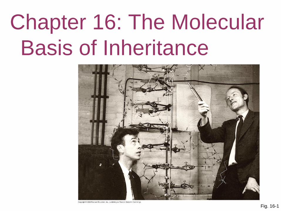
Fig. 16-1
The Molecular Basis of
Inheritance
Chapter 16: The Molecular
Basis of Inheritance
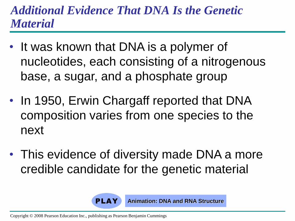
Additional Evidence That DNA Is the Genetic Material
• It was known that DNA is a polymer of
nucleotides, each consisting of a nitrogenous
base, a sugar, and a phosphate group
• In 1950, Erwin Chargaff reported that DNA
composition varies from one species to the
next
• This evidence of diversity made DNA a more
credible candidate for the genetic material
Copyright © 2008 Pearson Education Inc., publishing as Pearson Benjamin Cummings
Animation: DNA and RNA Structure
• Chargaff’s rule states that in any species
there is an equal number of A and T bases,
and an equal number of G and C bases
• Knowing what we already know about DNA
structure, why do you think that this rule would
be valid?
Copyright © 2008 Pearson Education Inc., publishing as Pearson Benjamin Cummings
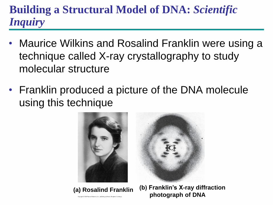
Building a Structural Model of DNA: Scientific Inquiry
• Maurice Wilkins and Rosalind Franklin were using a
technique called X-ray crystallography to study
molecular structure
• Franklin produced a picture of the DNA molecule
using this technique
(a) Rosalind Franklin (b) Franklin’s X-ray diffraction
photograph of DNA
• Franklin’s X-ray crystallographic images of
DNA enabled Watson to deduce that DNA was
helical
• The X-ray images also enabled Watson to
deduce the width of the helix and the spacing
of the nitrogenous bases
• The width suggested that the DNA molecule
was made up of two strands, forming a double
helix
Copyright © 2008 Pearson Education Inc., publishing as Pearson Benjamin Cummings
Animation: DNA Double Helix
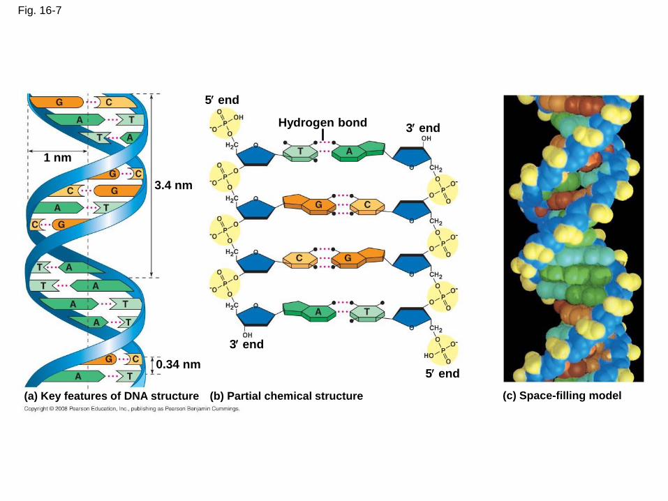
Fig. 16-7
(c) Space-filling model
Hydrogen bond 3 end
5 end
3.4 nm
0.34 nm
3 end
5 end
(b) Partial chemical structure (a) Key features of DNA structure
1 nm
• Watson and Crick built models of a double helix
to conform to the X-rays and chemistry of DNA
• Franklin had concluded that there were two
antiparallel sugar-phosphate backbones, with
the nitrogenous bases paired in the molecule’s
interior - (this means two separate strands
made up of A, G, C and T).
Copyright © 2008 Pearson Education Inc., publishing as Pearson Benjamin Cummings
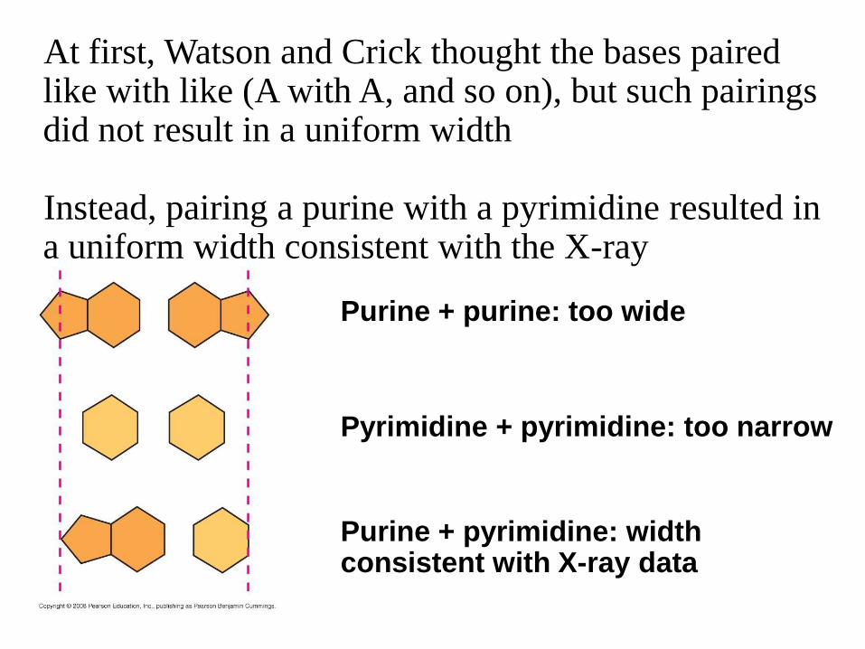
Purine + purine: too wide
Pyrimidine + pyrimidine: too narrow
Purine + pyrimidine: width consistent with X-ray data
At first, Watson and Crick thought the bases paired like with like (A with A, and so on), but such pairings did not result in a uniform width Instead, pairing a purine with a pyrimidine resulted in a uniform width consistent with the X-ray
• Watson and Crick reasoned that the pairing
was more specific, dictated by the base
structures
• They determined that adenine (A) paired only
with thymine (T), and guanine (G) paired only
with cytosine (C)
• The Watson-Crick model explains
Chargaff’s rules: in any organism the
amount of A = T, and the amount of G = C
Copyright © 2008 Pearson Education Inc., publishing as Pearson Benjamin Cummings
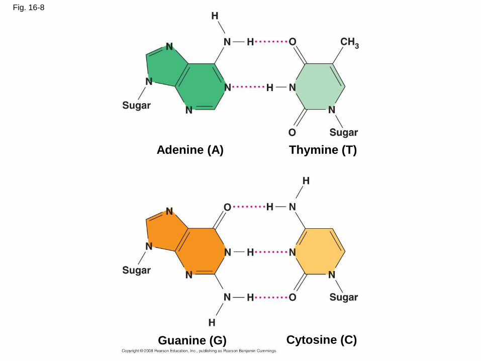
Fig. 16-8
Cytosine (C)
Adenine (A) Thymine (T)
Guanine (G)
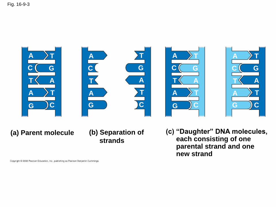
Fig. 16-9-3
A T
G C
T A
T A
G C
(a) Parent molecule
A T
G C
T A
T A
G C
(c) “Daughter” DNA molecules, each consisting of one parental strand and one new strand
(b) Separation of
strands
A T
G C
T A
T A
G C
A T
G C
T A
T A
G C
Getting Started
• Replication begins at special sites called
origins of replication (meaning where
replication starts), where the two DNA strands
are separated, opening up a replication
“bubble”
• A eukaryotic chromosome may have hundreds
or even thousands of origins of replication
• Replication proceeds in both directions from
each origin, until the entire molecule is copied
Copyright © 2008 Pearson Education Inc., publishing as Pearson Benjamin Cummings
Animation: Origins of Replication
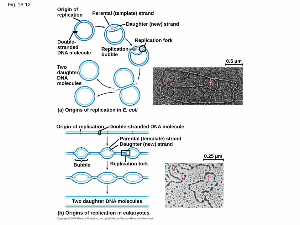
Fig. 16-12 Origin of replication Parental (template) strand
Daughter (new) strand
Replication fork
Replication bubble
Two daughter DNA molecules
(a) Origins of replication in E. coli
Origin of replication Double-stranded DNA molecule
Parental (template) strand Daughter (new) strand
Bubble Replication fork
Two daughter DNA molecules
(b) Origins of replication in eukaryotes
0.5 µm
0.25 µm
Double- stranded DNA molecule
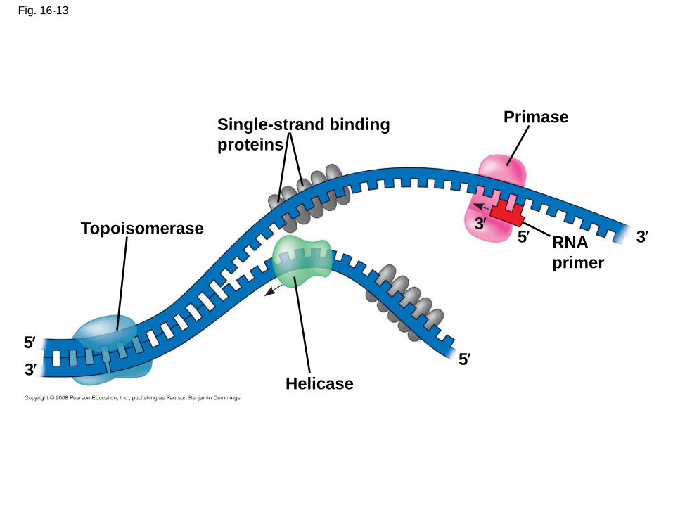
Fig. 16-13
Topoisomerase
Helicase
Primase Single-strand binding
proteins
RNA
primer
5 5
5 3
3
3
Synthesizing a New DNA Strand
• Enzymes called DNA polymerases catalyze
the elongation of new DNA at a replication fork
• Most DNA polymerases require a primer and a
DNA template strand
• The rate of elongation is about 500 nucleotides
per second in bacteria and 50 per second in
human cells
Copyright © 2008 Pearson Education Inc., publishing as Pearson Benjamin Cummings
Antiparallel Elongation
• The antiparallel structure of the double helix
(two strands oriented in opposite directions)
affects replication
• DNA polymerases add nucleotides only to the
free 3end of a growing strand
• Along one template strand of DNA, the DNA
polymerase synthesizes a leading strand
continuously, moving toward the replication
fork
Copyright © 2008 Pearson Education Inc., publishing as Pearson Benjamin Cummings
Animation: Leading Strand
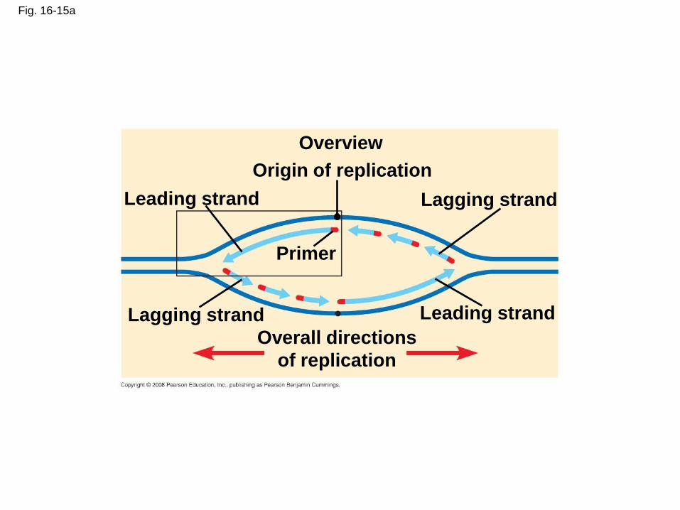
Fig. 16-15a
Overview
Leading strand
Leading strand Lagging strand
Lagging strand
Origin of replication
Primer
Overall directions
of replication
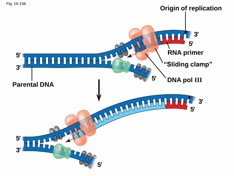
Fig. 16-15b
Origin of replication
RNA primer
“Sliding clamp”
DNA pol III Parental DNA
3
5
5
5
5
5
5
3
3
3
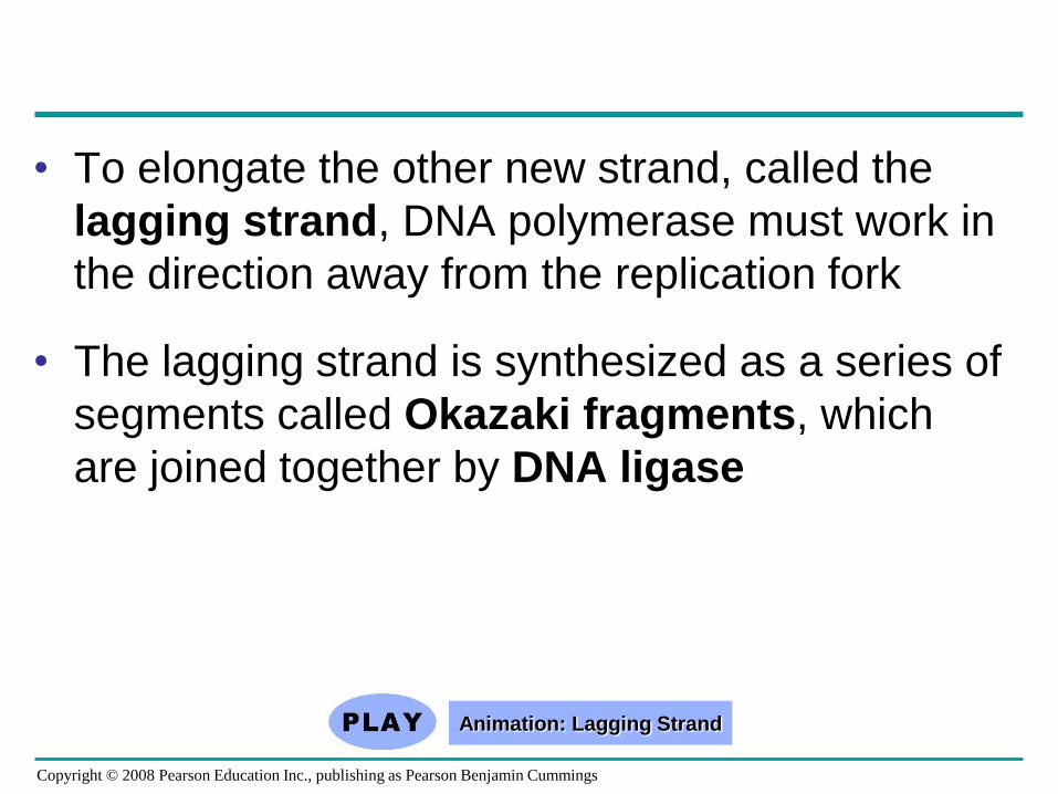
• To elongate the other new strand, called the
lagging strand, DNA polymerase must work in
the direction away from the replication fork
• The lagging strand is synthesized as a series of
segments called Okazaki fragments, which
are joined together by DNA ligase
Copyright © 2008 Pearson Education Inc., publishing as Pearson Benjamin Cummings
Animation: Lagging Strand
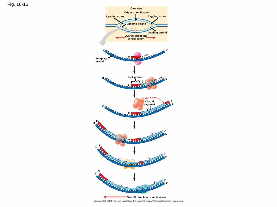
Fig. 16-16 Overview
Origin of replication
Leading strand
Leading strand
Lagging strand
Lagging strand
Overall directions of replication
Template strand
RNA primer
Okazaki fragment
Overall direction of replication
1 2
3
2
1
1
1
1
2
2
5
1 3
3
3
3
3
3
3
3
3
5
5
5
5
5
5
5
5
5
5
5 3
3
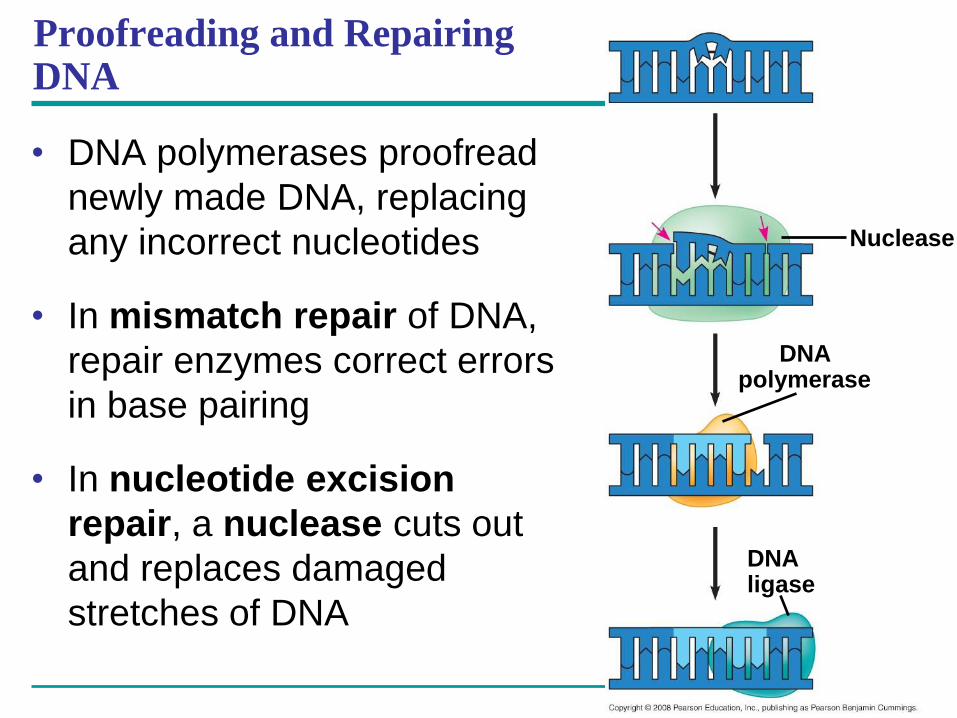
Proofreading and Repairing DNA
• DNA polymerases proofread
newly made DNA, replacing
any incorrect nucleotides
• In mismatch repair of DNA,
repair enzymes correct errors
in base pairing
• In nucleotide excision
repair, a nuclease cuts out
and replaces damaged
stretches of DNA
Nuclease
DNA polymerase
DNA ligase