Embed Size (px)
Citation preview
583
THE MODERN TREATMENT OF ANURIAAND OLIGURIABy DAVID K. BROOKS, M.B., B.S.
Research Fellow Surgical Unit, St. Mary's Hospital, London, W.z2
There are few conditions which offer a greaterchallenge to the medical and nursing staff thanthat of acute renal failure. There are also fewconditions where the application of modern tech-niques and careful clinical biochemical assess-ment are more essential to the achievement ofsuccess.True or complete anuria rarely occurs except
where there is mechanical obstruction and, asearly recognition of oliguria is essential in acuterenal failure, it is important to define it in arecognizable form.The normal output of urine varies between one
and two litres per day. On a mixed diet it isobligatory to pass a minimum volume of approxi-mately 500 ml. in 24 hours (Gamble, I947). Thisentails good renal function and the ability to con-centrate fully to a maximum specific gravity ofI.o35 (I,I55 m. osm./l.).
Oliguria has been defined as less than 700 ml. in24 hours. Where good renal function is presentsuch urine will concentrate sufficiently to producea specific gravity of i.oi8 (595 m. osm./l.) or more.Where renal damage is present, a low specificgravity of i.oo8 (265 m. osm./l.) to 1.o014 (460 m.osm./l.) is to be expected (Joekes, I957). Severeoliguria may be defined as a urine output of500 ml. or less in 24 hours.Where there is a urine output of 300 ml. or less,
such will be the inability of the kidneys to keeppace with the production of metabolites, thecondition might well be called metabolic anuria.The object of therapy in oliguria and anuria is
to tide the patient over until the kidneys have timeto recover their function.At this time, it is probably true to say that
suppression of urine due to malignant hyperten-sion, chronic nephritis and polycystic kidneysdeveloping into oliguria, and the rare condition ofbilateral cortical necrosis, is, in the main, notreversible.The reversible causes of acute renal suppression
may be listed as:(a) Mechanical-obstruction of the ureters, by:
operative procedures; bilateral renal calculi;carcinoma of the prostate and cervix; procidentia,etc.
(b) Dehydration and electrolyte depletion, e.g.low salt syndrome.
(c) Tubular necrosis such as follows: grossdehydration; prolonged hypertension; separationof placenta; abortion; crush syndrome; miss-matched blood transfusion; nephrotoxins, e.g.mercuric chlorides, bismuth, sulphonamides.
(d) Acute nephritis.The history of a successfully treated case of
acute suppression of urine normally falls into fourphases:
(a) The precipitating condition or cause.(b) The anuric phase.(c) The diuretic or pre-recovery phase.(d) The recovery phase.
The Precipitating Condition or CauseCareful clinical assessment to elucidate the
causation of anuria is important because of theundesirability of prolonging an irreversible condi-tion, and the need of remedying quickly, and tothe maximum, any condition that is immediatelytreatable.
Dehydration, electrolyte imbalance, low bloodpressure are all easily treatable. Sulphonamideanuria usually responds to alkalis and increasedfluids.The onset of renal failure, following poisoning
with bismuth or mercuric chloride, may be avoidedby the early use of British anti-lewisite or calciumdisodium versenate. These substances may betoxic, however, once anuria is established (Stock,1952; Merrill, 1955).Care must be observed in the treatment of de-
hydration and electrolyte imbalance in case renaldamage has already occurred.
The Anuric PhaseThree factors emerge as a consequence of the
acute suppression of urine:(a) The retention of water.
copyright. on M
ay 8, 2020 by guest. Protected by
http://pmj.bm
j.com/
Postgrad M
ed J: first published as 10.1136/pgmj.34.397.583 on 1 N
ovember 1958. D
ownloaded from
584 POSTGRADUATE MEDICAL JOURNAL November 1958
(b) The accumulation of the products of meta-bolism, urea, uric ,acid, creatinine, phosphate,sulphate, etc.
(c) The metabolic upsets arising from the above,as yet little understood, but involving the shift ofelectrolytes from the cells to the extra-cellularfluid compartments, and for want of a better termmay be called the poisoned-cell phenomenon.The consequences of fluid retention rapidly be-
come apparent when unrestricted fluid intake isallowed, or worse, when fluids are forced in themisguided attempt to invoke a diuresis. In eithercase, cardiac failure, oedema, pulmonary conges-tion, hypertension, cerebral oedema and the symp-toms of water intoxication will result.The most common cause of death within I4
days of the onset of oliguria due to renal diseaseis overhydration (Bull, Joekes and Lowe, I949).Even when overhydration is not a factor, the re-tained products of metabolism produce the gradualonset of lethargy, coma, vomiting, pericarditis andlowered resistance to infection. The rising con-centration of potassium in the body is reflected inhigh plasma-potassium levels and is associatedwith cardiac failure and muscle weakness.When the kidney begins to recover it secretes a
dilute urine of low concentration, which, on reach-ing a volume of a litre or more, begins to reducethe blood urea.The kidney at this stage still has an impaired
tubular function, unable to concentrate and todifferentiate between electrolytes. Thus, the pass-ing of a profuse dilute urine may soon lead todehydration, and electrolyte imbalance and de-pletion.Within a variable period of time, usually six to
eight weeks, full return of kidney function is to beexpected, sometimes with no detectable abnor-mality (Lowe, 1952; Oliver, 1953).
The Regulation of WaterThe fluid requirements in the anuric patient
need to be assessed accurately each 24 hours sothat the building up of a positive or negative im-balance is avoided as time progresses. These re-quirements amount to the fluid loss through theskin, lungs, urine, vomit and faeces.Although various formulae are available for the
assessment of insensible loss through the skin andlungs, none can be applied with confidence toevery patient.The simplest and most accurate method is to
record the daily change in weight, the weighingbeing performed at the same time each day and onthe same scales, with the bladder empty. Ideallythis is done on a bed weighing machine, butmobilization of the patient for weighing, where
this is clinically possible, is by no means contra-indicated.An accurate record of all additions to the patient
in the form of fluids, such as medicines, ionexchange resin, etc., and all losses in the form ofvomit, urine and faeces must be kept, in order forthis assessment to be made.The measured insensible loss of the average
adult is now accepted to be less than one litre in24 hours and generally falls as the time from theonset of anuria progresses, probably because ofendogenous production of water from fat meta-bolism (Swan and Merrill, I953).With this in view, and the probability that some
degree of overhydration may have occurred by thetime the oliguric state has been recognized, aslittle as 400 ml. in 24 hours can be recommendedin the early stages of treatment, and a graaual lossof weight, as food stores are used up, is to bedesired (Merrill, I955).The accurate measurement of urine output is of
paramount importance, not only for the assess-ment of fluid replacement, but for the determina-tion of the progress of the patient, and the amountof urea and electrolytes excreted.
If necessary, therefore, a self-retaining cathetershould be inserted into the bladder and, afteremptying, a measured quantity of fluid, containingan antibiotic, passed back into the bladder throughthe catheter: 30 ml. of fluid containing ioo mg. ofchloramphenical has been used successfully. Thisvolume is deducted from the volume of urineobtained on releasing the catheter.
In order to avoid the oral complications asso-ciated with oliguria and its treatment, i.e. parotitis,thrush and ulceration, part of the fluid intakeshould be taken by mouth. This regime should bereinforced by the sucking of pure glucose sweets,followed by bland antiseptic mouth washes.
The Suppression of Harmful MetabolitesThe main metabolic consequences the anuric
patient has to face arise from the metabolism ofprotein, producing a rising blood non-proteinnitrogen, cardiotoxic levels of plasma potassium,and release of unexcretable fixed acid.Therapy is therefore directed towards a com-
pletely protein-free intake and the reduction ofendogenous protein metabolism. Advantage istherefore taken of the protein-sparing effect ofglucose and, owing to the necessity of restrictingfluid intake, this entails the use of hypertonicsolutions. Up to 50 per cent. glucose can be safelyinjected into a large vein by means of a polythenecatheter (Bull, 1952). Although the procedure ofpassing the catheter is simple, in the condition ofanuria, where it may by necessity stay in situ forthree weeks, maximum precautions for sterility
copyright. on M
ay 8, 2020 by guest. Protected by
http://pmj.bm
j.com/
Postgrad M
ed J: first published as 10.1136/pgmj.34.397.583 on 1 N
ovember 1958. D
ownloaded from
November I958 BROOKS: The Modern Treatment of Anuria and Oliguria 585
LLOYD-LUKEBooks that enshrine profound thought
GENERAL PATHOLOGY INTRODUCTION TO FLUID BALANCE11tSURGERY IN SURGICAL PRACTICE
(2nd Edited by D. H. PATEY (2nd edit.)
M.D., F.R.C.P., F.R.S. (1958) Cloth-bound 17s. 6d. net D.M.(Oxon.), F.R.C.S.(Eng.)(1958) 84s. net (1958) Paper-covered 9s. 6d. net (1957) 20s. net
RECENT TRENDS MEDICAL ETHICSIN CHRONIC BRONCHITIS A Guide to Students POSTURAL DRAINAGEEdited by NEVILLE C. OSWALD and Practitioners by E. WINIFRED THACKER
T.D., M.D.(Cantab.), F.R.C.P.(Lond.) Edited by MAURICE DAVIDSON M.C.S.P.(1958)3.net
D.M.(Oxon.), F.R.C.P.(Lond.) (1956) 8s. 6d. net((I 957) 20s. net
OPHTHALMOLOGY THE RESPIRATORY MUSCLES
by P. D. TREVOR-ROPER BREATHING by V. KEATINGM.B., B.Chir.(Camb.), F.R.C.S., by E. J. MORAN CAMPBELL M.B., B.Ch., D.A., F.F.A.R.C.S.(Eng.)
D.O.M.S.(Eng.) M.D., Ph.D.(Lond.), M.R.C.P.(Lond.) (1956) 25s. net(1955) 75s. net (1958) 20s. net
Descriptive catalogue available on request
LLOYD-LUKE (MEDICAL BOOKS) LTD., 49 Newman Street, W.I
should be enforced and complete sterility main-tained at the changing of each bottle.To each 500 ml. of glucose solution are added
2,500 units of heparin, and one unit of insulin isadded for every 3 g. of glucose. The slow infusionof not less than 200 g. of glucose which should beaimed at, can be achieved in only 400 ml. of water.The advantages of this regime over the tube
feeding of fat and glucose, such as has been ad-vocated (Bull, Joekes and Lowe, 1949; Borst,I948), are seen in the complete control obtainedover fluid and calorie intake, the absence of anystimulation to vomiting, and the possible greaterprotein-sparing effect of glucose over that of fat(Engle, I952). In addition, the urea and electro-lyte content of vomit is such that it is often valuableas an excretory mechanism, and its return downthe stomach tube in an attempt to maintainbalance is contra-indicated.The use of testosterone proprionate to inhibit
protein catabolism has not proved very successful,but the newer synthetic non-virilising compoundsmay be of value (McSwiney and Prunty, I947).Electrolyte ImbalanceLowered plasma sodium, calcium, bicarbonate,
and pH, and raised phosphate and sulphate, are allfound in the uraemic state.Lowered serum sodium may be due either to
sodium depletion or more probably to dilution byoverhydration. The therapy for the latter isintense water restriction. The shift of sodium fromthe plasma into the cells, due to the poisoned cellphenomenon, is also a possible factor. The ad-ministration of the sodium ion is to be avoided. If,however, sodium depletion is considered to haveoccurred, small quantities of sodium in the form oflactate, bicarbonate, or chloride can be adminis-tered. If clinical improvement results, furtherquantities can be given until the deficit, as calcu-lated from the plasma deficiency and total bodywater, has been made good. This quantity shouldnever be exceeded.>
e.g. weight of patient = 70 kilo.Total body water estimated at 6o per cent.
of body weight = 42 1.Plasma sodium 12o mEq./l.42 X 20 = 840 mEq. of sodium are re-
quired to correct the observed deficit andbring the plasma sodium level toI40 mEq./l.
No marked clinical improvement is to be ex-
copyright. on M
ay 8, 2020 by guest. Protected by
http://pmj.bm
j.com/
Postgrad M
ed J: first published as 10.1136/pgmj.34.397.583 on 1 N
ovember 1958. D
ownloaded from
586 POSTGRADUATE MEDICAL JOURNAL November 1958
pected from attempts to modify the acidosisresulting from the accumulation of fixed acids,phosphate, sulphate, etc., by administration ofalkali in the form of bicarbonate or lactate(Grollman, 1954; Merrill, I955). Also tetany,due to a further lowering of the already decreasedlevel of plasma ionized calcium, may occur on theadministration of alkali.
Treatment of HyperkalaemiaA complete potassium-free intake should be
instituted and maintained throughout the periodof anuria. Milk, fruit, etc., are banned. Teawithout milk may relieve the monotony of glucosedrinks.A raised plasma potassium occurs mostly due
to its release from protein catabolism and probablypartly as an inherent metabolic consequence ofanuria. When toxic levels are reached, muscularweakness, cardiac irregularities, absence of tendonreflexes can be demonstrated. The E.C.G., whenplasma levels of 7 or 8 mEq./l. are reached, showshigh-peaked T waves, absent P waves, slurredwidened Q.R.S. complexes. Cardiotoxic levels ofpotassium vary with each individual, age andduration of the disease. Rapid onset of potassiumintoxication is to be expected following majoroperative procedures and where gross tissuedamage has occurred (Bywaters, 1944).Treatment for the control of potassium intoxica-
tion should be instituted early, before pathologicallevels appear. Intravenous glucose tends toalleviate the condition by the uptake of potassiumin its metabolism (Bull, 1952). Ion exchangeresins which exchange ammonia or sodium forpotassium may be used (Elkington et al., I950;Knowles, I953; Milne and Yellow Lees, I953).Fifteen g. of resin given in 30 ml. of 5 or io percent. glucose water is well tolerated orally whengiven early in the disease, and can be given, ifnecessary, three times a day: 30 to 60 g. of ionexchange resin can be given rectally in 2o0o ml. ofwater or I per cent. methyl cellulose. It may benecessary to administer a glycerine enema in orderto obtain return of the resin, as retained resin is ofonly limited value. The rectal route can be com-bined with oral administration or used alone whenthe patient is vomiting or unable to swallow.
Attempts to make use of the low potassium con-tent of stored packed cells have proved disappoint-ing, but there is some recent evidence that ex-change transfusion may be of some benefit in thisrespect (Hughes, I958).
It must be borne in mind, however, that trans-fusions of blood or plasma are associated withincreased protein metabolism. Attempts to correctanaemia should be avoided for this reason unlessfound to be essential, i.e. an haematocrit of less
than 25 per cent. The infusion of ioo ml. of io percent. calcium gluconate every 24 hours will do noharm and on physiological grounds may help tomitigate the effects of hyperkalaemia (Meroneyand Herndon, 1954). It may be added to hyper-tonic glucose solutions. Hyperkalaemia is anindication for dialysis. The rapid infusion ofhypertonic saline or of sodium bicarbonate willtemporarily, for one or two hours, reverse thecardio-toxic effect of potassium (Merrill, 1955) andmay be used, for example, prior to dialysis.
The Extra Renal Removal of MetabolitesWhen conservative treatment, as described
above, is inadequate to control or maintain thepatient until the pre-recovery stage develops, theextra renal removal of metabolites becomesessential.
This requires strict biochemical and physiologi-cal control and a well-trained team. The indica-tions for this procedure, which should be regardedas part of general therapy and not as a substitutefor the regime described above, may be sum-marized as follows:
(a) General clinical deterioration-' the uraemicstate.' No hard and fast rules can be laid down,as individual tolerance to raised-plasma non-protein nitrogen levels appears to vary so much,and are not necessarily in themselves indicationsfor dialysis. A careful clinical assessment of therate of progress as manifested by drowsiness, peri-carditis, vomiting, muscular irritability, etc., mustbe made.
(b) Hyperkalaemia-cardiotoxic plasma potas-sium levels of 7 mEq./l. or more as shown byE.C.G. recordings.
(c) Acidosis-a plasma bicarbonate of less thanI 5 m. mo./l.
(d) Gross overhydration.The therapeutic methods available are intestinal
irrigation, peritoneal lavage and dialysis.Intestinal Irrigation. The perfusing of hyper-
tonic fluids of varying composition through thegut at a constant rate is difficult to perform and maybe associated with complications of intestinalhaemorrhage, perforation, ileus and pulmonaryoedema.
It has not gained much popularity except onthe Continent.
Peritoneal Lavage. The use of the large peri-toneal surface for dialysing is, on physiologicalprinciples, sound and of value clinically (Grollman,1954).
Slightly hypertonic sterile solutions, varied ac-cording to the biochemical needs of the patient,are perfused through the abdominal cavity.
In hyperkalaemia, for example, no potassium isadded to the perfusing fluid and solution A would
copyright. on M
ay 8, 2020 by guest. Protected by
http://pmj.bm
j.com/
Postgrad M
ed J: first published as 10.1136/pgmj.34.397.583 on 1 N
ovember 1958. D
ownloaded from
November I958 BROOKS: The Modern Treatment of Anuria and Oliguria 587
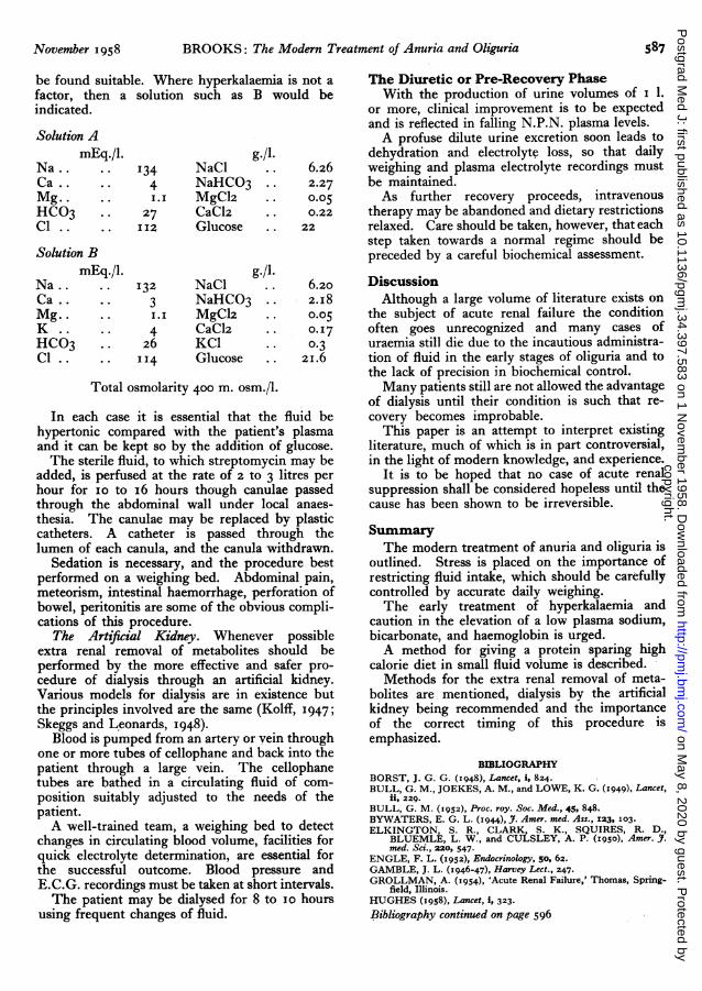
be found suitable. Where hyperkalaemia is not afactor, then a solution such as B would beindicated.
Solution AmEq./l. g./l.
Na .. .. i34 NaCl .. 6.26Ca .. .. 4 NaHC03 .. 2.27Mg . ... I.I MgCl2 .. o.o5HCO3 .. 27 CaCl2 .. 0.22C1 .. .. II2 Glucose .. 22
Solution BmEq./l. g./l.
Na... 132 NaCl .. 6.20Ca .. . 3 NaHCO3 .. 2.I8Mg.. . I. I MgClz .. 0.05K .. 4 CaC12 .. o.I7HCO3 .. 26 KC1 0. 3CI . .. II4 Glucose .. 21.6
Total osmolarity 400 m. osm./l.
In each case it is essential that the fluid behypertonic compared with the patient's plasmaand it can be kept so by the addition of glucose.The sterile fluid, to which streptomycin may be
added, is perfused at the rate of 2 to 3 litres perhour for io to x6 hours though canulae passedthrough the abdominal wall under local anaes-thesia. The canulae may be replaced by plasticcatheters. A catheter is passed through thelumen of each canula, and the canula withdrawn.
Sedation is necessary, and the procedure bestperformed on a weighing bed. Abdominal pain,meteorism, intestinal haemorrhage, perforation ofbowel, peritonitis are some of the obvious compli-cations of this procedure.
The Artificial Kidney. Whenever possibleextra renal removal of metabolites should beperformed by the more effective and safer pro-cedure of dialysis through an artificial kidney.Various models for dialysis are in existence butthe principles involved are the same (Kolff, 1947;Skeggs and Leonards, 1948).
Blood is pumped from an artery or vein throughone or more tubes of cellophane and back into thepatient through a large vein. The cellophanetubes are bathed in a circulating fluid of com-position suitably adjusted to the needs of thepatient.A well-trained team, a weighing bed to detect
changes in circulating blood volume, facilities forquick electrolyte determination, are essential forthe successful outcome. Blood pressure andE.C.G. recordings must be taken at short intervals.The patient may be dialysed for 8 to I0o hours
using frequent changes of fluid.
The Diuretic or Pre-Recovery PhaseWith the production of urine volumes of I 1.
or more, clinical improvement is to be expectedand is reflected in falling N.P.N. plasma levels.A profuse dilute urine excretion soon leads to
dehydration and electrolyte loss, so that dailyweighing and plasma electrolyte recordings mustbe maintained.As further recovery proceeds, intravenous
therapy may be abandoned and dietary restrictionsrelaxed. Care should be taken, however, that eachstep taken towards a normal regime should bepreceded by a careful biochemical assessment.
DiscussionAlthough a large volume of literature exists on
the subject of acute renal failure the conditionoften goes unrecognized and many cases ofuraemia still die due to the incautious administra-tion of fluid in the early stages of oliguria and tothe lack of precision in biochemical control.Many patients still are not allowed the advantage
of dialysis until their condition is such that re-covery becomes improbable.
This paper is an attempt to interpret existingliterature, much of which is in part controversial,in the light of modern knowledge, and experience.
It is to be hoped that no case of acute renalsuppression shall be considered hopeless until thecause has been shown to be irreversible.
SummaryThe modern treatment of anuria and oliguria is
outlined. Stress is placed on the importance ofrestricting fluid intake, which should be carefullycontrolled by accurate daily weighing.The early treatment of hyperkalaemia and
caution in the elevation of a low plasma sodium,bicarbonate, and haemoglobin is urged.A method for giving a protein sparing high
calorie diet in small fluid volume is described.Methods for the extra renal removal of meta-
bolites are mentioned, dialysis by the artificialkidney being recommended and the importanceof the correct timing of this procedure isemphasized.
BIBLIOGRAPHYBORST, J. G. G. (1948), Lancet, i, 824.BULL, G. M., JOEKES, A. M., and LOWE, K. G. (I949), Lancet,
ii, 229.BULL, G. M. (1952), Proc. roy. Soc. Med., 45, 848.BYWATERS, E. G. L. (I944), J. Amer. med. Ass., 123, 103.ELKINGTON, S. R., CLARK, S. K., SQUIRES, R. D.,
BLUEMLE, L. W., and CULSLEY, A. P. (1950), Amer. J.med. Sci., 220, 547.
ENGLE, F. L. (1952), Endocrinology, o50, 62.GAMBLE, J. L. (1946-47), Harvey Lect., 247.GROLLMAN, A. (I954), 'Acute Renal Failure,' Thomas, Spring-
field, Illinois.HUGHES (I958), Lancet, i, 323.Bibliography continued on page 596
copyright. on M
ay 8, 2020 by guest. Protected by
http://pmj.bm
j.com/
Postgrad M
ed J: first published as 10.1136/pgmj.34.397.583 on 1 N
ovember 1958. D
ownloaded from
596 POSTGRADUATE MEDICAL JOURNAL - November I9-58
SummaryAll the cases described had features which led
to the presumptive diagnosis being made clinically,and in every case L.E. cells were demonstratedin the peripheral blood. In one case the diagnosiswas confirmed at autopsy.High fever, loss of appetite and weight, painful
swollen joints and pleural involvement were com-mon to all. Splenic enlargement and ulceration ofthe mucous membrane of the mouth was noted inone case and peripheral vascular phenomena andhepatic enlargement in another. The characteristicbutterfly rash on the face occurred only once.Other interesting features have been discussed inthe footnote to each case. Although widespreadjoint involvement was common, X-ray failed todemonstrate any lesions in the affected joints.Examination of the blood showed mild to moderatehypochromic anaemia and a leucocyte count rang-ing from 4,00ooo to 5,ooo per c.mm. The B.S.R. wasraised in all cases, ranging from 63 to 105 mm.(Westergren one-hour reading). Plasma proteinswere raised (7 to io g./Ioo ml.), with the risemainly in the globulin fraction, a finding in keepingwith reported series. In those cases in whichplasma proteins were repeated they were found tobe uninfluenced by steroid therapy.
Since the L.E. cell phenomena was first demon-strated by Hargraves et al. (I948) several new tech-niques have been introduced. The test is nowbeing widely used and there are a number of con-ditions other than systemic lupus where it is foundto be positive. Positive L.E. cell tests have recentlybeen described in rheumatoid arthritis by Fried-man et al. (1957). One of our cases, which is notdescribed here, had painful deformity of the smalljoints of the hands of six years' duration. She had amild hypochromic anaemia, but no other visceralmanifestations were noted. L.E. cells were foundin the peripheral blood. She is still under observa-tion, but is at present considered to be a case ofrheumatoid arthritis.
Dubois (I956) states, on the other hand, that,despite recently introduced refinements of tests,
L.E. cells are not found in all patients with thedisease. In all the cases L.E. cells were found inthe peripheral blood and we felt that the test,taken in conjunction with the clinical findings, isof the greatest value.
Regarding the management of cases, the con-sensus of opinion at present favours the use ofconservative measures in milder cases. In theacute stages bed rest is essential with the necessarysymptomatic treatment. The joint manifestationsmay be helped by suitable splintage and salicylatetherapy. Pneumonic episodes should first betreated with appropriate antibiotics. In this seriesall cases were acutely ill and it was felt that steroidtherapy was indicated. Apart from the case whichlived only a short while after admission, all re-sponded to large doses of steroids and were able tobe discharged on a reduced dosage under out-patient supervision. An exacerbation of symptomsoccurring in case 5 while on maintenance therapywas controlled by increasing the steroid dosage.As symptoms abated it was found possible to oncemore reduce the dosage.While steroids have undoubtedly been life-
saving in the acute exacerbations, no evidence hasbeen found of alteration in the underlying patho-logical processes.We wish to thank Drs. R. M. Fulton and J. D.
Allan for their kindness in allowing us to see theirpatients and use the case notes, and we areespecially grateful to Dr. Fulton for his advice andencouragement.
REFERENCESBROWN, C. H., SHIRLEY, E. K., and HASERICK, J. B. (I957),
Gastroenterology, 3I, 649.DUBOIS, E. L. (I956), Ann. intern. Med., 45, I63.ELLMAN, P., and CUDKOWICZ, L. (i954), Thorax, 9,46.FRIEDMAN, A. I., et al. (xI957), Ann intern. Med., 46, "I I 3.GLASER, G. H. (I952), Arch. Neurol. and Psychiat, 67, 745.HARGRAVES, M. M., RICHMOND, H., and MORTON, R.
(I948), Proc. Mayo Clin., 23, 25.HILL, L. C. (xI957), Brit. med. Y., 2; 726.JOSKE, R. A. (I956), Proc. roy. Soc. Med., 49, 329.MUEHRCKE, et al. (I956), Proc. roy. Soc. Med., 49, 327.OSLER, W. (I885), Lancet, i, 415, 459, 504.OSLER, W. (I895), Amer. J. Med. Sci., IIo, 629.
Bibliography continuedfrom page 587-David K. Brooks, M.B., B.S.JOEKES, A. M. (I957), Proc. roy. Soc. Med., 50, 496.KNOWLES, H. C., and KAPLAN, S. A. (x953), Arch. intern. Med.,
92, I 89.KOLFF, W. J., and BERK, H. T. J. (x944), 'Artificial Kidney,
Dialyser with Great Area,' Geneesk, Gids., Vol. 2I.KOLFF, W. J. (I947), 'New Ways of Treating Uraemia,' J. and A.
Churchill Ltd., London.LOWE, K. G. (1952), Lancet, i, Io86.MATHE, G., and HAMBURGER, J., 'Ciba Foundation Sym-
posium on the Kidney.'
McSWINEY, P. R., and PRUNTY, F. T. G. (I957), J. Endocr.,x6, 28.
MERONEY, W. H., and HERNDON (I954), J. Amer. med. Ass.,I55, 877.
MERRILL, J. P. (I955), 'The Treatment of Renal Failure,' Gruneand Statton, New York.
MILNE, M. D., and YELLOW LEES, H. (I953), Lancet, ii, 791.OLIVER, J. (r953), Amer. J. Med., 15, 535.SKEGGS, L., and LEONARDS, J. R. (I948), Science, Io8, 212.STOCK, R. J. (I952), Bull. N.Y. Acad. Med., 28, 507.SWAN, R. C., and MERRILL, J. P. (I953), Medicine, 32, 215.
copyright. on M
ay 8, 2020 by guest. Protected by
http://pmj.bm
j.com/
Postgrad M
ed J: first published as 10.1136/pgmj.34.397.583 on 1 N
ovember 1958. D
ownloaded from