Embed Size (px)
Citation preview

1
Supporting Information
Cashmere-derived keratin for device manufacturing on the micro- and nanoscale
Benedetto Marelli and Fiorenzo G. Omenetto*
B. Marelli and F. G. Omenetto* Department of Biomedical EngineeringTufts University4 Colby St., Medford, MA, 02155, USAE-mail: [email protected]
F. G. OmenettoDepartment of PhysicsTufts University4 Colby St., Medford, MA 02155
Keywords: keratin, optic, photonic, sensing
Electronic Supplementary Material (ESI) for Journal of Materials Chemistry C.This journal is © The Royal Society of Chemistry 2015

2
Experimental details
Keratin extraction: Keratin is a family of proteins. Extraction of the proteins that form
cashmere fibers yields not a single protein but several proteins, which falls under the name of
keratins. For simplicity and clearness to non-technical experts, in the manuscripts we referred
to this pool of proteins as keratin, considering it as a single entity. Keratin extraction was
performed with a previously reported method 1, with some modifications. In brief, raw, white
cashmere wool (30g) was rinse in distilled water (6 liters, 30°C) for 30 minutes (three water
changes every 10 minutes), blotted and dried under vacuum for 6 hours. Lipid extraction was
then performed with 100% acetone for 24 hours to remove remaining unbound surface lipids.
The fibers were then washed (3x) with distilled water (6 liters, 30°C) for 3 hours (three water
changes every 45 min) and air-dried. Delipided cashmere fibers were then cut into short
fibers, 3 mm long. A mixture of 7M urea (500 ml), 2-mercaptoethanol (50 ml) and 0.5M
thiolurea (50 ml), was used to solubilized 30 g of cashmere at 50°C for 72h. The solution was
then filtered through a stainless steel sieve (#200) and dialyzed in dialysis tubes (3,500 MW
cut-off) against distilled water (8 liters) for 72 hours (changed every 6 hours). The so obtained
keratin solution was then centrifuged twice (5°C, 9000 rpm, 20 min per cycle) to remove
insoluble particles, resulting in a clear protein solution (0.70.3 wt%), which was then
concentrated to 2 wt% through a centrifugal evaporator. The total extraction yield was
475%.
Film Preparation and slow drying process: Keratin films were fabricated by solvent casting
keratin solution on PDMS molds. Slow drying process was achieved by controlling the
relative humidity environment (RH=30-95%) during keratin solution solvent casting by using
a custom made humidity chamber. Diffractive PDMS mold and PDMS-made multi-lens
arrays were fabricated using optical diffraction gratings (Edmund Optics, 300-1200 lines/mm)

3
or optical cards (Digital Optics Corp., Tessera Technologies Inc.) as masters. After drying,
films were let acclimated to RH=30% and then carefully lifted from the mold. Film thickness
was controlled by varying either keratin concentration or solution volume used during casting.
Inverse opal fabrication: PMMA nanospheres (=250 nm) were used to fabricate an opal
template (1% concentration dispersed in water, Phosphorex). The PMMA solution was
deposited onto a silicon wafer, which was then heated on a hotplate at 90 °C to generate the
PMMA opal by self-assembly induced by water evaporation. The keratin solution was then
added to the PMMA opal and filled the air voids by capillary infiltration. The solution was set
to dry in a film at room temperature and RH=95%. The so formed keratin film was soaked in
acetone for 24 h to allow for detachment from the silicon wafer and removal of the PMMA
nanospheres.
Measurement of regenerated keratin molecular weight and purity: Cashmere-derived keratin
was diluted 1:1 with a solution of 2x Laemmli sample buffer, 4% SDS, 20% glycerol, 10% 2-
mercaptoethanol and 0.125 M Tris–HCl. Keratins were then run in a vertical slab gel
electrophoretic system at 200 V, 80 mA, and 25W. A solution of 0.1% Coomassie brilliant
blue R-250 (Sigma),10% acetic acid, and 40% methanol was then used to stain the keratin in
the gel for 1 h. Excess of staining was then removed by rinsing the gel in deionized water
overnight.
Physical characterization: Scanning electron microscopy (SEM) was used to investigate
keratin film morphology and to determine surface patterning. Casted films were mounted on
carbon tape and sputter coated with platinum-palladium. The films were then imaged using a
scanning electron microscope (Supra55VP , Zeiss). Atomic force microscopy (AFM) was
used to investigate surface morphology and to determine surface roughness of non-patterned
and patterned keratin films. Micrographs of keratin films were acquired with a Digital
Instrument Dimension 3100 (Veeco Instruments, Inc.) in tapping mode. Keratin film spectrum

4
in the visible wavelengths was measured with a USB2000 Miniature Fiber Optic
Spectrometer. A Metricon waveguide instrument was used to evaluate refractive index of
keratin films. The measured indices of refraction and film thicknesses are evaluated at a
wavelength of λ = 633 nm, as previously reported for silk fibroin.
Spectroscopical characterization: Attenuated Total Reflectance Fourier Transform Infrared
(ATR-FTIR) spectroscopy of keratin films was performed with a Jasco FT/IR-6200
Spectrometer, equipped with a multiple reflection, horizontal MIRacle™ attachment (Ge
crystal, from Pike Tech., Madison, WI). Each collected spectrum was obtained as an average
of 128 scans with a wavenumber range of 4000-650 cm-1 and a nominal resolution of 4 cm-1.
To analyze keratin conformational changes as a function of slow drying processing, Amide I
and Amide III peak absorptions were analyzed as previously reported 2, 3. Micro-Raman
spectroscopy was performed with a Jasco NRS-3000 spectrometer in the 2000-400 cm-1 range
using a 733 laser and a 100x objective. Each spectra was collected as an average of 20 scans
(10s per scan) with a resolution of 1 cm-1. Cosmic rays removal, measurement of FWHM and
of I850/I830 ratio were performed with Jasco Spectra Analysis software. Ellman’s reagent was
used to determine the content of free thiol groups of cysteine residues in the cashmere-derived
regenerated keratin solution and in assembled films 4.
References
1. A. Nakamura, M. Arimoto, K. Takeuchi and T. Fujii, Biological and Pharmaceutical Bulletin, 2002, 25, 569-572.
2. A. Vasconcelos, G. Freddi and A. Cavaco-Paulo, Biomacromolecules, 2008, 9, 1299-1305.
3. G. Freddi, G. Pessina and M. Tsukada, International Journal of Biological Macromolecules, 1999, 24, 251-263.
4. G. L. Ellman, Archives of Biochemistry and Biophysics, 1959, 82, 70-77.
5
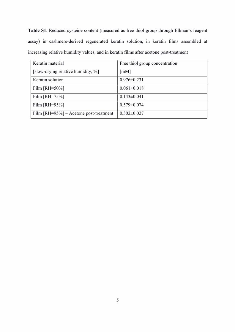
Table S1. Reduced cysteine content (measured as free thiol group through Ellman’s reagent
assay) in cashmere-derived regenerated keratin solution, in keratin films assembled at
increasing relative humidity values, and in keratin films after acetone post-treatment
Keratin material
[slow-drying relative humidity, %]
Free thiol group concentration
[mM]
Keratin solution 0.976±0.231
Film [RH=50%] 0.061±0.018
Film [RH=75%] 0.143±0.041
Film [RH=95%] 0.579±0.074
Film [RH=95%] – Acetone post-treatment 0.302±0.027

6
Figure S1
Figure S1. SDS-PAGE of standard protein molecular weight markers (left lane) and
cashmere-extracted keratin (right lane).

7
Figure S2
Figure S2. Photograph of projected patterns obtained from propagation of a green light laser
source through keratin-made 2D diffractive phase masks. The images are taken at a distance
of 10 cm from the keratin optical element. (Master from Digital Optics Inc., Tessera
Corporation).