Embed Size (px)
Citation preview

2015
UNIVERSIDADE DE LISBOA
FACULDADE DE CIÊNCIAS
DEPARTAMENTO DE QUÍMICA E BIOQUÍMICA
The mechanism of nonsense-mediated mRNA decay
Mestrado em Bioquímica
Especialização em Bioquímica
Nuno Filipe Teixeira Costa
Dissertação orientada por:
Doutora Luísa Romão
Doutora Juliane Menezes

ii

iii
Acknowledgments
Firstly, I would like to express my sincere thanks to Doutora Luísa Romão Loison
for giving me the opportunity to work in her research group. Her knowledge,
motivation and immense patience helped me in all the time of research.
I want to acknowledge Dr. João Lavinha and Dr. Glória Isidro for allowing me to
carry out this study at Departamento de Genética Humana from Instituto Nacional de
Saúde Doutor Ricardo Jorge.
To Juliane Menezes, I want to express my deep sense of gratitude. Always
available, even at cost of her own work, she provided excellent technical and
theoretical teaching and good advice that not only helped me during this research but
will surely help throughout my future career.
I also want to thank Gerson Asper, Paulo Costa, Rafaela Lacerda, Cláudia Onofre
and Ana Ramos who provided insight and expertise that greatly assisted this research.

iv
Index
Acknowledgments ........................................................................................................... iii
Index ................................................................................................................................. iv
Figures .............................................................................................................................. vi
Tables ............................................................................................................................... vi
Resumo ............................................................................................................................ vii
Palavras-chave .............................................................................................................. xi
Abstract ........................................................................................................................... xii
Keywords ..................................................................................................................... xiii
Abbreviations ................................................................................................................. xiv
1. Introduction ............................................................................................................... 16
1.1. Eukaryotic mRNA Translation .............................................................................. 17
1.1.1. Translation initiation – the scanning model ................................................. 17
1.1.2. Translation elongation .................................................................................. 20
1.1.3. Translation termination ................................................................................ 21
1.2. Surveillance mechanisms ..................................................................................... 23
1.2.1. Nonsense-mediated decay ............................................................................ 23
1.2.1.1. NMD factors and mechanism ................................................................. 24
1.2.1.2. PTC definition ......................................................................................... 27
1.2.1.3. Natural and aberrant targets of NMD .................................................... 28
1.2.1.4. Biological and medical significance of NMD .......................................... 30
1.3. The eukaryotic initiation factor 3 ........................................................................ 31
2. Aims ............................................................................................................................ 33
3. Materials and methods .............................................................................................. 35

v
3.1. Plasmid constructs ............................................................................................... 35
3.2. Cell culture, plasmid and siRNA transfection ...................................................... 36
3.3. Immunoprecipitation assay ................................................................................. 38
3.4. Cycloheximide treatment .................................................................................... 38
3.5. SDS-PAGE and Western blotting .......................................................................... 38
3.6. RNA isolation ........................................................................................................ 39
3.7. Reverse transcription ........................................................................................... 39
3.8. Semiquantitative PCR........................................................................................... 39
4. Results ........................................................................................................................ 41
4.1. ptre_βWT_3xMS2bs(45nt_CD39) construction by SOEing PCR .......................... 41
4.2. Anti-enterobacteriophage MS2 coat protein antibody test ................................ 43
4.3. Cycloheximide concentration test ....................................................................... 44
4.4. eIF3h, eIF3f and eIF3c subunits knockdown ........................................................ 45
4.5. Immunoprecipitation of mRNP complexes from HeLa cells treated with siRNAs
specific for the eIF3f subunit ...................................................................................... 46
4.6. Immunoprecipitation of mRNP complexes from HeLa cells treated with siRNAs
specific for the eIF3h subunit ..................................................................................... 49
4.7. eIF3h, eIF3f and eIF3c subunits knockdown with different siRNAs .................... 51
5. Discussion ................................................................................................................... 52
5.1. Proving that PABPC1/eIF4G remains associated with the ribosome, through eIF3
interaction, during the early stages of elongation ..................................................... 52
5.2. Identification and characterization of eIF3 interactions with eIF4G and the 40S
ribosomal subunit ....................................................................................................... 54
6. Conclusion and future directions .............................................................................. 57
7. References .................................................................................................................. 58

vi
Figures
Figure 1: Model of canonical eukaryotic translation initiation pathway ....................... 19
Figure 2: Model of eukaryotic translation elongation pathway ..................................... 21
Figure 3: Model of eukaryotic translation termination pathway ................................... 22
Figure 4: Model of NMD mechanism and degradation via SMG6 or SMG7/SMG5 ....... 26
Figure 5: Representation of the position-dependent effects of nonsense mutations in
the inheritance pattern and clinical severity of β-thalassemia ...................................... 31
Figure 6: Schematic diagram of SOEing PCR .................................................................. 42
Figure 7: ptre_βWT_3xMS2bs(45nt_CD39) construction by SOEing PCR ...................... 42
Figure 8: Test of anti-enterobacteriophage MS2 coat protein antibody ....................... 43
Figure 9: Test of cycloheximide concentration necessary to inhibit translation ........... 44
Figure 10: Test of the conditions for eIF3h, eIF3f and eIF3c subunits knockdown........ 45
Figure 11: Immunoprecipitation of mRNP complexes from HeLa cells treated with eIF3f
siRNAs ............................................................................................................................. 48
Figure 12: Immunoprecipitation of mRNP complexes from HeLa cells treated with eIF3h
siRNAs ............................................................................................................................. 50
Figure 13: Quantification by reverse transcription coupled semiquantitative PCR of
eIF3h knockdown efficiency ........................................................................................... 50
Figure 14: Analysis of eIF3h, eIF3f and eIF3c subunits knockdown efficiency ............... 51
Tables
Table 1: Sequences of the primers used in the current work ........................................ 37
Table 2: Sequences of the siRNAs used in the current work ......................................... 37

vii
Resumo
A expressão genética representa uma das vias mais complexas e importantes
para a célula, que culminam na síntese proteica. Como tal, quaisquer erros que possam
surgir durante estes eventos podem ser rapidamente amplificados e terem um
impacto severo na célula. Portanto surgiram diversos mecanismos de controlo de
qualidade para assegurar a fidelidade da expressão genética. Entre estes mecanismos
está o decaimento do mRNA mediado por mutações nonsense (na sigla inglesa NMD –
nonsense mediated decay) que permite rapidamente degradar transcritos que
contenham codões de terminação de tradução prematuros (na sigla inglesa PTC –
premature translation termination codon), reduzindo a sua abundância1. A eliminação
destes transcritos anómalos evita a formação e acumulação de proteínas truncadas no
C-terminal potencialmente tóxicas que, caso contrário, danificariam a célula. Assim, o
NMD tem um papel protector contra erros genéticos, muitos dos quais originam PTCs.
Recentemente, tem-se tornado óbvio que o NMD tem um papel mais abrangente, não
só funcionando como mecanismo de controlo de qualidade da expressão genética,
mas também apresenta uma importante função na regulação de transcritos
fisiológicos2,3. De facto foi demonstrado que o NMD controla a abundância de 3 a 10%
de todo o transcriptoma em Saccharomyces cerevisiae, Drosophila melanogaster e em
Homo sapiens, pois estes transcritos possuem várias características que os tornam
sensíveis à acção do NMD4.
Nas células humanas, este mecanismo envolve a acção dos factores UPF1
(upframe shift), UPF2, UPF3 e dos factores SMG1 (suppressor with morphogenetic
effects on genitalia), SMG5, SMG6 e SMG75. O NMD é activado assim que um PTC é
identificado. Os factores UPF1, UPF2, UPF3 e SMG1 são recrutados e interagem com o
ribossoma e com os factores de terminação de tradução eRF3 e eRF1 (eukaryotic
release factor), formando o complexo DECID (decay inducing complex)6,7. Este
complexo estimula a fosforilação do UPF18, o que conduz ao recrutamento dos
factores SMG5/SMG7 ou SMG6, que activam a degradação do ácido ribonucleico
(RNA) pela acção de exossomas5.

viii
Grande parte da pesquisa sobre o mecanismo do NMD tem-se focado na forma
como este mecanismo diferencia um PTC de um codão de terminação normal. De
acordo com o modelo prevalente, nas células dos mamíferos a capacidade do NMD
reconhecer um PTC depende dos complexos de junção exão-exão (na sigla inglesa EJC).
Os EJC são complexos multiproteicos depositados a 20 a 24 nucleótidos (nt) de
distância a montante dos locais de excisão de intrões (junções exão-exão) durante o
splicing do mRNA precursor9. Estes complexos mantêm-se associados ao mRNA
durante o seu transporte para o citoplasma, servindo como marcadores dos locais de
excisão de intrões e também como plataformas de ligação dos factores do NMD, UPF2
e UPF310–12. Durante o primeiro ciclo da tradução, se o transcrito não tiver PTC, o
complexo ribossomal dissocia os EJCs para fora da grelha de leitura até chegar ao
codão de terminação. No entanto, se o transcrito possuir um PTC e este se localizar a
pelo menos 50 a 55 nt de distância a montante da última junção exão-exão, a
elongação é terminada prematuramente e pelo menos um EJC fica associado ao
mRNA13. Nestas condições, o EJC facilita o recrutamento e a interacção dos factores do
NMD (UPF1, UPF2 e UPF3) com o complexo ribossomal, activando o NMD. Por outro
lado, se o PTC se localizar depois desta fronteira localizada a 55 nts a montante da
última junção exão-exão, o ribossoma consegue remover todos os EJCs, que não
poderão, assim, desencadear a degradação do transcrito14.
Contudo esta regra posicional do EJC não explica todos os transcritos
susceptíveis de serem degradados por NMD. Existem transcritos que mesmo estando
nas condições previstas pelo modelo (i.e.: no qual o PTC está localizado a pelo menos
50-55 nt de distância a montante da última junção exão-exão), conseguem resistir à
acção do NMD, como é o caso de mRNAs que contenham PTC próximos do codão de
iniciação15. De facto, verificou-se que existem outras características do mRNA, para
além dos EJC, que influenciam a activação do NMD. Por exemplo, trabalhos feitos pelo
grupo laboratorial onde este projecto experimental foi desenvolvido, apontam para a
distância física entre um PTC e a proteína citoplasmática de ligação à cauda poli-A do
mRNA (na sigla inglesa PABPC1) como sendo um factor que afecta a activação do
NMD16. Foi demonstrado que a PABPC1 consegue interagir com a proteína eRF3 e
pensa-se que a PABPC1 é necessária para estimular um processo de terminação
correcto e eficiente17,18. Para além disso, a PABPC1 consegue competir com a UPF1

ix
pela ligação ao eRF3, inibindo o NMD, mesmo na presença de um EJC a jusante16,19.
Propôs-se assim um novo modelo: se o ribossoma ao chegar ao PTC ficar fisicamente
perto da PABPC1, ocorre terminação correcta e não há indução do NMD. No caso em
que o ribossoma ao chegar ao PTC não possa interagir com a PABPC1, a UPF1 interage
com o complexo eRF3/eRF1 e inicia-se o NMD11. Portanto, tendo em consideração este
modelo e sabendo que o mRNA adquire uma conformação circular devido à interacção
do factor eucariótico de iniciação 4G (na sigla inglesa eIF4G) com a estrutura cap na
extremidade 5’, a PABPC1, o complexo eIF3 e o ribossoma, pode-se colocar a hipótese
de a PABPC1 ser arrastada para a vizinhança do codão de iniciação pelo ribossoma
durante o scanning. Nestas condições a PABPC1 encontra-se próxima o suficiente para
competir com a UPF1, caso ocorra terminação prematura na vizinhança do codão de
iniciação, permitindo assim explicar a resistência ao NMD dos transcritos contendo
PTCs próximos do codão de iniciação.
Esta tese teve como objectivo testar esta hipótese. Procedeu-se á construção
de plasmídeos necessários para estudar a interacção do PABPC1 com o ribossoma. Os
plasmídeos contêm a sequência da β-globina (que origina um pequeno transcrito de
três exões cujas mutações nonsense já foram extensivamente descritas) e várias
repetições da sequência do sítio de ligação da cápside do bacteriófago MS2. Estas
sequências são aptámeros de RNA naturais que em conjunto com uma proteína
recombinante contendo o N-terminal da proteína da cápside do MS2 e um anticorpo
específico, permitem a imunoprecipitação selectiva destes transcritos e de qualquer
proteína associada. A construção dos plasmídeos foi iniciada usando um processo de
SOEing PCR.
O factor de iniciação da tradução eIF3 é um complexo multiproteico que
interage com o ribossoma e a proteína eIF4G, funcionando como um ponte entre os
dois durante o processo de iniciação da tradução. Trabalhos feitos pelo nosso grupo
laboratorial mostraram que as subunidades eIF3h e eIF3f podem estar envolvidas na
interacção do ribossoma com a PABPC1 (através da eIF4G)20 e portanto podem ser
necessárias ao deslocamento da PABPC1 para a vizinhança do codão de iniciação pelo
ribossoma. Para testar o papel das subunidades eIF3h e eIF3f na ligação entre o
complexo ribossomal 40S e a PABPC1 (através do eIF4G), isolou-se por
imunoprecipitação, utilizando anticorpos anti-eIF3h ou eIF3f, os complexos

x
ribonucleoproteícos (na sigla inglesa mRNPs) de células HeLa tratadas com RNAs de
interferência (na sigla inglesa siRNA) específicos para as subunidades eIF3h ou eIF3f,
respectivamente. Os imunoprecipitados foram testados por Western Blot para
detectar a presença de PABPC1, eIF4G e proteínas do ribossoma. Nas
imunoprecipitações de mRNPs de células HeLa tratadas com siRNAs para a subunidade
eIF3f, verificou-se uma ligeira diminuição dos níveis proteicos tanto da PABPC1 como
do eIF4G, suportando um modelo no qual a eIF3f é responsável pela aproximação do
complexo PABPC1/eIF4G ao ribossoma. Já a imunoprecipitação de mRNPs de células
tratadas com siRNA para a subunidade eIF3h, não se observaram variações
significativas na expressão da proteína eIF4G e não foi detectada a proteína PABPC1.
Contudo a ausência de sinal da PABPC1 pode ter ocorrido devido à perca de proteína
durante a remoção dos anticorpos para reutilização da membrana de PVDF. Além
disso, a eficiência do silenciamento das subunidades não foi satisfatória, resultando
numa diminuição pouco apreciável dos níveis proteicos de eIF3h ou eIF3f. Assim
sendo, tanto o processo de imunoprecipitação e o método de silenciamento devem ser
optimizados para que se possam tirar conclusões definitivas.

xi
Palavras-chave
mRNA, tradução eucariótica, codão de terminação prematuro, decaimento do mRNA
mediado por codões nonsense, proteína de ligação à cauda poli-A, factor 3 de iniciação
da tradução

xii
Abstract
Nonsense-mediated mRNA decay (NMD) is a quality control pathway that
recognizes and rapidly degrades mRNAs containing a premature termination codon
(PTC). The prevalent model, proposes that in mammals a translation termination
codon is generally interpreted as premature if it is localized more than 50-55
nucleotides upstream of the last exon-exon junction. In these circumstances, during
translation, the ribosome is not able to displace the exon junction complexes (EJCs)
located downstream of the PTC; thus, when the ribosome reaches the PTC stalls and
triggers NMD. However, it has been reported that mRNAs containing PTCs in close
proximity to the translation initiation codon (AUG proximal PTCs) can substantially
evade NMD, despite the presence of downstream EJCs. Recently, it was demonstrated
that the cytoplasmatic poly(A)-binding protein 1 (PABPC1) is capable of inhibiting NMD
when placed close to a PTC, even in the presence of EJCs. Taking this into account and
that the mRNA acquires a circular conformation due to the interactions of PABPC1
with the scaffold protein eIF4G at the 5’-Cap, eIF3 (eukaryotic initiation factor 3) and
the 40S ribosome, we believe that the PABPC1 is relocated into the AUG vicinity during
40S ribosome scanning. In these conditions the PABPC1 would be placed near AUG
proximal EJCs and could be able to prevent NMD activation. To prove this hypothesis,
we tried to construct by SOEing PCR, the necessary plasmids to demonstrate how
PABPC1 is relocated to the AUG vicinity and that the interaction of PABPC1 and eIF4G
with the ribosome remains stable during the first steps of elongation. Furthermore, we
explored the eIF3h and eIF3f subunits role in bridging the 40S ribosome and the
PABPC1/eIF4G complex. For that, HeLa cells were treated with specific short
interfering RNAs for these eIF3 subunits and immunoprecipitation assays were
performed with antibodies against eIF3h and f, respectively, to test the proteins, such
as PABPC1, eIF4G and ribosomal proteins. However, the subunits silencing was not
satisfactory and therefore, the knockdown procedure should be optimized before any
decisive conclusions can be reached.

xiii
Keywords
mRNA, eukaryotic translation, premature termination codon, nonsense-mediated
mRNA decay, cytoplasmic poly-A binding protein 1, eukaryotic initiation factor 3

xiv
Abbreviations
A adenine A-site aminoacyl-site ATP adenosine triphosphate bp base pair BTZ Barentz C cytosine CBP cap binding protein cDNA complementary DNA CHX cycloheximide COP9 constitutive photomorphogenesis 9 C-terminal carboxyl-terminal DEAD Asp-Glu-Ala-Asp motif DECID decay-inducing complex DMEM Dulbecco’s modified Eagle medium DMSO dimethyl sulfoxide DNA deoxyribonucleic acid dNTP deoxynucleoside triphosphate eEF eukaryotic translation elongation factor eIF eukaryotic translation initiation factor EJC exon junction complex eRF eukaryotic translation release factor E-site exit-site FBS fetal bovine serum G guanine GAPDH glyceraldehyde-3-phosphate dehydrogenase GDP guanosine diphosphate GFP green fluorescent protein GTP guanosine triphosphate HRP horseradish peroxidase Ig immunoglobulin IP immunoprecipitation m7G 7-methylguanosine MAGOH mago-nashi homolog Met methionine Met-tRNAi methionine-loaded initiator tRNA MPN Mpr1-Pad1-N-terminal mRNA messenger ribonucleic acid mRNP messenger ribonucleoprotein particle MS2bs bacteriophage MS2 binding site MS2cp bacteriophage MS2 coat protein NMD nonsense-mediated mRNA decay NP40 nonidet-P40 nt nucleotide N-terminal amino-terminus

xv
ORF open reading frame PABP poly(A)-binding protein PABPC1 cytoplasmic poly(A)-binding protein 1 PABPN1 nuclear poly(A)-binding protein 1 PAGE polyacrylamide gel electrophoresis PAN poly(A) nuclease PCR polymerase chain reaction PCI proteasome, COP9 signalosome, eIF3 Pi inorganic phosphate PIC pre-initiation complex Poly(A) poly-adenylate PP2A protein phosphatase 2A Pre-mRNA messenger ribonucleic acid precursor P-site peptidyl-site PTC premature translation termination codon PVDF polyvinylidene difluoride RNA ribonucleic acid RNase ribonuclease RPS6 ribosomal protein small subunit 6 RT reverse-transcription SDS sodium dodecyl sulphate siRNA short interfering RNA SMG suppressor with morphogenetic effects on genitalia SQ-PCR semiquantitative PCR SOEing PCR gene splicing by overlap extension PCR ssDNA single stranded DNA SURF SMG1-UPF1-eRFs complex T thymine tRNA transfer ribonucleic acid U uracil UPF up-frameshift UTR untranslated region WT wild type XRN1 5’-3’ exoribonuclease 1 α-tub α-tubulin β15 human β‐globin gene with PTC at codon 15 β23 human β‐globin gene with PTC at codon 23 β25 human β‐globin gene with PTC at codon 25 β26 human β‐globin gene with PTC at codon 26 β39 human β‐globin gene with PTC at codon 39 βWT normal human β‐globin gene

16
1. Introduction
Eukaryotic gene expression consists of a multistep pathway that involves a
series of highly regulated and interconnected events in which the mRNA (messenger
ribonucleic acid) is a central player. Newly synthesized mRNA precursor transcripts
(pre-mRNA) are subjected to removal of introns (splicing) and assembling with a set of
RNA-binding proteins to form highly compact mRNA ribonucleoprotein particles
(mRNPs)21,22. The content of the mRNPs evolves dynamically throughout the transcript
lifetime, in response to diverse cellular signal networks, mediating mRNA cellular
localization, translation and decay. Such plasticity is highly important for cellular
homeostasis, because it provides the means to regulate gene expression, allowing the
cells to quickly adapt to changes in their environment by altering the patterns of gene
expression23,24.
During eukaryotic transcription, the pre-mRNA is rapidly modified into a
complex that contains a methylated cap structure bound to the cap-binding protein
(CBP) heterodimer CBP80/CBP20 complex at the 5’ end and a poly(A)-tail at the 3’ end,
bound to the nuclear poly(A)-binding protein 1 (PABPN1). These modifications not only
protect the newly synthesized RNA transcript from premature degradation, but also
facilitate translation initiation in the cytoplasm23,25. The pre-mRNA can also be
subjected to splicing to remove introns and ligate the exons together. During this
process, a complex of proteins called the exon junction complex (EJC) is deposited at
20–24 nuclotides (nt) upstream of every exon–exon splice site within the mRNP9. The
EJC is a dynamic structure with a heterogeneous protein composition that evolves
throughout the mRNA life cycle. Some of the EJC components dissociate before mRNA
export, while others only bind in the cytoplasm. However, it is thought that a core of
EJC components comprised of Y14, Mago–Nashi homologue (MAGOH), DEAD-box RNA
helicase eukaryotic initiation factor 4A3 (eIF4A3) and Barentsz (BTZ; also known as
metastatic lymph node 51: MLN51) remain associated with the mRNA escorting it to
the cytoplasm, serving as a marker of the intron excision location, for the translational
machinery10,11.

17
Once proper 5′ and 3′ modifications and splicing has been concluded, the
mRNA, assembled as an mRNP, migrates through the physical barrier between the
nucleus and cytoplasm, to reach the ribosomes and translation factors26. At this stage,
the 5’ cap of the majority of mRNAs are bound by the nuclear CBP80/CBP20 complex,
whereas the 3’ poly(A) tail carries a mixture of nuclear (PABPN1) and cytoplasmic
(PABPC1) poly(A) binding proteins. CBP80/CBP20 interacts with the eukaryotic
translation initiation factor 4G (eIF4G), promoting the recruitment of the small
ribosomal subunit 40S and initiate 5’3’ scanning along the 5’ untranslated region
(UTR) for a start codon (AUG). Once the start codon is identified, the large ribosomal
subunit 60S is engaged to form an 80S complex competent for protein synthesis,
initiating the “pioneering round” of translation.27 In this first passage, the ribosome
scans the mRNA, displacing any remaining nuclear-acquired mRNP proteins, such as
EJCs, residing inside the open reading frame (ORF)14,28,29. At some point, CBP80/CBP20
and PABPN1 are completely replaced by the major cytoplasmic cap binding protein
(eIF4E) and PABPC1, respectively, which directs the steady state of translation,
supporting the bulk of cellular protein synthesis30.
1.1. Eukaryotic mRNA Translation
The translation process can be divided into four distinct stages, initiation,
elongation, termination and ribosome recycling31, each requiring a particular set of
conditions and factors.
1.1.1. Translation initiation – the scanning model
Translation initiation is the rate-limiting step of translation and, in eukaryotic
cells, requires the participation of several eukaryotic initiation factors (eIF)32. The
initiation codon recognition generally occurs through a scanning mechanism, wherein
every triplet in the mRNA sequence is inspected for complementarity to the anticodon
of methionyl initiator transfer RNA (Met-tRNAi). This process begins with the assembly
of the Met-tRNAi and the GTP-bound (guanosine triphosphate) form of eIF2 into the

18
small ribosomal subunit 40S, in a process stimulated by eIF 1, 1A, 5, and the eIF3
complex resulting in the 43S pre-initiation complex (PIC)33,34. This structure is then
deposited near the 5’-cap with the support of the PABPC1, the eIF3 complex, and the
eIF4F complex (which is comprised by the cap-binding protein eIF4E, the eIF4G, and
the RNA helicase eIF4A) (fig. 1)33,35,36.
Mammalian eIF4G is a scaffold protein that harbors binding domains for both
PABP and eIF4E in its N-terminus and eIF4A and eIF3 in the middle and C-terminal
region, respectively37. The interaction between eIF4G and the eIF3 complex establishes
a protein bridge between the 43S PIC and the mRNA (fig. 1), while the ATP-dependent
helicase activity of eIF4A unwinds the mRNA secondary structure removing secondary
structures that impede ribosome attachment, promoting 43S PIC deposition in the
mRNA and subsequent scanning37,38. Simultaneous, the interaction of eIF4E and PABP
with eIF4G brings the 3’ UTR in close proximity with 5’ UTR, giving the mRNP a circular
conformation (fig. 1)39. This “closed-loop” structure is thought to protect both ends of
the transcript from the mRNA degradation machinery, promotes translational control
by regulatory elements in the 3’ UTR and stimulates ribosome initiation17,39.
Once bound near the 5’-cap, the 43S PIC scans the mRNA for an AUG codon in
an optimum/consensus context (Kozak context), typically the first AUG triplet with a
purine at the -3 and a G at the +4 positions, relative to the A of the AUG codon, which
is designated +140. Correct base-pairing between the anticodon of Met-tRNAi and the
AUG in the peptidyl-tRNA (P) site of the 40S subunit induces arrest of the scanning PIC.
The GTPase-activating factor eIF5 then triggers irreversible hydrolysis of the GTP
bound to eIF2, resulting in the release of eIF2-GDP (guanosine diphosphate) and
several other eIFs present in the PIC. Finally, loading of the large ribosomal subunit
60S is catalyzed by eIF5B, resulting in an 80S ribosome ready to begin the elongation
phase(fig. 1)31,41,42.

19
Figure 1: Model of canonical eukaryotic translation initiation pathway (modified from Hinnebusch,
2012)42
. This pathway begins with loading of the methionine-loaded initiator tRNA bound to the eIF2-
guanosine triphosphate (eIF2.GTP.Met-tRNA) into the 40S ribosomal complex, a process that is
stimulated by the eukaryotic initiation factors (eIF) 1, 1A, 3 and 5. The resulting 43S preinitiation
complex is then deposited near the 5’cap of an activated mRNA, where it scans the mRNA until an
AUG codon in a proper context is recognized. Subsequent downstream steps, including eIF2
hydrolysis, induce eIFs release and 60S joining.
Scanning
AUG recognition
Subunit joining

20
1.1.2. Translation elongation
In general, the mechanism of translation elongation is well conserved between
eukaryotes and bacteria, and most of our knowledge of the mechanistic details is
based on prokaryotic systems43,44. The elongation stage, is characterized by the
sequentially addition of amino acids to the carboxy-terminal end of the growing
polypeptide chain. The string of mRNA codons (read in the 5’→3’ direction) determines
the order in which amino acids are added to form the primary amino acid sequence of
each protein45.
The ribosome has three tRNA-binding sites: the A- (aminoacyl) site, which
receives the incoming aminoacyl-tRNA, the P- (peptidyl) site, which harbors the tRNA
with the nascent peptide chain and the E- (exit) site through which the deacylated
tRNA leaves the ribosome46. Following translation initiation, the 80S ribosome P-site,
holds an Met-tRNAi base-paired with the start codon31,47, while the A-site surrounds
the second codon of the ORF, awaiting binding of a cognate (carrying an anticodon
with a correct match for the codon) aminoacyl tRNA48 (fig. 2). Because the codon in the
A-site could represent any of the 64 candidates of the genetic code, a process of
aminoacyl-tRNA sampling occurs until a cognate tRNA is selected49. Various aminoacyl-
tRNAs, guided by an accompanying eukaryotic elongation factor 1A (eEF1A) in a GTP-
dependent manner, successively enter the A-site (fig. 2). If the tRNA is not a proper
match, the tRNA-eEF1a complex dissociates as a unit and aminoacyl-tRNA sampling
continues. If correct codon–anticodon interactions are achieved, GTP hydrolysis is
activated by eEF1A. This induces a major conformational change in eEF1A, releasing
the factor and enabling the aminoacyl-tRNA to be accommodated into the A-site48,49.
The nascent peptide chain, in P-site, is then transferred to the amino acid of the A-site
aminoacyl-tRNA and the translocation of peptidyl-tRNA from A- to P- and of deacylated
tRNA from P- to E- sites occurs (fig. 2). This translocation step is promoted by eEF2 in a
GTP dependent manner45. Once translocation occurs, the ribosomal A-site becomes
available for binding of the next aminoacyl-tRNA in a complex with eEF1A. The entire
process is then repeated, until a stop codon is encountered.

21
1.1.3. Translation termination
Notably, stop codon recognition is the only step where a protein, the
eukaryotic release factor 1 (eRF1), instead of a nucleic acid adaptor (tRNA) serves as
the recognizing factor. Translation termination begins when a stop codon (UAA, UGA,
or UAG) enters the A-site, stalling the ribosome (fig. 3). eRF1 adopts a tRNA-like
structure and is able to recognize the termination codon, through its N-terminus, while
the C-terminus forms a complex with the C-terminus of the GTPase eRF3. This
interaction triggers GTP hydrolysis, inducing a conformational change positioning eRF1
closer to the P-site. Thus, allowing eRF1 to stimulate hydrolysis of the ester bond of
Sampling
Codon
recognition
Peptide bond
formation
Ribosome
translocation
Figure 2: Model of eukaryotic translation elongation pathway (based on Keeling, 2014)49
. In this
pathway an aminoacyl-tRNA is carried by the eukaryotic elongation factor 1A (eEF1A) to the A-site of
the ribosome. If the aminoacyl-tRNA possesses a cognate anticodon for the codon enclosed in the
ribosomal aminoacyl-site (A-site), eEF1A hydrolysis is triggered allowing aminoacyl-tRNA
accommodation within the ribosome. Subsequently, the nascent peptide chain, in the peptidyl-site (P-
site), is transferred to the aminoacyl-tRNA in the A-site and ribosome translocation occurs.

22
the peptidyl-tRNA, facilitating the release of the completed polypeptide chain (fig.
3)5,50.
The eRF3 also interacts with the C-terminal domain of PABPC1, through its N-
terminal domain5. The functional consequence of the PABPC1-eRF3 interaction is not
yet fully understood. It has been shown in yeast that when the interaction between
the eRF3 orthologue Sup35 and Pab1p is impaired, the terminating ribosome cannot
efficiently dissociate from the mRNA51. It was also demonstrated that mammalian cells
lacking PABPC1 exhibited increased read-through of termination codons52. Therefore it
is believed that PABPC1 stimulates proper and efficient translation termination17,18.
When termination is completed and both eRF3 and the polypeptide chain are
released, the termination factor ABCE1 is able to interact with eRF1 and the stalled
ribosome, triggering dissociation and recycling of the ribosomal subunits53.
Stop codon
recognition
Peptide release
Figure 3: Model of eukaryotic translation termination pathway (based on Keeling, 2014)49
. This
process begins once a stop codon enters the ribosomal aminoacyl-site (A-site). The ribosome stalls
and the eukaryotic release factor (eRF1) recognize the termination codon. eRF3 hydrolysis forces
eRF1 to be positioned closer to the peptidyl-site (P-site), stimulating peptide release. The cytoplasmic
poly(A)-binding protein (PABP) stimulates proper termination by a process not yet known.

23
1.2. Surveillance mechanisms
Throughout the mRNA biogenesis several errors may occur. Naturally, cells
have developed surveillance mechanisms capable of detecting and stimulating
degradation of mRNPs that were not properly assembled and/or harboring mutations
within the ORF1. These surveillance mechanisms operate both in the nucleus and in the
cytoplasm, assessing the translatability of the mRNA54. So far, three different
translation-coupled mRNA surveillance systems have been identified in eukaryotes50.
The most recently discovered mechanism, the no-go mRNA decay (NGD) induces
degradation of mRNAs that have secondary structures that cause a complete block of
the translation elongation.1,55 The non-stop mRNA decay (NSD), appears to have
evolved to stimulate degradation of mRNAs that lack a stop codon56. In both cases,
there is no stop codon and hence no release factors engage with the stalled
ribosome1,50. The third and the best characterized mechanism in eukaryotes, in which
this work will focus, is the nonsense-mediated decay (NMD).
1.2.1. Nonsense-mediated decay
NMD was identified almost forty years ago, when it was observed that
nonsense codons truncating the ORF of ura3 mRNAs in Saccharomyces cerevisiae and
the β-globin mRNAs in β0-thalassemic patients dramatically reduced the RNA half-
life57,58. This resulted in decreased abundance of the affected mRNA transcripts, rather
than production of truncated proteins57,58. Fast degradation of mRNAs harboring
truncated ORFs due to premature translation termination codons (PTCs) was
subsequently reported in many other organisms, and it is believed to occur in most if
not all eukaryotes59. Although important aspects of the NMD mechanism can differ
between yeast, worms, flies and humans, the core process and proteins seem to be
conserved. Thus, NMD is described as an evolutionary conserved posttranscriptional
quality control mechanism that identifies and rapidly induces the degradation of faulty
transcripts containing PTCs. Therefore, preventing the synthesis and accumulation of

24
C-terminally truncated proteins and protecting the cell from potentially deleterious
dominant-negative or gain-of-function effects of these truncated proteins5,60 .
1.2.1.1. NMD factors and mechanism
NMD takes place once a PTC is identified, leading to the recruitment and
assembly of the surveillance complex. In human cells, this complex comprises the
factors up-frameshift 1 (UPF1), UPF2 and UPF3 (which represents the conserved core
of the NMD machinery and is thought to assemble in all organisms) and the suppressor
with morphogenetic effects on genitalia 1 (SMG1), SMG5, SMG6 and SMG75.
UPF1 is an ATP-dependent RNA helicase and is the most conserved NMD factor
and functionally the most important. It is currently unknown how UPF1 is recruited to
the terminating ribosome, and if it is present in all termination events. However, once
recruited, UPF1 interacts with eRF3 forming a complex with SMG1, eRF1 and eRF3,
called the SURF complex (SMG1-UPF1-eRFs complex)8. UPF1 also binds to UPF2 and
UPF3 forming the decay-inducing complex (DECID), which promotes UPF1
phosphorylation by the protein kinase SMG1 (fig. 4)6,7. As a result, UPF1 undergoes a
conformational change that increases its affinity for RNA and induces the dissociation
of eRF3 from UPF18. Phosphorylated UPF1 can then recruit either the SMG7/SMG5
heterodimer or the endonuclease SMG6.
Interaction of SMG7/SMG5 with the phosphorylated UPF1 promotes
deadenylation by the consecutive action of the complexes PAN2/PAN3 and
CCR4/CAF1, followed by exonucleolytic degradation of the transcript by the exosome
in 3’5’ direction61,62. While at the other end of the transcript, the heterodimer
DCP1/DCP2 removes the cap structure, leaving the 5’ end accessible for rapid
degradation by the exonuclease XRN1 in 5’3’ direction (fig. 4)24,63. Alternatively, if
SMG6 interacts with phosphorylated UPF1, endonucleolytic cleavage is elicited in the
vicinity of the PTC and the resulting intermediates are then rapidly degraded in the
3’5’ direction by the exosome and in the 5’3’ direction by XRN1 (fig. 4)64. SMG7
and SMG6 also recruit the protein phosphatase 2a (PP2A), which promotes UPF1
dephosphorylation and dissociation from SMG7 or SMG6, enabling the recycle of these
proteins for a new round of NMD54.
25
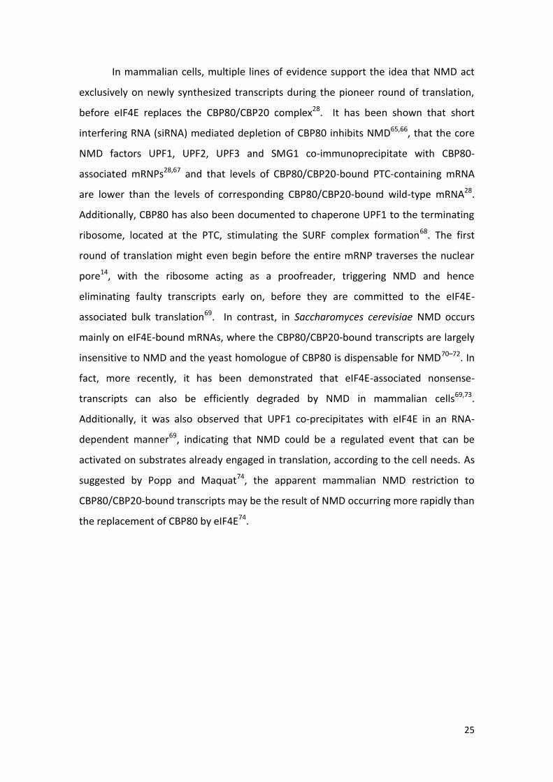
In mammalian cells, multiple lines of evidence support the idea that NMD act
exclusively on newly synthesized transcripts during the pioneer round of translation,
before eIF4E replaces the CBP80/CBP20 complex28. It has been shown that short
interfering RNA (siRNA) mediated depletion of CBP80 inhibits NMD65,66, that the core
NMD factors UPF1, UPF2, UPF3 and SMG1 co-immunoprecipitate with CBP80-
associated mRNPs28,67 and that levels of CBP80/CBP20-bound PTC-containing mRNA
are lower than the levels of corresponding CBP80/CBP20-bound wild-type mRNA28.
Additionally, CBP80 has also been documented to chaperone UPF1 to the terminating
ribosome, located at the PTC, stimulating the SURF complex formation68. The first
round of translation might even begin before the entire mRNP traverses the nuclear
pore14, with the ribosome acting as a proofreader, triggering NMD and hence
eliminating faulty transcripts early on, before they are committed to the eIF4E-
associated bulk translation69. In contrast, in Saccharomyces cerevisiae NMD occurs
mainly on eIF4E-bound mRNAs, where the CBP80/CBP20-bound transcripts are largely
insensitive to NMD and the yeast homologue of CBP80 is dispensable for NMD70–72. In
fact, more recently, it has been demonstrated that eIF4E-associated nonsense-
transcripts can also be efficiently degraded by NMD in mammalian cells69,73.
Additionally, it was also observed that UPF1 co-precipitates with eIF4E in an RNA-
dependent manner69, indicating that NMD could be a regulated event that can be
activated on substrates already engaged in translation, according to the cell needs. As
suggested by Popp and Maquat74, the apparent mammalian NMD restriction to
CBP80/CBP20-bound transcripts may be the result of NMD occurring more rapidly than
the replacement of CBP80 by eIF4E74.

26
Figure 4: Model of nonsense mediated mRNA decay (NMD) mechanism and degradation stimulation via suppressor with
morphogenetic effects on genitalia 6 (SMG6) or SMG7/SMG5 (modified from Nicholson, 2010)5. Once a premature translation
termination codon (PTC) is identified, the NMD factors up-frameshift 1 (UPF1), UPF2, UPF3 and SMG1 interact with the
eukaryotic release factor 3 (eRF3) and eRF1, forming the decay-inducing (DECID) complex. UPF2 and UPF3 recruitment and
interaction with UPF1 is facilitated by the presence of a downstream exon junction complex. The DECID complex triggers UPF1
phosphorylation (represented as a P) inducing eRF3 release and recruitment of either SMG7/SMG5 or SMG6. Interaction of
SMG7/SMG5 with UPF1 promotes deadenylation and deccaping, followed by mRNA degradation by exonucleases from the 5’ and
3’ ends. If SMG6 interacts with UPF1, the RNA is cleaved in the vicinity of the PTC and RNA is degraded in the direction of the 5’
and 3’ ends by exonucleases. Finally, protein phosphatase 2A (PP2A) recruitment enables UPF1, SMG7/5 and SMG6 recycling.
DECID formation
UPF1
phosphorylation

27
1.2.1.2. PTC definition
Trying to understand how exactly a stop codon is distinguished from a PTC has
been the subject of intense ongoing research and discussion. Despite the core NMD
factors appear to be conserved amongst species, several models for NMD PTC
definition have been proposed from studies in mammalian systems compared with
studies in Saccharomyces cerevisiae, Caenorhabditis elegans, Drosophila melanogaster
and plants75.
According to the prevailing model for mammalian NMD, a stop codon is
classified as aberrant if is located more than 50-55 nucleotides upstream of the last
exon-exon junction of a transcript. During the pioneer round of translation of a PTC
free transcript, the 80S ribosome displaces from the ORF any EJC deposited in the
mRNA, until it reaches a stop codon. Conversely, if the transcript possesses a PTC
localized at more than 50-55 nucleotides upstream of the last exon-exon junction, the
ribosome will stop before being able to remove the EJC13. Therefore, at least one EJC
will remain bound to the mRNA and trigger recruitment of the NMD factors. The EJC
also act as a binding platform for UPF2 and UPF3 (which is loaded onto mRNAs during
splicing and represents a genuine EJC component)12,76, greatly enhancing the
interaction between UPF2 and UPF3 with the SURF complex due to their close
proximity5. In contrast, if the PTC is located at less than 50-55 nucleotides upstream of
the last exon-exon junction of a transcript, all EJCs are removed and the NMD is not
activated13.
However, recent data has challenged the generality of this EJC-dependent
NMD model77,78. For example, the β-globin transcripts possessing a PTC near the AUG
fail to trigger NMD, despite the existence of downstream EJCs15,79. These exceptions
suggest that additional determinants may be involved. Indeed, there is evidence that,
similar to what happens in yeast, the decision of whether NMD is to be triggered or
not, relies upon competition between UPF1 and PABPC1 for binding to eRF3 on the
terminating ribosome19,80. If PABPC1 is in close proximity to a stop codon, it interacts
with the termination complex, stimulating proper translation termination, and
represses NMD18. On the contrary, in the event that the spatial distance between the
terminating ribosome and the poly(A) tail is too big, the interaction of PABPC1 with the

28
termination complex is reduced and UPF1 interacts with eRF3 triggering NMD16,52.
Supporting this model, it has been shown that tethering of PABPC1 in the vicinity of a
PTC abolishes NMD81, even in the presence of a downstream EJC16 and that native
stop codons were found to elicit NMD when 3’UTR length is increased82.
1.2.1.3. Natural and aberrant targets of NMD
Transcripts harboring PTCs can be generated at several stages during mRNA
biogenesis. At the DNA level, PTC containing mRNAs can arise from germline or
somatic alterations. Single base pair substitutions can change a sense codon to an in-
frame PTC (nonsense mutation) or more frequently frame-shifting deletions and
insertions alter the ribosomal reading frame, causing translation ribosomes to
encounter a PTC. Mutations at splice sites or splicing regulatory sequences may result
in inaccurate intron removal and create an intron-derived PTC or a frameshift83,84. In
addition, non-faulty regulated processes such as programmed DNA rearrangements
occurring in the immunoglobulin and T-cell receptor genes during lymphocyte
maturation generate PTCs at a high frequency, due to random deletions and the
addition of non-template nucleotides at the recombination sites5. NMD, therefore, has
an important role eliminating by-products of programmed DNA rearrangements and in
the differentiation and maintenance of hematopoietic cells85.
At the RNA level, alternative splicing constitutes a major source of PTC
containing mRNA. A genome wide analysis predicted that ∼3,100 of 16,000 human
genes examined should produce at least one alternative-splice product, one-third of
which would contain PTCs86. In some cases, it constitutes a form of auto-regulation of
the expression of the canonical protein-encoding isoforms, as the protein products of
these genes are responsible for NMD triggering87.
Other types of faulty mRNAs are transcripts from non-functional pseudogenes,
endogenous retroviral and transposon RNAs or mRNA-like non-protein coding RNAs
from intergenic regions2,4. This variety of targets infers an even broader role of the
NMD pathway in dampening the ‘‘transcriptional noise’’ of supposedly non-functional

29
RNAs. Altogether, this advocates that cells produce a large number of faulty PTC
mRNAs that are recognized and eliminated by NMD.
It has become clear during recent years that many physiological mRNAs that
encode full-length functional proteins are also NMD substrates, indicating a role for
NMD not only in mRNA quality control, but also as a translation-dependent post-
transcriptional regulator of the steady state level of gene expression2,3. Translation
regulation of existing mRNAs allows for a spatial and temporal fine-tuning of levels of
the encoded proteins, allowing the maintenance of cellular homeostasis. In fact,
several microarray studies comparing the mRNA levels of normal cells with NMD-
deficient cells in Saccharomyces cerevisiae, Drosophila melanogaster and Homo
sapiens) revealed that NMD directly and indirectly controls the abundance of 3–10% of
the transcriptome in the respective cells4.
Several features of physiological mRNAs can render them NMD-sensitive.
Introns in the 3’ UTR, transcripts containing regulatory ORFs that reside upstream of
the primary ORF41; programmed frameshifts; or long 3’ UTRs can activate NMD88.
Interestingly, mRNAs containing UGA triplets that direct selenocysteine incorporation
can also elicit NMD. When selenium is abundant, UGA codes for selenocysteine. But,
when the selenium concentration in the cell is low the UGA codon is interpreted as a
PTC89.
The natural NMD targets (identified so far) are involved in a variety of cellular
processes such as stress responses, hematopoietic stem cell development, regulation
of alternative splice forms90, genomic stability, cell-cycle, telomere length
maintenance91 and embryonic development4, potentially allowing NMD to adapt
protein expression in accordance with the cellular needs. Nevertheless, these
physiological substrates have one feature in common with their pathological
counterparts: they possess a translation termination codon that is, by NMD standards,
conceived as premature.

30
1.2.1.4. Biological and medical significance of NMD
NMD plays an important role as a modulator of the severity of the clinical
phenotype of many genetic diseases60. If translated, the mRNAs containing PTCs give
rise to truncated proteins that have either completely lost their function, have
acquired dominant-negative function, are still functional, or have gained new
functions. As a consequence of these different possibilities, NMD can either mitigate or
aggravate the disease outcome. NMD importance as a protective surveillance
mechanism is highlighted by the fact that one-third of all genetic disorders, including
many cases of cancer, are caused by nonsense mutations or frameshifts, which
generate nonsense codons60. β-thalassemia demonstrates the protective effects of
NMD against the production of faulty proteins. If a mutation causing premature
translation termination is localized within exons 1 and 2 of the transcript, the defective
β-globin mRNA is degraded by NMD and, therefore, synthesis of truncated β-globin is
limited. The resultant excess of free α-globin and any defective β-globin, which are
harmful to the cell, are degraded by proteolysis. But in the event, that the nonsense
codons resides within the third (final) exon, the transcript evades NMD allowing the
generation of truncated protein that overwhelms the cell’s proteolytic system and
causes toxic precipitation of insoluble globin chains92,93. In contrast, there are cases
(such as Ullrich disease or cystic fibrosis) in which NMD down-regulates mutant
proteins with residual activity that can partially retain normal protein function,
resulting in an augmentation of the defects caused by the original mutation. In these
cases, selective inhibition of NMD may provide a novel therapeutic method 94–96.

31
1.3. The eukaryotic initiation factor 3
The mammalian eIF3 is the largest (800 kDa molecular mass) and most complex
of the eukaryotic translation initiation factors, consisting of 13 different subunits
named from eIF3a-eIF3m97 that form three stable modules; a:b:g:i (module 1); c:d:e:l:k
(module 2) and f:h:m (module 3)98. Comparative studies of the cDNA sequences of the
mammalian eIF3 subunits with the entire genome of the Saccharomyces cerevisiae
have demonstrated that the subunits eIF3a, b, c, g, i and j are conserved across
species99–101. The eIF3a, b, c, g and i subunits are essential for translation in vivo97 and
eIF3j despite being nonessential, is capable of enhancing interactions with other
eIFs102, promoting binding of eIF3 to the 40S subunit103 and has an independent
function in 40S ribosome biogenesis104. These findings suggest that these essential
subunits provide most of the basic functions of eIF3 for translation initiation in vivo,
and the remaining nonessencial subunits (of the mammalian eIF3) appear to modulate
Figure 5: Representation of the position-dependent effects of nonsense mutations in the inheritance
pattern and clinical severity of β-thalassemia (based on Holbrook, 2004)60
. Premature translation
termination codons (PTCs) in close proximity to the start codon do not trigger NMD but any translated
β-chains are still small enough to be completely hydrolyzed by the red blood cell proteolytic system. If
the PTC is located downstream of codon 23 and more than 55 nucleotides (nt) upstream of the last
exon-exon junction, the transcript is targeted for NMD limiting protein production. In both cases the
organism is protected from the deleterious dominant-negative effects of the truncated peptides. In
contrast, transcripts bearing PTCs located less than 55 nt upstream of the last exon-exon junction
produce nonfunctional β-globin proteins that are too large to be efficiently degraded, overwhelming
the cellular proteolytic system and cause toxic precipitation of insoluble β-globin chains.

32
its activity99,105. This view is supported by the observations that module 1 can promote
mRNA binding to 40S ribosomal subunits on its own, but to achieve a maximal
efficiency, it requires additional support from module 2 and that downregulation of
eIF3c and eIF3a diminishes initiation rates106.
The mammalian subunits a, c, e, k, l, and m contain a PCI (proteasome, COP9,
eIF3) domain, a conserved structural motif shared by the functionally unrelated
complex proteasome lid and the COP9 (constitutive photomorphogenesis 9)
signalosome107. Intriguingly, the subunits f and h possess an MPN (Mpr1–Pad1 N-
terminal) domain also found in related proteins in the proteasome lid and COP9107.
These domains serve as a central structural scaffold that are involved in binding eIF3 to
the translation initiation factors eIF1, eIF1A, and eIF2, as well as to the 40S ribosomal
subunit108. The interactions between eIF3 and other translation initiation factors
control the binding of the GTP-eIF2/Met-tRNAi complex to the 40S ribosomal subunit,
positioning the mRNA on the 40S subunit, and modulate the stringency of start codon
selection42. Furthermore, subunits eIF3c, d, and e interact with eIF4G109, which is
known to promote mRNA loading onto the 40S ribosome37,38. Therefore, eIF3 plays a
central role in assembling the translation initiation complex.
In addition, recent data demonstrate a role of the eIF3 complex in NMD. In fact,
the mammalian eIF3e subunit, which is not required for general translation, impairs
NMD when silenced110. Our group has also demonstrated that human eIF3h and eIF3f
subunits are involved in the NMD-resistance of mRNAs of AUG proximal PTCs20. These
findings further support the notion that some subunits of this multiprotein complex
are involved in controlling different aspects of mRNA biogenesis rather than directly
participating in translation initiation.

33
2. Aims
Previous studies from our laboratory group revealed that mRNAs containing
PTCs in close proximity to the translation initiation AUG codon (AUG proximal PTCs)
escape NMD. This was initially surprising as these mRNAs would be expected to
contain residual EJCs and would situate the PTC quite far, in a linear sense, from the
poly(A) tail and PABPC115, a condition that could induce NMD. The observed NMD
resistance was attributed as a direct effect of the translation termination event being
located in close proximity to the AUG.
Knowing that the mRNA acquires a circular conformation due to the interaction
between eIF4G/PABPC1 and eIF4G/eIF4E and that the interaction between eIF4G and
the eIF3 complex establishes a protein bridge between the mRNA and the 43S PIC (fig.
1), we propose a model were the PABPC1 is relocated to the AUG vicinity during 43S
scanning. Consequently, if a premature termination event takes place in the vicinity of
the AUG, the PABPC1 might be able to compete with UPF1 for binding to eRF3,
stimulating proper translation termination, and repressing NMD. This model
conjectures that some initiation factors that promote scanning-dependent initiation
may remain ribosome associated during translation of first codons111. In fact, some
data have shown that mRNAs with AUG proximal PTCs become NMD sensitive when
translation elongation across the short ORF is slowed down16, indicating that PABPC1
interactions remain at the AUG vicinity during the first elongation steps, until the
ribosome reaches the short ORF stop codon. Furthermore, there are results
demonstrating that in HeLa cells treated with eIF3f or eIF3h subunits siRNAs, the AUG
proximal nonsense-mutated transcripts become sensitive to NMD20. This may imply
that these eIF3 subunits are responsible for pulling the PABPC1/eIF4G complex with
43S subunit during ribosomal scanning and translation initiation.
The aim of the work presented in this thesis was to clarify the mechanistic basis for
the NMD resistance of mRNAs carrying AUG proximal PTCs and expand the current

34
models for NMD and translation. In order to accomplish that objective, the following
goals were established:
Proving that mRNA circularization via PABPC1/eIF4G interactions, leads PABPC1
into the AUG codon vicinity, as a consequence of 43S scanning, and that this
interaction remains during the first elongation steps.
Identify and characterize how eIF3 interacts with eIF4G and with the 43S
ribosomal subunit.

35
3. Materials and methods
3.1. Plasmid constructs
A plasmid (pE_MS2-GFP) expressing a fusion protein that contains the N-
terminal portion of MS2 coat protein was previously described16. The wild-type β-
globin gene (βWT), as well as the β-globin variants β15 (UGG→UGA), β23
(GUU→UAG), β25 (GGU→UAG), β26 (GAG→UAG) and β39 (CAG→UAG) subcloned into
the pTRE2pur vector (BD Biosciences) were previously described79. A β-globin
containing three repeats of MS2 coat protein binding site in the 3’UTR
(pGEMβNins5’UTR-MS2) was previously constructed by the laboratory group, but not
published.
In order to produce a β-globin gene, as well as the β-globin variants β15, β23, β25,
β26 and β39 subcloned into a pTRE2pur vector with three repetitions of the MS2
phage coat protein binding site in the 3’UTR (pTre_β#_3xMS2bs(3’UTR); # = WT, 15,
23, 25, 26 or 39) the corresponding pTre_β# and pGEMβNins5’UTR-MS2 plamids
where used as templates. All plasmids were sequenced prior to use, to verify the β-
globin WT, β-globin variants and MS2 biding site sequence integrity with primers #1 -
#5 (table 1). pGEMβNins5’UTR-MS2 (3 µg) was subjected to an enzyme digestion with
1 µL of BstxI (New England Biolabs), 1 µL PciI (Roche), 0.5 µL BSA (100%), 5.0 µL
NEBuffer 3 (10X, New England Biolabs) in a total volume of 50 µL, at 37°C during 2h, to
cut and isolate a fragment containing the 3xMS2bs (estimated size = 491 bp) to be
used as an insert. The vector pTre_β# (# = WT, 15, 23, 25, 26 or 39) (2 µg) was also
digested following the same procedure (fragment estimated size = 5109bp). The
digestion product was resolved in a 1% agarose gel and the appropriate bands were
purified with the GeneJet Gel extraction kit (NZYtech) according to the manufacturer
protocol. The vector (pTre_β#) and the insert (3xMS2bs) were used in a ligation
reaction (molar ratio of 1:3) with 1 µL T4 DNA ligase reaction buffer (10X, Fermentas),
1 µg T4 DNA ligase enzyme (Fermentas) in a total volume of 10 µL, at 16°C overnight.
The ligation product was used to transform NZY5α competent E.coli cells according to

36
the manufacturer protocol. Random colonies were selected and grown in agar medium
overnight at 37°C, 220 rpm. The plasmid DNA was purified with the NYZtech miniprep
extraction kit according to the manufacturer protocol and sequenced with primers #1,
#2 and #5 (table 1). All plasmids with the correct sequence were amplified in NZY5α
competent E.coli cells and purified with NYZtech miniprep extraction kit.
In order to produce a β-globin gene into a pTRE2pur vector with three repetitions
of the MS2 phage coat protein binding site at forty five nucleotides of distance
downstream of the codon 39 (pTre_βWT_3xMS2bs(45nt_CD39)); the corresponding
pTre_βWT and pTreβWT_3xMS2bs(3’UTR) plamids where used as templates for gene
splicing by overlap extension (SOEing) PCR. pTreβWT (0.5 µg) was amplified with 0.5 µL
NZYSpeedy DNA polymerase (NZYtech), 5.0 µL reaction buffer (10X, NZYtech), 2.5 µL
MgCl2 (50 mM), 2.5 µL dNTPs (10 mM), 5.0 µL DMSO (100%), 1.0 µL BSA (100%) and
2.5 µL of primers (10 µM) #6-#7 (table 1) for Soeing 1 or #8-#9 (table 1) for Soeing 3, in
a total volume of 50 µL. Thermocycler conditions were 95°C for 2 min followed by 40
cycles of 95°C for 30 sec, 55°C for 1 min, and 70°C for 45 sec followed by a final
extension of 70°C for 5 min. For SOEing 2, pTre_βWT_3xMS2bs(3’UTR) was amplified
in with primers #10-#11 (table 1) following the same procedure as above, but with an
annealing temperature of 72°C for 1 min. The SOEing 1, 2 and 3 products were
analyzed by electrophoresis on a 2% agarose gels. The appropriate bands were purified
with the GeneJet Gel extraction kit (NZYtech) according to the manufacturer protocol
and quantified in a Nanodrop spectrophotometer (Thermo).
3.2. Cell culture, plasmid and siRNA transfection
HeLa cells were grown in Dulbecco’s modified Eagle’s medium (DMEM)
supplemented with 10% fetal bovine serum (FBS), at 37°C/5% CO2. Transfections of
cells with siRNAs were carried out when cells had a confluence of 30-40%, using 200
pmol of siRNA oligonucleotides and 4μl of Lipofectamine 2000 Transfection Reagent
(Invitrogen), following the manufacturer’s instructions. When indicated, a
supplemental siRNA transfection was made 24h after the initial siRNA transfection,
using 50 pmol of siRNA oligonucleotides and 4μl of Lipofectamine 2000. The siRNA

37
duplexes (Table 2) were designed as 19-mers with 3’-dTdT overhangs and purchased
from Thermo. Transfections of cells with plasmids were carried out when cells had a
confluence of 70-80%, using 200 ng of pTRE_β#_3xMS2bs(3’UTR), 800 ng pE_MS2-GFP
and 4μl of Lipofectamine 2000 Transfection Reagent (Invitrogen), following the
manufacturer’s instructions.
Table 1: Sequences of the primers used in the current work
Primer Sequences (5’-->3’)
1 ACATTTGCTTCTGACACAAC
2 GCAATGAAAATAAATGTTTTTTAT
3 GCTCCTGGGCAACGTGCT
4 GTGGATCCTGAGAACTTCAGGCT
5 GTTCATGTCATAGGAAGGGG
6 TCGGTACCCGGGGATCCTCTAGTC
7 CCTCATGTTAACAGCATCAGGAGTGGACAG
8 GATGCTGTTAACATGAGGATCACCCATGTT
9 GTTGCCCATCCCAAACATGGGTGATCCTCA
10 ATGTTTGGGATGGGCAACCCTAAGGTGAAG
11 GAAAGAAAACATCAAGCGTCC
12 CTGCTCATTGCAGGCCAGAT
13 GAGCCTGGGCCATGAAGAG
14 CACCCAGTCATTTTGGCCTC
15 CGACAGTTCCCAACAGGGTC
16 ACCAAGAGAGTTGTCCGCAGTG
17 TCATGGCATTACGGATGGTCC
18 CCATGAGAAGTATGACAACAGCC
19 GGGTGCTAAGCAGTTGGTG
Table 2: Sequences of the siRNAs used in the current work
siRNA Sequences (5’-->3’)
eIF3f1 AUACGCGUACUACGACACU
eIF3f2 GUGAAGGAGAAAUGGGUUU
eIF3h1 GAUCGGCUUGAAAUUACCA
eIF3h2 ACUGCCCAAGGAUCUCUCU
eIF3c UGACCUAGAGGACUAUCUU
GFP GGCUACGUCCAGGAGCGCAC

38
3.3. Immunoprecipitation assay
HeLa cells cultured in 35-mm dishes and treated with siRNAs were collected
48h after transfection. Cells were lysed in 150 μl of NP40 buffer [50 mM Tris-HCl
pH=7.5; 10 mM MgCl2; 100 mM NaCl; 10% (v/v) Glicerol; 1% (v/v) Nonidet P-40 and 1%
(v/v) protease inhibitor mixture (Sigma). Additionally, 0.4 µL of RNase inhibitor (40
U/µL; NZYtech) were added to the samples not intended to be treated with RNAse A.
Total lysates were cleared by centrifugation at 5000 rpm for 10 min at 4 °C and 20 μl
were collected for RNA extraction or protein analysis with 4 μl 5x SDS loading buffer
(Pre-IP). The remaining lysates were incubated overnight at 4°C with rabbit polyclonal
anti-eIF3F (Abcam) or rabbit monoclonal anti-eIF3H (Cell Signaling) at a 1:50 dilution in
a vertical rotator (Grant bio). Thirty μl of protein G-agarose beads (Roche) and 100
mg/mL of RNAse A (Quiagen) were then added to the corresponding samples. After an
incubation of 1 hour at 4°C, the samples were washed three times with excess NP40
buffer and ressuspended in 25 μl of 2x SDS sample buffer (IP).
3.4. Cycloheximide treatment
HeLa cells were grown in Dulbecco’s modified Eagle’s medium supplemented
with 10% fetal bovine serum, in 35-mm plates, at 37°C/5% CO2. Once they achieved 90-
100% confluence, the medium was replaced with a fresh one containing cycloheximide
(CHX, Sigma). The cells were in incubated at 37°C/5% CO2 for 2h and then lysated.
3.5. SDS-PAGE and Western blotting
Protein lysates were resolved, according to standard protocols, in 12% SDS-
PAGE electrophoresis and transferred to PVDF membranes (Bio-Rad). The membranes
were probed using mouse polyclonal anti-α-tubulin (Sigma) at 1:500 dilution (as a
loading control), rabbit polyclonal anti-PABPC1 (Cell Signaling) at 1:500 dilution, rabbit
monoclonal anti-eIF3H (Cell Signaling), rabbit polyclonal anti-CBP80 (generous gift
from E. Izaurralde), rabbit polyclonal anti-eIF4G (Cell Signaling), mouse monoclonal

39
anti-eIF4E (Santa Cruz), mouse monoclonal anti-RPS6 (Cell Signaling) at 1:250 dilution,
rabbit anti-c-myc (Santa Cruz) at 1:500 dilution and rabbit polyclonal anti-
enterobacteriophage MS2 coat protein antibody (Merck Millipore) at 1:250 dilution.
Detection was carried out using secondary peroxidase-conjugated anti-mouse IgG (Bio-
Rad), anti-rabbit IgG (Bio-Rad) antibodies followed by chemiluminescence.
3.6. RNA isolation
Total RNA from transfected cells was prepared using the Nucleospin RNA
extraction (Marcherey-Nagel) following the manufacturer’s instructions.
3.7. Reverse transcription
cDNA was prepared by incubating 1 µg of purified total mRNA with 1 µL of
dNTPs (10 µM) and 1 µL of random primers (0.25 µg/ µL), in a total volume of 15 µL,
during 5 min at 65°C. Then, the mixture was incubated in ice and 2 µL of reaction
buffer (10X; NZYtech), 0.1 µL of ribonuclease inhibitor (40 U/µL; NZYtech) and 0.5 µL of
reverse transcriptase (200 U/µL; NZYtech) was added, to a final volume of 20 µL.
Finally, the mixture was incubated in a thermocycler at 25°C for 10 min, 50°C for 50
min and 85°C for 5 min. cDNA was stored at 4°C.
3.8. Semiquantitative PCR
Three dilutions (1:1; 1:2 and 1:4) were prepared using the RT product. 5 µL of
each of the diluted samples were amplified with 0.2 µL of Taq DNA polymerase (5
U/µL; Ambion), 2.5 µL of 10X PCR buffer (with 15 mM MgCl2; Ambion), 0.5 µL of dNTPs
(10 mM) and 1 µL of each corresponding forward and reverse primer (table 1; #12-13
for eIF3H; #14-15 for eIF3F and #16-17 for eIF3C), in a total volume of 25 µL. The same
procedure was carried out in parallel, but using GADPH specific primers (table 1, #18-
19) as an internal standard. Thermocycler conditions were 95°C for 5 min followed by
27 cycles of 94°C for 45 sec, 55°C for 45 sec, and 72°C for 45 sec, followed by a final

40
extension of 72°C for 5 min. Ten-microliter aliquots from each sample were analyzed
by electrophoresis on 2% agarose gels.

41
4. Results
4.1. ptre_βWT_3xMS2bs(45nt_CD39) construction by SOEing PCR
To test the hypothesis that mRNA circularization via PABPC1/eIF4G
interactions, leads PABPC1 into the AUG codon vicinity, as a consequence of 43S
scanning, and that this interaction remains during the first elongation steps, we tried
to construct by SOEing PCR a plasmid containing the β-globin sequence and three
repetitions of MS2 coat protein binding site (3xMS2bs) 45 nt downstream of codon 39.
This construct would allow the capture of β-globin mRNPs to study PABPC1
interactions with the ribosome. Using ptre_βWT as a template the 5’UTR of βWT
(containing a ClaI restriction site) and codons 1-54 were amplified with primers #6-#7
(fig. 6 and 7; lane S1). Codon 55 until the beginning of exon 2 (containing a BbrPI
restriction site) was amplified with primers #8-#9 (fig. 6 and 7; lane S3). Both S1
(expected fragment size = 409 bp) and S3 (expected fragment size = 191 bp) fragments
were successfully amplified at annealing temperature of 55°C. The three repetitions of
MS2 binding site were amplified with primers #10-#11 using the plasmid
ptre_βWT_3xMS2bs(3’UTR) as a template (fig. 6 and 7; lane S2). The 3xMS2bs
(expected fragment size = 93 bp) was only successfully amplified at an annealing
temperature of 72°C. However, due to the repeated sequence of the MS2 binding site,
1x MS2bs was also amplified (expected fragment size = 43 bp) alongside other
unspecified products, resulting in a lower quantity of the desired fragment. This in
combination with the 3xMS2bs small size made the purification step very inefficient.
Therefore the 3xMS2bs fragment was not obtained with the quantity and quality
necessary to continue the plasmid construction.

42
Figure 7: ptre_βWT_3xMS2bs(45nt_CD39) construction by SOEing PCR. Agarose gel photos of the
amplification products of the ptre_βWT plasmid with primers #6-#7 (lane S1) or with primers #8-#9
(lane S3) and the ptre_βWT_3xMS2bs(3’UTR) plasmid with primers #10-#11 (lane S2).
S1 S3
50
150 200
400
50
100
150
S2
1x MS2bs
3x MS2bs
bp bp
Figure 6: Schematic diagram of SOEing PCR. Using ptre_βWT as a template, the 5’ untranslated
region (UTR) and codons 1-54 of the normal human β-globin gene (βWT) were amplified with primers
#6-#7 (Table 1), while the sequence from codon 55 (localized 45 nucleotides downstream of codon
39) until the beginning of exon 2 were amplified with primers #8-#9 (Table 1). The primers used for S1
and S3 contain overlapping sequences with the MS2 binding site (MS2bs) sequence. The three
repetitions of MS2bs were amplified with primers #10-#11 (Table 1) using the plasmid
ptre_βWT_3xMS2bs(3’UTR) as a template. The primers used for S2 amplification contained
overlapping sequences for the ptre_βWT, upstream and downstream of codon 55. Extension of these
overlaps, by DNA polymerase, creates the full-length mutant molecule of S1, S2 and S3 fragments.
S1 S2 S3

43
4.2. Anti-enterobacteriophage MS2 coat protein antibody test
The coat protein binding site of the MS2 bacteriophage is a naturally occurring
RNA aptamer112 that in conjunction with the MS2 coat protein (MS2cp) and a specific
antibody for the MS2cp can selectively immuneprecipitate the corresponding mRNA
transcripts and any protein attached, thus providing a robust method to study the
RNA–protein interactome113,114. Before proceeding with MS2 immunoprecipitations
the anti-enterobacteriophage MS2 coat protein antibody specificity and conditions
were tested (fig. 8). HeLa cells were transfected with pTre_BWT_3xMS2(3’UTR)
plasmid and the pE_MS2-GFP plasmid, which codifies a fusion protein containing
MS2cp. Lysates were generated 24 hours after transfection and probed with the anti-
MS2cp antibody or anti-α-tubulin as a loading control. According to the manufacturer
instructions, the MS2cp signal should appear around 13 kDa, however since the
protein expressed is a fusion protein the observed band size corresponding to MS2-
GFP was around 40 kDa (fig. 8). The bands at 35 kDa and 25 kDa correspond to
uncharacterized interactions. No band was detected in non-transfected HeLa cells (fig.
8; lane 6), confirming the antibody specificity.
Figure 8: Test of anti-enterobacteriophage MS2 coat protein antibody. Western blot analysis of HeLa
cells extracts transfected with a plasmid containing the β-globin sequence with three repetitions of
MS2 coat protein binding site at the 3’UTR and a plasmid that codifies for a MS2 coat protein fused
with the green fluorescent protein (GFP). Lysates were generated 24h after transfection and analyzed
with a specific antibody for MS2 coat protein and with anti-α-tubulin (α-tub) as a loading control.
α - tub
MS2-GFP 40 kDa
35 kDa
25 kDa
1 2 3 4 5 6 Lane

44
4.3. Cycloheximide concentration test
Besides using the ptre_βWT_3xMS2bs(45nt_CD39) to capture and study the
mRNPs, HeLa cells would also be treated (or untreated) with cycloheximide (CHX). This
is a common laboratory reagent that inhibits protein synthesis by binding the ribosome
in the E-site and blocking eEF2-mediated translocation. Due to its size, CHX has a
bigger affinity for the E-site when it is empty, i.e. when the ribosome is at the AUG. It
was shown that CHX can cause about half of the ribosomal population to stop at the
very first codon115. Taking advantage of this, CHX treatment would facilitate the
capture of the mRNPs near the AUG, allowing the analysis of the interaction of PABPC1
and eIF4G with the ribosome (through eIF3) near the start codon. The necessary
concentration of CHX to restrict protein synthesis was assessed in HeLa cells treated
with different concentration of CHX for 2 hours (fig. 9). Lysates were generated
afterwards and CHX effect on translation was evaluated by controlling C-myc protein
levels by Western blot. The c-Myc protein is normally degraded very rapidly with a
half-life of 20 to 30 min116. Therefore if translation elongation was blocked for 2h, it
should be expected to see a decrease of the C-my protein levels. In fact, the Western
blot shows a decrease of C-myc protein level of about 40% in the samples treated with
150 µg/mL of CHX (fig. 9; lane 6), indicating that at this concentration, the CHX should
be able to interfere with translation in HeLa cells.
62 kDa
α - tub
0 10 25 50 100 150
[Cycloheximide] µg/mL
c-myc
55 kDa
100 100 100 60 60 40 C-myc/α-tub (%) 1 2 3 4 5 6 Lane
Figure 9: Test of cycloheximide (CHX) concentration necessary to inhibit translation. Western blot
analysis of HeLa cells extracts treated with different concentrations of CHX. HeLa cells were
incubated with CHX for 2h. Lysates were analyzed with a specific antibody for c-myc and with anti-α-
tubulin (α-tub) antibody as a loading control. An estimative of the ratio (in %) of c-myc to α-tub is also
represented.

45
4.4. eIF3h, eIF3f and eIF3c subunits knockdown
Before proceeding to the immunoprecipitation (IP) experiments to assess the
eIF3h, eIF3f and eIF3c role in the interaction between PABPC1 and eIF4G with the
ribosome, the knockdown efficiency of these subunits was tested at the protein level
(fig. 10). HeLa cells were treated with 200 pmol of specific siRNAs for each subunit
(table 2 – siRNA eIF3f1, eIF3h1 or eIF3c respectively) or with siRNA GFP as a
transfection control. Lysates were generated 48 hours later and analyzed by Western
blot with specific antibodies or with α-tubulin as a loading control. Silencing was
achieved for both eiF3h (fig. 10A; lane 1) and eiF3c (fig. 10C; lane 1) when compared
with the lysates treated with siRNA GFP (fig. 10A and 10C; lane 2), with an estimated
knockdown efficiency of 50% and 80%, respectively. For eIF3f silence however, no
eiF3f signal was detected (fig. 10B; lane 1 and 2). This may indicate that the quantity or
the conditions utilized for the detection of eIF3 were not adequate.
C
α - tub
eIF3h GFP
eIF3h
siRNA
Lane 1 2
A
40 kDa
55 kDa α - tub
eIF3f GFP
eIF3f
siRNA
Lane 1 2
B
50 kDa
55 kDa α - tub
eIF3c GFP
eIF3c
siRNA
Lane 1 2
98 kDa
55 kDa
50 0 %KD 80 0 %KD
Figure 10: Test of the conditions for the eukaryotic initiation factor 3h (A), eIF3f (B) and eIF3c (C)
subunits knockdown. Western blot analysis of HeLa cells extracts transfected with siRNA eIF3h1 (A;
lane 1), siRNA eIF3f1 (B; lane 1), siRNA eIF3c (C; lane 1) or with a control green fluorescent protein
(GFP) siRNA (lane 2A, 2B and 2C). Cell lysates were obtained 48h post transfection and siRNA
silencing efficiency was analyzed with specific antibodies and with anti-α-tubulin (α-tub) as a loading
control. An estimative of the percentage of achieved knockdown (% KD) is represented below each
lane.

46
4.5. Immunoprecipitation of mRNP complexes from HeLa cells treated with
siRNAs specific for the eIF3f subunit
To test whether the eIF3f subunit mediates the interaction between PABPC1
and eIF4G with the 40S ribosome during initiation scanning, immunoprecipitation (IP)
of mRNPs from HeLa cells depleted of eIF3f subunits were carried out, in the
conditions previously tested (fig. 10). HeLa cells were transfected with 200 pmol of
siRNA GFP or with siRNA eIF3f1 (Table 2). The lysates were obtained 48 hours later and
the mRNP complexes were isolated by immunoprecipitation, using an antibody specific
for eIF3h. Immunoprecipitation was performed in the presence or absence of RNase A,
to test for protein interactions dependent or independent of mRNA. A 20 µL aliquot
from each sample, representing the total lysate, was taken before
immunoprecipitation (Pre-IP). The presence of PABPC1, RPS6 and other initiation
factors was assessed by Western blot (fig. 11). α-Tubulin was used as a loading control
and has expected, it was not detected in IP samples (fig. 11; lane 3-5), showing that the
washing procedure, to remove any nonspecifically bonded proteins, was effective.
Efficiency eIF3f knockdown was evaluated with an anti-eIF3f antibody.
Comparing the eIF3f band of the Pre-IP samples (fig. 11; lane 1 and 2) there is no
discernible differences, in terms of eIF3f protein level, between samples treated with
control siRNA (fig. 11; lane 1) or with siRNA eIF3f1 (fig. 11; lane 2). Both signals have
the same intensity and also there is no difference in the loading control. This indicates
that eIF3f silencing did not have any effect, at least at the protein level. In the IP
samples no eIF3f protein was detected (fig. 11; lane 3-5). Instead a band (50-55 kDa)
corresponding to the heavy chain of the immunoglobulin G (IgG) was observed. This
signal is detected because the anti-eIF3h antibody used during IP is also eluted from
the beads and size-fractionated by SDS-PAGE. Since the IP antibody and the Western
blot primary antibody are from the same species, the secondary antibody will also
recognize it. This signal was always detected when a primary antibody of the same
species was used in Western Blot and exclusively in the IP samples.
Despite the eIF3f subunit silencing apparently not working as expected, some
interesting variations can be pointed out. eIF4G, PABPC1 and CBP80 co-
immunoprecipitate with eIF3h and were detected in the IP samples even when treated

47
with RNase A (fig. 11; lane 4). These results imply that the interaction of these proteins
with eIF3h is independent of RNA. Both eIF4G and PABPC1 signals in IP samples
treated with control siRNA (fig. 11; lane 3) are stronger than the signals of siRNA eiF3f
(fig. 11; lane 4 and 5) treated samples. This suggests that eIF3f silencing interfered with
PABPC1 and eIF4G interaction with the eIF3h subunit. On the other hand, samples
treated with siRNA eIF3f (fig. 11; lanes 4 and 5) also exhibit a weaker eIF3h protein
signal when compared with the control (fig. 11; lane 3). Therefore the decreased
interaction of eIF4G and PABPC1 with eIF3h observed in samples treated with siRNA
eIF3f, may be due to eIF3f silencing destabilized eIF3h, and not because eIF3f mediates
the interaction between eIF4G and PABPC1 with eIF3h and the ribosome. The mRNA
levels of eIF3h would have to be assessed to see if the siRNA eIF3f1 is not affecting
eIF3h expression. Additionally, this differences seen could also be a case of lane 3 (fig.
11) having more sample quantity than lane 4 and 5 (fig. 11). Since α-tubulin was not
detected, has expected, in the IP samples, it is difficult to assess if the protein loading
was equal across lanes 3-5 (fig. 11).
RPS6, a component of the 40S ribosomal unit, was not detected in IP samples,
in either control (fig. 11; lane 3) or siRNA eIF3f (fig. 11; lane 4 and 5) treated cells. In
Pre-IP samples, however, the RPS6 signal of the control sample (fig. 11; lane 1) is more
intense than the siRNA eIF3f treated sample (fig. 11; lane 2), suggesting that the
silencing treatment interfered with RPS6 expression. RPS6 mRNA levels would have to
be analyzed to confirm this difference. The protein eIF4E was not detected in IP
samples (fig. 11; lane 3-5) and is barely seen in Pre-IP samples (fig. 11; lane 1 and 2).
This was unexpected since eIF4E supports the bulk of translation; therefore, it should
be more abundant than CBP80. Nonetheless, the absence of eIF4E and RPS6 in the IP
samples suggests that these proteins do not co-immunoprecipitate with eIF3h.
Conversely, this could be an experimental artifact. In fact, the PVDF membrane was
probed and reprobed several times and part of the protein may have been gradually
washed away during removal of primary and secondary antibodies, resulting in a weak
protein signal in Pre-IP samples and no signal in the IP samples (which had less protein
quantity than the Pre-IP samples to begin with). A newly prepared membrane would
have to be probed to assess if eIF4E and RPS6 does or does not co-immunoprecipitate
with eIF3h.
48
Figure 11: Immunoprecipitation of mRNP complexes from HeLa cells treated with siRNAs specific
for the eukaryotic initiation factor 3f (eIF3f). Western blot analysis of HeLa cells extracts transfected
with human eIF3f siRNA (lane 2, 4 and 5) or with a control green fluorescent protein (GFP) siRNA
(lane 1 and 3). HeLa cells lysates were obtained 48 hours after transfection with GFP siRNA or siRNA
targeting the eIF3f subunit. Immunoprecipitation (IP) was accomplished with an antibody specific to
eIF3h (lanes 3-5). Immunoprecipitation was carried out in the presence (lane 4) or absence (lane 5) of
RNAse A. Lanes 1 and 2 correspond to aliquots of cell lysates prior to IP (Pre-IP). α-tubulin (α-tub) was
used as a loading control.
siRNA GFP
siRNA eIF3f
Pre-IP
- +
siRNA GFP
siRNA eIF3f IP eIF3h
eIF3f
1 2
RNase A
eIF4G
PABPC1
IgG
eIF3h
α-tub
3 Lane
CBP80
RPS6
4 5
eIF4E
200 kDa
80 kDa
71 kDa
50 kDa
40 kDa
32 kDa
28 kDa
55 kDa

49
4.6. Immunoprecipitation of mRNP complexes from HeLa cells treated with
siRNAs specific for the eIF3h subunit
The eIF3h subunit role in the interaction of PABPC1 and eIF4G with the 40S
ribosome during translation initiation was also tested in cells depleted of eIF3h. HeLa
cells were either transfected with 200 pmol of siRNA GFP or with siRNA eIF3h1 (Table
2). The lysates were obtained 48 hours later and the mRNP complexes were isolated by
immunoprecipitation, using an antibody specific for eIF3f or with protein G-agarose
beads alone (mock) to control for nonspecific immunoprecipitation. IP was performed
in the presence or absence of RNase A. An aliquot from each sample, representing the
total lysate, was taken before immunoprecipitation (Pre-IP) for protein and RNA
analysis. The presence of PABPC1 and initiation factors was assessed by Western blot
and α-tubulin was used as a loading control (fig. 12). eIF3h silencing efficiency was
evaluated at the protein level using an anti-eIF3h antibody (fig. 12) and at the RNA
level by a semiquantitative PCR (fig. 13).
Western blot analysis of eIF3h knockdown shows no distinct differences
between GFP siRNA treated samples (fig. 12; lane 1) and siRNA eIF3h treated samples
(fig. 12; lane 2). Contrariwise, the eIF3h RNA endogenous levels in cells treated with
siRNA eiF3h (fig. 13; lane 9-11) are clearly lower than those treated with control siRNA
(fig. 13; lane 3-5). In fact the lane 9 band (dilution 1:1; fig. 13) intensity is weaker than
the band (dilution 1:2) in lane 4, indicating a silencing efficiency of at least 50% at the
RNA level.
Both eIF4G and PABPC1 were probed with specific antibodies. eIF4G was again
detected in an RNA independent manner in IP samples (fig. 12; lane 4-7). PABPC1,
however, was not detected in IP samples, suggesting that it does not co-precipitate
with eIF3f. This is surprising, because PABPC1 forms a specific complex with eIF4G, which
directly interacts with the eIF3 protein that co-immunoprecipitats with eIF3f. Additionally,
PABPC1 is also bound to the poly-A tail, so it was expected that at least the PABPC1 signal
could be observed in control IP samples not treated with RNAse A (fig. 12; lane 4). This can
be, once again, a case of the protein content loss due membrane washing and
reprobing. A newly prepared membrane would have to be probed to confirm these
findings.

50
siRNA GFP
siRNA eIF3h
Pre-IP
- + - +
siRNA GFP
MOCK
siRNA GFP siRNA eIF3h
IP eIF3f
RNase A
eIF4G
PABPC1
IgG eIF3f eIF3h
α-tub
Lane
200 kDa
71 kDa
50 kDa
40 kDa
55 kDa
1 2 3 4 5 6 7
Figure 12: Immunoprecipitation of mRNP complexes from HeLa cells treated with siRNAs specific for
the eukaryotic initiation factor 3h (eIF3h). Western blot analysis of HeLa cells extracts transfected
with a control green fluorescent protein (GFP) siRNA target (lane 1, 3, 4 and 5) or with human eIF3h
siRNA (lane 2, 6 and 7). Cell lysates were obtained 48 hours after transfection with GFP siRNA or
siRNA targeting eIF3h and immunoprecipitated with an antibody to eIF3f (lanes 4-7) or with protein G-
agarose beads only (lane 3). Immunoprecipitation was carried out in the presence (lane 5 and 7) or
absence (lane 4 and 6) of RNAse A. The lysates were analyzed for the presence of PABPC1 and
initiation factors by immunoblotting. Lanes 1 and 2 correspond to aliquots of cell lysates prior to IP
(pre-IP). α-tubulin (α-tub) was used as a loading control.
1:1 1:2 1:4 1:1 1:2 1:4 1:1 1:2 1:4 1:1 1:2 1:4
eIF3h GAPDH eIF3h GAPDH siRNA GFP siRNA eIF3h
1 2 3 4 5 6 7 8 9 10 11 12 13 14
Figure 13: Quantification by reverse transcription coupled semiquantitative (SQ) PCR of the
eukaryotic initiation factor 3h (eIF3h) knockdown efficiency. Representative SQ-PCR analyses of
cDNA obtained from RNAs extracted from green fluorescent protein (GFP; lane 3-8) or eIF3h (lane 9-
14) siRNA-treated HeLa cells. SQ-PCR was carried out with eIF3h mRNA specific primers to monitor
endogenous eIF3h expression or with glyceraldehyde-3-phosphate dehydrogenase (GADPH) mRNA
specific primers as an internal standard. Lane 1 and 2 represent blank samples with eIF3h primers or
GAPDH specific primers. Sample dilution is also represented.
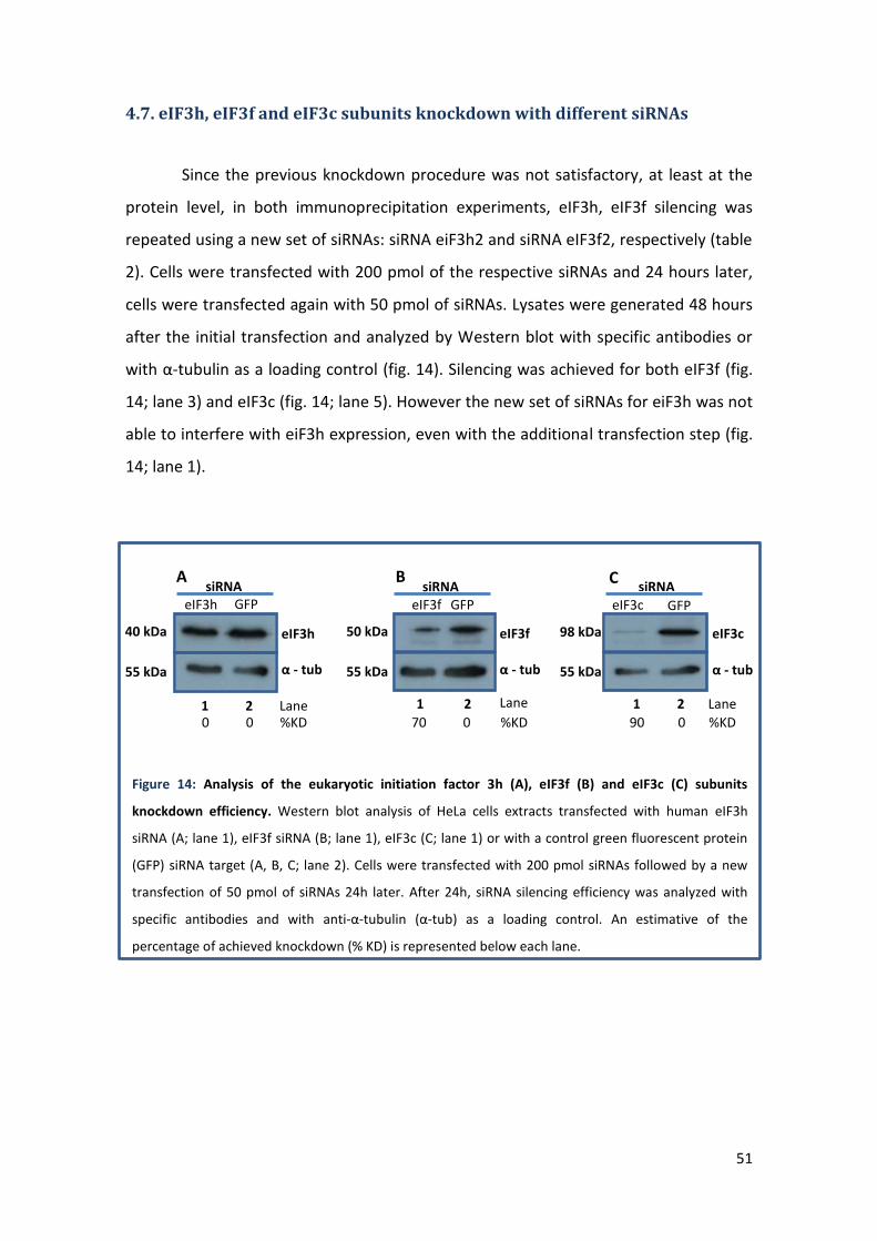
51
4.7. eIF3h, eIF3f and eIF3c subunits knockdown with different siRNAs
Since the previous knockdown procedure was not satisfactory, at least at the
protein level, in both immunoprecipitation experiments, eIF3h, eIF3f silencing was
repeated using a new set of siRNAs: siRNA eiF3h2 and siRNA eIF3f2, respectively (table
2). Cells were transfected with 200 pmol of the respective siRNAs and 24 hours later,
cells were transfected again with 50 pmol of siRNAs. Lysates were generated 48 hours
after the initial transfection and analyzed by Western blot with specific antibodies or
with α-tubulin as a loading control (fig. 14). Silencing was achieved for both eIF3f (fig.
14; lane 3) and eIF3c (fig. 14; lane 5). However the new set of siRNAs for eiF3h was not
able to interfere with eiF3h expression, even with the additional transfection step (fig.
14; lane 1).
α - tub
eIF3h GFP
eIF3h
siRNA
Lane 1 2
A
40 kDa
55 kDa
eIF3f GFP
Lane 1 2
α - tub
eIF3f
siRNA B
50 kDa
55 kDa
eIF3c
Lane 1 2
α - tub
GFP
eIF3c
siRNA C
98 kDa
55 kDa
0 0 %KD 90 0 %KD 70 0 %KD
Figure 14: Analysis of the eukaryotic initiation factor 3h (A), eIF3f (B) and eIF3c (C) subunits
knockdown efficiency. Western blot analysis of HeLa cells extracts transfected with human eIF3h
siRNA (A; lane 1), eIF3f siRNA (B; lane 1), eIF3c (C; lane 1) or with a control green fluorescent protein
(GFP) siRNA target (A, B, C; lane 2). Cells were transfected with 200 pmol siRNAs followed by a new
transfection of 50 pmol of siRNAs 24h later. After 24h, siRNA silencing efficiency was analyzed with
specific antibodies and with anti-α-tubulin (α-tub) as a loading control. An estimative of the
percentage of achieved knockdown (% KD) is represented below each lane.

52
5. Discussion
5.1. Proving that PABPC1/eIF4G remains associated with the ribosome,
through eIF3 interaction, during the early stages of elongation
Using β-globin mRNA (a small transcript of three exons, whose NMD profile has
been extensively characterized) as the primary model, it was shown that transcripts
containing PTCs in the latter half of the first exon, up to the third exon, are targeted for
NMD, which can be attributed to the existence of downstream EJCs. However β-globin
transcripts bearing PTCs localized up to 23 codons (AUG proximal PTCs) into the open
reading frame effectively evade NMD (fig. 5). This was unexpected since there are EJCs
still residing downstream. It was demonstrated that this resistance reflects a close
proximity of the PTC to the translation initiation codon and are not caused by defects
in transcript splicing, impaired translation, or reinitiation of translation 3’ to the
PTC15,79,117,118.
Recently, it has been verified that PABPC1 can compete with UPF1 for eRF3
binding, when a ribosome is poised in a stop codon, diminishing NMD response16,19 .
Additionally, it is also known that mRNAs form a closed-loop structure during
translation, as a consequence of eIF4G interaction with PABPC1, eIF4E, eIF3 and the
ribosome. With this findings, it was proposed that PABPC1 possesses a major role
protecting mRNAs harboring AUG proximal PTCs from the NMD surveillance
pathway16,24.
Assuming the retention of ribosome association with eIF3 and PABPC1 (through
eIF4G) during scanning and the early stages of elongation, we propose that PABPC1 is
repositioned in close proximity to an early PTC, during ribosome scanning, inhibiting
NMD and enhancing the efficiency of the translation termination. This model is
supported by the fact that tethering of PABPC1 in the vicinity of a PTC inhibited NMD16
and the observations that eIF3 can remain bound to the translating ribosome during
the initial phase of elongation97,119.

53
To investigate if PABPC1 is in fact translocated during initiation scanning and
that remains in the vicinity of the AUG during the first steps of elongation, the
pTre_βWT_3xMS2bs(45nt_CD39) was designed. The introduction of multiple MS2bs
into the β-globin sequence would facilitate the isolation of β-globin mRNPs, by
immunoprecipitation assays, allowing the analysis of PABPC1 interactions with the
ribosome and other proteins of the mRNP. The ptre_βWT_3xMS2bs(45nt_CD39)
plasmid assembly was initiated using a SOEing PCR approach (fig. 6), a method of
recombining sequences without depending on restriction sites or ligase. This process
involves using primers to introduce segments of identical sequence (called the overlap
region) in the amplified fragments. Then, during overlap PCR, the overlapping
segments anneal and act as primers of one another. Extension of this overlap by DNA
polymerase creates the full-length mutant molecule of both fragments. The 3xMS2bs
fragment (fig. 7; lane S2), however, proved to be rather difficult to amplify. Owing to
the presence of several repetitions of the MS2bs in the pTre_βWT_3xMS2bs(3’UTR),
the primer could anneal with any of them, resulting in a PCR product containing
fragments with 1 or several repetitions of MS2bs. Several PCR conditions were tested,
but none resulted in a satisfactory amount of the fragment of interest (containing
3xMS2bs). Moreover, the fragment is relatively small (93 bp) making purification
problematic.
An alternative process of construction could be using site directed mutagenesis
to flank 3xMS2bs with a pair of restriction sites, such as SalI and XhoI, at 5’ and 3’
respectively. These enzymes recognize very similar sequences and produce compatible
cohesive ends. The resulting fragment, containing the 3xMS2bs could then be
subcloned into the vector containing the β-globin sequence with a XhoI restriction site
at 45 nt downstream of the codon 39. The advantage of this method is that the ligation
of the insert into the XhoI site of the β-globin will produce an XhoI restriction site at 5’,
that can be cleaved to insert more 3xMS2bs if needed114. The addition of multiple
MS2bs admits more coat protein, which could improve pulldown efficiency during
immunoprecipitation assays.

54
5.2. Identification and characterization of eIF3 interactions with eIF4G and
the 40S ribosomal subunit
Human eIF3 is an 804 kDa protein complex containing 13 nonidentical subunits,
named from 3a to 3m. Six of these subunits (eIF3a, b, c, g, i and j) are conserved in
sequence across species, and are thought to provide most of the basic functions of
eIF3120. eIF3f and h are part of the nonconserved core and with eIF3m constitute a
stable module that is located on the periphery of the complex98. Both eIF3f and eIF3h
contain a MPN domain that is usually found in components of large protein complexes
and have been implicated in protein-protein interactions supporting a possible
interaction of these proteins with other initiation factor complexes121.
Our group has shown that AUG proximal nonsense-mutated transcripts are
NMD resistant but become sensitive to NMD when HeLa cells are treated with siRNAs
specific for the eIF3h, eIF3f20 and eIF3c (unpublished) subunits, supporting the notion
that eIF3 subunits might be involved in the delivery of eIF4G-associated PABPC1 to the
vicinity of the AUG proximal PTC.
To study the role of the eIF3h and eIF3f subunits, mRNPs from HeLa cells
treated with specific siRNAs for these subunits were isolated and examined. Overall
the IP procedure of mRNPs from HeLa cells treated with eIF3f specific siRNAs (fig. 11)
was successfully accomplished. The proteins expected to be bound to the mRNP were
detected, although some proteins (PABPC1, eIF4G and CBP80) were only visible (in IP
samples) with higher (5-10 min) exposure times to the X-ray film, resulting in higher
backgrounds. In comparison, IP pulldown efficiency in HeLa cells treated with eIF3h
siRNAs was even lower. As a result, the proteins detected in IP samples were barely
visible even with a 10 min exposure to the X-ray film. Nonetheless, in both IP
experiments, a bead and antibody quantity optimization can be performed to improve
precipitation efficiency. Furthermore, during Western blot analysis of the IP samples it
was visible the IgG heavy chain signal, around 50-55 kDa. This made eIF3f detection
harder. The IgG signal can be omitted simple using a probing antibody raised in a
different species than the one used for IP, or using protein G conjugated with
horseradish peroxidase (HRP), which binds only to the native IgG structure and not to
its reduced forms, instead of conventional HRP-secondary antibodies. Alternatively,

55
the antibody used for IP can be covalently linked to the agarose beads. Therefore
during elution the antibody remains with the beads. This also has the advantage of
increasing IP specificity, giving cleaner backgrounds. However, less represented
proteins may not be precipitated.
Surprisingly, in both IP experiments, eIF3h and eIF3f protein knockdown
efficiency was unexpectedly low. This may have been the result of transfecting an
insufficient quantity of siRNAs or using siRNAs that bind weakly to the target mRNA. As
such, the silencing procedure was carried out once more, using new siRNAs for eIF3h
(siRNA eIF3h2, table 2) and eIF3f (siRNA eIF3f2, table 2) and with an additional
transfection, 24h after the initial treatment (fig. 14). This resulted in the depletion of
eIF3f protein, but eIF3h knockdown failed, indicating that the siRNA eIF3h2 (fig. 14A) is
even more ineffective than siRNA eIF3h1 (fig. 10A versus fig.14A). Interestingly, eIF3c
silence using the same siRNA, but with an additional transfection step, had similar
results. This is easily explained by the fact that eIF3c protein has a half-life of 15.1h122
and therefore more time is needed between transfection and lysis to see any
difference. Additionally, in IP of mRNPs from HeLa cell lysates treated with eIF3h
siRNAs, a decrease of ≥ 50% of the eIF3h mRNA level was observed (fig. 13), while the
protein level remained unchanged (fig. 12). This could suggest that eIF3h has a high
stability and there was not enough time to decrease the protein population, between
the transfection and lysis.
Despite the silence procedure not being satisfactory in both IPs, there are still
some results that can be pointed out. Remarkably, in IP samples of HeLa cells treated
with eIF3f specific siRNAs (fig. 11), CBP80, eIF4G and PABPC1 co-precipitated with
eIF3h and were detected in a RNA independent manner. This supports the idea that
CBP80 bound mRNAs also acquire an mRNP circularization conformation analogous to
the well-characterized eIF4E-eIF4G-PABPC1 interaction that brings the 5′ and 3′ ends of
mRNAs close together39. Additionally, this data is in accordance with the results by
Lejeune and Chiu27,123 that suggests that eIF4G and PABPC1 have a function in the
pioneer initiation complex rather than being merely a presence during remodeling to
the steady-state complex, indicating that there is a degree of mechanistic conservation
between the pioneer round of translation and steady-state translation. Our findings
also show that treatment with eIF3f siRNAs leads to lower levels of PABPC1 and eIF4G

56
(fig. 11). This could support a model where the eIF3f subunit is involved in bringing
PABPC1 (through eIF4G) into the vicinity of the AUG. However, the difference is small
in nature (and arguable), as a result of the low silencing efficiency. If a better efficiency
could be obtained, perhaps a greater impact on PABPC1 and eIF4G protein levels
would be observed in IP samples treated with eIF3f siRNAs.

57
6. Conclusion and future directions
NMD is increasingly appreciated as one of the central mechanisms of mRNA
surveillance, with an important role both in the physiological control of gene
expression and in modulating diseases associated with PTCs. The detailed study of this
mechanism will certainly aid in the development of new tools to be implemented in
therapies for PTC related pathologies. But, despite having been thoroughly scrutinized
for several years, the molecular basis underlying the NMD surveillance pathway has
still not been fully elucidated.
PABPC1 has shown a major role in protecting AUG proximal PTCs from NMD. In
this thesis we tried to construct the tools necessary to investigate the interaction of
PABPC1 with the ribosome, during initiation and the first steps of elongation. Once
completed, the pTre_βWT_3xMS2bs(45nt_CD39) could be subjected to direct
mutagenesis to create β-globin variants with nonsense mutations in codons 15, 23, 26
and 39. Immunoprecipitation assays of the resultant mRNPs in the presence of RNAse
H and specific ssDNA probes, to specifically digest the mRNA in the 5’ UTR and
downstream of the MS2bs, would permit to capture the mRNA fragment with a
ribosome near the AUG or premature stop codon. The presence of PABPC1, ribosomal
complex proteins and NMD factors could then be assessed by Western blot. Treatment
with cycloheximide would facilitate the capture of the mRNPs near the AUG.
Furthermore, the role of the eIF3f and eIF3h subunits in pulling the
PABPC1/eIF4G complex with the 43S subunit during ribosomal scanning and
translation initiation was also explored. The data presented here point to a model
where eIF3f is indeed involved in this interaction, but both IP and silence procedures
should be optimized before solid results can be obtained.

58
7. References
1. Isken, O. & Maquat, L. E. Quality control of eukaryotic mRNA: Safeguarding cells from abnormal mRNA function. Genes Dev. 21, 1833–1856 (2007).
2. Mendell, J. T., Sharifi, N. A., Meyers, J. L., Martinez-Murillo, F. & Dietz, H. C. Nonsense surveillance regulates expression of diverse classes of mammalian transcripts and mutes genomic noise. Nat. Genet. 36, 1073–8 (2004).
3. Wittmann, J., Hol, E. M. & Jäck, H.-M. hUPF2 silencing identifies physiologic substrates of mammalian nonsense-mediated mRNA decay. Mol. Cell. Biol. 26, 1272–87 (2006).
4. Rehwinkel, J., Raes, J. & Izaurralde, E. Nonsense-mediated mRNA decay: Target genes and functional diversification of effectors. Trends Biochem. Sci. 31, 639–46 (2006).
5. Nicholson, P. et al. Nonsense-mediated mRNA decay in human cells: mechanistic insights, functions beyond quality control and the double-life of NMD factors. Cell. Mol. life Sci. 67, 677–700 (2010).
6. Denning, G., Jamieson, L., Maquat, L. E., Thompson, E. A. & Fields, A. P. Cloning of a novel phosphatidylinositol kinase-related kinase: characterization of the human SMG-1 RNA surveillance protein. J. Biol. Chem. 276, 22709–14 (2001).
7. Yamashita, A., Ohnishi, T., Kashima, I., Taya, Y. & Ohno, S. Human SMG-1, a novel phosphatidylinositol 3-kinase-related protein kinase, associates with components of the mRNA surveillance complex and is involved in the regulation of nonsense-mediated mRNA decay. Genes Dev. 15, 2215–28 (2001).
8. Kashima, I. et al. Binding of a novel SMG-1-Upf1-eRF1-eRF3 complex (SURF) to the exon junction complex triggers Upf1 phosphorylation and nonsense-mediated mRNA decay. Genes Dev. 20, 355–67 (2006).
9. Le Hir, H., Izaurralde, E., Maquat, L. E. & Moore, M. J. The spliceosome deposits multiple proteins 20-24 nucleotides upstream of mRNA exon-exon junctions. EMBO J. 19, 6860–9 (2000).
10. Bono, F., Ebert, J., Lorentzen, E. & Conti, E. The crystal structure of the exon junction complex reveals how it maintains a stable grip on mRNA. Cell 126, 713–25 (2006).
11. Mühlemann, O. Recognition of nonsense mRNA: towards a unified model. Biochem. Soc. Trans. 36, 497–501 (2008).
12. Le Hir, H., Gatfield, D., Izaurralde, E. & Moore, M. J. The exon-exon junction complex provides a binding platform for factors involved in mRNA export and nonsense-mediated mRNA decay. EMBO J. 20, 4987–97 (2001).

59
13. Nagy, E. & Maquat, L. E. A rule for termination-codon position within intron-containing genes: when nonsense affects RNA abundance. Trends Biochem. Sci. 23, 198–9 (1998).
14. Maquat, L. E. Nonsense-mediated mRNA decay: splicing, translation and mRNP dynamics. Nat. Rev. Mol. Cell Biol. 5, 89–99 (2004).
15. Inácio, A. et al. Nonsense mutations in close proximity to the initiation codon fail to trigger full nonsense-mediated mRNA decay. J. Biol. Chem. 279, 32170–80 (2004).
16. Silva, A. L., Ribeiro, P., Inácio, A., Liebhaber, S. a & Romão, L. Proximity of the poly(A)-binding protein to a premature termination codon inhibits mammalian nonsense-mediated mRNA decay. RNA 14, 563–576 (2008).
17. Mangus, D. A., Evans, M. C. & Jacobson, A. Poly(A)-binding proteins: multifunctional scaffolds for the post-transcriptional control of gene expression. Genome Biol. 4, 223 (2003).
18. Uchida, N., Hoshino, S.-I., Imataka, H., Sonenberg, N. & Katada, T. A novel role of the mammalian GSPT/eRF3 associating with poly(A)-binding protein in Cap/Poly(A)-dependent translation. J. Biol. Chem. 277, 50286–92 (2002).
19. Singh, G., Rebbapragada, I. & Lykke-Andersen, J. A competition between stimulators and antagonists of Upf complex recruitment governs human nonsense-mediated mRNA decay. PLoS Biol. 6, e111 (2008).
20. Peixeiro, I. et al. Interaction of PABPC1 with the translation initiation complex is critical to the NMD resistance of AUG-proximal nonsense mutations. Nucleic Acids Res. 40, 1160–73 (2012).
21. Moore, M. J. & Proudfoot, N. J. Pre-mRNA processing reaches back to transcription and ahead to translation. Cell 136, 688–700 (2009).
22. Singh, G. et al. The cellular EJC interactome reveals higher-order mRNP structure and an EJC-SR protein nexus. Cell 151, 750–64 (2012).
23. Moore, M. J. From birth to death: the complex lives of eukaryotic mRNAs. Science 309, 1514–8 (2005).
24. Silva, A. L. & Romão, L. The mammalian nonsense-mediated mRNA decay pathway: to decay or not to decay! Which players make the decision? FEBS Lett. 583, 499–505 (2009).
25. Miller, J. N. & Pearce, D. a. Nonsense-mediated decay in genetic disease: Friend or foe? Mutat. Res. Rev. Mutat. Res. 762, 52–64 (2014).
26. Burns, L. T. & Wente, S. R. Trafficking to uncharted territory of the nuclear envelope. Curr. Opin. Cell Biol. 24, 341–9 (2012).
27. Lejeune, F., Ranganathan, A. C. & Maquat, L. E. eIF4G is required for the pioneer round of translation in mammalian cells. Nat. Struct. Mol. Biol. 11, 992–1000 (2004).

60
28. Ishigaki, Y., Li, X., Serin, G. & Maquat, L. E. Evidence for a pioneer round of mRNA translation: mRNAs subject to nonsense-mediated decay in mammalian cells are bound by CBP80 and CBP20. Cell 106, 607–17 (2001).
29. Lejeune, F., Ishigaki, Y., Li, X. & Maquat, L. E. The exon junction complex is detected on CBP80-bound but not eIF4E-bound mRNA in mammalian cells: dynamics of mRNP remodeling. EMBO J. 21, 3536–45 (2002).
30. Maquat, L. E., Tarn, W.-Y. & Isken, O. The pioneer round of translation: features and functions. Cell 142, 368–374 (2010).
31. Sonenberg, N. & Hinnebusch, A. G. Regulation of translation initiation in eukaryotes: mechanisms and biological targets. Cell 136, 731–45 (2009).
32. Livingstone, M., Atas, E., Meller, A. & Sonenberg, N. Mechanisms governing the control of mRNA translation. Phys. Biol. 7, 021001 (2010).
33. Asano, K. et al. Multiple roles for the C-terminal domain of eIF5 in translation initiation complex assembly and GTPase activation. EMBO J. 20, 2326–37 (2001).
34. Algire, M. A. et al. Development and characterization of a reconstituted yeast translation initiation system. RNA 8, 382–97 (2002).
35. Korneeva, N. L., Lamphear, B. J., Hennigan, F. L. & Rhoads, R. E. Mutually cooperative binding of eukaryotic translation initiation factor (eIF) 3 and eIF4A to human eIF4G-1. J. Biol. Chem. 275, 41369–76 (2000).
36. LeFebvre, A. K. et al. Translation initiation factor eIF4G-1 binds to eIF3 through the eIF3e subunit. J. Biol. Chem. 281, 22917–32 (2006).
37. Lorsch, J. R. & Dever, T. E. Molecular view of 43 S complex formation and start site selection in eukaryotic translation initiation. J. Biol. Chem. 285, 21203–7 (2010).
38. Hinnebusch, A. G. Molecular mechanism of scanning and start codon selection in eukaryotes. Microbiol. Mol. Biol. Rev. 75, 434–67 (2011).
39. Wells, S. E., Hillner, P. E., Vale, R. D. & Sachs, A. B. Circularization of mRNA by Eukaryotic Translation Initiation Factors. Mol. Cell 2, 135–140 (1998).
40. Kozak, M. Structural features in eukaryotic mRNAs that modulate the initiation of translation. J. Biol. Chem. 266, 19867–70 (1991).
41. Barbosa, C., Peixeiro, I. & Romão, L. Gene expression regulation by upstream open reading frames and human disease. PLoS Genet. 9, e1003529 (2013).
42. Hinnebusch, A. & Lorsch, J. The mechanism of eukaryotic translation initiation: new insights and challenges. Cold Spring Harb. … 4, 1–25 (2012).
43. Rodnina, M. V & Wintermeyer, W. Recent mechanistic insights into eukaryotic ribosomes. Curr. Opin. Cell Biol. 21, 435–43 (2009).

61
44. Wohlgemuth, I., Pohl, C., Mittelstaet, J., Konevega, A. L. & Rodnina, M. V. Evolutionary optimization of speed and accuracy of decoding on the ribosome. Philos. Trans. R. Soc. Lond. B. Biol. Sci. 366, 2979–86 (2011).
45. Abbott, C. M. & Proud, C. G. Translation factors: in sickness and in health. Trends Biochem. Sci. 29, 25–31 (2004).
46. Proud, C. G. Peptide-chain elongation in eukaryotes. Mol. Biol. Rep. 19, 161–70 (1994).
47. Gebauer, F. & Hentze, M. W. Molecular mechanisms of translational control. Nat. Rev. Mol. Cell Biol. 5, 827–35 (2004).
48. Dever, T. & Green, R. The Elongation, Termination and Recycling Phases of Translation in Eukaryotes. Cold Spring Harb. Perspect. Biol. 4, 1–16 (2012).
49. Keeling, K. M., Xue, X., Gunn, G. & Bedwell, D. M. Therapeutics based on stop codon readthrough. Annu. Rev. Genomics Hum. Genet. 13, 371–94 (2014).
50. Schweingruber, C., Rufener, S. C., Zünd, D., Yamashita, A. & Mühlemann, O. Nonsense-mediated mRNA decay - mechanisms of substrate mRNA recognition and degradation in mammalian cells. Biochim. Biophys. Acta 1829, 612–23 (2013).
51. Amrani, N., Ganesan, R., Kervestin, S. & Mangus, D. A. A faux 3 0 -UTR promotes aberrant termination and triggers nonsense- mediated mRNA decay. Nature 432, 112–118 (2004).
52. Ivanov, P. V, Gehring, N. H., Kunz, J. B., Hentze, M. W. & Kulozik, A. E. Interactions between UPF1, eRFs, PABP and the exon junction complex suggest an integrated model for mammalian NMD pathways. EMBO J. 27, 736–47 (2008).
53. Pisarev, A. V et al. The role of ABCE1 in eukaryotic posttermination ribosomal recycling. Mol. Cell 37, 196–210 (2010).
54. Behm-Ansmant, I. & Izaurralde, E. Quality control of gene expression: a stepwise assembly pathway for the surveillance complex that triggers nonsense-mediated mRNA decay. Genes Dev. 20, 391–8 (2006).
55. Doma, M. K. & Parker, R. Endonucleolytic cleavage of eukaryotic mRNAs with stalls in translation elongation. Nature 440, 561–4 (2006).
56. Vasudevan, S., Peltz, S. W. & Wilusz, C. J. Non-stop decay--a new mRNA surveillance pathway. Bioessays 24, 785–8 (2002).
57. Losson, R. & Lacroute, F. Interference of nonsense mutations with eukaryotic messenger RNA stability. Proc. Natl. Acad. Sci. U. S. A. 76, 5134–7 (1979).
58. Maquat, L. E., Kinniburgh, A. J., Rachmilewitz, E. A. & Ross, J. Unstable beta-globin mRNA in mRNA-deficient beta o thalassemia. Cell 27, 543–53 (1981).
59. Culbertson, M. R. & Leeds, P. F. Looking at mRNA decay pathways through the window of molecular evolution. Curr. Opin. Genet. Dev. 13, 207–14 (2003).

62
60. Holbrook, J. a, Neu-Yilik, G., Hentze, M. W. & Kulozik, A. E. Nonsense-mediated decay approaches the clinic. Nat. Genet. 36, 801–808 (2004).
61. Yamashita, A. et al. Concerted action of poly(A) nucleases and decapping enzyme in mammalian mRNA turnover. Nat. Struct. Mol. Biol. 12, 1054–63 (2005).
62. Schmid, M. & Jensen, T. H. The exosome: a multipurpose RNA-decay machine. Trends Biochem. Sci. 33, 501–10 (2008).
63. Unterholzner, L. & Izaurralde, E. SMG7 acts as a molecular link between mRNA surveillance and mRNA decay. Mol. Cell 16, 587–96 (2004).
64. Gatfield, D. & Izaurralde, E. Nonsense-mediated messenger RNA decay is initiated by endonucleolytic cleavage in Drosophila. Nature 429, 575–8 (2004).
65. Hosoda, N., Kim, Y. K., Lejeune, F. & Maquat, L. E. CBP80 promotes interaction of Upf1 with Upf2 during nonsense-mediated mRNA decay in mammalian cells. Nat. Struct. Mol. Biol. 12, 893–901 (2005).
66. Kim, K. M. et al. A new MIF4G domain-containing protein, CTIF, directs nuclear cap-binding protein CBP80/20-dependent translation. Genes Dev. 23, 2033–45 (2009).
67. Lejeune, F., Ishigaki, Y., Li, X. & Maquat, L. The exon junction complex is detected on CBP80-bound but not eIF4E-bound mRNA in mammalian cells: dynamics of mRNP remodeling. EMBO J. 21, 3536–3545 (2002).
68. Hwang, J., Sato, H., Tang, Y., Matsuda, D. & Maquat, L. E. UPF1 association with the cap-binding protein, CBP80, promotes nonsense-mediated mRNA decay at two distinct steps. Mol. Cell 39, 396–409 (2010).
69. Rufener, S. C. & Mühlemann, O. eIF4E-bound mRNPs are substrates for nonsense-mediated mRNA decay in mammalian cells. Nat. Struct. Mol. Biol. 20, 710–7 (2013).
70. Gao, Q., Das, B., Sherman, F. & Maquat, L. E. Cap-binding protein 1-mediated and eukaryotic translation initiation factor 4E-mediated pioneer rounds of translation in yeast. Proc. Natl. Acad. Sci. U. S. A. 102, 4258–63 (2005).
71. Kuperwasser, N., Brogna, S., Dower, K. & Rosbash, M. Nonsense-mediated decay does not occur within the yeast nucleus. RNA 10, 1907–15 (2004).
72. Maderazo, A. B., Belk, J. P., He, F. & Jacobson, A. Nonsense-containing mRNAs that accumulate in the absence of a functional nonsense-mediated mRNA decay pathway are destabilized rapidly upon its restitution. Mol. Cell. Biol. 23, 842–51 (2003).
73. Durand, S. & Lykke-Andersen, J. Nonsense-mediated mRNA decay occurs during eIF4F-dependent translation in human cells. Nat. Struct. Mol. Biol. 20, 702–9 (2013).
74. Popp, M. W.-L. & Maquat, L. E. The dharma of nonsense-mediated mRNA decay in mammalian cells. Mol. Cells 37, 1–8 (2014).

63
75. Lejeune, F. & Maquat, L. E. Mechanistic links between nonsense-mediated mRNA decay and pre-mRNA splicing in mammalian cells. Curr. Opin. Cell Biol. 17, 309–15 (2005).
76. Kim, V. N., Kataoka, N. & Dreyfuss, G. Role of the nonsense-mediated decay factor hUpf3 in the splicing-dependent exon-exon junction complex. Science 293, 1832–6 (2001).
77. Bühler, M., Steiner, S., Mohn, F., Paillusson, A. & Mühlemann, O. EJC-independent degradation of nonsense immunoglobulin-mu mRNA depends on 3’ UTR length. Nat. Struct. Mol. Biol. 13, 462–4 (2006).
78. Eberle, A. B., Stalder, L., Mathys, H., Orozco, R. Z. & Mühlemann, O. Posttranscriptional gene regulation by spatial rearrangement of the 3’ untranslated region. PLoS Biol. 6, e92 (2008).
79. Pereira, F. J. C. et al. Resistance of mRNAs with AUG-proximal nonsense mutations to nonsense-mediated decay reflects variables of mRNA structure and translational activity. Nucleic Acids Res. 43, 6528–6544 (2015).
80. Amrani, N., Sachs, M. S. & Jacobson, A. Early nonsense: mRNA decay solves a translational problem. Nat. Rev. Mol. Cell Biol. 7, 415–25 (2006).
81. Amrani, N. et al. A faux 3’-UTR promotes aberrant termination and triggers nonsense-mediated mRNA decay. Nature 432, 112–8 (2004).
82. Behm-Ansmant, I., Gatfield, D., Rehwinkel, J., Hilgers, V. & Izaurralde, E. A conserved role for cytoplasmic poly(A)-binding protein 1 (PABPC1) in nonsense-mediated mRNA decay. EMBO J. 26, 1591–601 (2007).
83. Kuzmiak, H. A. & Maquat, L. E. Applying nonsense-mediated mRNA decay research to the clinic: progress and challenges. Trends Mol. Med. 12, 306–16 (2006).
84. Stalder, L. & Mühlemann, O. The meaning of nonsense. Trends Cell Biol. 18, 315–21 (2008).
85. Weischenfeldt, J. et al. NMD is essential for hematopoietic stem and progenitor cells and for eliminating by-products of programmed DNA rearrangements. Genes Dev. 22, 1381–96 (2008).
86. Lewis, B. P., Green, R. E. & Brenner, S. E. Evidence for the widespread coupling of alternative splicing and nonsense-mediated mRNA decay in humans. Proc. Natl. Acad. Sci. U. S. A. 100, 189–92 (2003).
87. Neu-Yilik, G. & Kulozik, A. E. NMD: multitasking between mRNA surveillance and modulation of gene expression. Adv. Genet. 62, 185–243 (2008).
88. Yepiskoposyan, H., Aeschimann, F., Nilsson, D., Okoniewski, M. & Mühlemann, O. Autoregulation of the nonsense-mediated mRNA decay pathway in human cells. RNA 17, 2108–2118 (2011).

64
89. Moriarty, P. M., Reddy, C. C. & Maquat, L. E. Selenium deficiency reduces the abundance of mRNA for Se-dependent glutathione peroxidase 1 by a UGA-dependent mechanism likely to be nonsense codon-mediated decay of cytoplasmic mRNA. Mol. Cell. Biol. 18, 2932–9 (1998).
90. Martins, R. et al. Alternative polyadenylation and nonsense-mediated decay coordinately regulate the human HFE mRNA levels. PLoS One 7, e35461 (2012).
91. Holstein, E.-M., Clark, K. R. M. & Lydall, D. Interplay between nonsense-mediated mRNA decay and DNA damage response pathways reveals that Stn1 and Ten1 are the key CST telomere-cap components. Cell Rep. 7, 1259–69 (2014).
92. Hall, G. W. & Thein, S. Nonsense codon mutations in the terminal exon of the beta-globin gene are not associated with a reduction in beta-mRNA accumulation: a mechanism for the phenotype of dominant beta-thalassemia. Blood 83, 2031–7 (1994).
93. Thein, S. L. et al. Molecular basis for dominantly inherited inclusion body beta-thalassemia. Proc. Natl. Acad. Sci. U. S. A. 87, 3924–8 (1990).
94. Usuki, F. et al. Specific inhibition of nonsense-mediated mRNA decay components, SMG-1 or Upf1, rescues the phenotype of Ullrich disease fibroblasts. Mol. Ther. 14, 351–60 (2006).
95. Usuki, F. et al. Inhibition of nonsense-mediated mRNA decay rescues the phenotype in Ullrich’s disease. Ann. Neurol. 55, 740–4 (2004).
96. Linde, L. & Kerem, B. Nonsense-mediated mRNA decay and cystic fibrosis. Methods Mol. Biol. 741, 137–54 (2011).
97. Hinnebusch, A. G. eIF3: a versatile scaffold for translation initiation complexes. Trends Biochem. Sci. 31, 553–62 (2006).
98. Zhou, M. et al. Mass spectrometry reveals modularity and a complete subunit interaction map of the eukaryotic translation factor eIF3. Proc. Natl. Acad. Sci. U. S. A. 105, 18139–44 (2008).
99. Asano, K. et al. Structure of cDNAs encoding human eukaryotic initiation factor 3 subunits. Possible roles in RNA binding and macromolecular assembly. J. Biol. Chem. 272, 27042–52 (1997).
100. Phan, L. et al. Identification of a translation initiation factor 3 (eIF3) core complex, conserved in yeast and mammals, that interacts with eIF5. Mol. Cell. Biol. 18, 4935–46 (1998).
101. Block, K. L., Vornlocher, H. P. & Hershey, J. W. Characterization of cDNAs encoding the p44 and p35 subunits of human translation initiation factor eIF3. J. Biol. Chem. 273, 31901–8 (1998).
102. Valásek, L., Phan, L., Schoenfeld, L. W., Valásková, V. & Hinnebusch, A. G. Related eIF3 subunits TIF32 and HCR1 interact with an RNA recognition motif in PRT1 required for eIF3 integrity and ribosome binding. EMBO J. 20, 891–904 (2001).

65
103. Nielsen, K. H., Valásek, L., Sykes, C., Jivotovskaya, A. & Hinnebusch, A. G. Interaction of the RNP1 motif in PRT1 with HCR1 promotes 40S binding of eukaryotic initiation factor 3 in yeast. Mol. Cell. Biol. 26, 2984–98 (2006).
104. Valásek, L., Hasek, J., Nielsen, K. H. & Hinnebusch, A. G. Dual function of eIF3j/Hcr1p in processing 20 S pre-rRNA and translation initiation. J. Biol. Chem. 276, 43351–60 (2001).
105. Asano, K., Phan, L., Anderson, J. & Hinnebusch, A. G. Complex formation by all five homologues of mammalian translation initiation factor 3 subunits from yeast Saccharomyces cerevisiae. J. Biol. Chem. 273, 18573–85 (1998).
106. Wagner, S., Herrmannová, A., Malík, R., Peclinovská, L. & Valášek, L. S. Functional and biochemical characterization of human eukaryotic translation initiation factor 3 in living cells. Mol. Cell. Biol. 34, 3041–52 (2014).
107. Querol-Audi, J. et al. Architecture of human translation initiation factor 3. Structure 21, 920–8 (2013).
108. Sun, C. et al. Functional reconstitution of human eukaryotic translation initiation factor 3 (eIF3). Proc. Natl. Acad. Sci. U. S. A. 108, 20473–8 (2011).
109. Villa, N., Do, A., Hershey, J. W. B. & Fraser, C. S. Human eukaryotic initiation factor 4G (eIF4G) protein binds to eIF3c, -d, and -e to promote mRNA recruitment to the ribosome. J. Biol. Chem. 288, 32932–40 (2013).
110. Morris, C., Wittmann, J., Jäck, H.-M. & Jalinot, P. Human INT6/eIF3e is required for nonsense-mediated mRNA decay. EMBO Rep. 8, 596–602 (2007).
111. Pestova, T. V et al. in Translational Control in Biology and Medicine (eds. Mathews, M. B., Sonenberg, N. & Hershey, J. W. B.) 48, 87–128 (Cold Spring Harbor Laboratory Press, 2007).
112. Peabody, D. S. The RNA binding site of bacteriophage MS2 coat protein. EMBO J. 12, 595–600 (1993).
113. Iioka, H., Loiselle, D., Haystead, T. A. & Macara, I. G. Efficient detection of RNA-protein interactions using tethered RNAs. Nucleic Acids Res. 39, e53 (2011).
114. Gehring, N. H., Hentze, M. W. & Kulozik, A. E. Tethering assays to investigate nonsense-mediated mRNA decay activating proteins. Methods Enzymol. 448, 467–82 (2008).
115. Schneider-Poetsch, T. et al. Inhibition of Eukaryotic Translation Elongation by Cycloheximide and Lactimidomycin. Nat. Chem. Biol. 6, 209–217 (2010).
116. Gregory, M. A. & Hann, S. R. c-Myc proteolysis by the ubiquitin-proteasome pathway: stabilization of c-Myc in Burkitt’s lymphoma cells. Mol. Cell. Biol. 20, 2423–35 (2000).
117. Romão, L. et al. Nonsense mutations in the human B-globin gene lead to unexpected levels of cytoplasmic mRNA accumulation. Blood 96, 2895–2901 (2000).

66
118. Silva, A. L. et al. The canonical UPF1-dependent nonsense-mediated mRNA decay is inhibited in transcripts carrying a short open reading frame independent of sequence context. RNA 12, 2160–2170 (2006).
119. Park, H. S., Himmelbach, A., Browning, K. S., Hohn, T. & Ryabova, L. A. A plant viral ‘reinitiation’ factor interacts with the host translational machinery. Cell 106, 723–33 (2001).
120. Hershey, J. W. B. The role of eIF3 and its individual subunits in cancer. Biochim. Biophys. Acta (2014). doi:10.1016/j.bbagrm.2014.10.005
121. Pick, E., Hofmann, K. & Glickman, M. H. PCI complexes: Beyond the proteasome, CSN, and eIF3 Troika. Mol. Cell 35, 260–4 (2009).
122. Roobol, A. et al. The chaperonin CCT interacts with and mediates the correct folding and activity of three subunits of translation initiation factor eIF3: b, i and h. Biochem. J. 458, 213–24 (2014).
123. Chiu, S.-Y., Lejeune, F., Ranganathan, A. C. & Maquat, L. E. The pioneer translation initiation complex is functionally distinct from but structurally overlaps with the steady-state translation initiation complex. Genes Dev. 18, 745–54 (2004).