Embed Size (px)
Citation preview

8/6/2019 The Many Manifestations of GERD
http://slidepdf.com/reader/full/the-many-manifestations-of-gerd 1/23
The Many Manifestationsof Gastroesophageal Reflux Disease: Presentation, Evaluation,and Treatment
Joel E. Richter, MD, FACP, MACGThe Richard L. Evans Chair, Department of Medicine, Temple University School of Medicine,3401 North Broad Street, 801 Parkinson Pavilion, Philadelphia, PA 19140, USA
Gastroesophageal reflux disease (GERD) is a common problem that isexpensive to diagnose and treat in primary and specialty care settings.The annual direct and indirect cost for managing this disease is esti-
mated to be more than $14 billion in the United States, of which 60% is spenton drugs [1]. There have been major advances in understanding and improving the diagnosis and treatment of GERD over the last 5 years, which are summa-rized in this article.
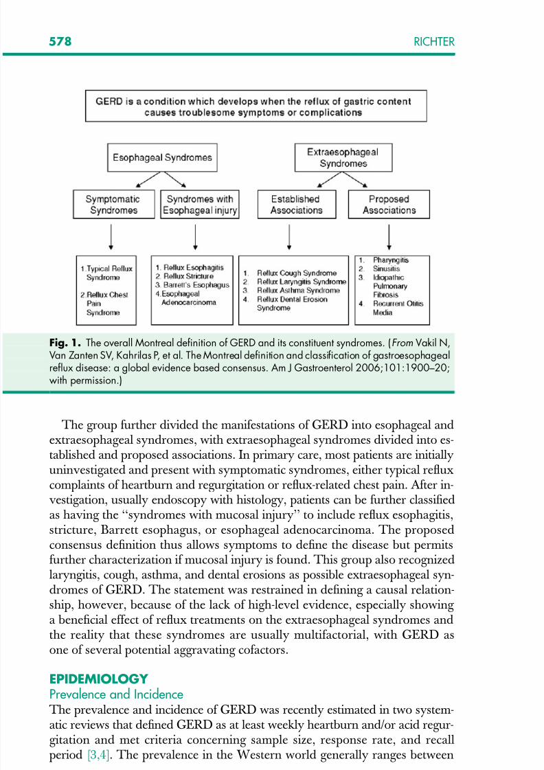
DEFINITIONUnfortunately there is no gold standard test for GERD. Because the reflux of acid, particularly after meals, is a physiologic process, the simple presence of gas-troesophageal reflux (GER) or occasional symptoms of heartburn or acid regur-gitation cannot be defined as a disease. Recently a group of 44 experts from 18countries used a modified Delphi process to develop a globally acceptable defi-nition and classification of GERD that can be applied in clinical practice and inresearch (Fig. 1) [2]. This international group defined GERD as ‘‘a condition
which develops when the reflux of stomach contents causes troublesome symp-toms and/or complications.’’ Troublesome symptoms are defined by the patientto affect their quality of life. Mild symptoms occurring 2 or more days per weekor moderate to severe symptoms occurring more than 1 day per week are oftenconsidered troublesome by patients. Patients may be diagnosed based on typicalsymptoms alone or on tests demonstrating reflux of stomach contents (eg, pHtesting, impedance monitoring) or the injurious effects of the refluxate (endos-copy, histology, electron microscopy), in the presence of typical or atypicalsymptoms or complications. This new definition also recognizes that the reflux-
ate causing symptoms may be weakly acidic or gaseous.
E-mail address: [email protected]
0889-8553/07/$ – see front matter ª 2007 Elsevier Inc. All rights reserved.doi:10.1016/j.gtc.2007.07.014 gastro.theclinics.com
Gastroenterol Clin N Am 36 (2007) 577–599
GASTROENTEROLOGY CLINICSOF NORTH AMERICA
8/6/2019 The Many Manifestations of GERD
http://slidepdf.com/reader/full/the-many-manifestations-of-gerd 2/23
The group further divided the manifestations of GERD into esophageal andextraesophageal syndromes, with extraesophageal syndromes divided into es-tablished and proposed associations. In primary care, most patients are initiallyuninvestigated and present with symptomatic syndromes, either typical refluxcomplaints of heartburn and regurgitation or reflux-related chest pain. After in-vestigation, usually endoscopy with histology, patients can be further classifiedas having the ‘‘syndromes with mucosal injury’’ to include reflux esophagitis,stricture, Barrett esophagus, or esophageal adenocarcinoma. The proposed
consensus definition thus allows symptoms to define the disease but permitsfurther characterization if mucosal injury is found. This group also recognizedlaryngitis, cough, asthma, and dental erosions as possible extraesophageal syn-dromes of GERD. The statement was restrained in defining a causal relation-ship, however, because of the lack of high-level evidence, especially showing a beneficial effect of reflux treatments on the extraesophageal syndromes andthe reality that these syndromes are usually multifactorial, with GERD asone of several potential aggravating cofactors.
EPIDEMIOLOGY Prevalence and IncidenceThe prevalence and incidence of GERD was recently estimated in two system-atic reviews that defined GERD as at least weekly heartburn and/or acid regur-gitation and met criteria concerning sample size, response rate, and recallperiod [3,4]. The prevalence in the Western world generally ranges between
Fig. 1. The overall Montreal definition of GERD and its constituent syndromes. (From Vakil N,Van Zanten SV, Kahrilas P, et al. The Montreal definition and classification of gastroesophagealreflux disease: a global evidence based consensus. Am J Gastroenterol 2006;101:1900–20;with permission.)
578 RICHTER
8/6/2019 The Many Manifestations of GERD
http://slidepdf.com/reader/full/the-many-manifestations-of-gerd 3/23
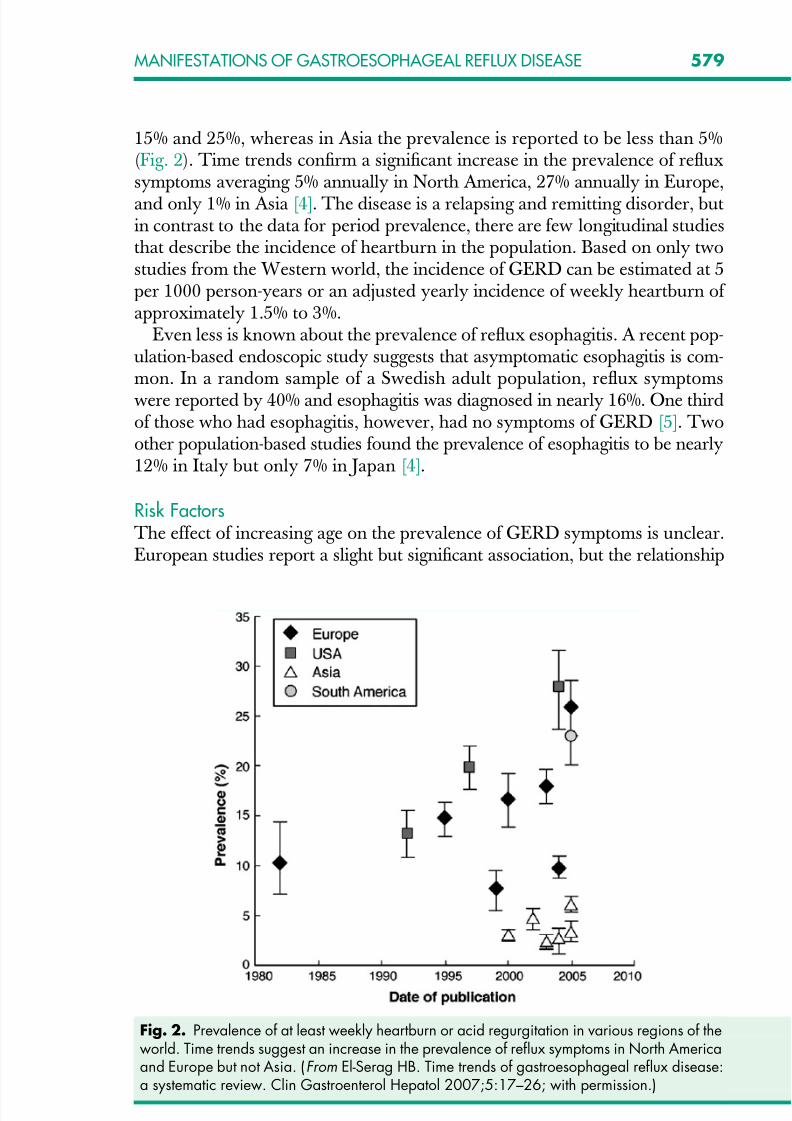
15% and 25%, whereas in Asia the prevalence is reported to be less than 5%(Fig. 2). Time trends confirm a significant increase in the prevalence of refluxsymptoms averaging 5% annually in North America, 27% annually in Europe,and only 1% in Asia [4]. The disease is a relapsing and remitting disorder, butin contrast to the data for period prevalence, there are few longitudinal studiesthat describe the incidence of heartburn in the population. Based on only twostudies from the Western world, the incidence of GERD can be estimated at 5per 1000 person-years or an adjusted yearly incidence of weekly heartburn of approximately 1.5% to 3%.
Even less is known about the prevalence of reflux esophagitis. A recent pop-ulation-based endoscopic study suggests that asymptomatic esophagitis is com-mon. In a random sample of a Swedish adult population, reflux symptomswere reported by 40% and esophagitis was diagnosed in nearly 16%. One thirdof those who had esophagitis, however, had no symptoms of GERD [5]. Twoother population-based studies found the prevalence of esophagitis to be nearly12% in Italy but only 7% in Japan [4].
Risk FactorsThe effect of increasing age on the prevalence of GERD symptoms is unclear.European studies report a slight but significant association, but the relationship
Fig. 2. Prevalence of at least weekly heartburn or acid regurgitation in various regions of theworld. Time trends suggest an increase in the prevalence of reflux symptoms in North Americaand Europe but not Asia. (From El-Serag HB. Time trends of gastroesophageal reflux disease:a systematic review. Clin Gastroenterol Hepatol 2007;5:17–26; with permission.)
579MANIFESTATIONS OF GASTROESOPHAGEAL REFLUX DISEASE
8/6/2019 The Many Manifestations of GERD
http://slidepdf.com/reader/full/the-many-manifestations-of-gerd 4/23
was not observed for heartburn with or without acid regurgitation in OlmsteadCounty, Minnesota [6]. A recent study suggested an association between ad-vancing age and milder reflux symptoms but more severe esophagitis [7]. Allstudies report a similar prevalence of heartburn in men and women [5]. Onthe other hand, endoscopy database studies find male sex a significant risk fac-tor for reflux esophagitis [8].
Cross-sectional studies and systematic reviews consistently find that obesityis associated with a statistically significant increase in the risk for reflux symp-toms, erosive esophagitis, Barrett esophagus, and esophageal adenocarcinoma [9–11]. In these studies, obesity (BMI >25) was associated with 2.5- to 3.0-foldincrease in these presentations of GERD. The causative mechanism for this re-lationship is unknown.
Helicobacter pylori infection is an environmental factor that has declined asGERD, Barrett esophagus, and esophageal adenocarcinoma have increasedin developed countries [12]. A systematic review of observational studies hasconfirmed that there is a negative association between H pylori and GERD, al-though this finding is most apparent in Asian countries [13]. The causativemechanism for this protective effect is the H pylori -induced gastritis, involving the antrum and corpus, which decreases the parietal cell mass, reduces acid se-cretion, and elevates gastric pH [14].
Along with environmental factors, the epidemiology of GERD may be af-
fected by genetics. There have been two studies [15,16] assessing the preva-lence of reflux symptoms in monozygotic versus dizygotic twins. Data fromthe Swedish Twin Registry [15] suggested that 31% (95% CI, 23%–39%) of GERD is caused by additive genetic factors, whereas a United KingdomTwin Registry study [16] reported that this value was 43% (32%–55%). Al-though one group defined a locus on chromosome 13 associated with severepediatric GERD [17], this has not been confirmed in adults. The geneticmechanisms are unknown but may be related to a smooth muscle disorderassociated with hiatal hernia, low LES pressure, and impaired esophageal
motility.
CLINICAL PRESENTATIONSHeartburn and acid regurgitation are the classic symptoms of GERD. Heart- burn describes a burning feeling, rising from the stomach or lower chest andradiating toward the neck, throat, and occasionally, the back [18]. It occurspostprandially, particularly after large meals or after eating spicy foods, citrusproducts, fats, chocolates, or drinking alcohol. The supine position or bending over may exacerbate heartburn. Nighttime heartburn may cause sleeping diffi-
culties and impair next-day function [19]. The frequency and severity of heart- burn does not predict the degree of esophageal damage [7]. The effortlessregurgitation of acidic fluid, especially after meals and worsened by stooping or the supine position, is suggestive of GERD. Among patients who have dailyregurgitation, LES pressure is usually low, many have associated gastroparesis,and esophagitis is common, making this symptom more difficult to treat than
580 RICHTER
8/6/2019 The Many Manifestations of GERD
http://slidepdf.com/reader/full/the-many-manifestations-of-gerd 5/23
classic heartburn. Symptoms such as dysphagia, odynophagia, globus sensa-tion, burping, water brash, and cough are other possible presentations of GERD, but their diagnostic yield is uncertain. Odynophagia may be seenwith severe reflux esophagitis, but usually suggests an infection or pill-relatedesophagitis. Water brash is the sudden appearance in the mouth of a salty fluid.It is not regurgitated fluid, but rather secretions from the salivary glands in re-sponse to acid reflux [20].
The clinical accuracy of heartburn or regurgitation in diagnosing GERD isdifficult to define. A recent systematic review [21] identified seven studiesthat assessed the accuracy of these reflux symptoms in diagnosing GERD inmore than 5000 patients. Endoscopy with the presence of esophagitis has excel-lent specificity; thus, it was used as the gold standard to assess the sensitivity of heartburn and regurgitation. Unfortunately the sensitivity of these classic re-flux symptoms was poor, with a range of 30% to 76% and a pooled sensitivityof 55% (95% CI, 45%–68%). Many patients who have atypical upper gastroin-testinal (GI) symptoms thus may have GERD.
Some patients who have GERD are asymptomatic. This is particularly truein elderly patients, perhaps because of reduced gastric acidity from chronic H pylori infection or decreased pain perception. Many elderly patients present firstwith complications of GERD because of long-standing disease with minimalcomplaints. For example, up to one third of patients who have Barrett esoph-
agus are insensitive to acid at the time of presentation [22].
DIAGNOSTIC TESTSA large number of tests are available for evaluating patients who have sus-pected GERD. Many times these tests are unnecessary, because the presenceof frequent heartburn and acid regurgitation are sufficiently accurate to identifythe disease and begin medical treatment. This is not always the case, however,and clinicians must decide which tests to choose so as to make the diagnosis in
a reliable, timely, and cost-effective manner, depending on the information de-sired (Table 1) [23].
Upper Endoscopy The identification of esophagitis at the time of endoscopy is highly specific(90%–95%) for GERD [24], but has a sensitivity of only approximately 50%.Multiple classification systems for esophagitis have been proposed, some areconfusing, and none have worldwide acceptance [25]. The most thoroughlyevaluated esophagitis classification is the Los Angeles (LA) system, which is
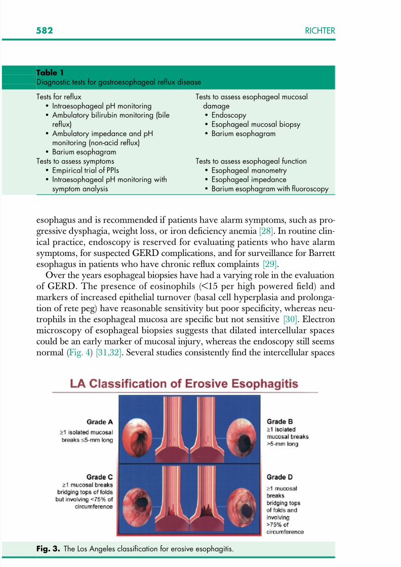
gaining acceptance in the United States and Europe (Fig. 3) [26]. In referral cen-ters, approximately 50% of patients have esophagitis, but in primary care andthe general population, the rate of esophagitis is more in the range of 10% to30% [4]. Most patients who have esophagitis have mild LA grade A-B diseaseand only 10% have the more severe LA grade C-D esophagitis [27]. Endoscopycan also evaluate complications of GERD, such as peptic strictures and Barrett
581MANIFESTATIONS OF GASTROESOPHAGEAL REFLUX DISEASE
8/6/2019 The Many Manifestations of GERD
http://slidepdf.com/reader/full/the-many-manifestations-of-gerd 6/23
esophagus and is recommended if patients have alarm symptoms, such as pro-gressive dysphagia, weight loss, or iron deficiency anemia [28]. In routine clin-ical practice, endoscopy is reserved for evaluating patients who have alarmsymptoms, for suspected GERD complications, and for surveillance for Barrettesophagus in patients who have chronic reflux complaints [29].
Over the years esophageal biopsies have had a varying role in the evaluationof GERD. The presence of eosinophils (<15 per high powered field) and
markers of increased epithelial turnover (basal cell hyperplasia and prolonga-tion of rete peg) have reasonable sensitivity but poor specificity, whereas neu-trophils in the esophageal mucosa are specific but not sensitive [30]. Electronmicroscopy of esophageal biopsies suggests that dilated intercellular spacescould be an early marker of mucosal injury, whereas the endoscopy still seemsnormal (Fig. 4) [31,32]. Several studies consistently find the intercellular spaces
Table 1Diagnostic tests for gastroesophageal reflux disease
Tests for reflux Intraesophageal pH monitoring Ambulatory bilirubin monitoring (bile
reflux) Ambulatory impedance and pH
monitoring (non-acid reflux) Barium esophagram
Tests to assess esophageal mucosaldamage Endoscopy Esophageal mucosal biopsy Barium esophagram
Tests to assess symptoms Empirical trial of PPIs Intraesophageal pH monitoring with
symptom analysis
Tests to assess esophageal function Esophageal manometry Esophageal impedance Barium esophagram with fluoroscopy
Fig. 3. The Los Angeles classification for erosive esophagitis.
582 RICHTER

8/6/2019 The Many Manifestations of GERD
http://slidepdf.com/reader/full/the-many-manifestations-of-gerd 7/23
at least two to three times greater in patients who have erosive and nonerosiveGERD compared with healthy control subjects [32]. Aggressive acid suppres-sion therapy seems to normalize the width of the intercellular spaces [32]. Un-fortunately these spaces are more difficult to define by light microscopy. Inclinical practice, biopsies are usually not taken in patients who have classic re-flux esophagitis unless necessary to exclude neoplasm, infection, pill injury, bul-lous skin disease, or eosinophilic esophagitis (>20 eosinophils per HPF). Thecurrent primary indication for esophageal biopsies is to determine the presenceof Barrett epithelium [29]. When this diagnosis is suspected, biopsies are man-datory and best done when esophagitis is healed.
Esophageal pH Monitoring
Ambulatory intraesophageal pH monitoring is the standard for establishing pathologic reflux [23]. Traditionally the pH probe is passed nasally, positioned5 cm above the manometrically determined LES, and connected to a battery-powered data logger capable of collecting pH values every 4 to 6 seconds for24 hours. Patients record meals, sleeping, and when symptoms are experi-enced. Acid reflux episodes are defined as a pH drop of less than 4. The totalpercent of time the pH is less than 4 is the most reproducible measure of GERD, with the reported upper limits of normal ranging from 4% to 5.5%[33]. The sensitivity of 24-hour pH monitoring in patients who have esophagitis
approaches 90% with a specificity of 85% to 100%. In patients who have nor-mal endoscopy in which pH testing may be most needed, the sensitivity is only60% and the specificity from 85% to 90% [34]. Clinical indications for ambula-tory pH monitoring include (1) before fundoplication to insure the presence of pathologic reflux in patients who have a normal endoscopy, (2) after antirefluxsurgery if heartburn symptoms persist, (3) patients who have reflux symptoms
Fig. 4. Intercellular spaces in esophageal mucosa from A) healthy subjects without refluxsymptoms and B) GERD patients without esophagitis identified by transmission electron micros-copy. (From Calabrese C, Fabbri A, Bortolotti M, et al. Dilated intercellular spaces as a markerof oesophageal damage: comparative results in gastro-oesophageal reflux disease with orwithout bile reflux. Aliment Pharmacol Ther 2003;18:525–32; with permission.)
583MANIFESTATIONS OF GASTROESOPHAGEAL REFLUX DISEASE

8/6/2019 The Many Manifestations of GERD
http://slidepdf.com/reader/full/the-many-manifestations-of-gerd 8/23
and a normal endoscopy not responding to PPI treatment, and (4) patients whohave suspected extraesophageal symptoms of GERD [34].
Two new advances are improving the role of pH testing in evaluating GERD. The first is a wireless device (Bravo pH probe, Medtronics, Minneap-olis, MN) the size of a vitamin pill attached, usually by endoscopy, 6 cm abovethe Z-line (Fig. 5) [35]. This decreases patient discomfort, allows for longer (48hours or more) monitoring, and may increase test sensitivity by allowing pa-tients to more comfortably carry out their usual activities [36]. The capsule de-taches and passes spontaneously within 2 weeks. Rare patients may note severepain after attachment, which resolves spontaneously with endoscopic removal.Two significant complications have occurred with this device—one report of esophageal bleeding requiring transfusion and one esophageal perforation [37].
The second technologic advancement combines multichannel intraluminal im-pedance monitoring with pH sensors to detect acid, weak acid, and non-acid re-flux using a transnasal catheter over 24 hours [38]. The number of respectivereflux episodes, rather than percentage of exposure time, is the critical measure-ment, with normal values established from United States and European studies[38]. The results of several studies suggest that impedance-pH monitoring is use-ful in the evaluation of patients who have PPI-resistant typical reflux symptoms,especially regurgitation complaints, and chronic unexplained cough [39–41].
Barium EsophagramThe barium esophagram is inexpensive and less invasive than endoscopy. It ismost useful in demonstrating strictures, rings, hiatus hernias, and major
Fig. 5. Wireless pH capsule attached to the esophagus 6 cm above the squamocolumnarjunction.
584 RICHTER

8/6/2019 The Many Manifestations of GERD
http://slidepdf.com/reader/full/the-many-manifestations-of-gerd 9/23
abnormalities in peristalsis. The barium esophagram’s ability to detect esopha-gitis varies, with sensitivities of 79% to 100% for moderate to severe esophagi-tis, whereas mild esophagitis is usually missed [23]. It is also not reliable fordetecting Barrett esophagus.
Esophageal Manometry Esophageal manometry allows assessment of LES pressure and relaxation andperistaltic activity, including contraction amplitude, duration, and velocity. It isgenerally not indicated in the evaluation of patients who have uncomplicatedGERD, however, because most have normal resting LES pressure [42]. Esoph-ageal manometry to document adequate esophageal peristalsis is traditionallyrecommended before antireflux surgery. If the study identifies ineffective peri-
stalsis (low amplitude or frequent failed peristalsis), then a complete fundopli-cation may be contraindicated. This assumption has recently been challenged,however, by several studies finding that reflux control was better and dyspha-gia no more common in patients who had weak peristalsis after a complete asopposed to partial fundoplication [43]. An improvement of traditional manom-etry, combining it with impedance testing, is helping to clarify this controversy.Using this technology, a recent study found that less than 50% of patients whohad ineffective peristalsis had a significant delay in esophageal bolus transitmeasured by impedance [44].
Proton Pump Inhibitor Test An empiric trial of acid suppression is the simplest method for diagnosing GERD and assessing its relationship to symptoms. With the advent of PPIs,this test has become the first diagnostic study used in patients who have classicor atypical reflux symptoms without alarming complaints. Symptoms usuallyrespond to a PPI test in 1 to 2 weeks. If symptoms disappear with therapyand then return when medication is discontinued, GERD is established. A sys-tematic review [45] identified 15 studies that assessed the accuracy of normal orhigh dose PPIs for 1 to 4 weeks in the diagnosis of GERD. The pooled sensi-
tivity was good at 78% (95% CI, 66%–86%), but the specificity was poor at 54%(95% CI, 44%–65%) when 24-hour ambulatory pH was used as a gold stan-dard. Nevertheless an empiric PPI trial for diagnosing GERD offers manyadvantages: the test is office-based, easily done, inexpensive (especially withover-the-counter PPIs), available to all physicians, and may avoid needless pro-cedures. For example, one study showed a savings of greater than $570 per av-erage patient because of a 59% reduction in the number of diagnostic tests(upper endoscopy, pH tests) [46]. Disadvantages are few, but include a placeboresponse and uncertain symptomatic endpoint if symptoms do not totally
resolve with extended treatment.
COMPLICATIONSThere are few data on the long-term outcome of patients who have varying se-verities of GERD. Severity and duration of symptoms seem to have a poor cor-relation with the presence or severity of esophagitis [7]. Furthermore, there is
585MANIFESTATIONS OF GASTROESOPHAGEAL REFLUX DISEASE
8/6/2019 The Many Manifestations of GERD
http://slidepdf.com/reader/full/the-many-manifestations-of-gerd 10/23
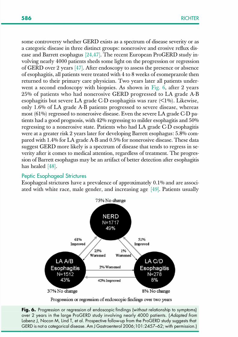
some controversy whether GERD exists as a spectrum of disease severity or asa categoric disease in three distinct groups: nonerosive and erosive reflux dis-ease and Barrett esophagus [24,47]. The recent European ProGERD study in-volving nearly 4000 patients sheds some light on the progression or regressionof GERD over 2 years [47]. After endoscopy to assess the presence or absenceof esophagitis, all patients were treated with 4 to 8 weeks of esomeprazole thenreturned to their primary care physician. Two years later all patients under-went a second endoscopy with biopsies. As shown in Fig. 6, after 2 years25% of patients who had nonerosive GERD progressed to LA grade A-Besophagitis but severe LA grade C-D esophagitis was rare (<1%). Likewise,only 1.6% of LA grade A-B patients progressed to severe disease, whereasmost (61%) regressed to nonerosive disease. Even the severe LA grade C-D pa-tients had a good prognosis, with 42% regressing to milder esophagitis and 50%regressing to a nonerosive state. Patients who had LA grade C-D esophagitiswere at a greater risk 2 years later for developing Barrett esophagus: 5.8% com-pared with 1.4% for LA grade A-B and 0.5% for nonerosive disease. These data suggest GERD more likely is a spectrum of disease that tends to regress in se-verity after it comes to medical attention, regardless of treatment. The progres-sion of Barrett esophagus may be an artifact of better detection after esophagitishas healed [48].
Peptic Esophageal StricturesEsophageal strictures have a prevalence of approximately 0.1% and are associ-ated with white race, male gender, and increasing age [49]. Patients usually
Fig. 6. Progression or regression of endoscopic findings (without relationship to symptoms)over 2 years in the large ProGERD study involving nearly 4000 patients. (Adapted fromLabenz J, Nocon M, Lind T, et al. Prospective follow-up from the ProGERD study suggests thatGERD is not a categorical disease. Am J Gastroenterol 2006;101:2457–62; with permission.)
586 RICHTER
8/6/2019 The Many Manifestations of GERD
http://slidepdf.com/reader/full/the-many-manifestations-of-gerd 11/23
present with dysphagia for solids, but unlike malignant strictures, weight loss isuncommon because their appetite is good. As dysphagia progresses, heartburnoften decreases, reflecting the stricture acting as a barrier to further reflux. Pep-tic strictures are smooth walled, tapered, circumferential narrowing in thelower esophagus that are usually less than 1 cm long. A mid to upper esopha-geal stricture should raise concern about Barrett esophagus or malignancy. Al-though once controversial, today a Schatzki ring is considered a forme fruste of an early peptic stricture [50]. All stricture patients should undergo endoscopy,at least initially, to confirm the benign nature of the disease and, if necessary, totake biopsies to exclude cancer and Barrett esophagus. Symptomatic patientscan be dilated by various bougies [51]. Dysphagia relief generally occurswhen the lumen is greater than 15 mm and associated esophagitis has healed[52].
Barrett EsophagusBarrett esophagus is the consequence of severe GERD in which the squamousepithelium of the distal esophagus is replaced by specialized columnar mucosa containing goblet cells (intestinal metaplasia). The disease is more common inwhite men, rare before age 50 years, and present in 1% to 2% of patients re-ferred for endoscopy over this age threshold [8]. Bile reflux and obesity have been associated with an increased risk for Barrett esophagus [9,53]. The diag-
nosis can be suspected at endoscopy and its circumferential involvement andmaximum proximal extension described using the new Prague classification[54]. Histology is required, however, to confirm the diagnosis and to definethe potentially premalignant intestinal metaplasia. Detection of Barrett esopha-gus is highest after suspected patients have been on PPIs for 8 to 12 weeks [48].In the era of PPIs, Barrett esophagus is easy to treat and only of major interest because of an increased risk for developing esophageal adenocarcinoma, esti-mated at between 0.5% and 1% each year [55]. Increased duration, frequency,and severity of reflux symptoms have been shown to be risk factors for this
cancer [56]. Further details on Barrett esophagus can be found in several excel-lent reviews [57,58].
Extraesophageal ManifestationsGastroesophageal reflux may be the cause of a wide spectrum of conditions,including non-cardiac chest pain, asthma, posterior laryngitis, chronic cough,recurrent pneumonitis, and even dental erosion [59]. GERD-related chestpain may mimic angina pectoris, even to the point of being induced by exercise.Most of these patients also have heartburn symptoms [60]. The mechanism of the pain is poorly understood, likely because of the volume and duration of
acid reflux, secondary esophageal spasm, or prolonged contractions of the lon-gitudinal muscle [61]. The causal link between GERD and pulmonary and ear,nose, and throat complaints is much more circumspect [59]. Although the pos-sible mechanisms from animal studies are plausible (ie, microaspiration and va-gal reflex), most suspected patients are heartburn-free and do not haveesophagitis, hiatus hernia, or low LES pressure. Unfortunately pH testing
587MANIFESTATIONS OF GASTROESOPHAGEAL REFLUX DISEASE
8/6/2019 The Many Manifestations of GERD
http://slidepdf.com/reader/full/the-many-manifestations-of-gerd 12/23
(distal or proximal), although frequently abnormal in these patients, does notpredict their response to medical or surgical therapies.
MEDICAL TREATMENTLifestyle and Over-the-Counter MedicationsNumerous dietary and lifestyle modifications are advocated for the treatmentof GERD. They are frequently first-line therapy for patients who have mild dis-ease and often adjunct therapy even for patients on PPIs.What is the evidence,however? In a recent evidence-based review, studies of smoking, alcohol, choc-olate, fatty foods, and citrus products showed physiologic evidence that theirintake can adversely affect symptoms or esophageal pH. There was little evi-dence, however, that cessation of these products predictably improvedGERD symptoms. Only elevation of the head of the bed, left lateral decubituspositioning, and weight loss was associated with GERD improvement in case-controlled studies [62].
Over-the-counter (OTC) antacids, alginate/antacid combinations, andH2RAs are useful in treating mild and infrequent heartburn symptoms, espe-cially when symptoms are brought on by lifestyle indiscretions. In one recentmeta-analysis [63], the relative benefit increase compared with the overall pla-cebo response was up to 41% with H2RAs, 60% with alginates, and 11% withantacids. Antacids rapidly relieve heartburn symptoms, the major reason these
drugs are so popular for mild, intermittent symptoms. Although their onset of relief is not as rapid as antacids, OTC H2RAs have a longer duration of action,up to 6 to 10 hours. From a practical standpoint, they are most useful whentaken before a potentially refluxogenic activity, such as a heavy meal, eating late at night, or exercise. Sales of these OTC medications in 2004 were consider-able: more than $350 million for antacids and nearly $200 million for H2RAs [1].
Proton Pump InhibitorsPPIs revolutionized the treatment of GERD and currently are the mainstay of
acute and maintenance treatment regimens. This class of drugs markedly di-minishes gastric acid secretion over a 24-hour period by inhibiting the finalcommon pathway of acid secretion, the HþKþ ATPase pump. Their superiorefficacy compared with H2RAs is based on their ability to maintain an intragas-tric pH of less than 4 between 15 and 21 hours, compared with approximately8 hours daily with H2RAs. In 2004, PPIs accounted for approximately 77% of the acid suppressant market, amounting to sales exceeding $9.5 billion [1].
In a recent Cochrane review [64], PPIs were more effective than placebo inhealing esophagitis (RR ¼ 0.23; 95% CI, 0.01–0.05) with a number to treat
(NNT) of 2 (95% CI, 1.4–2.5). The review also identified 26 trials involving 4064 patients that compared PPIs with H2RAs. PPIs were superior toH2RAs in healing esophagitis at 4 to 6 weeks (RR ¼ 0.47; 95% CI, 0.41–0.53) with an NNT of 3 (95% CI, 2.8–3.6). Another Cochrane systematicreview found that PPI therapy was superior to placebo and H2RAs in endos-copy-negative GERD and undiagnosed reflux symptoms in primary care,
588 RICHTER

8/6/2019 The Many Manifestations of GERD
http://slidepdf.com/reader/full/the-many-manifestations-of-gerd 13/23
although the effect was not as marked as with esophagitis [65]. Cochrane re-views also have identified the superiority of PPIs over H2RAs in maintaining the remission of esophagitis over 6 to 12 months [66]. Among 10 randomizedtrials, the relapse rate for esophagitis was 22% on PPIs compared with 58% onH2RAs, with an NNT of 2.5 (95% CI, 2.0–3.4).
Until recently the therapeutic efficacy between PPIs was similar. Recent largerandomized controled trial (1000–2500 patients), however, have found thenewest PPI, esomeprazole 40 mg, superior to omeprazole 20 mg and lansopra-zole 30 mg in healing esophagitis [67]. The therapeutic advantage is minimalwith mild LA grade A-B esophagitis (NNT 50 and 33, respectively) and great-est with severe LA grade C-D esophagitis (NNT 10 and 8, respectively). Thissuperiority is related to higher systemic bioavailability and less inter-patientvariability with esomeprazole.
Treatment of Complicated Gastroesophageal Reflux Diseaseand its Extraesophageal PresentationsThe extensive use of PPIs has markedly affected treatment of peptic stricturesand esophageal rings. Several studies in community and Veterans Affairs (VA)hospitals note an approximate 33% decline in the incidence of recurrent stric-tures. The timeline for this decrease parallels the marked increase in PPI usesince 1995 (Fig. 7) [68]. Another study convincingly shows that in patientswho have symptomatic Schatzki rings, maintenance PPI therapy after bougien-age markedly decreases future relapses of the rings [69]. In a randomized study,30 patients who had symptomatic rings without esophagitis were dilated andrandomized to placebo or omeprazole 20 mg per day. In the treated group,one patient relapsed after 13 months, whereas seven patients relapsed on pla-cebo after a mean of 20 months.
Fig. 7. Incidence of peptic esophageal stricture and use of PPIs between 1994 and 2000 inUnited Kingdom general practice. R, Spearman correlation test. (From Ruigomez A, RodriguezLAG, Wallender MA, et al. Esophageal stricture: incidence, treatment patterns and recurrencerate. Am J Gastroenterol 2006;101:2685–92; with permission.)
589MANIFESTATIONS OF GASTROESOPHAGEAL REFLUX DISEASE
8/6/2019 The Many Manifestations of GERD
http://slidepdf.com/reader/full/the-many-manifestations-of-gerd 14/23
The efficacy of PPI treatment in the extraesophageal presentations of GERDis more variable. There are two systemic reviews [70,71], both suggesting thatpatients who have non-cardiac chest pain respond to PPIs better than to pla-cebo. These reports identified eight RCTs that assessed 321 patients whohad a pooled relative risk for continued chest pain after PPI therapy, comparedwith placebo of 0.54 (95% CI, 0.41–0.71), with an NNT of 3 (95% CI, 2–4).Systemic reviews, however, do not support the efficacy of aggressive acid sup-pression, particularly with PPIs in other extraesophageal disorders, such aschronic cough [72], asthma [73], or ear, nose, and throat disorders [74].
Sleep disturbances may occur in up to 75% of patients who have GERD, im-pairing quality of life. In a large multicenter study, patients who had GERD-as-sociated sleep disturbances and nighttime heartburn were randomized to twodoses of esomeprazole (40 mg and 20 mg) or placebo for 4 weeks [75].GERD-related sleep disturbances resolved in significantly more patients on eso-meprazole 40 mg (73.7%) or 20 mg (73.2%) than those who received placebo(41.2%). These changes were associated with improved sleep quality and day-time productivity.
Refractory Gastroesophageal Reflux DiseaseTraditionally patients who have reflux symptoms no longer undergo initialendoscopy, but rather are given a 4- to 8-week trial of a PPI. Failure to improve
occurs in 25% to 42% of patients, thus placing them in a more difficult to man-age group. At this point the physician should insure patient compliance and re-view timing of the PPI dose (1/2 to 1 hour before meals). One recent studyfound that nearly 70% of primary physicians and 20% of gastroenterologistsgave the PPI at bedtime or did not believe the relationship to meals was impor-tant [76]. Switching to a second generation PPI (ie, pantoprazole, esomeprazole)may be a reasonable alternative. This was recently confirmed in a multicenterstudy of patients who had persistent heartburn symptoms while receiving lan-soprazole 30 mg once daily [77]. Switching to a single dose of esomeprazole (40
mg) was as effective as twice daily lansoprazole in relieving heartburn com-plaints over 8 weeks of therapy. Most physicians, however, increase the currentPPI to twice daily dosing (before breakfast and dinner), with up to 25% of pa-tients responding [78].
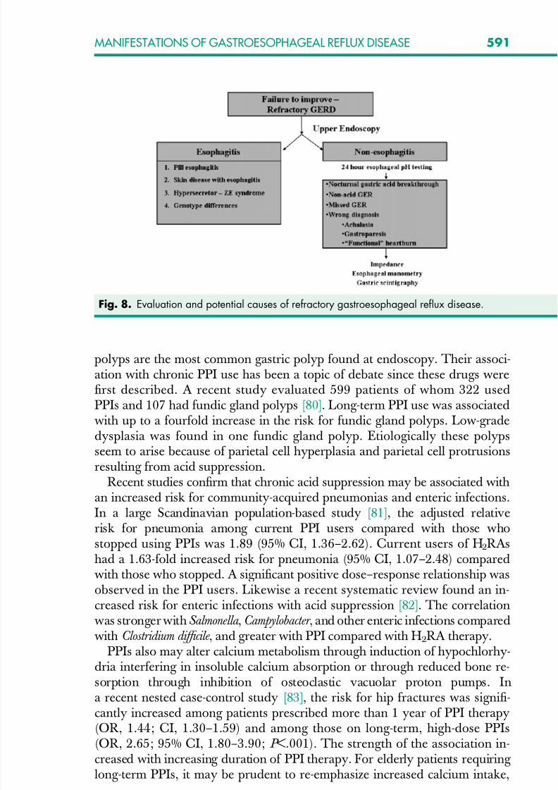
Those patients doing no better fall into the ‘‘refractory GERD’’ category[79]. As shown in Fig. 8, the critical diagnostic test is upper GI endoscopy,which identifies patients who have esophagitis or no esophagitis. The largestpercentage of these patients have refractory symptoms with no esophagitis.These patients may require 24-hour pH testing on PPI therapy, impedance test-
ing, and consideration of other diagnoses, such as achalasia, gastroparesis, andfunctional heartburn. Although much has been made recently of non-acidGER, there are no controlled data to help in treating this group of patients.
Safety ConcernsInitial concerns about PPIs causing gastric malignancies in rats have not beensubstantiated in other animal models or long-term patient studies. Fundic gland
590 RICHTER
8/6/2019 The Many Manifestations of GERD
http://slidepdf.com/reader/full/the-many-manifestations-of-gerd 15/23
polyps are the most common gastric polyp found at endoscopy. Their associ-ation with chronic PPI use has been a topic of debate since these drugs werefirst described. A recent study evaluated 599 patients of whom 322 usedPPIs and 107 had fundic gland polyps [80]. Long-term PPI use was associated
with up to a fourfold increase in the risk for fundic gland polyps. Low-gradedysplasia was found in one fundic gland polyp. Etiologically these polypsseem to arise because of parietal cell hyperplasia and parietal cell protrusionsresulting from acid suppression.
Recent studies confirm that chronic acid suppression may be associated withan increased risk for community-acquired pneumonias and enteric infections.In a large Scandinavian population-based study [81], the adjusted relativerisk for pneumonia among current PPI users compared with those whostopped using PPIs was 1.89 (95% CI, 1.36–2.62). Current users of H2RAs
had a 1.63-fold increased risk for pneumonia (95% CI, 1.07–2.48) comparedwith those who stopped. A significant positive dose–response relationship wasobserved in the PPI users. Likewise a recent systematic review found an in-creased risk for enteric infections with acid suppression [82]. The correlationwas stronger with Salmonella , Campylobacter , and other enteric infections comparedwith Clostridium difficile , and greater with PPI compared with H2RA therapy.
PPIs also may alter calcium metabolism through induction of hypochlorhy-dria interfering in insoluble calcium absorption or through reduced bone re-sorption through inhibition of osteoclastic vacuolar proton pumps. In
a recent nested case-control study [83], the risk for hip fractures was signifi-cantly increased among patients prescribed more than 1 year of PPI therapy(OR, 1.44; CI, 1.30–1.59) and among those on long-term, high-dose PPIs(OR, 2.65; 95% CI, 1.80–3.90; P <.001). The strength of the association in-creased with increasing duration of PPI therapy. For elderly patients requiring long-term PPIs, it may be prudent to re-emphasize increased calcium intake,
Fig. 8. Evaluation and potential causes of refractory gastroesophageal reflux disease.
591MANIFESTATIONS OF GASTROESOPHAGEAL REFLUX DISEASE
8/6/2019 The Many Manifestations of GERD
http://slidepdf.com/reader/full/the-many-manifestations-of-gerd 16/23
preferably from a dairy source, and co-ingestion of a meal when taking insol-uble calcium supplements.
New Drug TreatmentsNew drug treatments have primarily targeted transient LES relaxation, thecommon motility abnormality in all forms of GERD. Several agents, including cholecystokinin A agonists, anticholinergic drugs, nitric oxide synthase inhibi-tors, morphine, cannaboid, and gamma-aminobutyric acid B (GABA) agonistshave been shown to reduce transient LES relaxation and episodes of acid reflux[84]. The only agent available for oral therapy is baclofen, a GABA agonist.Several studies show that 10 to 20 mg of baclofen three to four times dailyfor up to 4 weeks reduces 24-hour esophageal acid and bilirubin reflux
[85,86]. Baclofen needs to be titrated upward slowly, beginning at 5 mg dailyand increased over 10 days to 40 to 60 mg per day. Side effects are commonand include drowsiness, nausea, and the lowering of the threshold for seizures.New compounds with more specific and better targeted action need to be de-veloped. Another approach has been to develop newer rapid-acting PPIs,such as potassium-competitive acid blockers (P-CAB). Unlike the traditionalPPIs that bind irreversibly to the proton pump, this new class of compounds blocks acid secretion by way of potassium-competitive inhibition of theHþKþ ATPase. This results in rapid onset with almost complete acid block-
ade achieved within 30 minutes of administration [84]. Unfortunately recentphase III studies found the P-CABs no more effective than esomeprazole inthe rapid relief of heartburn symptoms.
ENDOSCOPIC TREATMENTVarious endoscopic techniques for the treatment of GERD have been devel-oped as alternatives to antisecretory therapy or antireflux surgery [87]. Thesetechniques include the delivery of radiofrequency energy to the gastroesopha-geal junction (Stretta), injection of bulking agents (Eneryx), or implantation of a bioprosthesis (Gatekeeper) into the LES, and suture plication of the proximal
gastric folds (Endocinch, Endoscopic Plication System). Studies to date haveprimarily enrolled PPI-dependent patients who do not have severe esophagitisor large hiatus hernia.
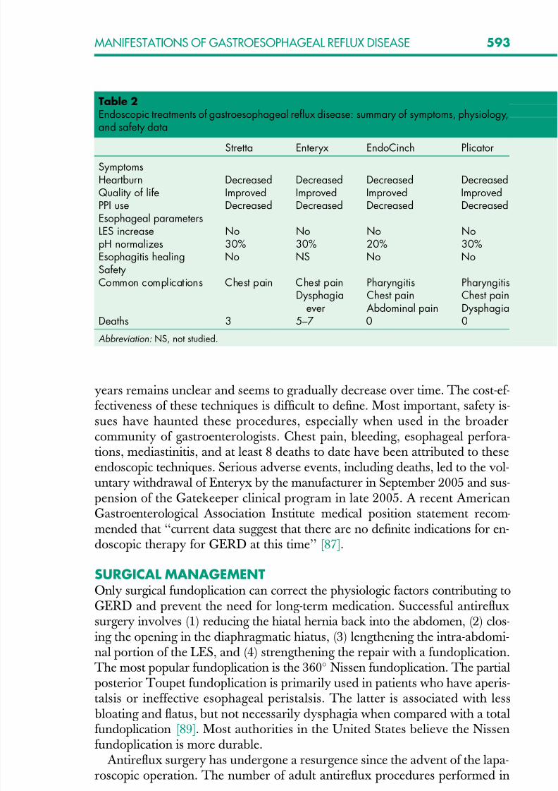
As shown in Table 2, each of these techniques decreases reflux symptoms,improves quality of life, and decreases the need for antisecretory medications.Physiologic studies, however, are much less impressive, with LES pressurerarely increasing, pH normalizing in only 30% of patients, and even mildesophagitis infrequently healing. Sham studies with Stretta, Enteryx, and thePlication system likewise show a decrease in heartburn symptoms and im-
proved quality of life after the active therapy compared with the sham groupafter 3 to 6 months [87]. Only the Plication study showed a significant decreasein pH values, by only 18%, whereas no change in pH or LES parameters wasobserved in studies using the other techniques [88].
Most studies of endoscopic therapy have only limited follow-up informationon a small number of patients. The durability of these techniques beyond 1 to 2
592 RICHTER
8/6/2019 The Many Manifestations of GERD
http://slidepdf.com/reader/full/the-many-manifestations-of-gerd 17/23
years remains unclear and seems to gradually decrease over time. The cost-ef-fectiveness of these techniques is difficult to define. Most important, safety is-
sues have haunted these procedures, especially when used in the broadercommunity of gastroenterologists. Chest pain, bleeding, esophageal perfora-tions, mediastinitis, and at least 8 deaths to date have been attributed to theseendoscopic techniques. Serious adverse events, including deaths, led to the vol-untary withdrawal of Enteryx by the manufacturer in September 2005 and sus-pension of the Gatekeeper clinical program in late 2005. A recent AmericanGastroenterological Association Institute medical position statement recom-mended that ‘‘current data suggest that there are no definite indications for en-doscopic therapy for GERD at this time’’ [87].
SURGICAL MANAGEMENTOnly surgical fundoplication can correct the physiologic factors contributing toGERD and prevent the need for long-term medication. Successful antirefluxsurgery involves (1) reducing the hiatal hernia back into the abdomen, (2) clos-ing the opening in the diaphragmatic hiatus, (3) lengthening the intra-abdomi-nal portion of the LES, and (4) strengthening the repair with a fundoplication.The most popular fundoplication is the 360 Nissen fundoplication. The partialposterior Toupet fundoplication is primarily used in patients who have aperis-
talsis or ineffective esophageal peristalsis. The latter is associated with less bloating and flatus, but not necessarily dysphagia when compared with a totalfundoplication [89]. Most authorities in the United States believe the Nissenfundoplication is more durable.
Antireflux surgery has undergone a resurgence since the advent of the lapa-roscopic operation. The number of adult antireflux procedures performed in
Table 2Endoscopic treatments of gastroesophageal reflux disease: summary of symptoms, physiology,and safety data
Stretta Enteryx EndoCinch Plicator
SymptomsHeartburn Decreased Decreased Decreased DecreasedQuality of life Improved Improved Improved ImprovedPPI use Decreased Decreased Decreased DecreasedEsophageal parametersLES increase No No No NopH normalizes 30% 30% 20% 30%Esophagitis healing No NS No NoSafetyCommon complications Chest pain Chest pain
Dysphagiaever
PharyngitisChest painAbdominal pain
PharyngitisChest painDysphagia
Deaths 3 5–7 0 0
Abbreviation: NS, not studied.
593MANIFESTATIONS OF GASTROESOPHAGEAL REFLUX DISEASE
8/6/2019 The Many Manifestations of GERD
http://slidepdf.com/reader/full/the-many-manifestations-of-gerd 18/23
the United States more than tripled from 11,000 per year in 1985 (open oper-ation) to 40,000 in 2001, a population-based annual rate of 12.0 per 100,000adults [90]. A systematic review identified six randomized controlled trials in-volving 449 patients that compared open and laparoscopic fundoplication[91]. There was no significant difference in recurrence rates between the proce-dures, and laparoscopic fundoplication was associated with lower operativemorbidity (NNT to prevent complication ¼ 8; 95% CI, 3–16) and shorter hos-pital stay.
What are the indications for surgical fundoplication in the era of inexpensivePPIs and proven long-term safety of these drugs? The most accepted indica-tions [89] are:
1. Patients who have typical or atypical GER symptoms who respond to PPIs butwho want surgery because of A desire for a permanent cure Patient preference An intolerance to PPIs
2. Failed medical therapy as a result of persistent volume regurgitation. Hereheartburn symptoms are controlled, but regurgitation is a persistent problem.
3. Recurrent peptic strictures in younger patients4. Respiratory complications related to regurgitation and recurrent aspiration
In patients who have Barrett esophagus, there is no convincing evidence thatfundoplication reduces the long-term risk for esophageal adenocarcinoma [92].Comparison studies of older medical treatments (antacids, H2RAs) consis-
tently find surgical fundoplication better at healing esophagitis and relieving symptoms. There are few studies comparing fundoplication with long-termPPI therapy, but one study [93] suggested that both were equally effective incontrolling symptoms over 5 years, provided patients in the medical treatmentgroup were allowed to increase the dose of the drug to twice daily if necessary.
Complications can occur after antireflux surgery, and many patients over
time continue to require antireflux medications. A recent database analysis of the surgical experience in all VA medical centers nationwide may more closelyreflect the surgical experience in community hospitals compared with highlyspecialized tertiary care centers [90]. In this report, more than 3000 patients un-dergoing antireflux surgery were identified between 1990 and 2001. Postoper-ative dysphagia was recorded in 19.4%, dilation was performed in 6.4%, anda repeat antireflux surgery was needed in 2.3% of patients. The surgical mor-tality rate was 0.8%. Approximately 50% of patients received multiple prescrip-tions for antireflux medications at a median of 5 years of follow-up evaluation
after their surgery.Tertiary specialized centers are seeing an increased rate of fundoplicationfailures [94]. The most common reasons for failure are herniation of the intactfundoplication into the chest, slipped fundoplication with a recurrent hiatal her-nia, probably caused by a short esophagus, paraesophageal hernia through anintact fundoplication, too tight a fundoplication, and malpositioned
594 RICHTER

8/6/2019 The Many Manifestations of GERD
http://slidepdf.com/reader/full/the-many-manifestations-of-gerd 19/23
fundoplication, usually on the cardia of the stomach. Total breakdown of thefundoplication is now rare. Revisional antireflux surgery needs to be per-formed by experienced surgeons, can be done laparoscopically but many preferan open approach, and has increased morbidity and mortality compared withthe initial operation.
SUMMARY Gastroesophageal reflux disease is a common problem that is expensive to di-agnose and treat in primary and specialty care settings. This review has empha-sized the major advances in understanding the diagnosis and treatment of GERD over the last 5 years. These are summarized below.
GERD is increasing in prevalence in the Western world, with important riskfactors being obesity and healthy stomachs resulting from H. pylori eradication.
The sensitivity of classic reflux symptoms is poor (55%) for the diagnosis of GERD. Response to the PPI test has good sensitivity (78%), but poor specificity(54%).
Ambulatory esophageal pH testing is the most sensitive test for GERD,whereas endoscopy is the most specific.
Medical treatment with PPIs and laparoscopic antireflux surgery have similarefficacy in the long-term treatment of GERD, but do have potential side-effects.
Currently endoscopic treatment of GERD should not be a clinical alternativeoutside of research trials.
Acknowledgment The author thanks Elizabeth Koniz for excellent secretarial assistance in thepreparation of this manuscript.
References[1] Shaheen NJ, Hansen RA, Morgan DR, et al. The burden of gastrointestinal and liver dis-
eases, 2006. Am J Gastroenterol 2006;101:2128–38.[2] Vakil N, Van Zanten SV, Kahrilas P, et al. The Montreal definition and classification of gas-
troesophageal reflux disease: a global evidence based consensus. Am J Gastroenterol2006;101:1900–20.
[3] Dent J, El Serag HB, Wallander MA, et al. Epidemiology of gastro-oesophageal reflux dis-ease: a systematic review. Gut 2005;54:710–71.
[4] El-Serag HB. Time trends of gastroesophageal reflux disease: a systematic review. Clin Gas-troenterol Hepatol 2007;5:17–26.
[5] Ronkainen J, Aro P, Storskrubb T, et al. High prevalence of gastroesophageal reflux symp-toms and esophagitis with or without symptoms in the general adult Swedish population:a Kalixandra study report. Scand J Gastroenterol 2005;40:275–85.
[6] Locke GR III, Talley NJ, Fett SL, et al. Prevalence and clinical spectrum of gastroesophagealreflux: a population-based study in Olmstead County, Minnesota. Gastroenterology1997;112:1448–56.
[7] Johnson DA, Fennerty MB. Heartburn severity underestimates erosive esophagitis severity inelderly patients with gastroesophageal reflux disease. Gastroenterology 2004;126:660–4.
[8] Ford AC, Forman D, Reynolds PD, et al. Ethnicity, gender and socioeconomic status as riskfactors for esophagitis and Barrett’s esophagus. Am J Epidemiol 2005;162:454–60.
595MANIFESTATIONS OF GASTROESOPHAGEAL REFLUX DISEASE
8/6/2019 The Many Manifestations of GERD
http://slidepdf.com/reader/full/the-many-manifestations-of-gerd 20/23
[9] Howard H, Abraham NS, El-Serag HB. Meta-analysis: obesity and the risk for gastroesoph-ageal reflux disease and its complications. Ann Intern Med 2005;143:199–211.
[10] Jacobson BC, Somers SC, Fuchs CS, et al. Body-mass index and symptoms of gastroesoph-
ageal reflux in women. N Engl J Med 2006;354:1340–8.[11] Corley DA, Kubo A. Body mass index and gastroesophageal reflux disease: a systematicreview and meta-analysis. Am J Gastroenterol 2006;108:2619–28.
[12] El-Serag HB, Sonnenberg AP. Opposing time trends of peptic ulcer and reflux disease. Gut1998;43:327–33.
[13] Raghunath A, Hungin AP, Woolf D, et al. Prevalence of Helicobacter pylori in patients withgastro-esophageal reflux disease: a systematic review. BMJ 2003;326:737–40.
[14] Labenz J, Malfertheiner P. Helicobacter pylori in gastro-oesophageal reflux disease: causalagent, independent or protective factor? Gut 1997;41:277–80.
[15] Cameron AJ, Lagergren J, Henriksson C, et al. Gastroesophageal reflux disease in monozy-gotic and dizygotic twins. Gastroenterology 2002;122:55–9.
[16] Mohammed I, Cherkas LF, Riley SA, et al. Genetic influences in gastro-oesophageal refluxdisease: a twin study. Gut 2003;52:1085–9.[17] Hu FZ, Preston RA, Post JC, et al. Mapping of a gene for severe pediatric reflux disease on
chromosome 13q14. JAMA 2000;284:325–30.[18] Carlsson R, Dent J, Bolling-Sternevold F, et al. The usefulness of a structured questionnaire in
the assessment of symptomatic gastroesophageal reflux disease. Scand J Gastroenterol1998;33:1023–7.
[19] Shaker R, Castell DO, Schoenfeld PS, et al. Nighttime heartburn is an under-appreciatedclinical problem that impacts sleep and daytime function: the results of a Gallup survey con-ducted on behalf of the American Gastroenterological Association. Am J Gastroenterol2003;98:1487–93.
[20] Helm JE, Dodds WJ, Hogan WJ, et al. Acid neutralizing capacity of human saliva. Gastro-enterol 1987;83:69–75.
[21] Moayyedi P, Talley NJ, Fennerty MB, et al. Can the clinical history distinguish between or-ganic and functional dyspepsia? JAMA 2006;295:1566–76.
[22] Johnson DA, Winters C, Spurling TJ, et al. Esophageal acid sensitivity in Barrett’s esopha-gus. J Clin Gastroenterol 1987;9:23–7.
[23] Richter JE. Diagnostic tests for gastroesophageal reflux disease. Am J Med Sci 2003;326:300–9.
[24] Richter JE. Severe reflux esophagitis. Gastrointest Endosc Clin N Am 1994;4:677–98.[25] Nayer DW, Vaezi MF. Classification of esophagitis: who needs it? Gastrointestinal Endos-
copy 2004;60:253–7.
[26] Lundell LR, Dent J, Bennett JR, et al. Endoscopic assessment of oesophagitis: clinical andfunctional correlation and further validation of the Los Angeles classification. Gut1999;45:172–80.
[27] Labenz J, Nocon M, Lind T, et al. Prospective follow-up from the ProGERD study suggests thatGERD is not a categorical disease. Am J Gastroenterol 2006;101:2457–62.
[28] Wo JM, Mendez C, Harrell S, et al. Clinical impact of upper endoscopy in the managementof patients with gastroesophageal reflux disease. Am J Gastroenterol 2004;99:2311–6.
[29] DeVault KR, Castell DO. American College of Gastroenterology updated guidelines for thediagnosis and treatment of gastroesophageal reflux disease. Am J Gastroenterol2005;100:190–200.
[30] Dent J. Microscopic esophageal mucosal injury in non-erosive reflux disease. Clin Gastro-enterol Hepatol 2007;5:4–16.
[31] Barlow WJ, Orlando RC. The pathogenesis of heartburn in non-erosive disease: a unifyinghypothesis. Gastroenterol 2005;128:771–8.
[32] Cavigilia R, Ribolsi M, Maggiano N, et al. Dilatedintercellular spaces as a marker of esoph-ageal damage: comparative results in GERD with or without bile reflux. Aliment PharmacolTher 2003;18:525–32.
596 RICHTER

8/6/2019 The Many Manifestations of GERD
http://slidepdf.com/reader/full/the-many-manifestations-of-gerd 21/23
[33] Wiener GJ, Morgan TM, Copper JB, et al. Ambulatory 24-hour esophageal pH monitoring:reproducibility and variability of pH parameters. Dig Dis Sci 1988;33:1127–33.
[34] Kahrilas PJ, Quigley EMM. Clinical esophageal pH recording: a technical review for prac-
tice guideline development. Gastroenterol 1996;110:1982–6.[35] Pandolfino JE, Richter JE, Ours T, et al. Ambulatory esophageal pH monitoring using a wire-less system. Am J Gastroenterol 2003;94:740–7.
[36] Prakash C, Clouse RE. Value of extended recording time with wireless pH monitoring in eval-uating gastroesophageal reflux disease. Clin Gastroenterol Hepatol 2005;3:329–34.
[37] US Food and Drug Administration, Center for Devices and Radiologic Health. Available at:http://www.accessdata.fda.gov/scripts/cdrh/cfdocs/cfMAUDE/search.cfm. AccessedFebruary 2, 2007.
[38] Bredenoord AJ, TutuianR, Smout AJPM, Castell DO. Technology review: esophageal imped-ance monitoring. Am J Gastroenterol 2007;102:187–94.
[39] Zerbid F, Roman S, Ropert A, et al. Esophageal pH-impedance monitoring and symptom
analysis in GERD: a study in patients off and on therapy. Am J Gastroenterol 2006;101:1956–63.[40] Mainie I, Tutuian R, Shay S, et al. Acid and non-acid reflux in patients with persistent symp-
toms despite acid suppressive therapy. A multicenter study using combined ambulatory im-pedance-pH monitoring. Gut 2006;55:1398–402.
[41] Sifrim D, Dupont L, Blondeau K, et al. Weakly acidic reflux in patients with chronic unex-plained cough during 24 hour pressure, pH and impedance monitoring. Gut 2005;54:449–54.
[42] Kahrilas PJ, Dodds WJ, Hogan WJ, et al. Esophageal peristaltic dysfunction in peptic esoph-agitis. Gastroenterol 1986;91:897–902.
[43] Oleynikov D, Eubanks TR, Oelschlager BK, et al. Total fundoplication is the operation of
choice for patients with gastroesophageal reflux and defective peristalsis. Surg Endosc2002;16:909–16.
[44] Tutuian R, Castell DO. Classification of the esophageal function defect in patients with man-ometric ineffective esophageal motility: studies using combined impedance-manometry.Gastroenterology 2004;2:230–9.
[45] Numans ME, Lau J, deWit NJ, et al. Short-term treatment with PPIs as a test for gastroesoph-ageal reflux disease: a meta-analysis of diagnostic test characteristics. Ann Intern Med2004;140:518–27.
[46] Fass R, Fennerty MB, Offman JJ. The clinical and economic value of a short course of ome-prazole in patients with non-cardiac chest pain. Gastroenterol 1998;115:42–7.
[47] Fass R, Ofman JJ. Gastroesophageal reflux disease—should we adopt a new conceptual
framework? Am J Gastroenterol 2002;97:1901–9.[48] Hanna S, Rastogi A, Weston AP, et al. Detection of Barrett’s esophagus after endoscopic
healing of erosive esophagitis. Am J Gastroenterol 2006;101:1416–20.[49] Richter JE. Peptic strictures of the esophagus. Gastroenterol Clin North Am 1999;28:
875–92.[50] Marshall JP, Kretchman JM, Kiaz-Arias AA. Gastroesophageal reflux as a pathogenic factor
in the development of symptomatic lower esophageal rings. Arch Intern Med 1990;150:1669–72.
[51] Riley SA, Attwood SEA. Guidelines on the use of esophageal dilatation in clinical practice.Gut 2004;55:1–7.
[52] Dakkak M, Hoare RC, Maslin SC, et al. Oesophagitis is as important as oesophageal stric-ture diameter in determining dysphagia. Gut 1993;34:152–7.
[53] Vaezi MF, Richter JE. Role of acid and duodenogastroesophageal reflux in gastroesopha-geal reflux disease. Gastroenterol 1996;111:1192–9.
[54] Sharma P, Dent J, Armstrong D, et al. The development and validation of an endoscopicgrading system for Barrett’s esophagus: the Prague C&M criteria. Gastroenterology2006;131:1392–9.
597MANIFESTATIONS OF GASTROESOPHAGEAL REFLUX DISEASE
8/6/2019 The Many Manifestations of GERD
http://slidepdf.com/reader/full/the-many-manifestations-of-gerd 22/23
[55] Shaheen N, Ransohoff N. Gastroesophageal reflux Barrett’s esophagus and esophagealcancer: scientific review. JAMA 2002;287:1972–81.
[56] Lagergren J, Bergstrom R, Lindgren A, et al. Symptomatic gastroesophageal reflux as a risk
factor for esophageal adenocarcinoma. N Engl J Med 1999;340:825–31.[57] Sharma P, McQuaid K, Dent J, et al. A critical review of the diagnosis and management of Barrett’s esophagram: the AGA Chicago workshop. Gastroenterology 2004;127:310–30.
[58] Pallone F, Galmiche JP. Barrett’s oesophagus and oesophageal adenocarcinoma. Gut2005;54(Suppl 1):1–42.
[59] Richter JE. Ear, nose and throat and respiratory manifestations of gastro-oesophageal refluxdisease: an increasing conundrum. Eur J Gastroenterol Hepatol 2004;16:837–45.
[60] Hewson EG, Sinclair JW, Dalton CB, et al. Twenty-four hour esophageal pH monitoring. Themost useful test for evaluating non-cardiac chest pain. Am J Med 1991;90:576–82.
[61] Balaban D, Yamamoto Y, Liu J, et al. Sustained esophageal contraction: a marker of esoph-
ageal chest pain identified by intraluminal ultrasonography. Gastroenterology 1999;116:29–35.[62] Kaltenback T, Crockett S, Gerson LB. Are lifestyle measures effective in patients with gastro-
esophageal reflux disease? An evidence-based approach. Arch Intern Med 2006;166:965–71.
[63] Tran T, Lowry AM, El-Serag HB. Meta-analysis: the efficacy of over-the-counter gastro-oeso-phageal reflux disease therapies. Ailment Pharmacol Ther 2006;25:143–53.
[64] Moayyedi P, Talley N. Gastroesophageal reflux disease. Lancet 2006;367:2086–100.[65] van Pinxteren B, Numans ME, Bonis PA, Lau J. Short term treatment with proton pump inhib-
itors, H2RAs and prokinetics for gastro-oesophageal reflux disease-like symptoms andendoscopy negative reflux disease. Cochrane Database Syst Rev 2004;3:CD002095.
[66] Donnellan C, Sharma N, Preston C, Moayyedi P. Medical treatments for the maintenancetherapy of reflux oesophagitis and endoscopic negative reflux disease. Cochrane DatabaseSyst Rev 2004;3:CD003245.
[67] Gralnek IM, Dulai GS, Fennerty MB, et al. Esomeprazole versus other proton pump inhibi-tors in erosive esophagitis: a meta-analysis of randomized clinical trials. Clin GastroenterolHepatol 2006;4:1452–8.
[68] Ruigomez A, Rodriguez LAG, Wallender MA, et al. Esophageal stricture: incidence, treat-ment patterns and recurrence rate. Am J Gastroenterol 2006;101:2685–92.
[69] Sgouros SN, Vlachogiannakos J, Karamanolis G, et al. Long-term acid suppression therapymay prevent the relapse of lower esophageal (Schatzki’s) rings: a prospective, randomized,placebo-controlled study. Am J Gastroenterol 2005;100:1929–34.
[70] Cremmini F, Wise J, Moayyedi P, et al. Diagnostic and therapeutic use of proton pumpinhibitors in non-cardiac chest pain: a meta-analysis. Am J Gastroenterol 2005;100:1226–32.
[71] Wang WH, Huang JQ, Zheng GK, et al. Is proton pump inhibitor testing an effective ap-proach to diagnose gastroesophageal reflux disease in patients with non-cardiac chestpain? A meta-analysis. Arch Intern Med 2005;165:1222–8.
[72] Chang AB, LassersonTJ, GaffneyJ, ConnorFL, Garske LA. Gastroesophageal reflux diseasefor prolonged non-specific cough in children and adults. Cochrane Database Syst Rev2005;2:CD004823.
[73] Gibson PG, Henry RI, Coughlan JL. Gastro-oesophageal reflux treatment for adults and chil-dren with asthma. Cochrane Database Syst Rev 2003;1:CD001496.
[74] Qadeer MA, Philips CO, Lopez AR, et al. Proton pump inhibitor therapy suspected GERD-related chronic laryngitis: a meta-analysis of randomized controlled trials. Am J Gastroen-terol 2006;101:2646–54.
[75] Johnson DA, Orr WC, Crawley JA, et al. Effect of esomeprazole on nighttime heartburn andsleep quality in patients with GERD: a randomized, placebo-controlled trial. Am J Gastroen-terol 2005;100:1914–22.
598 RICHTER
8/6/2019 The Many Manifestations of GERD
http://slidepdf.com/reader/full/the-many-manifestations-of-gerd 23/23
[76] Barrison AF, Jarke LA, Weinberg MD, et al. Patterns of proton pump inhibitors in clinicalpractice. Am J Med 2001;111:469–73.
[77] Fass R, Sontag SJ, Traxler B, et al. Treatment of patients with persistent heartburn symptoms:
a double-blind randomized trial. Clin Gastroenterol Hepatol 2006;4:50–6.[78] Martinez SD, Malagon IB, Garewal HS, et al. Non-erosive reflux disease, acid reflux andsymptom pattern. Aliment Pharmacol Ther 2003;17:537–45.
[79] Richter JE. The patient with refractory gastroesophageal reflux disease. Dis Esophagus2006;19:443–7.
[80] Jalving M, Koornstra JJ, Wesseling J, et al. Increased risk of fundic gland polyps during long-term proton pump inhibitor therapy. Aliment Pharmacol Ther 2006;24:1341–8.
[81] Laheij RJF, Sturkenboom MCJM, Hassing RJ, et al. Risk of community-acquired pneumoniaand use of gastric acid-suppression drugs. JAMA 2004;292:1955–60.
[82] Leonard J, Marshall JK, Hassing P. Systemic review of the risk of enteric infections in patientstaking acid suppression. Am J Gastroenterology 2007;102:2047–56.
[83] Yang YX, Lewis JD, Epstein S, et al. Long-term proton pump inhibitor therapy and risk of hipfracture. JAMA 2006;296:2947–53.[84] Richter JE. New investigational therapies for gastroesophageal reflux disease. Thorac Surg
Clin 2005;15:377–84.[85] Ciccaglione AF, Marzio L. Effect of acute and chronic administration of the GABA B agonist
baclofen on 24 hr pH-metry and symptoms in control subjects and patients with gastro-oe-sophageal reflux disease. Gut 2003;52:464–70.
[86] Koek GH, Sifrim D, Lerut T, et al. Effect of the GABA B agonist baclofen in patients with symp-toms and duodeno-gastro-oesophageal reflux refractory to proton pump inhibitors. Gut2003;52:1397–402.
[87] FalkGW,FennertyMB,RothsteinRI.AGAInstituteTechnicalReviewontheuseofendoscopic
therapy for gastroesophageal reflux disease. Gastroenterology 2006;131:1351–36.[88] Rothstein R, Filipi C, Caca K, et al. Endoscopic full-thickness plication for the treatment of
gastroesophageal reflux disease: a randomized, sham-controlled trail. Gastroenterology2006;131:704–12.
[89] Watson DI. Laparoscopic treatment of gastro-oesophageal reflux disease. Best Practice andResearch Clinical Gastroenterology 2004;18:19–35.
[90] Dominitz JA, Dire CA, Billingsley KG, et al. Complications and anti-reflux medication useafter antireflux surgery. Clin Gastroenterol Hepatol 2006;4:299–305.
[91] Allgood PC, Bachmann M. Medical or surgical treatment for chronic gastroesophagealreflux? A systematic review of the published evidence of effectiveness. Eur J Surg2000;166:713–21.
[92] Tran T, Spechler SJ, Richardson P, et al. Fundoplication and the risk of esophageal cancer ingastroesophageal reflux disease: a Veterans Affair Cohort Study. Am J Gastroenterol2005;100:1002–8.
[93] Lundell L, Meittiner P, Myrvold HE, et al. Continued (5 yr) follow-up of a randomized clinicalstudy comparing antireflux surgery and omeprazole in gastroesophageal reflux disease.
J Am Coll Surg 2001;192:172–81.[94] Hatch KF, Daily MF, Christiansen BJ, et al. Failed fundoplication. Am J Surg 2004;188:
786–91.
599MANIFESTATIONS OF GASTROESOPHAGEAL REFLUX DISEASE