Embed Size (px)
Citation preview
Copyright © 2010 Pearson Education, Inc.
C h a p t e r
14
The Lymphoid System and Immunity
PowerPoint® Lecture Slides
prepared by Jason LaPres
Lone Star College - North Harris
Copyright © 2010 Pearson Education, Inc.

Copyright © 2010 Pearson Education, Inc.
14-1 Anatomical barriers and
defense mechanisms
constitute nonspecific
defense, and
lymphocytes provide specific
defense

Copyright © 2010 Pearson Education, Inc.
Overview of the Lymphoid System
• Pathogens
– Microscopic organisms that cause disease:
• Viruses
• Bacteria
• Fungi
• Parasites
– Each attacks in a specific way

Copyright © 2010 Pearson Education, Inc.
Overview of the Lymphoid System
• The Lymphoid System
– Protects us against disease
– Lymphoid system cells respond to:
• Environmental pathogens
• Toxins
• Abnormal body cells, such as cancers
Copyright © 2010 Pearson Education, Inc.
Overview of the Lymphoid System
• Specific Defenses
– Lymphocytes:
• Part of the immune response
• Identify, attack, and develop immunity:
– to a specific pathogen

Copyright © 2010 Pearson Education, Inc.
Overview of the Lymphoid System
• The Immune System
– Immunity:
• The ability to resist infection and disease
– All body cells and tissues involved in
production of immunity:
• Not just lymphoid system
Copyright © 2010 Pearson Education, Inc.
Overview of the Lymphoid System
• Nonspecific Defenses
– Block or attack any potential infectious
organism
– Cannot distinguish one attack from another
Immunity: Nonspecific Defenses

Copyright © 2010 Pearson Education, Inc.
14-2 Lymphatic vessels,
lymphocytes, lymphoid
tissues, and lymphoid organs
function in body defenses

Copyright © 2010 Pearson Education, Inc.
Components of the Lymphoid System
• Lymphatic vessels (lymphatics)
– Carries lymph from peripheral tissues to the venous system
• Fluid (Lymph)
– A fluid similar to plasma but does not have plasma proteins
• Lymphocytes, phagocytes, and other immune
system cells
• Lymphoid tissues and lymphoid organs

Copyright © 2010 Pearson Education, Inc.
Components of the Lymphoid System
Figure 14-1

Copyright © 2010 Pearson Education, Inc.
Functions of the Lymphoid System
• To produce, maintain, and distribute
lymphocytes
• Return fluid and solutes to the blood
• Distribute hormones, nutrients, and waste
products from their tissues of origin to the
general circulation
Copyright © 2010 Pearson Education, Inc.
Lymphatic Vessels
• Are vessels that carry lymph
– Lymphoid system begins with the smallest
vessels:
• Lymphatic capillaries (terminal lymphatics):
– differ from blood capillaries in four ways:
» start as pockets rather than tubes
» have larger diameters
» have thinner walls
» are flat or irregular in section
Copyright © 2010 Pearson Education, Inc.
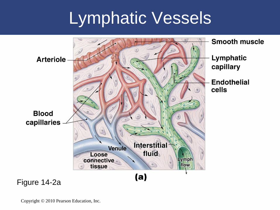
Lymphatic Vessels
Figure 14-2a
Copyright © 2010 Pearson Education, Inc.
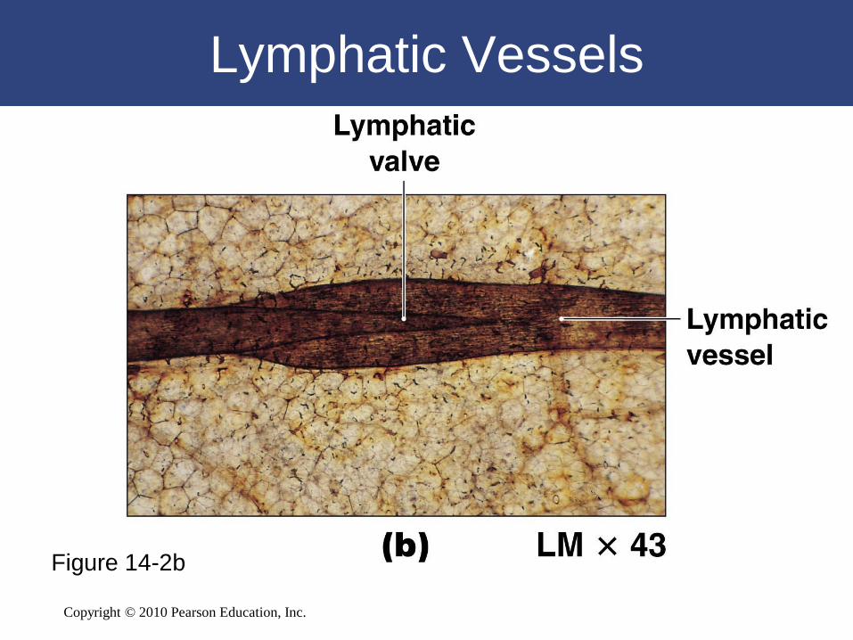
Lymphatic Vessels
Figure 14-2b

Copyright © 2010 Pearson Education, Inc.
Lymphatic Vessels
• Major Lymph-Collecting Vessels
– The base of the thoracic duct:
• Expands into cisterna chyli
– Cisterna chyli receives lymph from:
• Right and left lumbar trunks
• Intestinal trunk

Copyright © 2010 Pearson Education, Inc.
Lymphatic Vessels
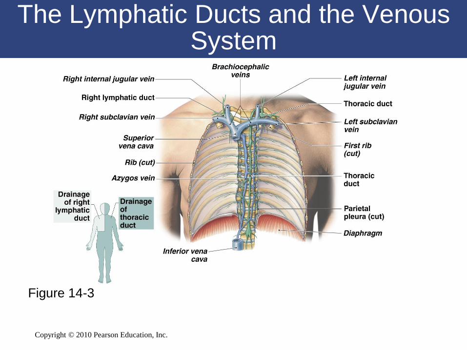
• The Inferior Segment of Thoracic Duct
– Collects lymph from:
• Left bronchiomediastinal trunk
• Left subclavian trunk
• Left jugular trunk
– Empties into left subclavian vein

Copyright © 2010 Pearson Education, Inc.
Lymphatic Vessels
• The Right Lymphatic Duct
– Collects lymph from:
• Right jugular trunk
• Right subclavian trunk
• Right bronchiomediastinal trunk
– Empties into right subclavian vein
Copyright © 2010 Pearson Education, Inc.
The Lymphatic Ducts and the Venous System
Figure 14-3

Copyright © 2010 Pearson Education, Inc.
Lymphocytes
• Three Classes of Circulating Lymphocytes
– T cells:
• Thymus dependent
– B cells:
• Bone marrow derived
– NK cells:
• Natural killer cells

Copyright © 2010 Pearson Education, Inc.
Lymphocytes
• T Cells
– Make up 80% of circulating lymphocytes
• Three Main Types of T Cells
– Cytotoxic T cells
– Helper T cells
– Suppressor T cells

Copyright © 2010 Pearson Education, Inc.
Lymphocytes
• B Cells
– Make up 10% to 15% of circulating lymphocytes
– Differentiate (change) into plasma cells
– Plasma cells:
• Produce and secrete antibodies (immunoglobulin proteins)
– Antibody-mediated immunity:
• A chain of events that destroys the target compound or organism

Copyright © 2010 Pearson Education, Inc.
Lymphocytes
• Natural Killer (NK) Cells
– Also called large granular lymphocytes
– Make up 5% to 10% of circulating lymphocytes
– Responsible for immunological surveillance
– Attack foreign cells, virus-infected cells, and cancer
cells

Copyright © 2010 Pearson Education, Inc.
Figure 14-4

Copyright © 2010 Pearson Education, Inc.
The Origins of Lymphocytes
• T Cells and B Cells
– Migrate throughout the body:
• To defend peripheral tissues
– Retain their ability to divide:
• Is essential to immune system function

Copyright © 2010 Pearson Education, Inc.
The Origins of Lymphocytes
• Differentiation
– B cells differentiate:
• With exposure to hormone called cytokine (interleukin-7)
– T cells differentiate:
• With exposure to several thymic hormones

Copyright © 2010 Pearson Education, Inc.
Lymphoid Nodules
• Areolar tissue with densely packed
lymphocytes
• Germinal center contains dividing
lymphocytes

Copyright © 2010 Pearson Education, Inc.
The Tonsils
Figure 14-5

Copyright © 2010 Pearson Education, Inc.
Lymphoid Nodules
• Distribution of Lymphoid Nodules
– Lymph nodes
– Spleen
– Respiratory tract (tonsils)
– Along digestive and urinary tracts

Copyright © 2010 Pearson Education, Inc.
Lymphoid Organs
• Lymph nodes
• Thymus
• Spleen
• Are separated from surrounding tissues by a
fibrous connective tissue capsule
Copyright © 2010 Pearson Education, Inc.
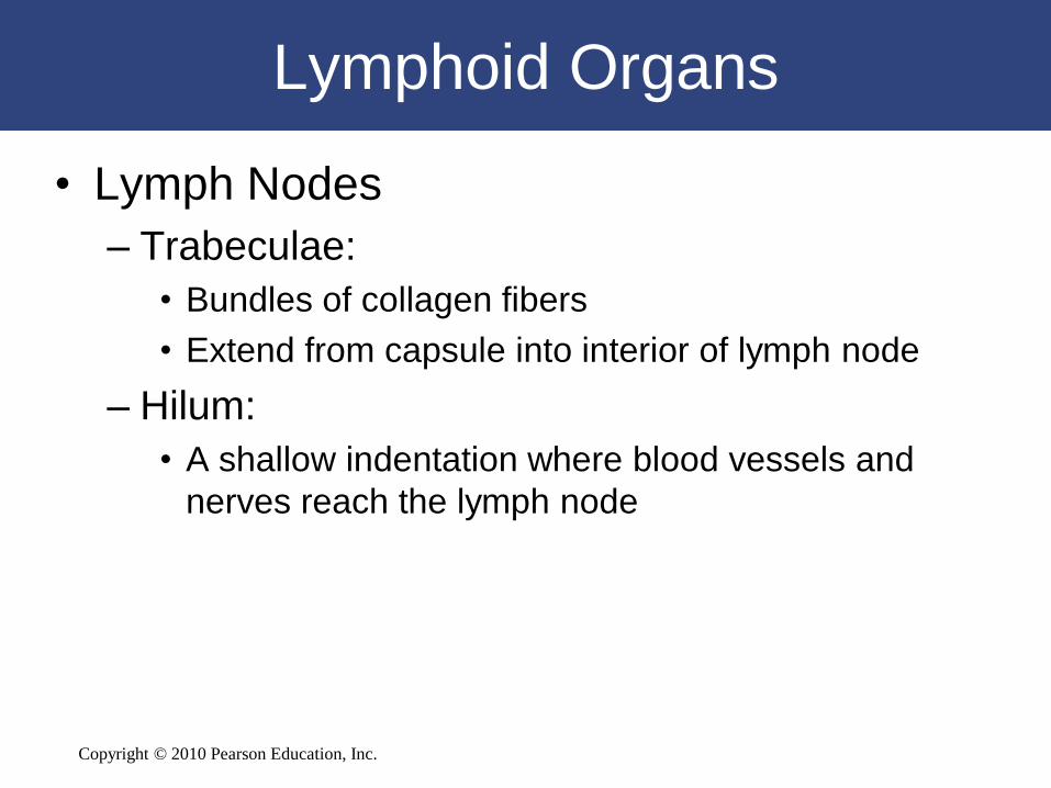
Lymphoid Organs
• Lymph Nodes
– Trabeculae:
• Bundles of collagen fibers
• Extend from capsule into interior of lymph node
– Hilum:
• A shallow indentation where blood vessels and
nerves reach the lymph node

Copyright © 2010 Pearson Education, Inc.
Lymphoid Organs
• Lymph Nodes
– Afferent lymphatic vessels:
• Carry lymph:
– from peripheral tissues to lymph node
– Efferent lymphatic vessels:
• Leave lymph node at hilum
• Carry lymph to venous circulation

Copyright © 2010 Pearson Education, Inc.
The Structure of a Lymph Node
Figure 14-6
Copyright © 2010 Pearson Education, Inc.
Lymphoid Organs
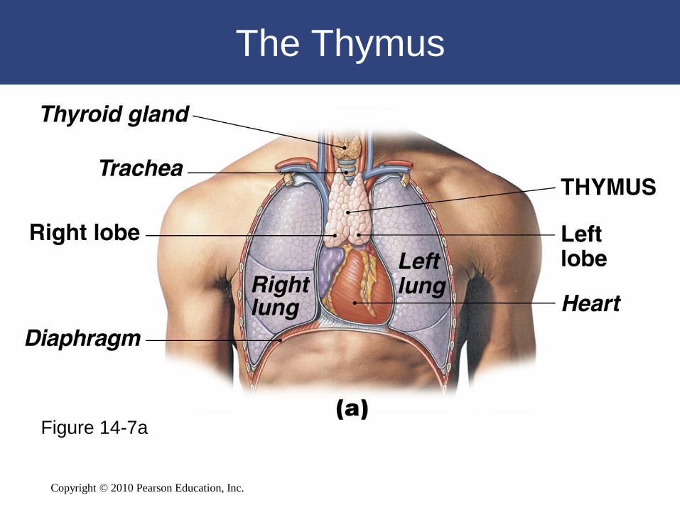
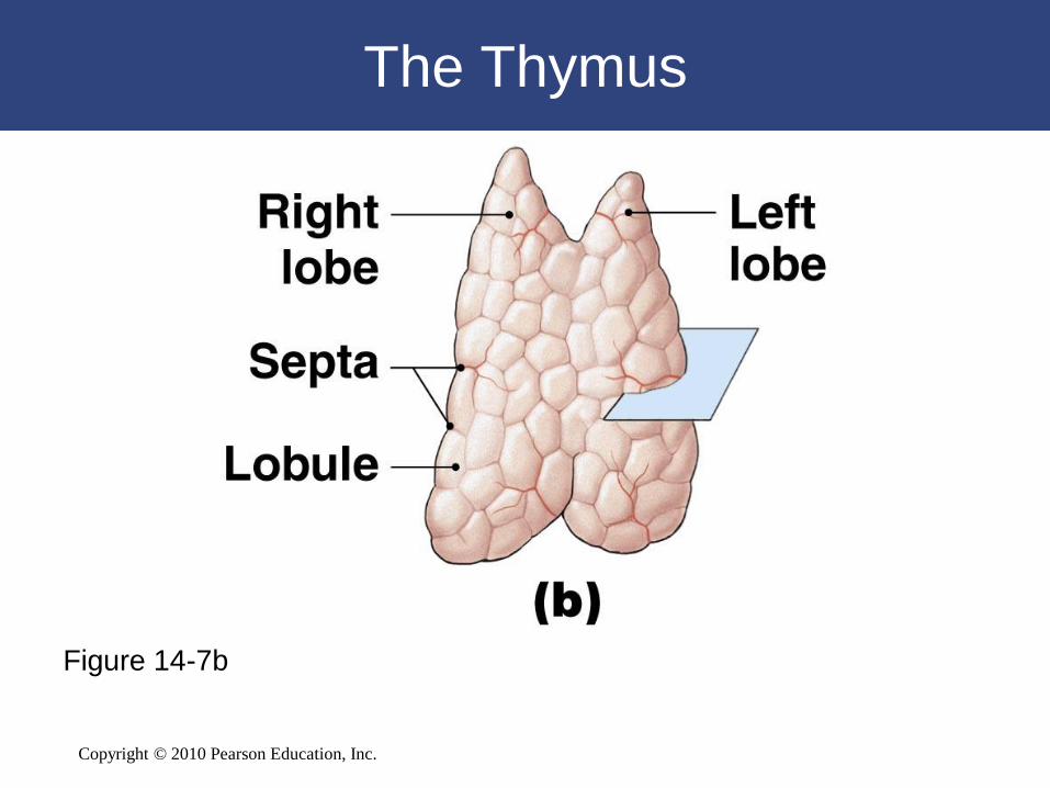
• The Thymus
– Located in mediastinum
– Atrophies after puberty:
• Diminishing effectiveness of immune system
• Divisions of the Thymus
– Thymus is divided into two thymic lobes
– Septa divide lobes into smaller lobules
Copyright © 2010 Pearson Education, Inc.
The Thymus
Figure 14-7a
Copyright © 2010 Pearson Education, Inc.
The Thymus
Figure 14-7b

Copyright © 2010 Pearson Education, Inc.
The Thymus
Figure 14-7c

Copyright © 2010 Pearson Education, Inc.
Lymphoid Organs
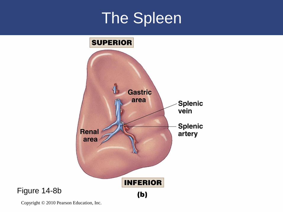
• Three Functions of the Spleen
– Removal of abnormal blood cells and other
blood components by phagocytosis
– Storage of iron recycled from red blood cells
– Initiation of immune responses by B cells and
T cells:
• In response to antigens in circulating blood

Copyright © 2010 Pearson Education, Inc.
Lymphoid Organs
• Structure of the Spleen
– Inside fibrous capsule:
• Red pulp: contains many red blood cells
• White pulp: resembles lymphoid nodules

Copyright © 2010 Pearson Education, Inc.
The Spleen
Figure 14-8a
Copyright © 2010 Pearson Education, Inc.
The Spleen
Figure 14-8b

Copyright © 2010 Pearson Education, Inc.
The Spleen
Figure 14-8c

Copyright © 2010 Pearson Education, Inc.
Roles of the Lymphoid System in Body
Defenses
• Body defenses provide resistance to fight infection,
illness, and disease
• Two categories of defenses
– Nonspecific defenses
– Specific defenses
• Nonspecific and specific defenses operate together to
provide resistance to infection and disease

Copyright © 2010 Pearson Education, Inc.
14-3 Each nonspecific
defense responds in a
characteristic way
regardless of the potential
threat
Copyright © 2010 Pearson Education, Inc.
Nonspecific Defenses
• Seven major categories of nonspecific defenses
– Physical barriers
– Phagocytes
– Immunological surveillance
– Interferons
– Complement
– Inflammatory response
– Fever
Copyright © 2010 Pearson Education, Inc.
Nonspecific Defenses
• Physical Barriers
– Keep hazardous materials outside the body
• Phagocytes
– Attack and remove dangerous microorganisms
• Immunological Surveillance
– Constantly monitors normal tissues:
• With natural killer cells (NK cells)
Copyright © 2010 Pearson Education, Inc.
Nonspecific Defenses
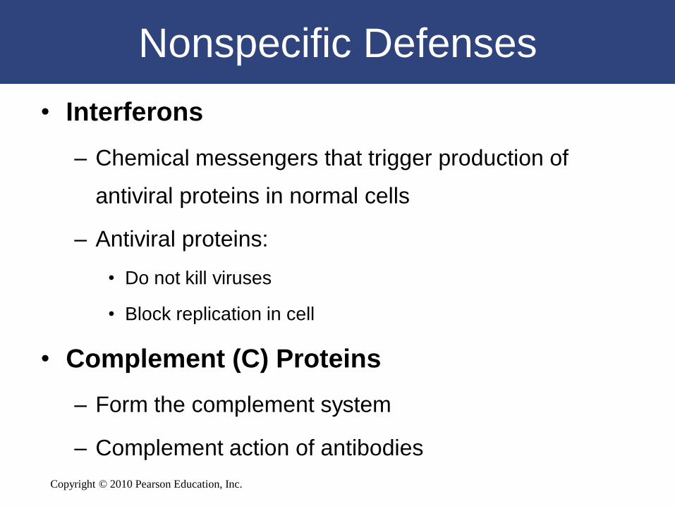
• Interferons
– Chemical messengers that trigger production of
antiviral proteins in normal cells
– Antiviral proteins:
• Do not kill viruses
• Block replication in cell
• Complement (C) Proteins
– Form the complement system
– Complement action of antibodies
Copyright © 2010 Pearson Education, Inc.
Nonspecific Defenses
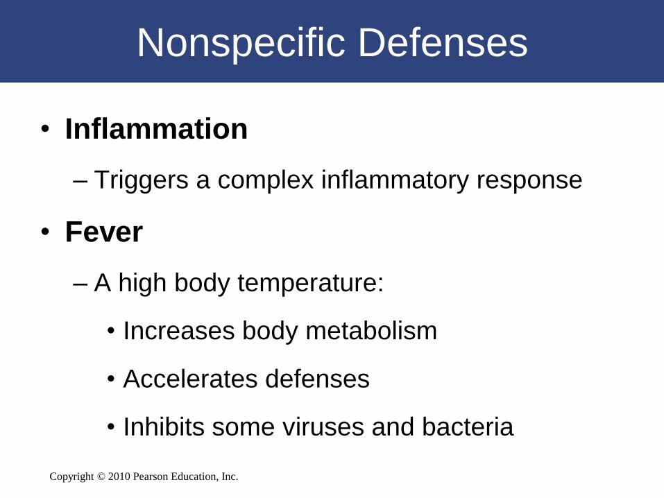
• Inflammation
– Triggers a complex inflammatory response
• Fever
– A high body temperature:
• Increases body metabolism
• Accelerates defenses
• Inhibits some viruses and bacteria

Copyright © 2010 Pearson Education, Inc.
Nonspecific Defenses
Figure 14-9

Copyright © 2010 Pearson Education, Inc.
Nonspecific Defenses
Figure 14-9

Copyright © 2010 Pearson Education, Inc.
Figure 14-10

Copyright © 2010 Pearson Education, Inc.
14-4 Immunity (specific
defenses) responds to specific
threats and is either cell
mediated or antibody
mediated
Copyright © 2010 Pearson Education, Inc.
Types of Immunity
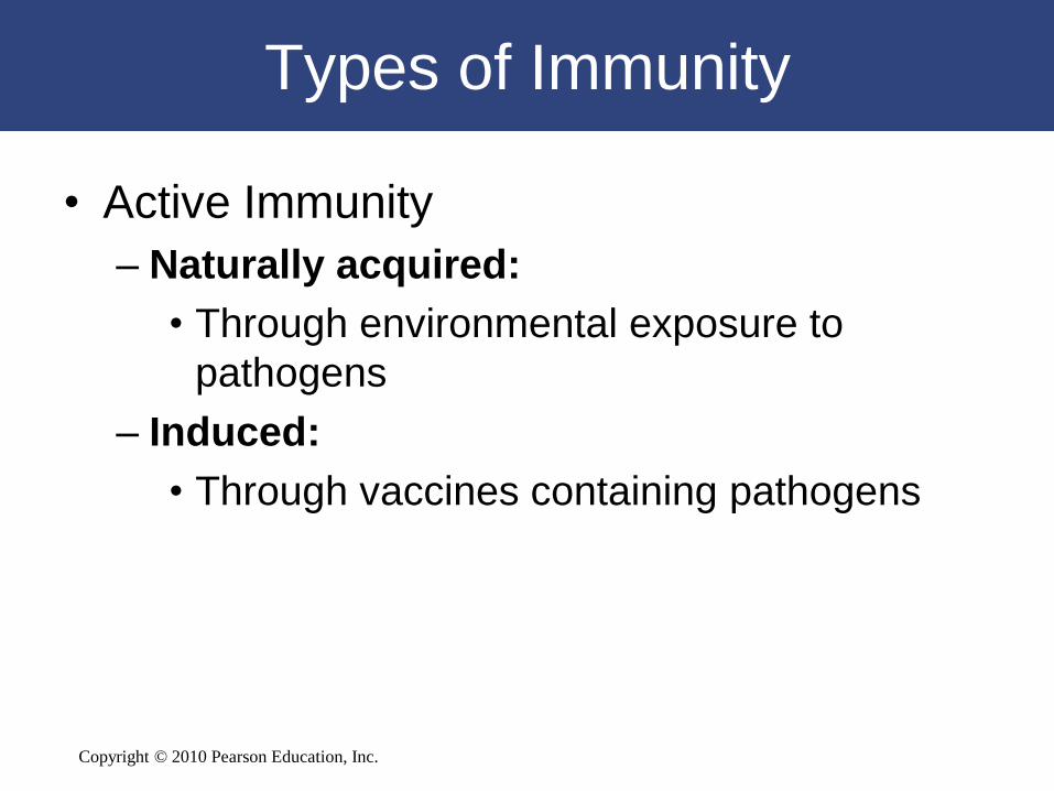
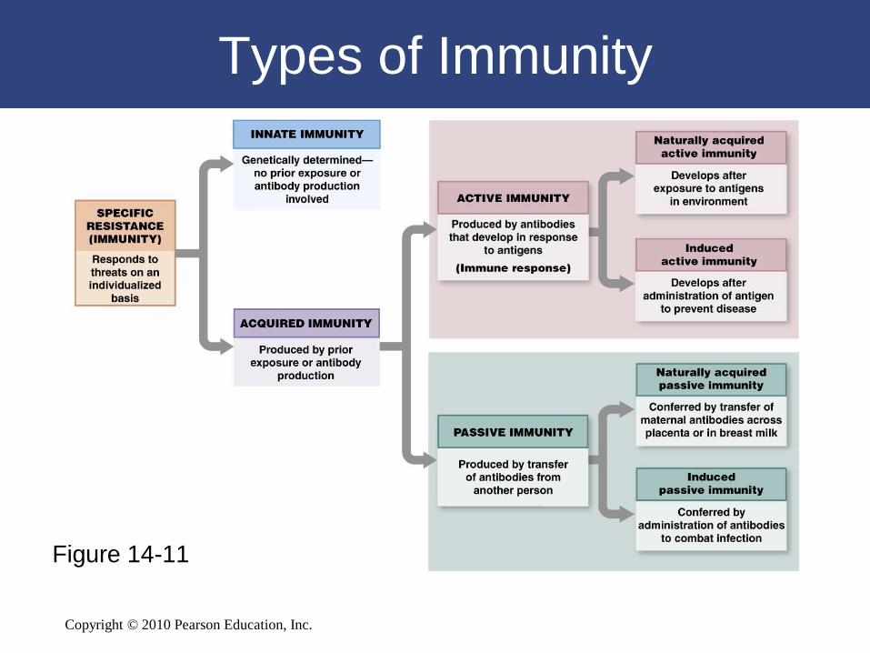
• Active Immunity
– Naturally acquired:
• Through environmental exposure to
pathogens
– Induced:
• Through vaccines containing pathogens

Copyright © 2010 Pearson Education, Inc.
Types of Immunity
• Passive Immunity
– Naturally acquired:
• Antibodies acquired from the mother
– Induced:
• By an injection of antibodies
Copyright © 2010 Pearson Education, Inc.
Types of Immunity
Figure 14-11

Copyright © 2010 Pearson Education, Inc.
Properties of Immunity
• Specificity
– Each T or B cell responds only to a specific antigen and ignores all others
• Versatility
– The body produces many types of lymphocytes:
• Each fights a different type of antigen
• Active lymphocyte clones itself to fight specific antigen
• Memory– Some active lymphocytes (memory cells):
• Stay in circulation
• Provide immunity against new exposure
• Tolerance– Immune system ignores “normal” (self) antigens

Copyright © 2010 Pearson Education, Inc.
An Overview of the Immune Response
Figure 14-12

Copyright © 2010 Pearson Education, Inc.
14-5 T cells play a role in the
initiation and control of the
immune response

Copyright © 2010 Pearson Education, Inc.
Antigen Presentation
• Antigen Recognition
– T cells only recognize antigens that are bound
to glycoproteins in plasma membranes
Copyright © 2010 Pearson Education, Inc.
Antigen Presentation
• MHC Proteins
– The membrane glycoproteins that bind to
antigens
– Genetically coded in chromosome 6:
• The major histocompatibility complex (MHC)
• Differs among individuals

Copyright © 2010 Pearson Education, Inc.
Antigen Presentation
• Two Classes of MHC Proteins
– Class I:
• Found in membranes of all nucleated cells
– Class II:
• Found in membranes of antigen-presenting cells
(APCs)
• Found in lymphocytes

Copyright © 2010 Pearson Education, Inc.
Antigen Presentation
• Class I MHC Proteins
– Pick up small peptides in cell and carry them
to the surface:
• T cells ignore normal peptides
• Abnormal peptides or viral proteins activate T cells
to destroy cell

Copyright © 2010 Pearson Education, Inc.
Antigen Presentation
• Class II MHC Proteins
– Antigenic fragments:
• From antigenic processing of pathogens
• Bind to Class II proteins
• Inserted in plasma membrane to stimulate T cells

Copyright © 2010 Pearson Education, Inc.
Antigen Presentation
• Antigen-Presenting Cells (APCs)
– Responsible for activating T cells against foreign cells
and proteins
• Phagocytic APCs
– Free and fixed macrophages:
• In connective tissues
– Kupffer cells:
• Of the liver
– Microglia:
• In the CNS
Copyright © 2010 Pearson Education, Inc.
T Cell Activation
• Cytotoxic T Cells
– Also called killer T cells
– Seek out and immediately destroy target cells
Copyright © 2010 Pearson Education, Inc.
T Cell Activation
• Actions of Cytotoxic T Cells
1. Release perforin:
• To destroy antigenic plasma membrane
2. Secrete poisonous lymphotoxin:
• To destroy target cell
3. Activate genes in target cell:
• That cause cell to die

Copyright © 2010 Pearson Education, Inc.
T Cells and Immunity
• Helper T Cells
– Activated CD4 T cells divide into:
• Active helper T cells (TH cells):
– secrete cytokines
• Memory TH cells:
– remain in reserve

Copyright © 2010 Pearson Education, Inc.
T Cells and Immunity
• Memory TC Cells
– Produced with cytotoxic T cells
– Stay in circulation
– Immediately form cytotoxic T cells if same
antigen appears again

Copyright © 2010 Pearson Education, Inc.
T Cells and Immunity
• Suppressor T Cells
– Secrete suppression factors
– Inhibit responses of T and B cells
– Act after initial immune response
– Limit immune reaction to single stimulus

Copyright © 2010 Pearson Education, Inc.
T Cell Activation
Figure 14-13

Copyright © 2010 Pearson Education, Inc.
14-6 B Cells respond to
antigens by producing
specific antibodies

Copyright © 2010 Pearson Education, Inc.
B Cells and Immunity
• B Cells
– Responsible for antibody-mediated immunity
– Attack antigens by producing specific
antibodies
– Millions of populations, each with different
antibody molecules

Copyright © 2010 Pearson Education, Inc.
The Sensitization and Activation of
B Cells
Figure 14-14
Copyright © 2010 Pearson Education, Inc.
Antibody Structure
• Two parallel pairs of polypeptide chains
– One pair of heavy chains
– One pair of light chains
• Each chain contains
– Constant segments
– Variable segments

Copyright © 2010 Pearson Education, Inc.
Antibody Structure
Figure 14-12

Copyright © 2010 Pearson Education, Inc.
Copyright © 2010 Pearson Education, Inc. Figure 22–22
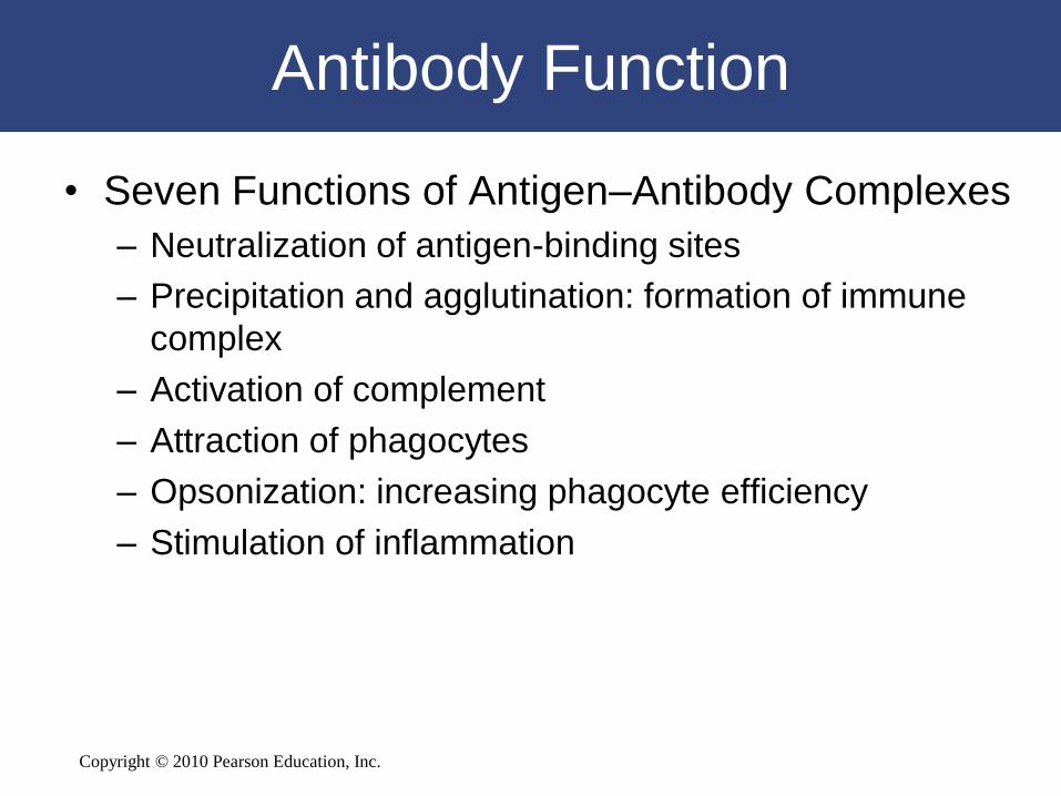
Antibody Function
• Seven Functions of Antigen–Antibody Complexes
– Neutralization of antigen-binding sites
– Precipitation and agglutination: formation of immune
complex
– Activation of complement
– Attraction of phagocytes
– Opsonization: increasing phagocyte efficiency
– Stimulation of inflammation

Copyright © 2010 Pearson Education, Inc.
Primary and Secondary Responses to
Antigen Exposure
• First exposure
– Produces initial primary response
• Next exposure
– Triggers secondary response
– More extensive and prolonged
– Memory cells already primed

Copyright © 2010 Pearson Education, Inc.
Primary and Secondary Responses to
Antigen Exposure
• The Primary Response
– Takes time to develop
– Antigens activate B cells
– Plasma cells differentiate
– Antibody titer (level) slowly rises

Copyright © 2010 Pearson Education, Inc.
Primary and Secondary Responses to
Antigen Exposure
• The Primary Response
– Peak response:
• Can take 2 weeks to develop
• Declines rapidly
– IgM:
• Is produced faster than IgG
• Is less effective
Copyright © 2010 Pearson Education, Inc.
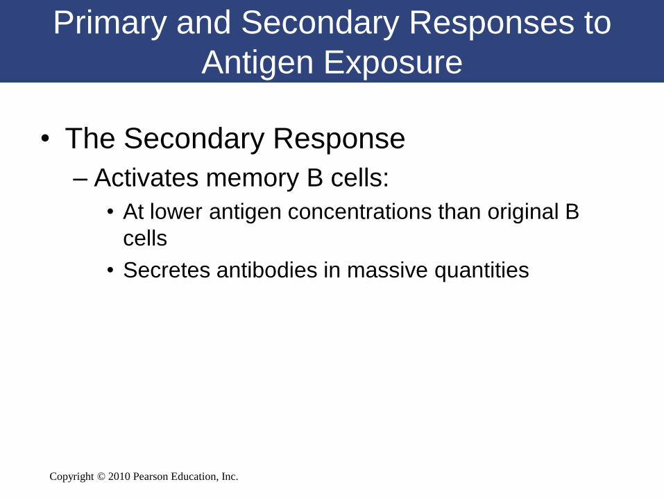
Primary and Secondary Responses to
Antigen Exposure
• The Secondary Response
– Activates memory B cells:
• At lower antigen concentrations than original B
cells
• Secretes antibodies in massive quantities

Copyright © 2010 Pearson Education, Inc.
Primary and Secondary Responses
Figure 14-16

Copyright © 2010 Pearson Education, Inc.
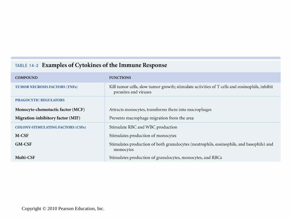
Hormones of the Immune System
• Six Groups of Hormonal Cytokines
– Interleukins
– Interferons
– Tumor necrosis factors (TNFs)
– Chemicals that regulate phagocytic activities
– Colony-stimulating factors (CSFs)
– Miscellaneous cytokines

Copyright © 2010 Pearson Education, Inc.
Copyright © 2010 Pearson Education, Inc.

Copyright © 2010 Pearson Education, Inc.
14-7 Abnormal immune
responses result in immune
disorders
Copyright © 2010 Pearson Education, Inc.
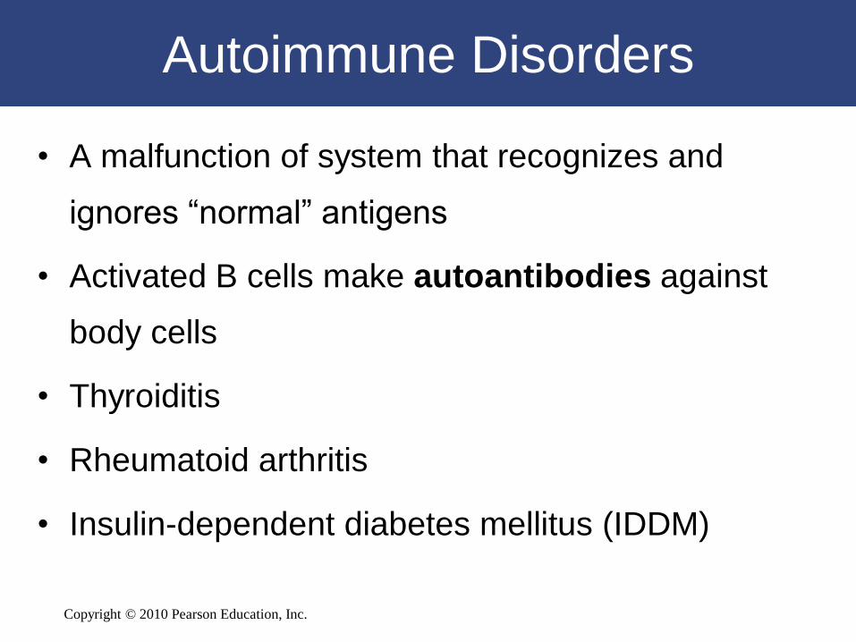
Autoimmune Disorders
• A malfunction of system that recognizes and
ignores “normal” antigens
• Activated B cells make autoantibodies against
body cells
• Thyroiditis
• Rheumatoid arthritis
• Insulin-dependent diabetes mellitus (IDDM)

Copyright © 2010 Pearson Education, Inc.
Immunodeficiency Diseases
• Problems with embryological development of lymphoid
tissues
– Can result in severe combined immunodeficiency disease
(SCID)
• Viral infections such as HIV
– Can result in AIDS
• Immunosuppressive drugs or radiation treatments
– Can lead to complete immunological failure

Copyright © 2010 Pearson Education, Inc.
Allergies
• Inappropriate or excessive immune
responses to antigens
– Allergens:
• Antigens that trigger allergic reactions

Copyright © 2010 Pearson Education, Inc.
Allergies
• Four Categories of Allergic Reactions
– Type I:
• Immediate hypersensitivity
– Type II:
• Cytotoxic reactions
– Type III:
• Immune complex disorders
– Type IV:
• Delayed hypersensitivity

Copyright © 2010 Pearson Education, Inc.
Allergies
• Type I Allergy
– Also called immediate hypersensitivity
– A rapid and severe response to the presence
of an antigen
– Most commonly recognized type of allergy
– Includes allergic rhinitis (environmental
allergies)
Copyright © 2010 Pearson Education, Inc.
Allergies
• Type I Allergy
– Sensitization leads to:
• Production of large quantities of IgE antibodies
distributed throughout the body
– Second exposure leads to:
• Massive inflammation of affected tissues
Copyright © 2010 Pearson Education, Inc.
Allergies
• Type I Allergy
– Severity of reaction depends on:
• Individual sensitivity
• Locations involved
– Allergens (antigens that trigger reaction) in
bloodstream may cause anaphylaxis

Copyright © 2010 Pearson Education, Inc.
Allergies
• Anaphylaxis
– Can be fatal
– Affects cells throughout the body
– Changes capillary permeability:
• Produces swelling (hives) on skin
– Smooth muscles of respiratory system contract:
• Make breathing difficult
– Peripheral vasodilatation:
• Can cause circulatory collapse (anaphylactic shock)
Copyright © 2010 Pearson Education, Inc.
14-8 The immune response
diminishes with advancing age

Copyright © 2010 Pearson Education, Inc.
Immune System and Aging
• Four Effects of Aging on the Immune Response
– Thymic hormone production is greatly reduced
– T cells become less responsive to antigens
– Fewer T cells reduces responsiveness of B cells
– Immune surveillance against tumor cells declines
Copyright © 2010 Pearson Education, Inc.
14-9 For all body systems, the
lymphoid system provides
defenses against infection and
returns tissue fluid to the
circulation

Copyright © 2010 Pearson Education, Inc.
The Lymphoid System
in Perspective
Functional Relationships Between
the Lymphoid System and Other Systems
Copyright © 2010 Pearson Education, Inc.

Copyright © 2010 Pearson Education, Inc.
• The Integumentary System provides physical barriers to pathogen entry; macrophages in dermis resist infection and present antigens to trigger immune response; mast cells trigger inflammation, mobilize cells of lymphoid system
• The Lymphoid System provides IgA antibodies for secretion onto integumentary surfaces
The Integumentary System

Copyright © 2010 Pearson Education, Inc.
The Skeletal System
• The Skeletal System’s bone marrow produces and stores lymphocytes and other cells involved in the immune response
• The Lymphoid System assists in repair of bone after injuries; macrophages fuse to become osteoclasts

Copyright © 2010 Pearson Education, Inc.
The Nervous System
• The Nervous System’s microglia present antigens that stimulate specific defenses; glial cells secrete cytokines; innervation stimulates antigenpresenting cells
• The Lymphoid System’s cytokines affect hypothalamic production of CRH and TRH

Copyright © 2010 Pearson Education, Inc.
The Endocrine System
• The Endocrine System’s
glucocorticoids have anti-
inflammatory effects; thymosins
stimulate development and
maturation of lymphocytes;
many hormones affect immune
function
• The Lymphoid System’s thymus
secretes thymosins; cytokines
affect cells throughout the body

Copyright © 2010 Pearson Education, Inc.
The Cardiovascular System
• The Cardiovascular System distributes WBCs; carries antibodies that attack pathogens; clotting response helps restrict spread of pathogens; hemocytoblasts give rise to stem cells that produce WBCs
• The Lymphoid System fights infections of cardiovascular organs; returns tissue fluid to bloodstream
Copyright © 2010 Pearson Education, Inc.
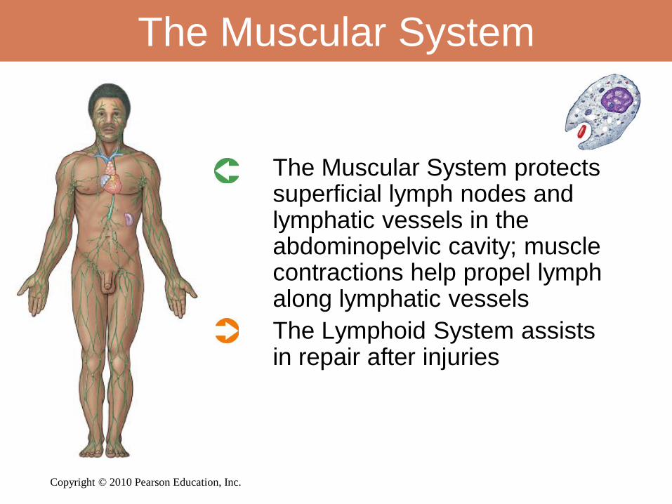
The Muscular System
• The Muscular System protects superficial lymph nodes and lymphatic vessels in the abdominopelvic cavity; muscle contractions help propel lymph along lymphatic vessels
• The Lymphoid System assists in repair after injuries

Copyright © 2010 Pearson Education, Inc.
The Respiratory System
• The Respiratory System’s alveolar phagocytes present antigens and trigger specific defenses; provides oxygen and eliminates carbon dioxide for the lymphoid system
• The Lymphoid System’s tonsils protect against infection at entrance to respiratory tract

Copyright © 2010 Pearson Education, Inc.
The Digestive System
• The Digestive System provides
nutrients required by lymphatic
tissues; digestive acids and enzymes
provide nonspecific defense against
pathogens
• The Lymphoid System’s tonsils and
intestinal lymphoid nodules defend
against infection and toxins absorbed
from the digestive tract; lymphatics
carry absorbed lipids to venous
system

Copyright © 2010 Pearson Education, Inc.
The Urinary System
• The Urinary System eliminates metabolic wastes generated by cellular activity; acid pH of urine provides nonspecific defense against urinary tract infection
• The Lymphoid System provides specific defenses against urinary tract infections
Copyright © 2010 Pearson Education, Inc.
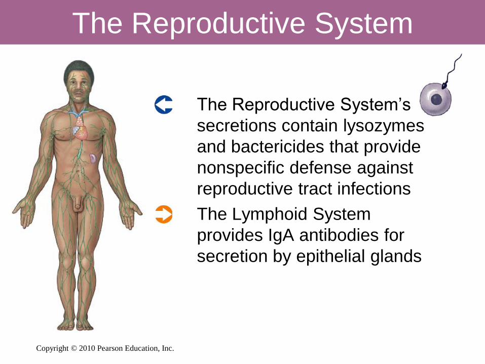
The Reproductive System
• The Reproductive System’s
secretions contain lysozymes
and bactericides that provide
nonspecific defense against
reproductive tract infections
• The Lymphoid System
provides IgA antibodies for
secretion by epithelial glands