Embed Size (px)
Citation preview

www.wjpr.net Vol 8, Issue 3, 2019.
Makinde et al. World Journal of Pharmaceutical Research
130
THE LYMPHOCYTE BLAST TRANSFORMATION (BT) TEST
Taiwo Mary Makinde*1, Prof A. I. Bozhkov
2
1Department of Biological Sciences, National University of Life and Environmental Sciences
of Ukraine, Heroiv Oborony St, 15, Kyiv, 03041.
2Department of Biology, V.N. Karazin Kharkiv National University, Svobody Square, 4,
Kharkiv, Kharkivs'ka oblast, 61000.
ABSTRACT
The purpose is to show the rate of conversion of lymphocyte blast in
different samples of human blood infected with myasthenia gravis (an
autoimmune disease). This reaction was based on the ability of normal
lymphocytes transformed in undifferentiated germ cells of blast type
after culturing in vitro in the presence of specific allergens and
nonspecific stimulants, mitogens (PHA or PHA, concanavalinA,
lipopolysaccharides, and other substances). To determine the rate of
conversion of the blast, the number of explosions and intermediate
forms determined on 100 cells and a similar calculation is made in the
control as well. The morphology of the human blood cells
(lymphocytes and blasts) in the presence and absence of mitogen
(phytohemagglutinin), which viewed under a microscope. As we all
know, it was previously shown that under normal conditions of 40 to 90 percent of the
lymphocyte can turn into explosions and reduce the conversion of the explosion in the
presence of allergens, or PHA indicate any abnormality. Necessity biosafety diagnostic
laboratory for analysis in each step of the test tube was fully considered in order to prevent /
avoid the risk of any form especially for diagnostics of human blood at various ill patients.
Therefore, in this document, different blood samples and serum were analyzed separately;
Templates colony, the number of blood cells and their morphology were studied by light
microscopy, to lead us to more understanding of the mechanism underlying
immunopathology / immunodeficiency and their risks in the human immune system.
Biosecurity measures to avoid infection and to avoid invalid results were fully met.
World Journal of Pharmaceutical Research SJIF Impact Factor 8.074
Volume 8, Issue 3, 130-146. Research Article ISSN 2277– 7105
*Corresponding Author
Taiwo Mary Makinde
Department of Biological
Sciences, National
University of Life and
Environmental Sciences of
Ukraine, Heroiv Oborony
St, 15, Kyiv, 03041.
Article Received on
31 Dec. 2018,
Revised on 21 Jan. 2019, Accepted on 10 Feb. 2019
DOI: 10.20959/wjpr20193-14255

www.wjpr.net Vol 8, Issue 3, 2019.
Makinde et al. World Journal of Pharmaceutical Research
131
KEYWORDS: Biotechnology, Recombinant DNA technology, Transformation of
lymphocytes blast mitogen.
INTRODUCTION
The twentieth century was marked by intensive development of functional science and
particularly in the development of molecular biology and biotechnology.
Latest technologies allow the biological reconstruction of genetic systems, create new genetic
constructs and widely used microorganisms in practice. Creating transgenic organisms
allowed for progress in getting new resistance to various diseases of plants and animals. The
development of biotechnology has led not only to the appearance of previously existing
process, but also to change our outlook, medical practice and housekeeping.
However, the latest advanced technologies involve new biological risks and threats. This in
turn triggered protests among the population with further development of biotechnology.
A new challenge to our civilization is associated with the understanding and analysis of the
prospects for further development on one hand and the risks of biotechnology and biological
hazards potentially in new technologies. In this connection is the analysis of the potential
risks of working with the microorganisms, the DNA technology and the development of
medical practice, including studies of autoimmune pathologies.[54,55,56,57,58]
Autoimmune disease is one of the common diseases that occur when the body's immune
system turns against the body itself, attacking as if it were a foreign pathogen. Examples of
autoimmune disorders include rheumatoid arthritis, multiple sclerosis, juvenile diabetes,
cardiomyopathy, antiphospholipid syndrome, Guillain-Barre syndrome, Crohn's disease,
Graves' disease, Sjogren's syndrome, alopecia, myasthenia gravis, lupus erythematous, and
psoriasis.[39,59]
In addition, such disorders complicate other diseases that are not autoimmune in origin, such
as atherosclerosis. A key challenge to developing therapies for autoimmune disorders is the
cross-reactivity between normal body proteins and those of a variety of pathogenic agents.
Therefore, it is important to allow as much independent function and self-care as possible and
to promote appropriate activities to ensure a sense of normalcy in the body system.[60]

www.wjpr.net Vol 8, Issue 3, 2019.
Makinde et al. World Journal of Pharmaceutical Research
132
The development of modern biotechnology is characterized by intense development of
biosafety guidelines when working on the genetic, cellular, tissue and organism level.
Today, about special attention is given to the criteria and indicators, biosafety, and in all
countries of the world, there is a state legal regulation in various areas of biotechnology.
High biotechnology based on modification of genetic structures with the purpose of
improvement of biological objects.
Management molecular and genetic events affecting indigenous mechanisms of the most
important properties of living organisms, such as heredity, variability, energy and mass
transfer, adaptation, resistance, productivity and production quality.
Molecular-genetic engineering and the effects of such interventions today carry to dual-use
technology, as their results cannot always be predicted.
Artificial changes in mass and energy can also be related to changes in information
characteristics of realization of genetic information. In society there is a certain caution in the
use of genetically-modified plants, animals, microorganisms and viruses. And in some cases,
lack of knowledge in this area can have a negative impact on the pace of development of
biotechnology and especially in its strategic component, which is bioengineering. Today,
experts in the field focused biotechnology are working to strengthen or weaken the virulence
or other characteristics of bacteria, solving the problem of health safety and protection of the
state against biological weapons and terrorism. It is necessary to take into account the
relationship between the kinds of biosafety and the impact on their biotech production:[6]
1) In public health - in the production of medicines, vaccines, diagnostics, human
reproduction, gene therapy and xenotransplantation;
2) When the production of new food products Modern biotechnology is aimed at the
development of balanced food rations, the production of food products and dietary
productive use of microbial producers for the production of bread, cheese, wine, food
additives, enzymes;
3) Agriculture is important to obtain new transgenic plants and animals with desired
productive, adaptive and autonomic properties and the development of biological
protection of plants and animals and the creation of bacterial fertilizers;
4) In livestock - in vitro fertilization and cloning of embryos;

www.wjpr.net Vol 8, Issue 3, 2019.
Makinde et al. World Journal of Pharmaceutical Research
133
5) Providing a virus-free planting material for plants;
6) Establishment of recombinant microorganisms for the utilization of agricultural, domestic
and industrial waste, for destruction of pollutants (pesticides, polymers, oil). Science,
society and the State must develop and efficiently use the system of measures for the
protection of human health and the environment from the harmful effects of hazardous
factors.
It is necessary to develop a system of effective measures to combat and prevent the abusive
use of microbiological science to achieve. The world scientific community and mainly
specialists in the field of molecular biology have come to the conclusion that bioengineering
is currently the only option for the efficient use of its results and achievements, and to
address food security of the world population, diagnosis and treatment of diseases of
civilization.
OBJECTIVES
We need to determine; - Viral persistence (which is a risk leading to infection occurrence)
and immune-resistance (on the functional response of lymphocytes) in patients with different
clinical phenotypes of myasthenia gravis.
The Task
1. To determine the presence of specific antibodies to the herpes virus in patients with
different clinical phenotypes of myasthenia gravis.
2. Determine the ratio of subpopulations of blood cells as a potential reserve for the
implementation of the immune response.
3. Identify potential mitogenic activity of serum components of blood of patients with
different clinical phenotypes of myasthenia gravis.
4. Determine the proliferative potential of peripheral blood leukocytes of patients with
different clinical phenotypes of myasthenia.
5. Biosafety measures to prevent bio-risks, contamination and invalid results in the
diagnostic lab.

www.wjpr.net Vol 8, Issue 3, 2019.
Makinde et al. World Journal of Pharmaceutical Research
134
MATERIALS AND METHODS
LIST OF MATERIALS USED
a. Glass wares
The glass wares used are the conical flasks, cover slips, beakers, and disposable petri-dishes,
glass rods, Nikiforov slide glass, measuring cylinder, and pipette, test tubes, and glass test
bottles.
b. Equipment’s
Autoclave, Bunsen burner, light microscope, thermostat, centrifuge, cell colony counter.
c. Reagents
Alcohol (95%), human blood, human plasma, penicillin, streptomycin, fetal calf serum,
isotonic solution, heparin (anticoagulant), mitogen or PHA (phytohemagglutinin).
d. Media
Antibiotics: - penicillin and streptomycin, nutrient medium 199.
e. Other materials
Marker, sterile syringe, paper tape, Aluminum foil, cotton wool, vials, test tube rack,
disinfectant, Microfluidic pipette.
METHOD
First Stage
1. Peripheral blood was taken aseptically and incubated for 30-40 minutes at room
temperature until a visible clear separation of plasma and erythrocytes.
2. In sterile vials make 4 ml of nutrient medium 199 containing 200 units of penicillin and
100 units of streptomycin per 1 ml and 0.5 ml of inactivated fetal calf serum.
3. 0.3-0.5 ml of plasma containing cells was added in flasks with medium 199.
4. In the test sample was added 0.1 ml of an isotonic solution containing 100 mkg of PHA.
In the control samples, 0.1 ml of isotonic solution was added (NO MITOGEN WAS
ADDED).
5. Bottles are rightly placed for 48-72 hours in a thermostat at a temperature of 37digree
Celsius (Room temperature).
6. After incubation, blasts were seen but they were in very small amount under the
microscope.

www.wjpr.net Vol 8, Issue 3, 2019.
Makinde et al. World Journal of Pharmaceutical Research
135
Second Stage
7. Centrifuged for 10 min at 1000 rev / min, and the supernatant was removed (the excess
liquid dispelled), and sediments were seen. After centrifugation, the solution was
thoroughly mixed.
Fixation
8. 2ml of acetic acid and 6ml of methanol (1:3) were added to the sediments in the 2 test
tubes (the test and the control). This mixture will help us in viewing the blast cells
perfectly under the microscope. They were mixed thoroughly and kept for 10 minutes in
an open place on a working bench while the fixation process is taking place.
9. Then the mixture undergoes centrifugation again for 10 minutes and no sediments are
seen because of excess liquid. Therefore, excess liquid is dispelled again and the blast
cells which are the residue at the bottom of the glass tubes.
10. The supernatant was re-mixed, and a quantity of 100 ml is removed with a pipette and
smears are prepared on a sterile glass slide.
11. The slides are left to dry in a ventilation box for 72 hours.
12. Smear stained with azure-eosin by Romanowsky Giemsa for 30 min. Smear stained with
azure-eosin by Romanowsky Giemsa for 30 min.
13. Microscopic view and cell count of blood cells (Lymphocytes and blasts).
14. Microscopic reading of dry drops of serum.
RESULTS AND DISCUSSION
The blasts and white blood cells were viewed under the microscope. White blood cells look
smaller in size and blasts look bigger in size.
TEST
WITH MITOGEN
(PHA)
WITHOUT MITOGEN
(PHA)
LYMPHOCYTES
(%) 71.7% 62.5%
BLASTS
(%) 28.3% 41.9%

www.wjpr.net Vol 8, Issue 3, 2019.
Makinde et al. World Journal of Pharmaceutical Research
136
CONTROL
WITH MITOGEN
(+ PHA)
WITHOUT MITOGEN
(- PHA)
BLAST CONTROL 65% 10%
The blasts and white blood cells were viewed under the microscope. White blood cells look
smaller in size and blasts look bigger in size.
It has been proven that under normal conditions 40 to 90 per cent of lymphocytes can
transform into blasts. Decrease in blast transformation in the presence of allergens or PHA
points to some abnormality. Therefore, we can say that there is a decreased functioning of the
immune system. This could be as a result of different diseases such as inflammation,
oncology diseases, and auto-immune diseases like Myasthenia gravis etc.
In addition, lymphocytes with mitogen have a spontaneous level of blast transformation.
When mitogen is added there is an observable modulation in the action of different antigens.
Therefore, mitogen helps the immune system for better functioning.[41, 42, and 43]
Heparin helps
to determine the osmotic resistance of erythrocytes in venous blood, it is also an
anticoagulant.
Blast Index And Lymphocyte Index
PHA-Phytohaematogglutinin.
(+ PHA)- Proliferation of cell culture in the presence of mitogen.
(- PHA)- Spontaneous proliferation of cell culture without mitogen.
Blast index
Blast index = (+ PHA) - (- PHA) × 100
(- PHA)
I Blast = (28.3%) - (-41.9%) × 100
(-41.9) %
I = 70.2% × 100
-41.9%
I = ≃ - 167.54%

www.wjpr.net Vol 8, Issue 3, 2019.
Makinde et al. World Journal of Pharmaceutical Research
137
Lymphocyte Index
Lymphocyte index = (+ PHA) - (- PHA) × 100
(- PHA)
I Lymphocyte = (71.7%) – (- 62.5%) × 100
(-62.5%)
I = 134.2% × 100
-62.5%
I ≃ - 214.72%
In a situation where we have (+ PHA) > (- PHA), there is a normal situation or normal
healthy condition of the immune system of a person. And if reverse is the case, then there is a
problem.
In the table above, in the blast transformation (+ PHA) < (- PHA) this proves the occurrence
of a disease condition such as autoimmune disease (Myasthenia gravis). While in
lymphocytes it is observed that (+ PHA) > (- PHA) and it shows the lymphocytes are in a
normal condition.
Determination of Viral Persistence And Immune-Resistance (On The Functional
Response Of Lymphocytes) In Patients With Different Myasthenia Clinical Phenotypes
Objective: To determine viral persistence and immune-resistance (on the functional response
of lymphocytes) in patients with different myasthenia clinical phenotypes.
THE TASKS
1. To determine the presence of specific antibodies to the herpes virus in patients with
different myasthenia clinical phenotypes. (Table 1)
Table 1: The concentration of IgG (mmol/l) antibodies to viral infection in patients with
myasthenia on morphological and functional changes of thymus.
Index Control
group
Surveyed groups
Myasthenia
without defeat
in the thymus
(M)
Myasthenia against
the backdrop of
thymic hyperplasia
(MG)
Myasthenia
against the
backdrop of a
thymoma (MT)
IgG against
SMV
0,80 ±
0,05 22,8 ± 2,1 * 18,7 ± 3,7 * 23,1 ± 1,6 *
IgG antibodies to
EBV
0,24 ±
0,03 2,01 ± 0,18 * 2,94 ± 0,09 * 2,36 ± 0,11 *
Note: * - p<0.05

www.wjpr.net Vol 8, Issue 3, 2019.
Makinde et al. World Journal of Pharmaceutical Research
138
Findings
1. The concentration of IgG antibodies to viral infection in patients with myasthenia on
morphological and functional changes of thymus.
2. A high incidence of viral persistence could serve as a basis for the consideration of a viral
infection as a trigger factor of the disease.
3. IgG against CMV (cytomegalovirus) showed the highest value when surveyed in
Myasthenia without defeat in the thymus (M) and the lowest value when surveyed in
Myasthenia against the backdrop of thymic hyperplasia (MG).
4. IgG antibodies against EBV (Epstein-Barr virus) showed the highest value when
surveyed in Myasthenia against the backdrop of thymic hyperplasia (MG) and the
lowest value in Myasthenia without defeat in the thymus (M).
2. Determine the ratio of subpopulations of blood cells as a potential reserve for the
implementation of the immune response. (Table 2)
Table 2: The differentiation of white blood cells (lymphocytes and the ratio neutrophils)
in the patient groups with different clinical phenotypes of myasthenia gravis.
The clinical
phenotype of
myasthenia gravis
WBC counts The ratio
L / H *
100
White blood cells,
abs. X10[6]
Neutrophils
%
Lymphocytes
, %
control
M 6.07 59.5 25.2 42.35
MG 5.75 61.04 22.1 36.2
MT 6.68 65.65 29.05 44.24
Findings
1. It can be concluded that lymphocytes had the lowest count in the clinical phenotype of
myasthenia gravis.
2. Also, White blood cells have the lowest count in the clinical phenotype of myasthenia
gravis.
www.wjpr.net Vol 8, Issue 3, 2019.
Makinde et al. World Journal of Pharmaceutical Research
139
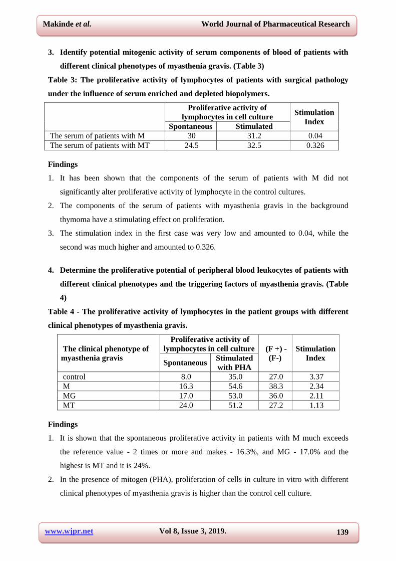
3. Identify potential mitogenic activity of serum components of blood of patients with
different clinical phenotypes of myasthenia gravis. (Table 3)
Table 3: The proliferative activity of lymphocytes of patients with surgical pathology
under the influence of serum enriched and depleted biopolymers.
Proliferative activity of
lymphocytes in cell culture Stimulation
Index Spontaneous Stimulated
The serum of patients with M 30 31.2 0.04
The serum of patients with MT 24.5 32.5 0.326
Findings
1. It has been shown that the components of the serum of patients with M did not
significantly alter proliferative activity of lymphocyte in the control cultures.
2. The components of the serum of patients with myasthenia gravis in the background
thymoma have a stimulating effect on proliferation.
3. The stimulation index in the first case was very low and amounted to 0.04, while the
second was much higher and amounted to 0.326.
4. Determine the proliferative potential of peripheral blood leukocytes of patients with
different clinical phenotypes and the triggering factors of myasthenia gravis. (Table
4)
Table 4 - The proliferative activity of lymphocytes in the patient groups with different
clinical phenotypes of myasthenia gravis.
The clinical phenotype of
myasthenia gravis
Proliferative activity of
lymphocytes in cell culture (F +) -
(F-)
Stimulation
Index Spontaneous
Stimulated
with PHA
control 8.0 35.0 27.0 3.37
M 16.3 54.6 38.3 2.34
MG 17.0 53.0 36.0 2.11
MT 24.0 51.2 27.2 1.13
Findings
1. It is shown that the spontaneous proliferative activity in patients with M much exceeds
the reference value - 2 times or more and makes - 16.3%, and MG - 17.0% and the
highest is MT and it is 24%.
2. In the presence of mitogen (PHA), proliferation of cells in culture in vitro with different
clinical phenotypes of myasthenia gravis is higher than the control cell culture.

www.wjpr.net Vol 8, Issue 3, 2019.
Makinde et al. World Journal of Pharmaceutical Research
140
3. For a standardized results stimulation index was calculated, which compares the
proliferative activity without PHA with proliferative activity with PHA. But the
magnitude of the pre-proliferative activity with PHA subtracted the value of proliferative
activity without PHA.
4. The stimulation index = (+ PHA) - (-PGA)/(-PGA) %
(+ PHA)- Proliferation of cell culture in the presence of mitogen in the cytoplasm.
(- PHA)- Spontaneous proliferation of cell culture without mitogen.
In recent years, there was a reversal of viral persistence. In patients with myasthenia detected
with the population frequency of 95%, herpes viruses, Epstein-Barr virus and
cytomegalovirus.
The possible error may occur: -
1. When drawing blood:
Violation of the procedure of blood sampling;
2. When setting the method:
Not enough clean dishes;
The use of old or improperly prepared reagents;
Bad staining.
Safety Measures And Disposal
Wear rubber gloves, lab coats, safety goggles. It is forbidden to eat, smoke.
All samples for analysis are considered potentially infectious material. Therefore, the
remnants of samples and reagents must be disposed of in accordance with the established
rules.
BLAST CELLS
Blast cells are immature cells found in bone marrow. They are not fully developed and
therefore cannot carry out any function within the body yet. In normal humans, up to 5% of
the cells found in the bone marrow are blats cells. When a higher percentage of them are
found, further testing may be needed as this is an indication of one of several disorders which
affect the blood and the bones. Normally, blast cells continue to mature within the bone
marrow and then begin to carryout set functions. But when a higher than normal ratio of blast
cell is found within the bone marrow, a problem may exist.[65,66,67]

www.wjpr.net Vol 8, Issue 3, 2019.
Makinde et al. World Journal of Pharmaceutical Research
141
Lymphocytes
Lymphocytes are small white blood cells that play a role in the body’s immune response (i.e.,
in the body they fight against germs and diseases). There are two main types of Lymphocytes
known as the B lymphocytes (B-cells) and the T lymphocytes (T cells).
B cells- Produce antibodies that attack foreign molecules (germs and the toxins they
produce).
T cells- They are more complicated, but they can attack the body’s own cells when they are
diseased. For example: - when the cells have been invaded by cancer or viruses.
DISCUSSION
1. The levels of biohazards in biotechnology research are divided into 4 classes. In order to
reduce biological risks in establishments, modern biotechnology should strictly enforce
biosecurity measures.
2. Big role in reducing biological risks of infectious diseases play a role in personal hygiene.
3. Viral infections can lead to the development of various pathologies, particularly in
myasthenia. In myasthenia there can be a formation of different clinical phenotypes.
4. Development of myasthenia gravis is accompanied by a multiple increase in the content
of immunoglobulin G, the change in the ratio of lymphocyte / neutrophil changes the
proliferative activity of lymphocytes.[64,69,76]
CONCLUSION
Biotechnology is set to play a vital role in the future of the medical, agricultural,
environmental, food pharmaceutical and industrial sectors of the world at large. The concern
should be on how to tap into modern biotechnology, maximize the benefits of the technology
and minimize the risks in terms of environmental harm and human health risks. New
innovations in modern in modern biotechnology can as well serve as an eye-opener for
biotechnologists to find a lasting solution to autoimmune diseases and other types of diseases
now and in the future, thereby giving a better life and living standard to humans from
generation to generation. Biosafety laws must be put in place to ensure products released are
environmentally safe. Biosafety units should be set up at Institutions & Universities utilizing
modern biotechnology. The public need to be educated on modern biotechnology.[74,75]
In addition, in the course of carrying out a blood test to determine the presence of an
autoimmune disease or any other type of disease, very important biological safety must be

www.wjpr.net Vol 8, Issue 3, 2019.
Makinde et al. World Journal of Pharmaceutical Research
142
observed and strictly adhered to. This will enable valid results and avoid infections of any
sort and risks in the laboratory as well as throughout the lab work.
Finally, it is important to allow as much independent function and self-care as possible and to
promote appropriate activities to ensure a sense of normalcy in the body system.[21]
REFERENCES
1. Science and Security in Russia. M, 2000.
2. Laboratory Biosafety Manual, second edition, WHO, Geneva, 1993.
3. Donchenko LV, H and d s to t and VD Food safety. M., 2001.
4. PI Filimonov On national security and sovereign way of revival of Russia. M., 2000.
5. Shevelukha B.C. Nuclear biology - a strategic reserve of producing environmentally
friendly products. Proc.: Your food. Moscow, 2001, 1.
6. Shevelukha B.C. Biotechnology and biosafety. Natural-resource statements, 2001.25 (80).
7. http://www.biosafety.be.
8. http://www.hc-sc.qc.ca/hpb/lcdc/biosafety/msds/index.html.
9. http://www.ebsa.be.
10. http://www.cdc.gov/od/ohs.
11. American Industrial Hygiene Association 1986.
12. © Kenneth Todar, PhD, (2005) (www.textbookofbacteriology.net).
13. http://ujponline.com/wp-content/uploads/2013/03/5-UJP-14359-Rv1.pdf.
14. (http://www.news-medical.net/health/Cholera-Epidemiology.aspx).
15. (http://www.medindia.net/patients/waterborne/cholera-
pathogenicity.htm#ixzz3UMrOjIj2).
16. Pollitzer R. Plague. Geneva, World Health Organization, 1954.
17. (Lehmann & Neumann, 1896) van Loghem 1944 «Genome sequence of Yersinia pestis,
the causative agent of plague». Nature 413. Pg. 523–527.
18. (Harwood, R. F. and M. T. James. 1979. Entomology in human and animal health.
Macmillan, New York) & (Peterson, R. K. D. Insects, disease, and military history: the
Napoleonic campaigns and historical perception. American Entomologist, 1995; 41: 147-
160.
19. Butler, T. Plague into the 21st century. Clinical Infect Dis., 2009; 49: 736–742.
20. San Francisco Department of Public Health ▪ Communicable Disease Control &
Prevention ▪ www.sfdph.org/cdcp.

www.wjpr.net Vol 8, Issue 3, 2019.
Makinde et al. World Journal of Pharmaceutical Research
143
21. R D Perry and J D Fetherston. Yersinia pestis--etiologic agent of plague. Author’s
affiliation-Department of Microbiology and Immunology, University of Kentucky,
Lexington 40536, USA. [email protected].
22. Wilson ME, Chen LH, Barnett ED. Yellow fever immunizations: indications and risks.
Curr Infect Dis Rep, 2004; 6: 34-42.
23. Monath TP, Cetron MS. Prevention of yellow fever in persons traveling to the tropics.
Clin Infect Dis, 2002; 34: 1369-78.
24. Virigina Tech. Yellow fever virus.
http://pathport.vbi.vt.edu/pathinfo/pathogens/YFV.html.
25. Infection Research. Yellow Fever. Available at: http://www.infection-
research.de/infectious_diseases/yellow_fever/. Accessed on November 1, 2009.
26. Monath TP. Yellow fever. In: Plotkin SA, Orenstein WA, eds. Vaccines. 3rd ed.
Philadelphia, Pennsylvania: WB Saunders, 1999; 815-79.
27. World Health Organization. Yellow fever, 1998--1999. Wkly Epidem Rec., 2000; 75:
322-8.
28. World Health Organization. Outbreak news: imported case of yellow fever, Belgium
(update). Wkly Epidem Rec, 2001; 76: 365.
29. (http://emedicine.medscape.com/article/232244-diagnosis. November 19, 2009.).
30. http://www.who.int/mediacentre/news/ebola/22-august-2014/en/.
31. http://apps.who.int/ebola/current-situation/ebola-situation-report-11-march-2015.
32. http://www.ncbi.nlm.nih.gov/pubmed?term=8712894.
33. Pathology Division, Unites States Army Medical Research Institute of Infectious
Diseases, Fort Detrick, MD 21702-5011, USA.
34. http://www.cdc.gov/vhf/ebola/diagnosis/.
35. http://studentreforminiti.wix.com/ebola-project.
36. http://www.cdc.gov/vhf/ebola/prevention/.
37. Institute of Medicine (US) Forum on Microbial Threats. Washington (DC): National
Academies Press (US); 2010.
38. http://labtestsonline.org/understanding/conditions/myasthenia-gravis/.
39. Myasthenia gravis- A manual for the health care provider. James F. Howard, Jr., M.D.
editor ISBN 0981888305, 2008. Myasthenia gravis foundation of America.
40. (MGFA’s Medical Advisory Board, November 2010).
41. Medical microbiology /Patrick R. Murray, 2009; 948.

www.wjpr.net Vol 8, Issue 3, 2019.
Makinde et al. World Journal of Pharmaceutical Research
144
42. W. Levinson, E. Jawetz/ Medical Microbiology and Immunology/ International edition,
2001; 582.
43. http://intranet.tdmu.edu.ua/data/kafedra/theacher/micbio/tvorko/english/Lectures/Microbi
ology,%20virology%20and%20immunology/medical/2%20year/Immune%20status.%20I
mmunodeficiency.Specific.ppt.
44. Garrett, Reginald H., and Grisham, Charles M. Principles of Biochemistry: With a
Human Focus. Fort Worth, TX: Harcourt College Publishers, 2002.
45. National Institutes of Health. Office of Biotechnology Activities.
(http://www4.od.nih.gov/oba/).
46. http://www.chemistryexplained.com/Pr-Ro/Recombinant-DNA.html#ixzz3Q2k4mx64.
47. Day PR. Genetic modification of proteins in food. Crit Rev Food Sci Nutr. 1996; 36
Supply: S49-67. Losey JE; Rayor LS; Carter ME. Transgenic pollen harms monarch
larvae [letter]. Nature, 1999; 399(6733): 214.
48. Tangley L. How safe is genetically modified food? (Transgenic foods could trigger
allergies, antibiotic resistance, and other health problems). U.S. News & World Report,
July 26, 1999; 127(4): 40.
49. http://en.wikipedia.org/wiki/Restriction_enzyme.
50. http://web.mit.edu/esgbio/www/rdna/cloning.html.
51. http://faculty.plattsburgh.edu/donald.slish/Transformation.html.
52. Agar, Science in the Twentieth Century and Beyond, p. 436.
53. Morange, A History of Molecular Biology, chapters 15 and 16.
54. Bud, The Uses of Life, chapter 8; Gottweis, Governing Molecules, chapter.
55. Morange, A History of Molecular Biology, chapter 16.
56. Morange, A History of Molecular Biology, chapter 16.
57. Morange, A History of Molecular Biology, chapter 17.
58. Krimsky, Biotechnics and Society, chapter 2; on the race for insulin, see: Hall, Invisible
Frontiers; see also: Thackray (ed.), Private Science.
59. American Autoimmune Related Diseases Association (http://www.aarda.org).
60. Willenborg, D.O.et al. J. Immunol, 1996; 157: 1973– 1980. | PubMed | ISI | CAS.
61. Science Service. (2006). ISEF rules: Potentially hazardous biological agents. Retrieved
October 30, 2006, from Science Service:
http://www.societyforscience.org/Page.aspx?pid=319.
62. James, Daniel E., 2008. Nine Safe Practices for the Microbiology Laboratory Carolina
Biological Supply, Burlington, NC.

www.wjpr.net Vol 8, Issue 3, 2019.
Makinde et al. World Journal of Pharmaceutical Research
145
63. SGM, Microbiology On-line: Safety, Society for General Microbiology (SGM), UK,
2002.
64. CDC, Biosafety in Microbiological and Biomedical Laboratories (BMBL) 5th Edition,
Centers for Disease Control and Prevention, Office of Health and Safety (CDC), 2007.
65. Texas university at Austin, Administrative Code 30 TAC 330.1207(c).
66. Tyagi, S., Sharma, P. K., Kumar, N., Visht, S. Hybridoma technique in pharmaceutical
science. Int.J. of Pharm. Tech. Res., 2011; 3(1): 459-464.
67. Pandey, S. Hybridoma technology for production of monoclonal antibodies. Int .J. of
Pharm. sci. Res., 2010; 1(2): 88-94.
68. Satyanarayana, U., Biotechnology (Indian Reprint, 2011). Books and allied (P) Ltd,
Kolkata, 2005; 213- 226.
69. Goodman & Gilman’s The Pharmacological. Basis of Therapeutics 12th edn.
70. Katzung Basic & clinical Pharmacology 11th edn.
71. Harrison’s Internal Medicine 17th edn.
72. Dipiro’s pathophysiologic Approach 8th edn.
73. http://users.rcn.com/jkimball.ma.ultranet/BiologyPages/V/Vaccines.html.
74. http://www.biotechnologyforums.com/thread-1802.html.
75. Johnson, I. S. "Human insulin from recombinant DNA technology”. Science, 1983;
219(4585): 632.
76. Sindelar RD. Pharmaceutical biotechnology. In: Foyes WO, Lemke TL, Williams DA,
eds. Principles of Medicinal Chemistry. 4th ed. Media, PA: Williams & Wilkins, 1995;
637.
77. Paoletti, Enzo, Bernard R. Lipinskas, Carol Samsonoff, Susan Mercer, and Dennis
Panicali, "Construction of Live Vaccines Using Genetically Engineered Poxviruses:
Biological Activity of Vaccinia Virus Recombinants Expressing the Hepatitis B Virus
Surface Antigen and the Herpes Simplex Virus Glycoprotein D" Proc. Natl. Acad. Sci.
USA, 1984; 81: 193-197.
78. American Society of Health-System Pharmacists (2009-02-01). "Insulin Injection".
PubMed Health. National Center for Biotechnology Information, U.S. National Library of
Medicine. Retrieved 2012-10-12.
79. Alarcon JB, Waine GW, McManus DP. "DNA vaccines: technology and application as
anti-parasite and anti-microbial agents". Adv. Parasitol, 1999; 42: 343–410.
80. Boundless. “Genetically Engineered Vaccines.” Boundless Microbiology. Boundless, 01
Jun. 2015. Retrieved 02 Jun. 2015 from

www.wjpr.net Vol 8, Issue 3, 2019.
Makinde et al. World Journal of Pharmaceutical Research
146
https://www.boundless.com/microbiology/textbooks/boundless-microbiology-
textbook/microbial-genetics-7/transgenic-organisms-94/genetically-engineered-vaccines-
502-10778/
81. Compare Chargaff, On the Dangers of Genetic Meddling, 192 SCIENCE 938 (June 4,
1976) with Cohen, Recombinant DNA: Fact and Fiction, 195 SCIENCE 654 (Feb. 18,
1977). See also 196 SCIENCE NO. 4286 (Apr. 8, 1977), which devotes an entire issue to
recombinant DNA research.
82. See generally Subcommittee on Science, Research and Technology of the House
Committee on Science and Technology, Genetic Engineering, Human Genetics, and Cell
Biology: DNA Recombinant Molecule Research (Supplemental Report II) 32-42 (1976).
83. Berg, Baltimore, Brenner, Roblin & Singer, Summary Statement of the Asilomar
Conference on Recombinant DNA Molecules, 72 PROC. NAT'L ACADEMY OF
SCIENCES USA 1981 (1975).