Embed Size (px)
Citation preview
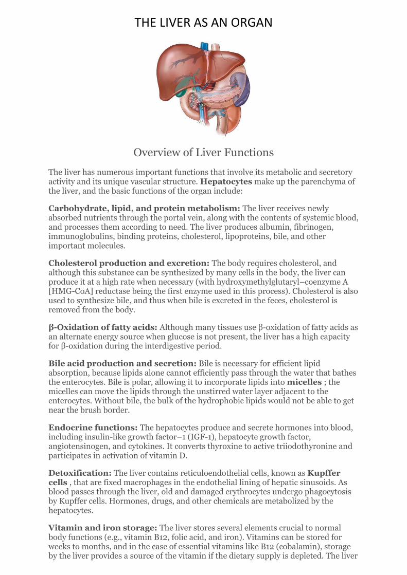
THE LIVER AS AN ORGAN
Overview of Liver Functions
The liver has numerous important functions that involve its metabolic and secretory activity and its unique vascular structure. Hepatocytes make up the parenchyma of the liver, and the basic functions of the organ include:
Carbohydrate, lipid, and protein metabolism: The liver receives newly absorbed nutrients through the portal vein, along with the contents of systemic blood, and processes them according to need. The liver produces albumin, fibrinogen, immunoglobulins, binding proteins, cholesterol, lipoproteins, bile, and other important molecules.
Cholesterol production and excretion: The body requires cholesterol, and although this substance can be synthesized by many cells in the body, the liver can produce it at a high rate when necessary (with hydroxymethylglutaryl–coenzyme A [HMG-CoA] reductase being the first enzyme used in this process). Cholesterol is also used to synthesize bile, and thus when bile is excreted in the feces, cholesterol is removed from the body.
β-Oxidation of fatty acids: Although many tissues use β-oxidation of fatty acids as an alternate energy source when glucose is not present, the liver has a high capacity for β-oxidation during the interdigestive period.
Bile acid production and secretion: Bile is necessary for efficient lipid absorption, because lipids alone cannot efficiently pass through the water that bathes the enterocytes. Bile is polar, allowing it to incorporate lipids into micelles ; the micelles can move the lipids through the unstirred water layer adjacent to the enterocytes. Without bile, the bulk of the hydrophobic lipids would not be able to get near the brush border.
Endocrine functions: The hepatocytes produce and secrete hormones into blood, including insulin-like growth factor–1 (IGF-1), hepatocyte growth factor, angiotensinogen, and cytokines. It converts thyroxine to active triiodothyronine and participates in activation of vitamin D.
Detoxification: The liver contains reticuloendothelial cells, known as Kupffer cells , that are fixed macrophages in the endothelial lining of hepatic sinusoids. As blood passes through the liver, old and damaged erythrocytes undergo phagocytosis by Kupffer cells. Hormones, drugs, and other chemicals are metabolized by the hepatocytes.
Vitamin and iron storage: The liver stores several elements crucial to normal body functions (e.g., vitamin B12, folic acid, and iron). Vitamins can be stored for weeks to months, and in the case of essential vitamins like B12 (cobalamin), storage by the liver provides a source of the vitamin if the dietary supply is depleted. The liver

is also the site of large iron stores, bound to the protein ferritin (the liver contains 25% of the body's iron). When needed, the iron (bound to transferrin) is released into the blood and enters the bone marrow for the production of hemoglobin.
Because it performs this great variety of functions, the liver is a crucial organ for maintaining proper blood glucose levels, excreting waste products, processing proteins, contributing to immune function, and even indirectly regulating blood pressure and fluid homeostasis (through synthesis of plasma proteins. When part of the liver is damaged, it has the ability to regenerate functional hepatocytes, and this compensatory action allows it to maintain adequate metabolic function. In addition, the ability of the liver to achieve a high level of metabolic activity is dependent on the blood flow to and from the liver.
The regenerative capacity of the liver is so great that one donor liver can be used for two liver transplant patients, with the liver sections regaining almost 100% of the original liver size. Because the demand for donor livers is far greater than the supply, since 1989, living donor liver transplantation (using one lobe) has been performed widely. The remnant liver in the donor recovers its original size and function in 4 to 6 weeks, as does the donated lobe in the patient.

Locate your liver:
• Upper right quadrant deep to inferior ribs • Left/right lobes • Gall bladder- thin muscular sac on inferior surface where bile
collects
Dual blood supply to liver:
Where the two blood supplies mix?
Triads: branches of three vessels: hepatic portal vein, hepatic artery, bille drainage ductiles

• Sinusoids—special liver capillaries where blood mixes and liver cells act…by-products leave as bile in caniliculi which merge to form bile ducts
Bilirubin production and excretion
Basic Metabolism of Carbohydrates, Lipids, and Proteins
Carbohydrates
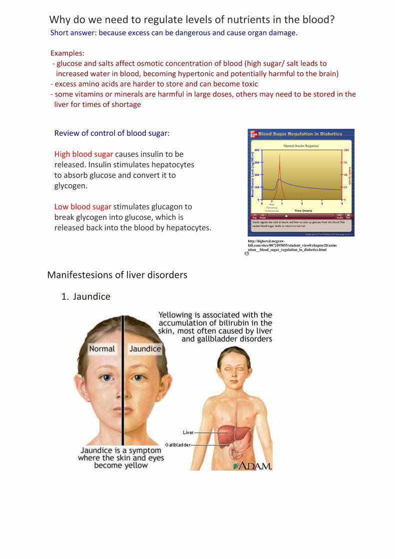
The liver acts as a blood glucose reservoir, storing glucose as glycogen and releasing it when blood levels are low. Carbohydrates are absorbed in the intestine as monosaccharides and are carried in the portal blood to the liver. Most of the glucose passes through the liver rapidly and is released into the systemic blood, where elevated insulin will facilitate its entry into tissues. In the liver, excess monosaccharides are handled by the following processes:
Conversion of other monosaccharides to glucose: Fructose and galactose can be converted into glucose.
Glycogen synthesis and storage: Excess glucose is polymerized and stored as glycogen. Stored hepatic glycogen can provide glucose for 12 to 17 hours during fasting. When blood glucose levels are low, glucagon and other hyperglycemic hormones such as epinephrine and growth hormone stimulate glycolysis to break down glycogen and release glucose into the blood. If the glycogen stores are not used, excess glucose (which is not released into the blood) will eventually be converted to triglycerides (TGs) and transported to adipose tissue for storage (see the “ Lipids ” section)
Gluconeogenesis: The liver (and to a lesser extent, the kidney) has the ability to make glucose from substrates such as glycerol, pyruvate, and the amino acids glutamine and alanine. Gluconeogenesis provides an alternate energy source and occurs primarily during fasting and starvation.
Formation of chemical compounds: Excess glucose can also be converted into other chemical compounds (e.g., pyruvic acid, lactic acid, and acetyl CoA) that can be used in metabolic pathways such as the citric acid cycle.
The ingestion of carbohydrates raises blood glucose levels, but the effects can vary according to the type of carbohydrate and its quantity in the food. The glycemic index of a specific food refers to the degree to which it raises blood glucose in comparison with pure glucose, which has a glycemic index of 100.Glycemic load is a parameter that takes into account the glycemic index of a food and puts it into the context the quantity of food consumed. Thus, glycemic load is the glycemic index of a food times the grams of carbohydrate per serving. Diets that produce high glycemic loads are believed to be associated with development of type II diabetes. Although fructose has a relatively low glycemic index, the addition of high fructose corn syrup, a common ingredient in prepared foods, can greatly increase the glycemic load of food and beverages, as is the case with the addition of other sugars.
Lipids
Most lipids are packaged into chylomicrons in the enterocytes (see Chapter 26 ). The chylomicrons enter the lymph lacteals in the small intestine, ultimately entering the systemic blood in the large vessels in the thoracic cavity. Thus the first entry of absorbed lipid into the liver is from the systemic, not portal, circulation. Liver lipid metabolism includes the following:
β -Oxidation of fatty acids: Although many tissues use β-oxidation as a source of energy when the need arises, the rate of use of β-oxidation is very high in the liver.
Formation of most lipoproteins: Very low density lipoprotein (VLDL), low-density lipoprotein (LDL), and high-density lipoprotein (HDL) are formed in the liver. VLDL and LDL transport TGs and cholesterol to tissues. LDL is implicated in development of cardiovascular disease because it is incorporated into atherosclerotic plaques. HDL transports lipids from tissues to the liver and is considered beneficial in terms of cardiovascular health.

Synthesis of cholesterol and phospholipids: Cholesterol and phospholipids are necessary for making membranes, and cholesterol is also the precursor for steroid hormones and bile. Because of these important functions, the liver ensures a supply of these substrates by forming them from other lipids. A liver enzyme, HMG-CoA reductase, catalyzes the rate-limiting step in cholesterol synthesis, and pharmacologic intervention by statins (cholesterol-lowering drugs) inhibits this enzyme.
Conversion of unused glycogen to TGs: When liver glycogen is not used, it is converted to TGs, which are transported to adipose tissues in VLDLs.
Proteins
Protein metabolism in the liver is essential for survival; the liver processes dietary amino acids for systemic use and participates in the processing of nitrogen wastes for excretion. The major functions of the liver are:
Deamination of amino acids: Deamination is the first step in removal of excess amino acids; aminotransferases remove the amino group from the amino acids, creating ammonia (NH 3 ).
Production of urea: The NH 3 combines with CO 2 to form urea, thereby buffering NH 3 , a toxin, and allowing urinary excretion.
Synthesis of plasma proteins: About 90% of plasma proteins are made in the liver. These proteins include:
albumin, which contributes to oncotic pressure of plasma.
immunoglobulins, which contribute to immune functions.
fibrinogen, which is necessary for blood clotting.
Interconversion of amino acids: Needed amino acids are synthesized from other available amino acids.
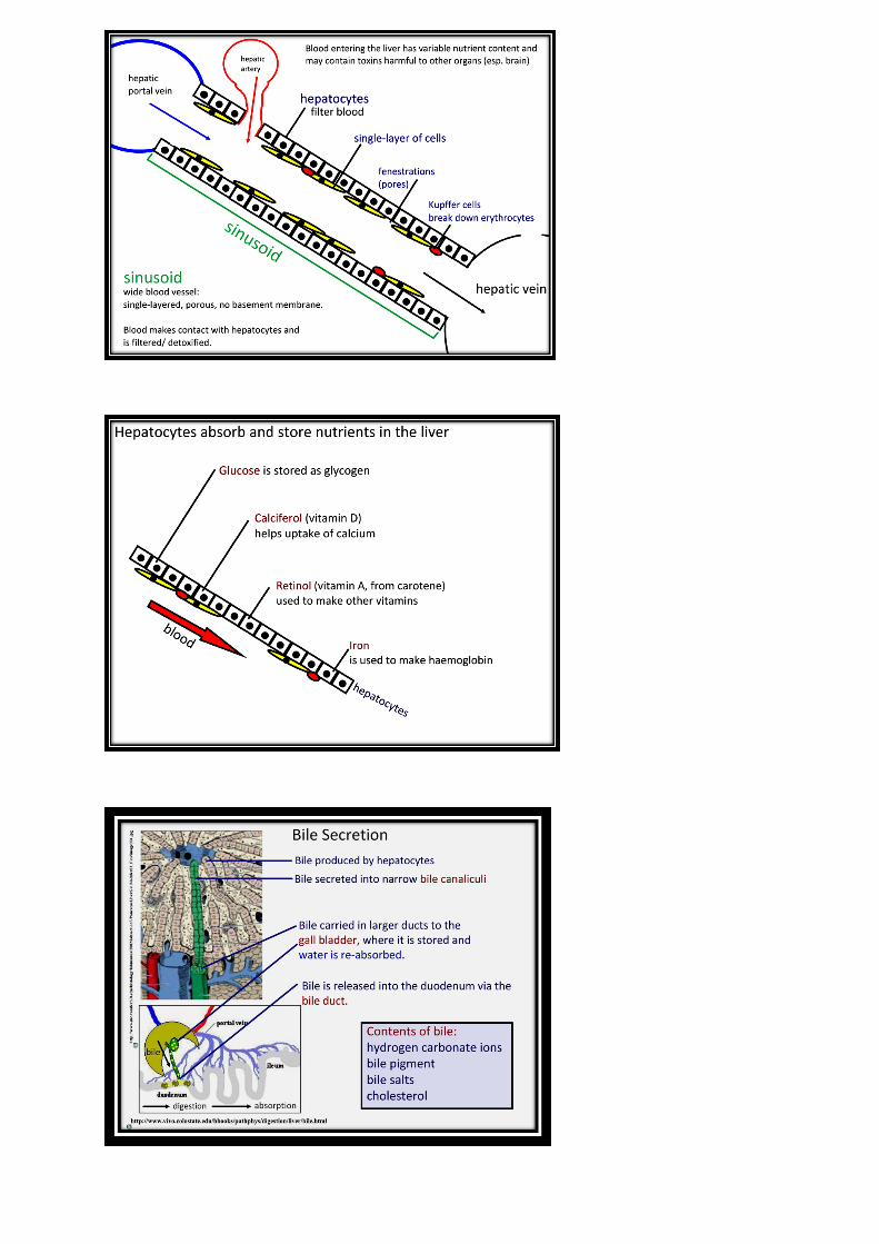
Production and Secretion of Bile
Bile is critical for transport of lipids through the unstirred water layer to the enterocytes. The unstirred water layer is the area in the gut lumen closest to the villous lining. Little flow occurs near the cells, and the water and mucus create a barrier that impedes access of the hydrophobic lipids to the enterocytes (carbohydrates and proteins have no trouble passing through this area). This challenge of transporting hydrophobic lipids is met by bile, which is amphipathic (i.e., it has both hydrophilic and hydrophobic regions) and thus is able to move lipids across the unstirred water. Bile and lipids form micelles , which act like taxis to shuttle the lipids through the unstirred water layer to the enterocytes. Bile solids are composed of bile salts (50%), phospholipids (40%), and smaller amounts of cholesterol (∼4%), bilirubin (∼2%), and water and electrolytes. Secreted bile is composed of the bile solids, water, and electrolytes.
In the liver, primary bile acids (e.g., cholic and chenodeoxycholic acids) are synthesized in the hepatocytes from sterol rings (from cholesterol). Bile synthesis and secretion has several important aspects:
In the liver, one side of the primary bile acid is conjugated with an amino acid (either taurine or glycine), forming a bile salt . This conjugation increases the water solubility of bile in the lower pH found in the duodenum. The primary bile salts are secreted into the bile canaliculi, or ductules, and into the common bile duct.

Bile salts are osmotic, and their secretion will draw water and then solutes (e.g., sodium chloride and HCO3
− ) from the cells; this process is called solvent drag and contributes to the buffering capacity of the bile when it enters the duodenum.
After micelles are formed and the lipids are dropped off at the enterocytes, the majority of the bile remains in the lumen of the small intestine until the terminal ileum, where Na + -dependent transporters recycle the primary bile into the portal vein back to the liver. This bile recycling will occur three to five times for each meal and permits efficient absorption of the lipids without synthesis of large amounts of bile, because it is reused.
However, with each cycle about 10% of the bile is not absorbed but is lost in the feces, which is the major pathway by which cholesterol is removed from the system. Other substances, including bilirubin, are also excreted. The synthesis of bile acids is under feedback control by bile salts that enter the liver from the portal circulation. A reduced level of bile salts will increase cholesterol 7α-hydroxylase , the rate-limiting enzyme in bile synthesis.
In the intestines, some of the primary bile acids undergo dehydroxylation by bacteria, formingsecondary bile acids (deoxycholic and lithocholic acids), which are less efficient at crossing the unstirred water layer and hence are less readily absorbed.
When the stomach is emptied and chyme is no longer present in the duodenum, the sphincter of Oddi closes. As the bile is recycled through the portal system back through the liver, it will stop at the sphincter of Oddi and back up into the relaxed gallbladder, where it will be stored until the next meal.
CLINICAL CORRELATE 25.2
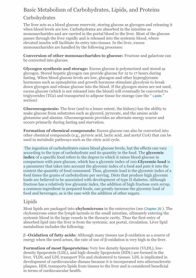
Portal Hypertension and Esophageal Varices in Obstructive Liver Disease Increased blood pressure in the portal vein can result from suprahepatic pathology (e.g., an increase in systemic venous pressures, as with congestive heart failure) or hepatic disease (e.g., obstructive liver disease). Obstructive liver disease is most commonly a result of cirrhosis or fibrous scarring of the liver, which severely decreases the flow of blood through the organ. In the United States, alcoholism and hepatitis C infection are the most common causes of cirrhosis. Interestingly, alcoholism causes cirrhosis of the liver or pancreatitis, but not both diseases, in individual patients.
In the cirrhotic liver, the obstruction of blood flow through the liver increases pressure in the portal system. This portal hypertension results in increased back pressure in vessels coming from the stomach and esophagus, causing enlargement and thinning of these vessel walls, thus forming varices . The thin walls, high pressure, and increased radius in the varices make them susceptible to rupture, and because of the superficial nature of the vessels serving the esophagus, rupture may cause severe bleeding into the esophageal lumen, requiring immediate medical attention. Varices are treated either by sclerotherapy (i.e., injecting a solution to block the vessels) or by rubber-band ligation of the varices, a procedure in which a band is wound around the varices, cutting off blood flow. The atrophied area sloughs off, leaving a healed scar. Portal hypertension can also result in ascites and hemorrhoids , which also are caused by the backup in pressure in the portal venous system.
Basic Endocrine Functions
The liver produces or modifies several endocrine and paracrine substances, including:
IGF-1 , which is released by the liver into the circulation in response to growth hormone. IGF-1 mediates many of the somatic effects of growth hormone.
Angiotensinogen , which is the precursor to angiotensin I and II (angiotensin II plays an important role in fluid and electrolyte homeostasis).
Thrombopoietin , which stimulates stem cells in bone marrow to differentiate to megakaryocytes, which give rise to platelets. Platelets participate in blood clotting.
Hepatocyte growth factor , which acts locally to stimulate regeneration of liver cells and is especially important when the organ is damaged.
Vitamin D metabolism : The liver hydroxylates cholecalciferol (from diet or synthesized in skin) to form 25-hydroxycholecalciferol. This substance is still inactive and must be further hydroxylated in the kidney to become the active form of vitamin D, 1,25-dihydroxycholecaliferol. Vitamin D is a key regulator of intestinal calcium absorption.
Liver manufacturing functions
• Protein- for the bloodstream (albumin) • Glycogen- storage form of glucose for energy
• Bile- to help digest fats that are needed for cell structure and energy)
• Cholesterol – and special proteins to carry fat through the blood
Liver-storage facility
• Glycogen-released when our bodies need energy (this includes during sleep for basic metabolism)
• Iron- most is stored in the liver
Liver-waste disposal • Ammonia - from the breakdown of dietary
protein and muscle tissue
• Bilirubin - from the breakdown of red blood cells
• Bacteria – removed from the bloodstream
• Drugs and Alcohol - are metabolized in the liver
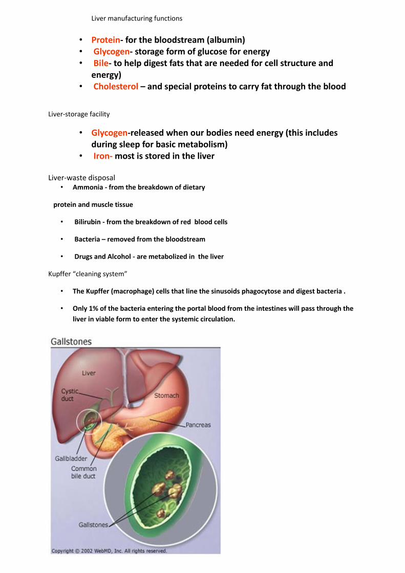
Kupffer “cleaning system”
• The Kupffer (macrophage) cells that line the sinusoids phagocytose and digest bacteria .
• Only 1% of the bacteria entering the portal blood from the intestines will pass through the
liver in viable form to enter the systemic circulation.
Cholesterol
• Our body needs cholesterol for
– Cell membranes
– Vitamin D
– Hormones—progesterone and testosterone
– Myelin (neuron axonal “wrapping”)
– Component of bile salts
• 85% of cholesterol in our blood is “endogenous” or manufactured by our own cells
(mostly liver)
• 15% comes from the food we eat
• So, is zero-cholesterol good…or even healthy?
Cholesterol in the liver:
• Liver constantly manufactures cholesterol using acetyl-CoA as substrate
• Some cholesterol to gut via bile for emulsification of dietary fats
• Some cholesterol to blood for cell membranes, myelin, hormones, vitamins
“good” and “bad” cholesterol
• Two ways cholesterol is “packed”
– LDL—low density lipo-proteins (“bad”)
– HDL—high density lipo-proteins (“good”)
• LDL is component of arterial plaques that can lead to “blocked arteries”
• HDL can help to clear LDL from arterial walls
Manifestesions of liver disorders
1. Jaundice
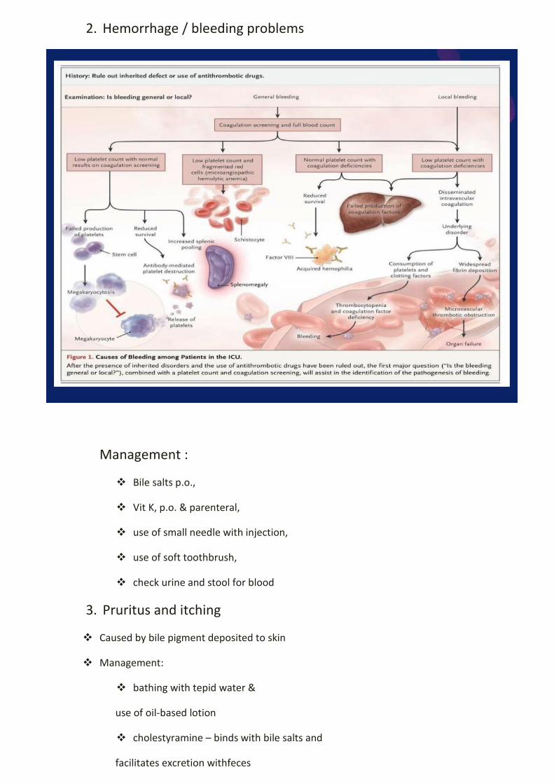
2. Hemorrhage / bleeding problems
Management :
Bile salts p.o.,
Vit K, p.o. & parenteral,
use of small needle with injection,
use of soft toothbrush,
check urine and stool for blood
3. Pruritus and itching
Caused by bile pigment deposited to skin
Management:
bathing with tepid water &
use of oil-based lotion
cholestyramine – binds with bile salts and
facilitates excretion withfeces

Use soft linen
Short fingernails
4. Ascites
Management :
daily weight & abdominal girth
low Na diet, fluid restriction, diuretics
relieve symptoms from pressure of ascites :
high fowler’s
turning & positioning
IV albumin,
Paracentesis
Peritoneovenous Shunt

5. Generalized Edema
- Insufficient albumin
6. Intolerance of Sedation
- Most sedatives are metabolized in the liver except
phenobarbital
Diseases of the liver:
- Cirrhosis
- hepatitis
Viral Hepatitis types A, B, C
Toxic Hepatitis – exposure to hepatotoxin : carbon tetrachloride. Morphine, barbiturates

HEPATIC COMA
DEGENERATIVE DISEASE OF THE BRAIN FROM LIVER FAILURE
DUE TO INABILITY OF THE LIVER TO CONVERT AMMONIA TO UREA
CHANGES IN PERSONALITY AND BEHAVIOR
LETHARGY
CONFUSION
TREMORS
STUPOR
DIZZINESS
COMA
FETOR HEPATICUS – FRUITY ODOR BREATH
SPIDER TELANGIECTASIA
ELEVATED SERUM AMMONIA LEVELS
LIVER FUNCTION TESTS
What for to do them?
1. to detect the presence of liver diseases
2. to distinguish among the different types of liver diseases
3. to follow response to treatment
TESTS BASED ON DETOXIFICATIONS AND EXCRETORY FUNCTIONS OF LIVER:
1. SERUM BILIRUBIN – a breakdown product of porphyrin ring of heme containing proteins
(conjugated,unconjugated)
unconjugated/indirect – insouble in water; bound to albumin in the blood – high in
case of hemolysis, when liver is not able to combine bilirubin
5-7% population – Gilbert’s syndrome – higher bilirubin
conjugated/direct – water soluble; excreted in kidneys – high means bile duct
dysfuction
Normal serum bilirubin concentration: <17 micromol/L (1mg/dl)
Up to 30% of the total is direct-reacting
2. URINE BILIRUBIN:
presence of bilirubinuria liver diseases almost 100% accurate

3. BLOOD AMMONIA: elevated in advanced liver diseases with significant muscle wasting
Hyperammonimea
There is a poor correlation between either the presence or the degree of acute enceph and elevation
of blood ammonia
There is a poor correlation of the blood serum ammonia and hepatic function
Too much ammonia in urea – too much protein ingested
4. SERUM ENZYMES
2 categories of serum enzymes:
Enzymes whose elevation in serum reflects damage to hepatocytes
Enzymes whose elevation in serum reflect cholestasis (when bile is not able to flow form
the liver to duodenum
Enzymes that do not fit precisely into either patterns
ENZYMES THAT REFLECT DAMAGE
TO HEPATOCYTES
AMINOTRANSFERASES - sensitive indicator of liver cell injury and most helpful in recognizing acute
hepatocellular diseases such as hepatitis
AST (Aspartate aminotransferase): liver, cardiac, skeletal, kidneys, brain, pancreas, lungs, leukocytes
and erythrocytes – when high can show disease of this organs – not specific
ALT (Alanine aminotransferase): found primarily in the liver
Very high AST I ALT – hepatitis
Mild high AST I ALT – fatty liver
AST high, ALT ok, ALP ok – no liver disease
AST:ALT = 2:1 – dysfunction of lever because of alkohol
ENZYMES THAT REFLECT CHOLESTASIS
• Alkaline phosphatase and 5’ nuleotidase: found near the bile canalicular membrane of
hepatocytes
• Gamma Glutamyl Transpeptidase: Endoplasmic reticulum and in bile duct epithelial cells
lacks specificity
TESTS THAT MEASURE BIOSYNTHETIC FUNCTION OF THE LIVER
SERUM ALBUMIN: synthesized exclusively by hepatocytes* has a slow turn over not a good indicator
of acute or mild hepatic dysfunction. When low – infection, inflamatory state
• SERUM GLOBULINS: alpha and beta globulins produced primarily in hepatocytes
gamma globulins (immunoglobulins) produced by B lymphocytes.
When low – dysfuction of protein absorption, liver damage
COAGULATION FACTORS:
With the exception of Factor VIII, the blood clotting factors are made exclusively in
hepatocytes.
Because of their rapid turnover, measurement of the clotting factors is the single best acute measure
of hepatic synthetic function and helpful in both the diagnosis and assessing the prognosis of acute
parenchymal liver disease.