Embed Size (px)
Citation preview

The life of the peroxisome: from birth to deathAlison Baker and Rupesh Paudyal
Available online at www.sciencedirect.com
ScienceDirect
Peroxisomes are dynamic and metabolically plastic organelles.
Their multiplicity of functions impacts on many aspects of plant
development and survival. New functions for plant
peroxisomes such as in the synthesis of biotin, ubiquinone and
phylloquinone are being uncovered and their role in generating
reactive oxygen species (ROS) and reactive nitrogen species
(RNS) as signalling hubs in defence and development is
becoming appreciated. Understanding of the biogenesis of
peroxisomes, mechanisms of import and turnover of their
protein complement, and the wholesale destruction of the
organelle by specific autophagic processes is giving new
insight into the ways that plants can adjust peroxisome function
in response to changing needs.
Addresses
Centre for Plant Sciences, School of Biology, University of Leeds, Leeds
LS2 9JT, UK
Corresponding author: Baker, Alison ([email protected])
Current Opinion in Plant Biology 2014, 22:39–47
This review comes from a themed issue on Cell biology
Edited by Shaul Yalovsky and Viktor Zarsky
http://dx.doi.org/10.1016/j.pbi.2014.09.003
1369-5266/# 2014 Published by Elsevier Ltd.
IntroductionPlant peroxisomes possess astonishing metabolic versati-
lity that encompasses degradation of storage oil, recycling
of glycolate produced by photorespiration, pro-hormone
metabolism, redox signalling and a range of biosynthetic
capabilities. The biogenesis and the functions of plant
peroxisomes are the subject of a recent comprehensive
review to which the reader is referred [1]. In this article
we highlight recent advances in the cell biology of plant
peroxisomes in particular that update and extend the
conclusions of the earlier article.
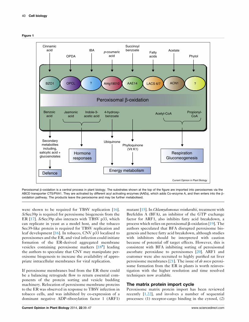
Update on peroxisome functionsBeta-oxidation of a wide range of substrates is one of the
hallmarks of plant peroxisomes [1,2]. This central path-
way integrates peroxisomes into the cellular signalling
network and energy metabolism pathways. Diverse sub-
strates for b-oxidation are imported via the PEROXI-
SOME ABC TRANSPORTER 1/COMATOSE (PXA1/
CTS) which can accept CoA esters and may cleave them
www.sciencedirect.com
upon transport [3��] followed by reactivation by a perox-
isomal acyl-CoA synthetase with the appropriate sub-
strate specificity (Figure 1). The CTS/PXA1
interacting protein COMPARATIVE GENE IDENTI-
FICATION-58 (CGI-58) may regulate CTS/PXA1
activity in non-lipid storing tissues [4]. Peroxisomal b-
oxidation participates in the synthesis of benzoic acid,
which acts as a precursor to various secondary metab-
olites, (see [5�]) and 4-hydroxybenzoic acid, an intermedi-
ate in one of two independent pathways for ubiquinone
synthesis [6�] (Figure 1). Peroxisomes also play a role in
synthesis of polyamines, biotin and isoprenoid com-
pounds [2]. Because b-oxidation is so central, mutants
often have pleotropic effects. A small molecule inhibitor
that blocks oil body breakdown but did not impact on pro-
auxin metabolism [7] may be a useful tool separating out
effects on fatty acid degradation from metabolism of other
b-oxidation substrates.
Peroxisomes are one of the most significant generators of
ROS in cells and their role as both signalling hubs and
scavengers of ROS under normal and stress situations in
plants and animals is increasingly appreciated [8,9]. Per-
oxisomes also produce RNS [10] and nitric oxide (NO)
[11]. Superoxide and peroxynitrite were also shown to be
produced endogenously by Arabidopsis peroxisomes and
their production was increased under conditions of Cd2+
stress [12]. NO can be produced in several cell compart-
ments, and peroxisomal Xanthine oxidoreductase can
reduce nitrite to NO [11]. Production of NO leads to
S-nitrosylation of some peroxisome proteins including
those involved in b-oxidation, photorespiration and
ROS metabolism [13].
Birth of peroxisomesPeroxisomes are classed as semi-autonomous organelles
[1], as they are capable of forming de novo from the
endoplasmic reticulum (ER) and they also replicate by
growth and division (Figure 2). De novo peroxisome
biogenesis has been studied in yeast and mammals but
evidence from plants is more limited and less direct,
although several plant peroxisomal membrane proteins
(PMPs) have been reported to traffic via the ER [1,14].
Viruses are extremely powerful tools to study cell biology
and are providing insight into possible de novo routes of
peroxisome formation in plants. Tombus viruses such as
Cucmber Necrosis Virus (CNV) and Tomato Bushy Stunt Virus(TBSV) are known to highjack peroxisomes for replica-
tion through the action of replication protein p33 [15].
Recently, the ER-localised vesicle transport protein
Sec39p in S. cerevisiae and Sec39-like protein in tobacco
Current Opinion in Plant Biology 2014, 22:39–47
40 Cell biology
Figure 1
Cinnamicacid
Benzoicacid
Secondarymetabolitesincluding,
salicylic acid +glucosinolates
Jasmonicacid
Indole-3-acetic acid
4-hydroxy-benzoate
UbiquinonePhylloquinone
(Vit K1)Hormoneresponses
DefenceEnergy metabolism
RespirationGluconeogenesis
Acetyl-CoA Propionyl-CoA
OPDA
IBA p-coumaricacid
Succinoylbenzoate
Fattyacids
Acetate
Phytol
?ACN1LACS 6/7AAE14At4g19010?OPCLBZO1
Peroxisomal β-oxidation
Current Opinion in Plant Biology
Peroxisomal b-oxidation is a central process in plant biology. The substrates shown at the top of the figure are imported into peroxisomes via the
ABCD transporter CTS/PXA1. They are activated by different acyl activating enzymes (AAEs), which adds Co-enzyme A, and then enters into the b-
oxidation pathway. The products leave the peroxisome and may be further metabolised.
were shown to be required for TBSV replication [16].
ScSec39p is required for peroxisome biogenesis from the
ER [17]. ScSec39p also interacts with TBSV p33, which
can replicate in yeast as a model host, and the tobacco
Sec39-like protein is required for TBSV replication and
leaf development [16]. In tobacco, CNV p33 localised to
peroxisomes and the ER, and viral infection could initiate
formation of the ER-derived aggregated membrane
vesicles containing peroxisome markers [18�] leading
the authors to speculate that CNV may manipulate per-
oxisome biogenesis to increase the availability of appro-
priate intracellular membranes for viral replication.
If peroxisome membranes bud from the ER there could
be a balancing retrograde flow to return essential com-
ponents of the protein sorting and vesicle budding
machinery. Relocation of peroxisome membrane proteins
to the ER was observed in response to TBSV infection in
tobacco cells, and was inhibited by co-expression of a
dominant negative ADP-ribosylation factor 1 (ARF1)
Current Opinion in Plant Biology 2014, 22:39–47
mutant [15]. In Chlamydomonas reinhardtii, treatment with
Brefeldin A (BFA), an inhibitor of the GTP exchange
factor for ARF1, also inhibits fatty acid breakdown, a
process which relies on peroxisomal b-oxidation [19]. The
authors speculated that BFA disrupted peroxisome bio-
genesis and hence fatty acid breakdown, although studies
with inhibitors should be interpreted with caution
because of potential off target effects. However, this is
consistent with BFA inhibiting sorting of peroxisomal
ascorbate peroxidase to peroxisomes [20]. ARF1 and
coatomer were also recruited to highly purified rat liver
peroxisome membranes [21]. The issue of de novo peroxi-
some formation from the ER in plants is worth reinves-
tigation with the higher resolution and time resolved
techniques now available.
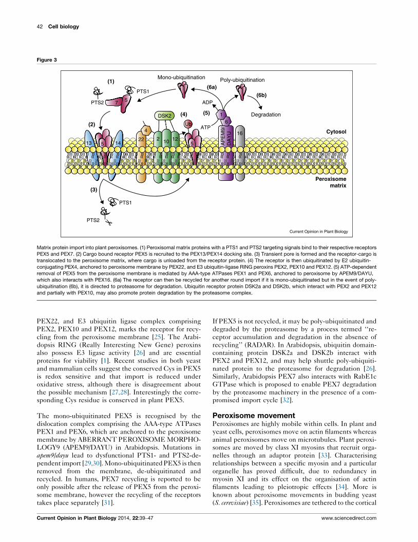
The matrix protein import cyclePeroxisome matrix protein import has been reviewed
recently [1,22], and involves a number of sequential
processes: (1) receptor-cargo binding in the cytosol, (2)
www.sciencedirect.com

The birth and death of peroxisomes Baker and Paudyal 41
Figure 2
Fusion
Growth
Macropexophagy
H2O2 stress
Micropexophagy
Vacuole
Autophagosome
Protein import:- PTS1- PTS2
Division:
Peroxisome biosynthesisand division
Vacuolar proteintrafficking
Pexophagy
Autophagosome
pre-vacuolarcompartment
ATG8
Damagedperoxisome
Pre-peroxisomevesicle
Peroxisome
de novo synthesisedperoxisome
trans-Golginetwork
Golgi
NucleusEndoplasmicreticulum
- Elongation- Constriction- Fission
Current Opinion in Plant Biology
Protein trafficking, peroxisome biogenesis and pexophagy. Proteins are transported from the ER en route to Golgi, trans-Golgi network (TGN) and the
pre-vacuolar compartment (PVC) before reaching the vacuole. Vesicles termed as pre-peroxisome vesicles also bud off from the ER that may contain
peroxisomal membrane proteins. The fusion of these pre-peroxisomal vesicles causes the formation of de novo synthesised peroxisomes. Extra
membrane materials and phospholipids are also obtained from the ER for the growth of peroxisome. Peroxisomal matrix proteins with peroxisome
targeting signal (PTS) 1 and PTS2 are imported into the peroxisome to generate a fully functional mature peroxisome. These peroxisomes undergo
division, which involves elongation, constriction and finally fission. Obsolete, damaged or dysfunctional peroxisomes can be degraded by autophagic
process termed pexophagy. Autophagy-related protein 8 is found in the PAS and is generally used as an autophagosome marker. In macro-
pexophagy, a double membrane structure engulfs the dysfunctional peroxisome to generate a compartment known as autophagosome, which fuses
with the vacuole releasing engulfed peroxisomes to be degraded by the vacuolar proteases. Alternatively, peroxisomes can also be surrounded by
vacuolar membrane and subsequently degraded in the vacuole lumen.
docking of the receptor-cargo at the peroxisome mem-
brane docking complex, (3) translocation into the peroxi-
some and cargo release into the peroxisome matrix, (4)
mono-/poly-ubiquitination of the receptor, (5) receptor
release from the membrane, and (6) effective recycling/
degradation of the receptor (Figure 3). Proteins destined
for import into the peroxisome matrix have one of two
peroxisome targeting signal (PTS) sequences, PTS1 or
PTS2, within their primary sequence. These proteins
once synthesised and folded are recognised by their
respective receptors PEX5 and PEX7 in the cytosol
www.sciencedirect.com
[23]. Receptor-bound PTS-containing proteins are
recruited to the docking complex (PEX13/PEX14) at
the peroxisome membrane, and a transient pore is formed
which allows translocation of receptor and cargo into the
peroxisome.
Subsequently, the cargo is unloaded from the receptor
and PEX14 may play a role in unloading PTS1- [24] or
PTS2-cargoes [23]. Mono-ubiquitination of PEX5 on a
conserved cysteine by the E2 ubiquitin-conjugating
PEX4, anchored at the peroxisome membrane by
Current Opinion in Plant Biology 2014, 22:39–47
42 Cell biology
Figure 3
(1)
(2)
(3)
(4) (5)
(6a)(6b)
Cytosol
Degradation
Poly-ubiquitinationMono-ubiquitination
PTS2
PTS2
PTS1
PTS1
ADP
ATP
DSK2
57
13 5 1422
4
2 10 12
Ub
5
61
16
AP
EM
9/D
AY
U
Peroxisomematrix
Current Opinion in Plant Biology
Matrix protein import into plant peroxisomes. (1) Peroxisomal matrix proteins with a PTS1 and PTS2 targeting signals bind to their respective receptors
PEX5 and PEX7. (2) Cargo bound receptor PEX5 is recruited to the PEX13/PEX14 docking site. (3) Transient pore is formed and the receptor-cargo is
translocated to the peroxisome matrix, where cargo is unloaded from the receptor protein. (4) The receptor is then ubiquitinated by E2 ubiquitin-
conjugating PEX4, anchored to peroxisome membrane by PEX22, and E3 ubiquitin-ligase RING peroxins PEX2, PEX10 and PEX12. (5) ATP-dependent
removal of PEX5 from the peroxisome membrane is mediated by AAA-type ATPases PEX1 and PEX6, anchored to peroxisome by APEM9/DAYU,
which also interacts with PEX16. (6a) The receptor can then be recycled for another round import if it is mono-ubiquitinated but in the event of poly-
ubiquitination (6b), it is directed to proteasome for degradation. Ubiquitin receptor protein DSK2a and DSK2b, which interact with PEX2 and PEX12
and partially with PEX10, may also promote protein degradation by the proteasome complex.
PEX22, and E3 ubiquitin ligase complex comprising
PEX2, PEX10 and PEX12, marks the receptor for recy-
cling from the peroxisome membrane [25]. The Arabi-
dopsis RING (Really Interesting New Gene) peroxins
also possess E3 ligase activity [26] and are essential
proteins for viability [1]. Recent studies in both yeast
and mammalian cells suggest the conserved Cys in PEX5
is redox sensitive and that import is reduced under
oxidative stress, although there is disagreement about
the possible mechanism [27,28]. Interestingly the corre-
sponding Cys residue is conserved in plant PEX5.
The mono-ubiquitinated PEX5 is recognised by the
dislocation complex comprising the AAA-type ATPases
PEX1 and PEX6, which are anchored to the peroxisome
membrane by ABERRANT PEROXISOME MORPHO-
LOGY9 (APEM9/DAYU) in Arabidopsis. Mutations in
apem9/dayu lead to dysfunctional PTS1- and PTS2-de-
pendent import [29,30]. Mono-ubiquitinated PEX5 is then
removed from the membrane, de-ubiquitinated and
recycled. In humans, PEX7 recycling is reported to be
only possible after the release of PEX5 from the peroxi-
some membrane, however the recycling of the receptors
takes place separately [31].
Current Opinion in Plant Biology 2014, 22:39–47
If PEX5 is not recycled, it may be poly-ubiquitinated and
degraded by the proteasome by a process termed ‘‘re-
ceptor accumulation and degradation in the absence of
recycling’’ (RADAR). In Arabidopsis, ubiquitin domain-
containing protein DSK2a and DSK2b interact with
PEX2 and PEX12, and may help shuttle poly-ubiquiti-
nated protein to the proteasome for degradation [26].
Similarly, Arabidopsis PEX7 also interacts with RabE1c
GTPase which is proposed to enable PEX7 degradation
by the proteasome machinery in the presence of a com-
promised import cycle [32].
Peroxisome movementPeroxisomes are highly mobile within cells. In plant and
yeast cells, peroxisomes move on actin filaments whereas
animal peroxisomes move on microtubules. Plant peroxi-
somes are moved by class XI myosins that recruit orga-
nelles through an adaptor protein [33]. Characterising
relationships between a specific myosin and a particular
organelle has proved difficult, due to redundancy in
myosin XI and its effect on the organisation of actin
filaments leading to pleiotropic effects [34]. More is
known about peroxisome movements in budding yeast
(S. cerevisiae) [35]. Peroxisomes are tethered to the cortical
www.sciencedirect.com

The birth and death of peroxisomes Baker and Paudyal 43
ER in mother cells by Inp1p [36]. Inp2p acts as an adaptor
that mediates peroxisome recruitment by the myosin
Myo2 for transport to the daughter cells, where regulated
degradation of Inp2p causes release of peroxisome [35].
Myo2 also transports vacuoles through binding the
vacuole specific adaptor Vac17, which is degraded follow-
ing ubiquitination mediated by a novel E3-ubiquitin
ligase Dma1 and its paralogue Dma2, to release the cargo.
Dma1 and Dma2 are also required for peroxisome move-
ments in yeast [37]. GTP hydrolysis by associated Rabs is
also proposed as a mechanism for release of exocytic
vesicles from myosin V in yeast [38], so it remains to
be seen if either or both mechanisms function in plants to
release peroxisomes from their molecular motors. Plant
peroxisomes are observed to cluster around fungal
penetration sites and are linked to the plant immune
response [39,40]. Clearly, understanding mechanisms of
peroxisomal movement and positioning will be important
to fully elucidate host–pathogen responses.
Death of peroxisomes; protein and organelleturnoverCellular homeostasis requires mechanisms for the syn-
thesis and turnover of individual macromolecules as well
as organelles. In the case of peroxisomes, changes in
biochemical and physiological function can take place
by overlapping and complementary mechanisms;
peroxisome associated protein degradation (PexAD)
and by a specific autophagic process termed ‘pexophagy’
(Figure 4).
Peroxisomes contain a number of different proteases. A
cysteine protease, Resistant to drought 21A-like 1
(RDL1) was identified in peroxisomes isolated from
etiolated Arabidopsis seedlings and rdl1 mutants have
mild peroxisome deficiency phenotypes and reduced
seed viability, but RDL1 substrates are unknown [41].
Greening of etiolated seedlings is accomplished by
turnover of the glyoxylate cycle enzymes isocitrate
lyase (ICL) and malate synthase (MLS), and they are
stabilised in mutants of pex4 and pex6, components
required for PEX5 recycling (‘The matrix protein
import cycle’ section). A genetic screen for mutants
showing persistent GFP-ICL fluorescence ( pfl mutants)
identified mutants in pex14, and a novel allele of pex6suggesting that matrix proteins must be imported in
order to be degraded and are most likely ubiquitinated
and re-exported in a process paralleling ER associated
degradation (ERAD) [42�]. Interestingly ICL has been
identified as an ubiquitinated protein in a recent pro-
teomic study [43].
Peroxisomal protein degradation or protein export for
degradation by the proteasome facilitates peroxisome
remodelling in response to environmental or develop-
mental cues (Figure 4). Peroxisomes in mammalian and
fungal cells can be degraded by a selective autophagic
www.sciencedirect.com
process termed pexophagy [44]. Autophagy is a process of
bulk recycling of cellular components, often under con-
ditions of nutrient limitation. Cytosol and organelles
become sequestered in autophagosomes, double mem-
brane compartments, which originate from a phagophore
assembly site (PAS) followed by fusion with and degra-
dation in the vacuole. This process has been shown to
occur in plants (Figure 2) [45,46]. Very recently four
studies [47��,48,49��,50] have shown selective autophagy
of peroxisomes also occurs in plants and intriguingly
LON2, a peroxisomal protease, plays a role in both
pexophagy and PexAD [47��,51].
Larger but fewer peroxisomes are observed in lon2mutants which exhibit an age-dependent defect in
PTS2-protein import and peroxisome metabolism [52].
A screen for suppressors of the lon2 mutation identified
multiple alleles of AUTOPHAGY RELATED (ATG) genes
which encode components of the autophagic pathway
[47��]. This study identified atg2, atg3 and atg7 as sup-
pressors of lon2 defects in protein import and of indole
butyric acid (IBA) response, which is indicative of per-
turbed b-oxidation. Double mutants of lon2 atg showed
stabilisation of ICL and MLS, but a single mutant did not
show this phenotype. These results suggest that PexAD,
mediated by LON2, and autophagy can function together
to remove obsolete proteins. ICL and MLS stabilisation
and increased peroxisome number was also seen in hypo-
cotyls of atg7-2 and atg5-1 mutants [48�]. Light triggered
ATG7-dependent pexophagy, and the blocking of
vacuolar degradation allowed visualisation of bodies
labelled with peroxisomal-targeted CFP within the
vacuole in wild-type but not in the atg7-2 mutant [48�].ATG8a which marks the pre-autophagosomal structure,
partially overlapped with a peroxisomal marker. Similar
results were reported for peroxisomes in the leaves of atg2mutants [50].
A screen for peroxisomal unusual positioning ( peup)
showed that peup1 (atg2), peup2 (atg18a) and peup4(atg7) mutants displayed aggregated peroxisomes. Sim-
ilarly, peroxisome aggregates were also induced by hydro-
gen peroxide treatment in wild-type plants and in the
catalase (cat2) mutant suggesting that oxidatively
damaged peroxisomes are cleared by pexophagy [49��].Similar results to those of [47��] were reported for the
apem10 mutant (an allele of lon2) which was supressed by
peup1 [51]. Interestingly a mutant in the AAA domain of
LON2 gave a phenotype like the apem10 peup1 double
mutant leading the authors to propose that the LON2
AAA domain has a role in suppressing autophagy [51].
Collectively these studies show that plant peroxisomes,
like their yeast and mammalian counterparts, can be
turned over by pexophagy but that an alternative inde-
pendent pathway exists for removal of peroxisomal
matrix proteins and LON2 functions in both pathways
(Figure 4).
Current Opinion in Plant Biology 2014, 22:39–47

44 Cell biology
Figure 4
(a)REM
REM
ATG2
ATG18a
ATG8
ATG8
ATG7
ATG8-ATG3
ATG5
+PE?
CAT
REM
DTM DTM
EnvironmentalMetabolic
DevelopmentalSignals
DTM
LON2/APEM10
LON2/APEM10
LON2/APEM10
LON2/APEM10
(b)
Current Opinion in Plant Biology
Model for Peroxisome remodelling and turnover. (A) On the left hand side PEX5 (brown square) imports cargo proteins (grey hexagon) via the docking
and translocation machinery (DTM) and is recycled by the receptor export module (REM). LON2 and other peroxisomal proteases may turnover
proteins within peroxisomes or they may be exported for cytosolic degradation analogous to ER associated degradation (ERAD) using components of
the REM such as PEX4 and PEX6. Signals arising from environmental, developmental or metabolic cues may promote turnover of obsolete proteins,
while the import of new proteins with different functions (orange hexagon) allows remodelling of peroxisome capabilities such as in the transition from
heterotrophy to photoautotrophy. (B) Proteins are imported as in A, and may undergo turnover by PexAD (not shown). However when peroxisomes age
or become otherwise damaged, (represented here by catalase aggregation, although catalase aggregation is not necessary or sufficient since
pexophagy is induced in the cat2 mutant) they can be destroyed by pexophagy. Autophagy genes shown to be involved in pexophagy in plants are
shown. ATG7 (PEUP4) acts as an E1 ligase for activation of ATG8, an ubiquitin-like protein. ATG3 is the corresponding E2 and ATG5 is needed to
conjugate phosphatidylethanolamine (PE) to ATG8. ATG2 (PEUP1) and ATG18a (PEUP2) are thought to be involved in the recruitment of membrane to
the PAS. LON2 is required for suppression of pexophagy via its AAA domain although the mechanism is not known.
Recruitment to pexophagy requires a specific receptor (as
yet unknown in plants) which interacts with ATG8 via an
ATG8 interacting motif (AIM) containing the consensus
amino acid sequence (W/Y/F)XX(L/V) [45]. In mammals,
PEX14 is bound by the ATG8 homologue LC3-II and
this binding is competitive with that of PEX5 [53]. PEX5
binds to PEX14 via closely related WX3F/Y repeats and
the LVXEF amino acid sequence in the N-terminus of
PEX5 [54]. In plants Arabidopsis PEX5 and PEX14 bind
one another in a similar manner [23] and it may therefore
Current Opinion in Plant Biology 2014, 22:39–47
be significant that alleles of AtPEX14 were uncovered in
the screen for pfl mutants [42�]. Variation between species
is also observed. In Pichia pastoris, the pexophagy receptor
Atg30 interacts with PEX3 and PEX14 on the peroxiso-
mal membrane and the autophagy machinery on the PAS
[44]. The P. pastoris integral PMP, Atg37, an acyl-CoA
binding protein, also binds Pex3p and is recruited by
Atg30 to the pexophagic receptor complex and binding to
Atg30 and Acyl-CoA is competitive. The human Atg37
homologue, Abcd5, is also required for pexophagy [55].
www.sciencedirect.com
The birth and death of peroxisomes Baker and Paudyal 45
Meanwhile in S. cerevisiae, Atg36 acts as an adapter
between PEX3, ATG8 and ATG11 on the pre-autopha-
gosomal structure, and ScPex14p is dispensable for pex-
ophagy [56]. In mammalian cells ubiquitinated PMPs
recognised by p62 or its homologue NBR1 (neighbour
of BRCA1) trigger pexophagy [44,57]. However, ubiqui-
tinated PMPs were not detected in Arabidopsis atg2 [50]
mutants leading to the suggestion that oxidatively
damaged membrane components could be a signal for
pexophagy in plants [49��,50].
Conclusions and future perspectivesPeroxisome protein import and export are linked via the
process of ubiquitination and proteolysis. This allows
restocking of existing organelles with new activities as
proposed by the original two population hypothesis [58].
At the same time the capacity to synthesise and degrade
whole peroxisomes fits to the one population hypothesis
[58]. This flexibility may allow peroxisomes and their
functions to adjust rapidly in response to both internal and
external perturbations, which is likely to be of importance
in surviving in a fluctuating environment. Recent
research emphasises the central role of peroxisomes as
generators of redox signalling molecules and also as the
targets of oxidative damage and turnover. Future research
will identify the pathways and missing components in this
process. New functions are still being discovered which
integrate peroxisomes into virtually all aspects of plant
biology, providing potential new targets for crop improve-
ment.
AcknowledgementsWork in the authors’ laboratory is funded by grants BB/L001012/1 from theBBSRC and RPG-2012-516 from the Leverhulme Trust.
References and recommended readingPapers of particular interest, published within the period of review,have been highlighted as:
� of special interest�� of outstanding interest
1. Hu J, Baker A, Bartel B, Linka N, Mullen RT, Reumann S,Zolman BK: Plant peroxisomes: biogenesis and function. PlantCell 2012, 24:2279-2303.
2. Linka N, Theodoulou FL: Metabolite transporters of the plantperoxisomal membrane: known and unknown. SubcellBiochem 2013, 69:169-194.
3.��
De Marcos Lousa C, van Roermund CW, Postis VL, Dietrich D,Kerr ID, Wanders RJ, Baldwin SA, Baker A, Theodoulou FL:Intrinsic acyl-CoA thioesterase activity of a peroxisomal ATPbinding cassette transporter is required for transport andmetabolism of fatty acids. Proc Natl Acad Sci U S A 2013,110:1279-1284.
Provides evidence that the peroxisomal ABC transporter responsible forimport of diverse substrates posseses an acyl-CoA esterase activity,thereby resolving the paradox that the transporter has a preference forCoA ester substrates but peroxisomal acyl CoA synthetases are essentialfor further metabolism.
4. Park S, Gidda SK, James CN, Horn PJ, Khuu N, Seay DC,Keereetaweep J, Chapman KD, Mullen RT, Dyer JM: The alpha/beta hydrolase CGI-58 and peroxisomal transport proteinPXA1 coregulate lipid homeostasis and signaling inArabidopsis. Plant Cell 2013, 25:1726-1739.
www.sciencedirect.com
5.�
Bussell JD, Reichelt M, Wiszniewski AA, Gershenzon J, Smith SM:Peroxisomal ATP-binding cassette transporter COMATOSEand the multifunctional protein abnormal INFLORESCENCEMERISTEM are required for the production of benzoylatedmetabolites in arabidopsis seeds. Plant Physiol 2014,164:48-54.
Through measurement of metabolite levels in a range of mutant plants therole peroxisomes in the production of benozoic acid a key intermediate inthe production of secondary metabolites involved in plant defence waselucidated.
6.�
Block A, Widhalm JR, Fatihi A, Cahoon RE, Wamboldt Y,Elowsky C, Mackenzie SA, Cahoon EB, Chapple C, Dudareva Net al.: The origin and biosynthesis of the benzenoid moiety ofubiquinone (Coenzyme Q) in Arabidopsis. Plant Cell 2014.
Using network modelling and gene knockouts two pathways for ubiqui-none synthesis in arabidopsis were delineated. The peroxisomal ABCtransporter PXA1/CTS and a peroxisomal p-coumarate CoA ligase arerequired for the phenyalanine derived branch of the ubiquinone biosynth-esis pathway, identifying yet another metabolic role for peroxisomes.
7. Brown LA, Larson TR, Graham IA, Hawes C, Paudyal R,Warriner SL, Baker A: An inhibitor of oil body mobilization inArabidopsis. New Phytol 2013, 200:641-649.
8. Mhamdi A, Noctor G, Baker A: Plant catalases: peroxisomalredox guardians. Arch Biochem Biophys 2012, 525:181-194.
9. Nordgren M, Fransen M: Peroxisomal metabolism and oxidativestress. Biochimie 2014, 98:56-62.
10. Del Rio LA: Peroxisomes as a cellular source of reactivenitrogen species signal molecules. Arch Biochem Biophys 2011,506:1-11.
11. Yu M, Lamattina L, Spoel SH, Loake GJ: Nitric oxide function inplant biology: a redox cue in deconvolution. New Phytol 2014,202:1142-1156.
12. Corpas FJ, Barroso JB: Peroxynitrite (ONOO-) is endogenouslyproduced in arabidopsis peroxisomes and is overproducedunder cadmium stress. Ann Bot 2014, 113:87-96.
13. Ortega-Galisteo AP, Rodriguez-Serrano M, Pazmino DM,Gupta DK, Sandalio LM, Romero-Puertas MC: S-Nitrosylatedproteins in pea (Pisum sativum L.) leaf peroxisomes: changesunder abiotic stress. J Exp Bot 2012, 63:2089-2103.
14. Theodoulou FL, Bernhardt K, Linka N, Baker A: Peroxisomemembrane proteins: multiple trafficking routes and multiplefunctions? Biochem J 2013, 451:345-352.
15. McCartney AW, Greenwood JS, Fabian MR, White KA, Mullen RT:Localization of the tomato bushy stunt virus replicationprotein p33 reveals a peroxisome-to-endoplasmic reticulumsorting pathway. Plant Cell 2005, 17:3513-3531.
16. Sasvari Z, Gonzalez PA, Rachubinski RA, Nagy PD: Tombusvirusreplication depends on Sec39p endoplasmic reticulum-associated transport protein. Virology 2013, 447:21-31.
17. Perry RJ, Mast FD, Rachubinski RA: Endoplasmic reticulum-associated secretory proteins Sec20p, Sec39p, and Dsl1p areinvolved in peroxisome biogenesis. Eukaryot Cell 2009,8:830-843.
18.�
Rochon D, Singh B, Reade R, Theilmann J, Ghoshal K, Alam SB,Maghodia A: The p33 auxiliary replicase protein of Cucumbernecrosis virus targets peroxisomes and infection induces denovo peroxisome formation from the endoplasmic reticulum.Virology 2014, 452–453:133-142.
CNV replication protein p33 was shown to be targeted to the peroxisomesin tobacco. This study also reports that upon p33 infection, dramatic re-modelling of the ER is observed, which results in formation of compart-ments similar to peroxisomes that co-localises with proteins containingPTS1 signal. CNV p33 was reported to be targeted to the inducedperoxisome-like structures.
19. Kato N, Dong T, Bailey M, Lum T, Ingram D: Triacylglycerolmobilization is suppressed by brefeldin A in Chlamydomonasreinhardtii. Plant Cell Physiol 2013, 54:1585-1599.
20. Mullen RT, Lisenbee CS, Miernyk JA, Trelease RN: Peroxisomalmembrane ascorbate peroxidase is sorted to a membranousnetwork that resembles a subdomain of the endoplasmicreticulum. Plant Cell 1999, 11:2167-2185.
Current Opinion in Plant Biology 2014, 22:39–47
46 Cell biology
21. Lay DB, Heid LG, Gorgas H, Just KWW: Binding and functions ofADP-ribosylation factor on mammalian and yeastperoxisomes. J Biol Chem 2005, 280:34489-34499.
22. Smith JJ, Aitchison JD: Peroxisomes take shape. Nat Rev MolCell Biol 2013, 14:803-817.
23. Lanyon-Hogg T, Hooper J, Gunn S, Warriner SL, Baker A: PEX14binding to Arabidopsis PEX5 has differential effects on PTS1and PTS2 cargo occupancy of the receptor. FEBS Lett 2014,588:2223-2229.
24. Freitas MO, Francisco T, Rodrigues TA, Alencastre IS, Pinto MP,Grou CP, Carvalho AF, Fransen M, Sa-Miranda C, Azevedo JE:PEX5 protein binds monomeric catalase blocking itstetramerization and releases it upon binding the N-terminaldomain of PEX14. J Biol Chem 2011, 286:40509-40519.
25. Francisco T, Rodrigues TA, Pinto MP, Carvalho AF, Azevedo JE,Grou CP: Ubiquitin in the peroxisomal protein import pathway.Biochimie 2014, 98:29-35.
26. Kaur N, Zhao Q, Xie Q, Hu J: Arabidopsis RING peroxins are E3ubiquitin ligases that interact with two homologous ubiquitinreceptor proteins. J Integr Plant Biol 2013, 55:108-120.
27. Apanasets O, Grou CP, Van Veldhoven PP, Brees C, Wang B,Nordgren M, Dodt G, Azevedo JE, Fransen M: PEX5, the shuttlingimport receptor for peroxisomal matrix proteins, is a redox-sensitive protein. Traffic 2014, 15:94-103.
28. Ma C, Hagstrom D, Polley SG, Subramani S: Redox-regulatedcargo binding and release by the peroxisomal targeting signalreceptor, Pex5. J Biol Chem 2013, 288:27220-27231.
29. Goto S, Mano S, Nakamori C, Nishimura M: ArabidopsisABERRANT PEROXISOME MORPHOLOGY9 is a peroxin thatrecruits the PEX1-PEX6 complex to peroxisomes. Plant Cell2011, 23:1573-1587.
30. Li XR, Li HJ, Yuan L, Liu M, Shi DQ, Liu J, Yang WC: ArabidopsisDAYU/ABERRANT PEROXISOME MORPHOLOGY9 is a keyregulator of peroxisome biogenesis and plays critical rolesduring pollen maturation and germination in planta. Plant Cell2014, 26:619-635.
31. Rodrigues TA, Alencastre IS, Francisco T, Brites P, Fransen M,Grou CP, Azevedo JE: A PEX7-centered perspective on theperoxisomal targeting signal type 2-mediated protein importpathway. Mol Cell Biol 2014.
32. Cui S, Fukao Y, Mano S, Yamada K, Hayashi M, Nishimura M:Proteomic analysis reveals that the Rab GTPase RabE1c isinvolved in the degradation of the peroxisomal proteinreceptor PEX7 (peroxin 7). J Biol Chem 2013, 288:6014-6023.
33. Sparkes IA: Motoring around the plant cell: insights from plantmyosins. Biochem Soc Trans 2010, 38:833-838.
34. Madison SL, Nebenfuhr A: Understanding myosin functions inplants: are we there yet? Curr Opin Plant Biol 2013, 16:710-717.
35. Fagarasanu A, Mast FD, Knoblach B, Rachubinski RA: Molecularmechanisms of organelle inheritance: lessons fromperoxisomes in yeast. Nat Rev Mol Cell Biol 2010, 11:644-654.
36. Knoblach B, Sun X, Coquelle N, Fagarasanu A, Poirier RL,Rachubinski RA: An ER-peroxisome tether exerts peroxisomepopulation control in yeast. EMBO J 2013, 32:2439-2453.
37. Yau RG, Peng Y, Valiathan RR, Birkeland SR, Wilson TE,Weisman LS: Release from myosin V via regulated recruitmentof an E3 ubiquitin ligase controls organelle localization. DevCell 2014, 28:520-533.
38. Donovan KW, Bretscher A: Myosin-V is activated by bindingsecretory cargo and released in coordination with Rab/exocyst function. Dev Cell 2012, 23:769-781.
39. Lipka V, Dittgen J, Bednarek P, Bhat R, Wiermer M, Stein M,Landtag J, Brandt W, Rosahl S, Scheel D et al.: Pre- andpostinvasion defenses both contribute to nonhost resistancein Arabidopsis. Science 2005, 310:1180-1183.
40. Srhagen K, Laxa M, Peterhansel C, Reumann S: The emergingrole of photorespiration and non-photorespiratory
Current Opinion in Plant Biology 2014, 22:39–47
peroxisomal metabolism in pathogen defence. Plant Biol(Stuttg) 2013, 15:723-736.
41. Quan S, Yang P, Cassin-Ross G, Kaur N, Switzenberg R, Aung K,Li J, Hu J: Proteome analysis of peroxisomes from etiolatedArabidopsis seedlings identifies a peroxisomal proteaseinvolved in beta-oxidation and development. Plant Physiol2013, 163:1518-1538.
42.�
Burkhart SE, Lingard MJ, Bartel B: Genetic dissection ofperoxisome-associated matrix protein degradation inArabidopsis thaliana. Genetics 2013, 193:125-141.
This study identified mutants defective in degradation of isocitrate lyase inphotosynthetic tissues. The mutants included pex14, ped1, and pex6,suggesting that proteins that need to be turned over for peroxisometransition from heterotrophy to photoautotrophy, must first enter peroxi-somes before they are exported for degradation in a similar manner to thePEX5 receptor by the dislocase complex that includes PEX6.
43. Kim DY, Scalf M, Smith LM, Vierstra RD: Advanced proteomicanalyses yield a deep catalog of ubiquitylation targets inArabidopsis. Plant Cell 2013, 25:1523-1540.
44. Till A, Lakhani R, Burnett SF, Subramani S: Pexophagy: theselective degradation of peroxisomes. Int J Cell Biol 2012,2012:512721.
45. Li F, Vierstra RD: Autophagy: a multifaceted intracellularsystem for bulk and selective recycling. Trends Plant Sci 2012,17:526-537.
46. Liu Y, Bassham DC: Autophagy: pathways for self-eating inplant cells. Annu Rev Plant Biol 2012, 63:215-237.
47.��
Farmer LM, Rinaldi MA, Young PG, Danan CH, Burkhart SE,Bartel B: Disrupting autophagy restores peroxisome functionto an Arabidopsis lon2 mutant and reveals a role for the LON2protease in peroxisomal matrix protein degradation. Plant Cell2013, 25:4085-4100.
A genetic screen for suppressors of a lon2 mutant which encodes adefective peroxisomal matrix protease identified multiple genes involvedin autophagy. Analysis of double mutant phenotypes showed LON2 to beinvolved in peroxisomal matrix protein degradation. In the absence ofLON2, pexophagy takes place and peroxisome number and function isreduced. Accumulation of glyoxylate cycle enzymes in atg mutantssuggests that pexophagy and LON2 work in parallel to maintain peroxi-some function and protein/peroxisome degradation.
48.�
Kim J, Lee H, Lee HN, Kim SH, Shin KD, Chung T: Autophagy-related proteins are required for degradation of peroxisomesin Arabidopsis hypocotyls during seedling growth. Plant Cell2013, 25:4956-4966.
This study showed autophagy related proteins ATG7 and ATG5 are requiredfor peroxisome degradation in the vacuole of Arabidopsis hypocotyl cells.This was further reinforced by co-localisation of ATG8 with peroxisomemarkers in wild type but not in autophagy defective mutants. Autophagydefective atg7 and atg5 mutants also accumulated peroxisomal enzymesinvolved in the glyoxylate cycle but they were degraded in older seedlings.
49.��
Shibata M, Oikawa K, Yoshimoto K, Kondo M, Mano S, Yamada K,Hayashi M, Sakamoto W, Ohsumi Y, Nishimura M: Highlyoxidized peroxisomes are selectively degraded via autophagyin Arabidopsis. Plant Cell 2013, 25:4967-4983.
A screen for mutants with unusually positioned peroxisomes lead toidentification of alleles of autophagy related genes ATG2, ATG18 andATG7. In these mutants peroxisomes contained aggregated inactivecatalase and were often clustered together. The peroxisome clusteringeffect and cellular oxidation could be mimicked in the cat2 mutant or byaddition of exogenous H2O2, suggesting oxidative damage may signalautophagic destruction of peroxisomes.
50. Yoshimoto K, Shibata M, Kondo M, Oikawa K, Sato M, Toyooka K,Shirasu K, Nishimura M, Ohsumi Y: Organ-specific qualitycontrol of plant peroxisomes is mediated by autophagy. J CellSci 2014, 127:1161-1168.
51. Goto-Yamada S, Mano S, Nakamori C, Kondo M, Yamawaki R,Kato A, Nishimura M: Chaperone and protease functions of LONprotease 2 modulate the peroxisomal transition anddegradation with autophagy. Plant Cell Physiol 2014,55:482-496.
52. Lingard MJ, Bartel B: Arabidopsis LON2 is necessary forperoxisomal function and sustained matrix protein import.Plant Physiol 2009, 151:1354-1365.
www.sciencedirect.com

The birth and death of peroxisomes Baker and Paudyal 47
53. Hara-Kuge S, Fujiki Y: The peroxin Pex14p is involved in LC3-dependent degradation of mammalian peroxisomes. Exp CellRes 2008, 314:3531-3541.
54. Neuhaus A, Kooshapur H, Wolf J, Meyer NH, Madl T, Saidowsky J,Hambruch E, Lazam A, Jung M, Sattler M et al.: A novel Pex14protein-interacting site of human Pex5 is critical for matrixprotein import into peroxisomes. J Biol Chem 2014,289:437-448.
55. Nazarko TY, Ozeki K, Till A, Ramakrishnan G, Lotfi P, Yan M,Subramani S: Peroxisomal Atg37 binds Atg30 or palmitoyl-CoA
www.sciencedirect.com
to regulate phagophore formation during pexophagy. J CellBiol 2014, 204:541-557.
56. Motley AM, Nuttall JM, Hettema EH: Pex3-anchored Atg36 tagsperoxisomes for degradation in Saccharomyces cerevisiae.EMBO J 2012, 31:2852-2868.
57. Williams C: Going against the flow: a case for peroxisomalprotein export. Biochim Biophys Acta 2014, 1843:1386-1392.
58. Beevers H: Microbodies in higher-plants. Annu Rev Plant PhysiolPlant Mol Biol 1979, 30:159-193.
Current Opinion in Plant Biology 2014, 22:39–47