Embed Size (px)
Citation preview
The Leaf Epidermome of Catharanthus roseus Reveals ItsBiochemical Specialization W OA
Jun Murata,a,1 Jonathon Roepke,a Heather Gordon,b and Vincenzo De Lucaa,2
a Department of Biological Sciences, Brock University, St. Catharines, Ontario L2S3A1 Canadab Department of Chemistry, Brock University, St. Catharines, Ontario L2S3A1 Canada
Catharanthus roseus is the sole commercial source of the monoterpenoid indole alkaloids (MIAs), vindoline and catharanthine,
components of the commercially important anticancer dimers, vinblastine and vincristine. Carborundum abrasion technique
was used to extract leaf epidermis–enriched mRNA, thus sampling the epidermome, or complement, of proteins expressed in
the leaf epidermis. Random sequencing of the derived cDNA library established 3655 unique ESTs, composed of 1142 clusters
and 2513 singletons. Virtually all known MIA pathway genes were found in this remarkable set of ESTs, while only four known
genes were found in the publicly available Catharanthus EST data set. Several novel MIA pathway candidate genes were
identified, as demonstrated by the cloning and functional characterization of loganic acid O-methyltransferase involved in
secologanin biosynthesis. The pathways for triterpene biosynthesis were also identified, and metabolite analysis showed that
oleanane-type triterpenes were localized exclusively to the cuticular wax layer. The pathways for flavonoid and very-long-
chain fatty acid biosynthesis were also located in this cell type. The results illuminate the biochemical specialization of
Catharanthus leaf epidermis for the production of multiple classes of metabolites. The value and versatility of this EST data set
for biochemical and biological analysis of leaf epidermal cells is also discussed.
INTRODUCTION
Plant tissues are composed of various cell types with unique
sizes, shapes, and biological functions that play different roles in
normal plant growth, development, and reproduction. Each of
these different roles calls for a different complement of proteins
produced in each cell type. Among these cell types, the epider-
mis, composed of epidermal cells, trichomes, and guard cells,
constitutes the surface layer of the plant that is directly exposed
to the outside environment. The leaf epidermis usually consti-
tutes a single layer of cells that serves as a protective barrier to
environmental factors (i.e., UV light, water loss, herbivory, and
pathogen attack). Similarly, the leaf epidermis also has special-
ized cells, such as glandular trichomes, that possess highly
specialized systems for biosynthesis, secretion, and/or accu-
mulation of toxic phytochemicals to defend plants against in-
sects and pathogens. By contrast, guard cells are the important
physical openings that control gas exchange between the plant
tissue and the atmosphere. In underground plant parts, the root
epidermis is involved in water uptake from soil, where these cells
also encounter a variety of microorganisms in the rhizosphere.
Recently, the developmental regulation of epidermal layer cell
patterning has been extensively studied to identify various genes
that trigger the formation of guard cells and trichomes from pa-
rental epidermal cells (Martin and Glover, 2007). Unfortunately, the
mechanisms involved in biochemical differentiation and global
gene expression of epidermal cells remain largely unknown, partly
due to perceived difficulties associated with selective isolation of
leaf epidermal cells that are intimately associated with adjacent
cells within the tissue. However, several protocols have been de-
veloped for the isolation of other leaf surface cells, like glandular
trichomes that protrude on the leaf surface (Lange et al., 2000;
Gang et al., 2002; Wagner et al., 2004).
Some biochemical features of epidermal cells have been well
studied in the last few decades, mainly by chemical analysis and
microscopy. Two key characteristics of epidermal cells include
(1) the ubiquitous biosynthesis of cuticular wax for producing the
protective barrier of the surface (Kunst and Samuels, 2003;
Shepherd and Griffiths, 2006) and (2), with the exception of guard
cells, the apparent lack of chlorophylls. The cuticle is composed
of wax containing a mixture of very-long-chain fatty acids
(VLFAs), primary and secondary alcohols, aldehydes, ketones
and esters, and cutin polymer esters that are primarily composed
of C16 and C18 unsaturated hydroxy fatty acid monomers. The
extremely hydrophobic wax layer minimizes not only water loss
from the plant body but also the growth of fungi or bacteria on the
surface of the plant. By contrast, the chemistry of the cutin layer
is slightly more hydrophilic than the wax layer, but its biological
roles remain to be studied in detail. The chemical composition of
wax and cutin layers as well as the morphology of wax crystal-
loids are quite variable among different plant species and there-
fore have been used for taxonomical analysis (Barthlott et al.,
1 Current address: Nara Institute of Science and Technology, GraduateSchool of Biological Sciences, 8916-5 Takayama Ikoma, Nara 630-0192, Japan.2 Address correspondence to [email protected] author responsible for distribution of materials integral to thefindings presented in this article in accordance with the policy describedin the Instructions for Authors (www.plantcell.org) is: Vincenzo De Luca([email protected]).W Online version contains Web-only data.OA Open Access articles can be viewed online without a subscription.www.plantcell.org/cgi/doi/10.1105/tpc.107.056630
The Plant Cell, Vol. 20: 524–542, March 2008, www.plantcell.org ª 2008 American Society of Plant Biologists
1998). Apart from guard cells, other epidermal cells do not
usually contain significant levels of chlorophyll, but they have
nongreen leucoplasts whose detailed biological functions have
yet to be elucidated.
In most plant species, the epidermis also plays specialized
roles in the biosynthesis and accumulation of a wide range of
secondary metabolites, including flavonoids, terpenes, and al-
kaloids, as illustrated in recent localization studies (Dudareva
et al., 2005; Kutchan, 2005; Murata and De Luca, 2005; Mahroug
et al., 2006). In the case of Catharanthus roseus (Madagascar
periwinkle), leaf epidermal cells appear to be specialized for
monoterpenoid indole alkaloid (MIA) biosynthesis (St-Pierre
et al., 1999; Murata and De Luca, 2005; Mahroug et al., 2006).
This medicinal plant is the only commercial source of the valu-
able dimeric MIAs with anticancer activities vinblastine and
vincristine, which are derived from combining vindoline and
catharanthine monomers. Remarkably, studies using in situ RNA
hybridization and immunolocalization techniques showed that
vindoline biosynthesis occurred in at least three cell types
(epidermal cells, idioblast cells, and laticifer cells) within the
leaf (St-Pierre et al., 1999), with early steps occurring in leaf
epidermal cells and terminal steps occurring in idioblast and
laticifer cells found in the mesophyll leaf layer. More recently,
laser capture microdissection (LCM) and carborundum abrasion
technique (CA) (Murata and De Luca, 2005) were used to expand
the importance of the epidermal cells as the primary site for
biosynthesis of 16-methoxytabersonine from the secoiridoid,
secologanin, and tryptamine. Further studies using in situ RNA
hybridization showed that three genes involved in the plastid
localized 2-C-methyl-D-erythritol-4-phosphate (MEP) pathway
and geraniol-10-hydroxylase (G10H) that commits this pathway
to the production of secoiridoids were preferentially expressed in
the internal phloem-associated parenchyma (IPAP) of leaves.
These latter studies implied that phloem parenchyma cells
participated in MIA biosynthesis by supplying an unknown iso-
prenoid pathway intermediate that would be translocated to the
leaf epidermis for elaboration into secologanin and into MIAs
(Burlat et al., 2004; Mahroug et al., 2006).
To obtain novel genes involved in MIA biosynthesis, a useful
approach for nonmodel systems like Catharanthus involves ran-
dom sequencing and development of ESTs from cDNA libraries
prepared from selected tissues. Random sequencing of Cathar-
anthus cDNA libraries prepared from whole organs (Murata et al.,
2006; Shukla et al., 2006) or from induced cell cultures (Rischer
et al., 2006) have produced potentially valuable candidate genes
for MIA biosynthesis; virtually no MIA pathway genes have been
identified and functionally characterized using these approaches.
These results suggest that the MIA pathway was poorly repre-
sented in the RNA populations derived from leaf, root, and
induced cell cultures and that new approaches like LCM and CA
technique (Murata and De Luca, 2005; Murata et al., 2006) were
required to harvest cell types specialized in MIA biosynthesis.
This report applies CA to produce a leaf epidermis–enriched
cDNA library that samples the Catharanthus epidermome that is
the complement of genes expressed in the leaf epidermis.
Because of the unique developmental and metabolic roles of
the epidermis, the epidermome is predicted to contain genes for
important pathways not expressed in other tissues of the plant.
This work shows that the moderate population of 3655 ESTs
(CROLF1NG data set) produced by random sequencing contains
essentially all the known MIA pathway genes that are known to
be preferentially expressed in Catharanthus leaf epidermis, in-
cluding tabersonine-16-hydroxylase (T16H) (Schroder et al.,
1999), but not deacetylvindoline-4-hydroxylase (D4H) nor deace-
tylvindoline-4-O-acetyltransferase (DAT ), which are known to be
preferentially expressed in Catharanthus idioblasts and laticifers
(St. Pierre et al., 1999). Furthermore, the EST data set has been
used to identify, clone, and functionally characterize loganic acid
O-methyltransferase (LAMT ) involved in secologanin biosyn-
thesis described here, as well as 16-hydroxytabersonine 16-
O-methyltransferase (16OMT) that catalyzes the fifth to last step
in the tabersonine-to-vindoline pathway (Levac et al., 2008). The
CROLF1NG data set also contains genes involved in the meva-
lonic acid (MVA) pathway, together with post-isopentenyl di-
phosphate (IPP) biosynthesis genes and oleanane triterpene
biosynthesis genes. The exclusive localization of the triterpenes
ursolic and oleanolic acid to the cuticular wax layer provided a
basis for the leaf epidermis–localized expression of this pathway.
These data suggest that epidermal cells are involved in the
biosynthesis of triterpenes as well as the monoterpene compo-
nent of MIAs. The identification the entire pathways for flavonoid
and VLFA biosynthesis further demonstrated the high value of
CA-based leaf epidermis–enriched cDNA libraries. The results
clearly illustrate the biochemical specialization of Catharanthus
leaf epidermis for the production of multiple classes of metab-
olites (i.e., MIAs, flavonoids, triterpenes, and VLFAs) and predict
the putative mechanisms for directed transport of various me-
tabolites from the epidermal cells to proper destinations: some
MIAs to laticifer/idioblast cells, the triterpenes and VLFAs to the
leaf surface, and the flavonoids to the plant vacuole. The value
and versatility of this EST data set for biochemical and biological
analysis of leaf epidermal cells also highlights the versatility of CA
as a unique tool to identify the complex biochemical pathways
associated with leaf epidermal cells.
RESULTS
Construction of the Leaf Epidermis–Enriched
Catharanthus cDNA Library
To study the biochemical specialization of Catharanthus leaf epi-
dermal cells, the CA technique was used to extract mRNA (Murata
and De Luca, 2005) for producing a leaf epidermis–enriched cDNA
library. Briefly, the upper and lower surface of young Catharanthus
leaves (1.5 cm in length) were selectively abraded using a cotton
swab coated with carborundum particles, and the leaf was dipped
into Trizol reagent to release mRNA from leaf tissue. This RNA was
then used to make a leaf epidermis–enriched cDNA library, and
8527 colonies were randomly chosen for sequencing to produce
3655 unique sequences composed of 1142 clusters and 2513 sin-
gletons. The average length of unique sequences from this library
was 431.5 bp after removing low quality clones and vector se-
quences. Genes were annotated accord to their putative functions
based on their similarity to sequences in the GenBank database
using the BLASTX program coordinated by the FIESTA gene an-
notation system (http://bioinfo.pbi.nrc.ca/napgen.beta//login.html)
Leaf Epidermome Specialized Biochemistry 525
developed at the Plant Biotechnology Institute (Saskatoon, Canada).
This data set was named CROLF1NG according to rules devel-
oped by the Natural Products Genomic Resource (NAPGEN)
consortium (http://pbi-ibp.nrc-cnrc.gc.ca/en/CEHH/napgen.htm)
at the Plant Biotechnology Institute. The validity of putative anno-
tations was checked manually through the FIESTA system.
Data Mining of the CROLF1NG Data Set and Categorizing
the Genes of Interest
After annotations for each gene were verified manually, they
were categorized into genes involved in the biosynthesis of (1)
MIAs, (2) isoprenoid precursors, (3) other terpenoids, (4) flavo-
noids, and (5) lipids. Since many MIA biosynthetic genes remain
to be cloned, additional genes encoding certain classes of pu-
tative enzymes were also annotated, including (6) methyltrans-
ferases, (7) acyltransferases, (8) cytochrome P450-dependent
monooxygenases (CYPs), (9) glycosyltransferases, (10) dioxy-
genases, and (11) transcription factors.
The CROLF1NG Data Set Contains Virtually All Known MIA
Biosynthetic Genes
Detailed analysis established that the CROLF1NG data set was a
particularly rich source of genes for MIA biosynthesis, based on
the representation of most functionally characterized genes,
from the pathway for secoiridoid biosynthesis to the early steps
in the conversion of tabersonine into vindoline. This included
10-hydroxygeraniol oxidoreductase (10HGO), secologanin syn-
thase (SLS), cytochrome P450 reductase (CPR), tryptophan
decarboxylase (TDC), strictosidine synthase (STR), strictosidine
b-glucosidase (SGD), T16H, octadecanoid-derivative responsive
Catharanthus AP2-domain3 (ORCA3), box P binding factor
1 (BPF1), and zinc finger Catharanthus transcription factor 2
(ZCT2) (Figure 1, Table 1). It was quite significant that SLS, TDC,
SGD, and T16H, which have been shown by various localization
studies to be expressed exclusively in Catharanthus leaf epider-
mal cells (St-Pierre et al., 1999; Irmler et al., 2000; Murata and De
Luca, 2005), were also identified several times in the CROLF1NG
data set. This is particularly striking with the SLS contig
(CL19Contig4), which is represented by 25 ESTs and is consid-
erably enriched in the leaf epidermis. These results dramatically
show the effectiveness and the value of CA technique in con-
structing a leaf epidermis–enriched cDNA library and the spe-
cialization of this cell type for MIA biosynthesis.
The representation of functionally characterized MIA pathway
genes in the CROLF1NG data set also strongly suggests that other
novel and uncharacterized pathway genes are very likely to be
represented. For example, there are four different clones that
belong to the CYP72 family of cytochrome P450 monooxygenases
(Table 1). Since these genes have sequences that are extremely
similar to (>95% identity at the amino acid level), but slightly
different from SLS, they may encode 7-deoxyloganin hydroxylase
that preceeds SLS (Yamamoto et al., 1999, 2000; Irmler et al.,
2000). The functional characterization of these candidate genes
will help to complete our understanding of the terminal two steps in
secologanin biosynthesis in Catharanthus and in other species of
plants that accumulate these secoiridoid compounds.
It is interesting that a single EST for G10H (CAC80883; Collu
et al., 2001) was represented in the CROLF1NG data set as well
as two additional CYP ESTs with significant similarity to a gene
annotated as G10H (CAC27827; Meijer et al., 1993) but whose
biochemical function has yet to be described. However, this
clone (CAC27827) is not likely to catalyze the G10H reaction
since it was more similar in sequence to other CYPs, including
T16H, than to the functionally characterized G10H clone
(CAC80883). In general, the ESTs that encoded MIA biosynthesis
enzymes were represented several times compared with those
of the three transcription factors (ORCA3, BPF1, and ZCT2) (van
der Fits and Memelink, 2000; van der Fits et al., 2000; Pauw et al.,
2004) that were only represented once. These relative differ-
ences implied that transcription factors were expressed at lower
levels than those of the MIA pathway enzymes that they regulate
(Table 1) and that the overall profile of mRNA expression in the
leaf epidermal cells of Catharanthus was largely maintained in the
CROLF1NG data set.
The CROLF1NG Data Set Contains the Genes for the MVA
and Triterpenoid Biosynthesis Pathways
It is well known that plants possess two different biosynthetic
pathways for making IPP, the key building block of all isopre-
noids, including pigments (chlorophylls and carotenoids), phy-
tohormones (gibberellins), sterols, and other terpenes (Rohmer
et al., 1993; Lichtenthaler, 1999). It has been suggested that the
cytosolic MVA pathway produces precursors for sterols, triter-
penes, and polyterpenes, while the plastidic MEP pathway supplies
IPP precursors for chlorophyll, carotenoid, phytol, monoterpene,
and gibberellin (Lichtenthaler, 1999) biosynthesis. It is remark-
able that one MEP pathway EST (4-diphosphocytidyl-2-C-methyl-
D-erythritol synthase), four MVA pathway ESTs (HMGR, AACT1,
AACT2, and HMGS), and three late isoprenoid pathway ESTs
(GPS, GPPS, and IPPI) (see Supplemental Table 1 online) were
represented in the CROLF1NG data set.
The enrichment in MVA pathway genes (13 ESTs in total)
compared with the single MEP pathway EST identified strongly
suggests that the leaf epidermis of Catharanthus expressed
higher levels of the cytosolic MVA pathway. Since Catharanthus
accumulates considerable amounts of the oleanane-type triter-
penes, oleanolic and ursolic acid (Usia et al., 2005), it is possible
that the MVA pathway in leaf epidermis is involved in their
biosynthesis. The CROLF1NG data set also contained squalene
monooxygenase (five ESTs), a key enzyme for sterol and triter-
pene biosynthesis and 13 b-amyrin synthase-like ESTs that may
commit oxidosqualene to the formation of oleanane-type triter-
penes (see Supplemental Table 2 online). The representation
of these transcripts strongly implies that the leaf epidermis of
Catharanthus may also be specialized for triterpene biosynthesis
and accumulation.
The CROLF1NG Data Set Contains the Genes for Flavonoid
Biosynthesis-Related Genes
Many genes involved in flavonoid biosynthesis were also found in
the CROLF1NG data set (see Supplemental Table 3 online).
Among the ESTs reported, only cinnamate 4-hydroxylase (C4H)
526 The Plant Cell

had complete sequence identity to a functionally characterized
C4H previously reported in Catharanthus (Hotze et al., 1995),
whereas the other ESTs are novel with considerable similarity
to known flavonoid pathway genes described in other plant spe-
cies that include phenylalanine ammonia lyase (PAL), chalcone
synthase, flavanone 3-hydroxylase, and 29-hydroxy isoflavone/
dihydroflavonol reductase (DFR). These data were not surprising
since flavonoids appear to play vital roles in the control of epi-
dermal cell fate (reviewed in Broun, 2005), have long been pro-
posed to protect plants against UV-B radiation (Schmitz-Hoerner
and Weissenbock, 2003), and have been shown to accumulate
within the leaf epidermis of Catharanthus by microscopy (Mahroug
et al., 2006). Together, these data suggest that Catharanthus
epidermis is a primary site for flavonoid biosynthesis.
It is also interesting that another EST (CL314Contig1) is
represented four times in the CROLF1NG data set and is anno-
tated as a putative flavonoid O-methyltransferase (OMT) with
77% similarity to a previously functionally characterized flavo-
noid 49-OMT (AAR02420; Schroder et al., 2004). By contrast, this
EST was not represented in the CrUniGene set established from
sequencing of cDNA libraries prepared from the base part of the
leaf and from the root tip (Murata et al., 2006). The public
database did contain one EST sequence (EG555799) from a C.
roseus flower bud cDNA library identical to CL314Contig1,
Figure 1. Vindoline Biosynthesis and Its Proposed Localization within the Leaf in C. roseus.
Among ;30 enzymatic steps involved in vindoline biosynthesis, 17 genes encoding known MIA biosynthesis enzymes and transcription factors and the
recently cloned and characterized 16OMT (Levac et al., 2008) and LAMT described in this article are represented in the CROLF1NG data set. The leaf
epidermal localization of 16OMT and LAMT (highlighted in gray) expands the list of genes and enzymes expressed in this tissue and highlights it as the
major site of MIA biosynthesis up to 16-methoxytabersonine. Putative numbers of the enzymatic reactions in each cell type are shown. CPR is believed
to be involved in all the reactions catalyzed by cytochrome P450 monooxygenases. DXS, 1-deoxy-D-xylulose 5-phosphate synthase; DXR, 1-deoxy-D-
xylulose 5-phosphate reductoisomerase; MECS, 2-C-methyl-D-erythritol-2,4-cyclodiphosphate synthase; G10H, geraniol 10-hydroxylase; LAMT,
loganic acid methyltransferase; SGD, strictosidine b-glucosidase; T16H, tabersonine-16-hydroxylase; 16OMT, 16-hydroxytabersonine-O-methyl-
transferase; BPF1, box P binding factor 1; ZCT2, zinc finger Catharanthus transcription factor 2.
Leaf Epidermome Specialized Biochemistry 527

whereas the root tip library was only represented with OMT2 and
OMT4 that had been previously cloned and functionally charac-
terized as flavonoid OMTs. This novel OMT contig turned out to
encode 16OMT (Levac et al., 2008).
The CROLF1NG Data Set Contains Many Genes for
Lipid Biosynthesis
Leaf epidermal cells are important sites of biosynthesis of VLFA
lipids that are secreted into cell walls to form the impermeable
cuticular wax layer. Many fatty acid biosynthesis-related genes
were represented in the CROLF1NG data set, including: fatty
acyl-ACP thioesterase, fatty acyl-CoA synthases, fatty acid
desaturase, carboxylic acid ester hydrolase, phospholipase D,
and lipid transfer protein (LTP) (see Supplemental Table 4 online).
Among these, the number of ESTs encoding LTP was quite high
(121 ESTs), suggesting that this type of protein might be very
abundant in the epidermal cells of young leaves. LTPs are
composed of two subfamilies, LTP1 and LTP2, which are re-
motely related to each other by sequence similarity. LTPs bind to
different types of fatty acids and hydrophobic substances in their
hydrophobic groove in a noncovalent manner. Although there is
much to be studied to assign in vivo functions of LTPs, this class
of proteins is implicated in various biological processes, includ-
ing cuticle biosynthesis. It has been claimed that some LTPs are
localized preferentially to the cell wall of epidermal cells or in
cuticular wax layer of various plants (Sterk et al., 1991; Pyee et al.,
1994; Thoma et al., 1994). Moreover, barley (Hordeum vulgare)
seedlings accumulate cuticular wax and express LTPs in re-
sponse to the abiotic stress caused by heavy metals (Hollenbach
et al., 1997). These previous observations support our result that
LTPs are abundantly expressed in the leaf epidermis of Cathar-
anthus. In addition, the putative ortholog (92% identity at the
amino acid level) to Arabidopsis thaliana 3-ketoacyl CoA synthase
(CUT1) was represented with seven ESTs in CL135Contig1.
Previous studies showed that CUT1 was involved in VLFA bio-
synthesis and that it was expressed specifically in leaf epidermal
cells (Millar et al., 1999).
The CROLF1NG Data Set Contains Genes for
S-Adenosyl-L-Methionine–Dependent Methyltransferases
Several classes of genes were also identified that could be po-
tential candidates for identifying novel MIA genes that remain
to be characterized. In addition to 16OMT (CL314Contig1) de-
scribed above, 20 putative OMTs were annotated, including
three ESTs with identical sequences to known Catharanthus
genes: methionine synthase (CL69Contig1) (Eichel et al., 1995),
S-methyltransferase (169-E03) (Coiner et al., 2006), and caffeic
acid OMT (101-B09) (Schroder et al., 2002). It is well known that
the MIA pathway leading to vindoline biosynthesis involves three
separate methyltransferases, loganic acid OMT (LAMT; Madyastha
Table 1. CROLF1NG Genes with Significant Similarity to Known MIA Biosynthetic Genes
Gene Category Name Description Hit ID E-Value No. of ESTs
SLS (CYP72A1) CYP CL19Contig4 Cytochrome P-450 protein (Catharanthus) gij167484j 0 25
G10H CYP 258-D02 Geraniol-10-hydroxylase gij17065916j 1.00E-121 1
10HGO-type Dehydrogenase CL24Contig1 10-Hydroxygeraniol oxidoreductase (Catharanthus) gij34013695j 1.00E-114 24
G10H-like CYP CL34Contig1 Cytochrome P450 (Catharanthus) gij12657333j 1.00E-138 18
10HGO-type Dehydrogenase CL36Contig1 10-Hydroxygeraniol oxidoreductase (Catharanthus) gij34013695j 1.00E-103 17
TDC Decarboxylase CL123Contig1 TDC (Catharanthus) gij18226j 1.00E-145 8
CYP72B CYP CL19Contig3 Cytochrome P450 gij404688j 1.00E-101 5
CPR CYP CL224Contig1 NADPH–ferrihemoprotein reductase (Catharanthus) gij18139j 1.00E-110 5
SGD b-Glucosidase CL267Contig1 Strictosidine b-glucosidase (Catharanthus) gij6840855j 3.00E-99 4
G10H-like CYP CL241Contig1 Cytochrome P450 (Catharanthus) gij12657333j 1.00E-102 4
T16H CYP CL288Contig1 Cytochrome P450 (Catharanthus) gij5921278j 1.00E-109 4
CYP72C CYP CL19Contig2 Cytochrome P450 gij404690j 1.00E-45 3
CYP72C CYP CL19Contig1 Cytochrome P450 gij404690j 1.00E-102 2
ZCT3-type TF, ZF CL972Contig1 Zinc finger DNA binding protein (Catharanthus) gij55734108j 2.00E-78 2
PR Reductase CL578Contig1 Perakine reductase (R. serpentina) gij59896631j 1.00E-168 4
G10H-like CYP 027-H04 Cytochrome P450 (Catharanthus) gij12657333j 3.00E-78 1
CYP72C CYP 110-F10 Cytochrome P450 gij404690j 1.00E-130 1
10HGO-like Dehydrogenase 145-C09 Geraniol dehydrogenase (Ocimum basilicum) gij62461968j 9.00E-75 1
10HGO Dehydrogenase 056-A09 10-Hydroxygeraniol oxidoreductase (Catharanthus) gij34013695j 4.00E-26 1
10HGO-like Dehydrogenase 165-G01 10-Hydroxygeraniol oxidoreductase
(Camptotheca acuminata)
gij33519154j 2.00E-49 1
STR Others 032-G06 STR (Ophiorrhiza pumila) gij13928598j 9.00E-31 1
STR Others 164-E07 STR precursor (Catharanthus) gij18222j 1.00E-100 1
ORCA3-type TF, AP2 015-D04 AP2-domain DNA binding protein (Catharanthus) gij8980315j 6.00E-29 1
ORCA3 TF, AP2 118-A07 AP2-domain DNA binding protein (Catharanthus) gij8980315j 4.00E-75 1
BPF1 TF, MYB 128-A08 MYB-like DNA binding protein (Catharanthus) gij12043533j 8.00E-78 1
ZCT2 TF, ZF 158-E02 Zinc finger DNA binding protein (Catharanthus) gij55734106j 2.00E-19 1
Genes that are shown in bold are the clones with perfect identity at amino acid level with known and functionally characterized Catharanthus genes.
528 The Plant Cell
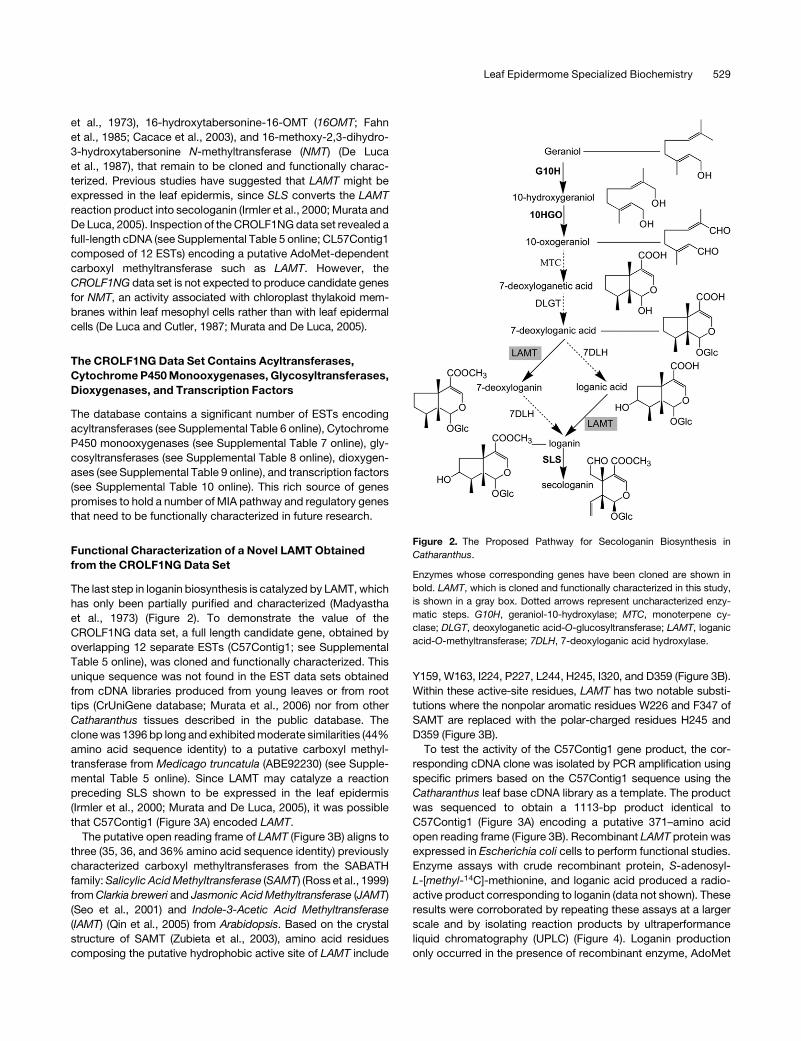
et al., 1973), 16-hydroxytabersonine-16-OMT (16OMT; Fahn
et al., 1985; Cacace et al., 2003), and 16-methoxy-2,3-dihydro-
3-hydroxytabersonine N-methyltransferase (NMT) (De Luca
et al., 1987), that remain to be cloned and functionally charac-
terized. Previous studies have suggested that LAMT might be
expressed in the leaf epidermis, since SLS converts the LAMT
reaction product into secologanin (Irmler et al., 2000; Murata and
De Luca, 2005). Inspection of the CROLF1NG data set revealed a
full-length cDNA (see Supplemental Table 5 online; CL57Contig1
composed of 12 ESTs) encoding a putative AdoMet-dependent
carboxyl methyltransferase such as LAMT. However, the
CROLF1NG data set is not expected to produce candidate genes
for NMT, an activity associated with chloroplast thylakoid mem-
branes within leaf mesophyl cells rather than with leaf epidermal
cells (De Luca and Cutler, 1987; Murata and De Luca, 2005).
The CROLF1NG Data Set Contains Acyltransferases,
Cytochrome P450 Monooxygenases, Glycosyltransferases,
Dioxygenases, and Transcription Factors
The database contains a significant number of ESTs encoding
acyltransferases (see Supplemental Table 6 online), Cytochrome
P450 monooxygenases (see Supplemental Table 7 online), gly-
cosyltransferases (see Supplemental Table 8 online), dioxygen-
ases (see Supplemental Table 9 online), and transcription factors
(see Supplemental Table 10 online). This rich source of genes
promises to hold a number of MIA pathway and regulatory genes
that need to be functionally characterized in future research.
Functional Characterization of a Novel LAMT Obtained
from the CROLF1NG Data Set
The last step in loganin biosynthesis is catalyzed by LAMT, which
has only been partially purified and characterized (Madyastha
et al., 1973) (Figure 2). To demonstrate the value of the
CROLF1NG data set, a full length candidate gene, obtained by
overlapping 12 separate ESTs (C57Contig1; see Supplemental
Table 5 online), was cloned and functionally characterized. This
unique sequence was not found in the EST data sets obtained
from cDNA libraries produced from young leaves or from root
tips (CrUniGene database; Murata et al., 2006) nor from other
Catharanthus tissues described in the public database. The
clone was 1396 bp long and exhibited moderate similarities (44%
amino acid sequence identity) to a putative carboxyl methyl-
transferase from Medicago truncatula (ABE92230) (see Supple-
mental Table 5 online). Since LAMT may catalyze a reaction
preceding SLS shown to be expressed in the leaf epidermis
(Irmler et al., 2000; Murata and De Luca, 2005), it was possible
that C57Contig1 (Figure 3A) encoded LAMT.
The putative open reading frame of LAMT (Figure 3B) aligns to
three (35, 36, and 36% amino acid sequence identity) previously
characterized carboxyl methyltransferases from the SABATH
family: Salicylic Acid Methyltransferase (SAMT) (Ross et al., 1999)
from Clarkia breweri and Jasmonic Acid Methyltransferase (JAMT)
(Seo et al., 2001) and Indole-3-Acetic Acid Methyltransferase
(IAMT) (Qin et al., 2005) from Arabidopsis. Based on the crystal
structure of SAMT (Zubieta et al., 2003), amino acid residues
composing the putative hydrophobic active site of LAMT include
Y159, W163, I224, P227, L244, H245, I320, and D359 (Figure 3B).
Within these active-site residues, LAMT has two notable substi-
tutions where the nonpolar aromatic residues W226 and F347 of
SAMT are replaced with the polar-charged residues H245 and
D359 (Figure 3B).
To test the activity of the C57Contig1 gene product, the cor-
responding cDNA clone was isolated by PCR amplification using
specific primers based on the C57Contig1 sequence using the
Catharanthus leaf base cDNA library as a template. The product
was sequenced to obtain a 1113-bp product identical to
C57Contig1 (Figure 3A) encoding a putative 371–amino acid
open reading frame (Figure 3B). Recombinant LAMT protein was
expressed in Escherichia coli cells to perform functional studies.
Enzyme assays with crude recombinant protein, S-adenosyl-
L-[methyl-14C]-methionine, and loganic acid produced a radio-
active product corresponding to loganin (data not shown). These
results were corroborated by repeating these assays at a larger
scale and by isolating reaction products by ultraperformance
liquid chromatography (UPLC) (Figure 4). Loganin production
only occurred in the presence of recombinant enzyme, AdoMet
Figure 2. The Proposed Pathway for Secologanin Biosynthesis in
Catharanthus.
Enzymes whose corresponding genes have been cloned are shown in
bold. LAMT, which is cloned and functionally characterized in this study,
is shown in a gray box. Dotted arrows represent uncharacterized enzy-
matic steps. G10H, geraniol-10-hydroxylase; MTC, monoterpene cy-
clase; DLGT, deoxyloganetic acid-O-glucosyltransferase; LAMT, loganic
acid-O-methyltransferase; 7DLH, 7-deoxyloganic acid hydroxylase.
Leaf Epidermome Specialized Biochemistry 529
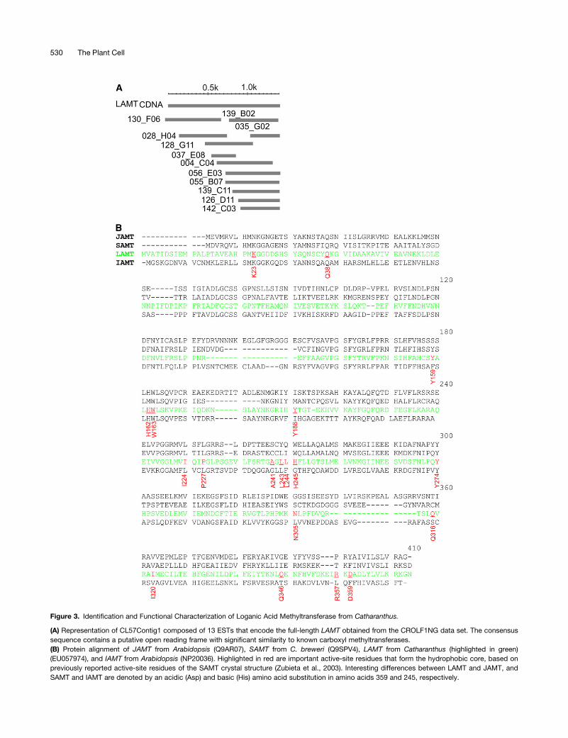
Figure 3. Identification and Functional Characterization of Loganic Acid Methyltransferase from Catharanthus.
(A) Representation of CL57Contig1 composed of 13 ESTs that encode the full-length LAMT obtained from the CROLF1NG data set. The consensus
sequence contains a putative open reading frame with significant similarity to known carboxyl methyltransferases.
(B) Protein alignment of JAMT from Arabidopsis (Q9AR07), SAMT from C. breweri (Q9SPV4), LAMT from Catharanthus (highlighted in green)
(EU057974), and IAMT from Arabidopsis (NP20036). Highlighted in red are important active-site residues that form the hydrophobic core, based on
previously reported active-site residues of the SAMT crystal structure (Zubieta et al., 2003). Interesting differences between LAMT and JAMT, and
SAMT and IAMT are denoted by an acidic (Asp) and basic (His) amino acid substitution in amino acids 359 and 245, respectively.
530 The Plant Cell
and loganic acid (Figure 4, profile A), but not in reactions with
boiled recombinant enzyme (Figure 4, profile B), in reactions
lacking loganic acid (Figure 4, profile C), or in reactions with E.
coli cell-free extracts expressing the empty vector (Figure 4,
profile D). Mass spectra analysis (Figure 4, table) illustrated that
the reaction product (Figure 4, profile A) is loganin based on its
base peak at 435.1 (mass-to-charge ratio [m/z]), consistent with
the mass obtained with loganin standard.
Recombinant LAMT Is a Highly Specific OMT
The recombinant LAMT (rLAMT) showed striking specificity for
loganic acid since it was not active with other similar iridoids,
including deoxyloganic, dehydrologanic, epiloganic, loganetic acid,
or with the reaction product, loganin (Figure 5). Various benzoic
acids (salicylic, benzoic, p-hydroxybenzoic, and anthranillic
acids), phenylpropanoids (caffeic, ferulic, sinapic, gallic, chloro-
genic, p-coumaric, and trans-cinammic acids), dicarboxylic
acids (malic and tartaric), and hormones (indole acetic and
abscisic acids) were not accepted as substrates by rLAMT nor
was it active with MIAs (16-OH tabersonine and 3-OH taberso-
nine) (data not shown). Together, these results confirm that the
12 ESTs used to assemble C57Contig1 encoded authentic
LAMT, that Catharanthus leaf epidermis is specialized for MIA
biosynthesis, and that the CROLF1NG data set will be a valuable
resource for cloning many more unknown and uncharacterized
genes in this pathway.
Biochemical and Kinetic Properties of rLAMT
The pH optimum of rLAMT was 7.5 with loganic acid as a
substrate, and cofactors such as Mg2þ or Kþ were not required
for activity nor did they enhance enzyme activity (data not
shown). The Km for loganic acid of rLAMT was 14.76 6 1.7 mM
(Table 2), similar to the results obtained for the partially purified
enzyme isolated from cell suspension cultures of C. roseus (Km¼12.5 mM) (Madyastha et al., 1973). While the Km value for the
cosubstrate AdoMet was not previously reported for LAMT,
rLAMT showed a remarkably high Km (742.1 6 37 mM) compared
with JAMT (Seo et al., 2001) and SAMT (Ross et al., 1999) whose
Km values were 6.3 and 9 mM, respectively.
Figure 4. Production of Loganin by E. coli Cell-Free Extracts Expressing
Recombinant LAMT.
Loganin production only occurred in the presence of recombinant
enzyme, AdoMet and loganic acid (A), but not in reactions with boiled
recombinant enzyme (B), in reactions lacking loganic acid (C) nor in
reactions with E. coli cell-free extracts expressing the empty vector (D).
Note the loss of AdoMet and loganic acid with the appearance of loganin
in (A). The table confirms that the reaction product in (A) is loganin based
on its base peak at 435.1 (m/z). Analyses for A to D were performed by
UPLC. The reaction products obtained in A were also analyzed by liquid
chromatography–mass spectrometry (LC-MS) using a Bruker HCTþ
System as described in Methods.
Figure 5. Substrate Specificity of rLAMT.
The LAMT only accepted loganic and secologanic acids as possible
substrates but was not active with other similar compounds shown.
Asterisk, the 100% value is related to the specific activity of the enzyme
for loganic acid (310 pmol loganin/mg protein/h).
Leaf Epidermome Specialized Biochemistry 531

The high Km values for both substrate and cosubstrate suggest
that the catalytic efficiency of rLAMT appears to be very low
(Table 2) compared with other plant OMTs. The turnover number
of rLAMT for loganic acid was quite low (0.327 s�1) compared
with nonhistidine-tagged (2.8 s�1) and histidine-tagged (14 s�1)
rSAMT (Ross et al., 1999) and to JAMT (25 s�1) (Seo et al., 2001).
Remarkably, inhibition studies with the reaction products re-
vealed that loganin is a strong inhibitor for rLAMT with a Ki of
215 6 30 mM, since it has 50 times greater affinity for loganin
compared with loganic acid. By contrast, the Ki (400 6 47 mM) for
S-adenosyl-L-homocysteine (AdoHys) was only twofold higher
then that of AdoMet. Since the AdoHys inhibition constant was
approximately twofold higher than that of AdoMet, inhibition of
LAMT by AdoHys would depend on the ability of Catharanthus
cells to recycle AdoHys into AdoMet through the AdoMet cycle.
LAMT Enzyme Activity Is Preferentially Expressed in
Catharanthus Leaf Epidermis
To localize LAMT in Catharanthus leaf, the enzyme activity of
LAMT was measured using extracts either from whole leaves in
various developmental stages or epidermis extracts of corre-
sponding leaves obtained by CA. Previous studies with 16OMT
(Levac et al., 2008) showed that this enzyme activity was highly
enriched in the epidermis of young Catharanthus leaves,
whereas the activity of the chloroplast thylakoid-associated
NMT (De Luca and Cutler, 1987; Murata and De Luca, 2005) is
found within cells inside the leaf. In accordance with these
findings, NMT activity was only detected in whole leaves but not
in epidermis-enriched extracts in all developmental stages
tested (Figure 6A). By contrast, 16OMT-specific enzyme activity
was 10 times higher in leaf epidermis–enriched extracts com-
pared with the activity found in whole young leaves (Figure 6A).
The 10-fold higher specific activity of LAMT in the leaf epidermis
than in whole leaves strongly suggests that this enzyme is also
preferentially localized to the leaf epidermis (Figure 6A). How-
ever, the activity of 16OMT was 35 times lower in epidermis-
enriched leaf extracts of L2 compared with those of L1 leaves,
whereas the activity of LAMT only decreased by ;50% in
epidermis-enriched leaf extracts of L2, L3, and L4 leaves com-
pared with those of L1 leaves. These data suggest that unlike
Table 2. CROLF1NG Is Enriched in ESTs for Enzymes in the MIA
Biosynthetic Pathway
LF (3655) LB (1231) RT (1546) Pub (9850)
G10H 1 0 3 1
10HGO 1 0 1 0
LAMT 12 0 0 1
SLS 25 2 3 0
TDC 8 0 0 0
STR 1 0 0 0
SGD 4 1 1 2
T16H 4 0 0 0
16OMT 4 0 0 1
D4H 0 0 0 0
DAT 0 0 0 0
CPR 5 0 0 0
ORCA3 1 0 0 0
ZCT2 1 0 3 0
BPF1 1 0 0 0
Comparison of the numbers of the MIA-related ESTs that were obtained
by sequencing cDNA libraries produced from leaf epidermis–enriched
mRNA (LF) in this study compared with previous studies involving random
sequencing of cDNA libraries produced from mRNA extracted from the
leaf base of young Catharanthus leaves (LB; Murata et al., 2006) from hairy
root tips (RT; Murata et al., 2006) or from EST sequences found in the
GenBank database (Pub). The numbers of unique EST sequences found
in each case are shown in parentheses.
Figure 6. Differential Distribution of LAMT Enzyme Activity in Cathar-
anthus Leaf Epidermis.
(A) Catharanthus leaves of different ages (L1 to L4) were extracted in trip-
licate either as whole leaves or by CA treatment to obtain epidermis-
enriched leaf extracts as described in Methods. The two sets of desalted
extracts from each stage of leaf development were assayed for NMT, 16OMT,
or LAMT to identify the differential distribution of these enzyme activities.
(B) Whole Catharanthus leaves, hairy roots, flowers, siliques, and stems
were extracted in triplicate as described in Methods. Desalted extracts
were assayed for LAMT and 16OMT to compare the distribution of these
enzyme activities within different plant organs. Each point represents the
mean of three assays 6 SD.
532 The Plant Cell
highly regulated 16OMT, significant LAMT activity is maintained
as Catharanthus leaves age. The reasons for this difference in
regulation remain to be determined. Leaf, hairy root, stem,
silique, and flower extracts contained both 16OMT and LAMT
activities in all these tissues (Figure 6B). However, the levels of
LAMT (Figure 6B) in hairy roots were 4 to 8 times lower than the
activities found in the other plant organs, respectively.
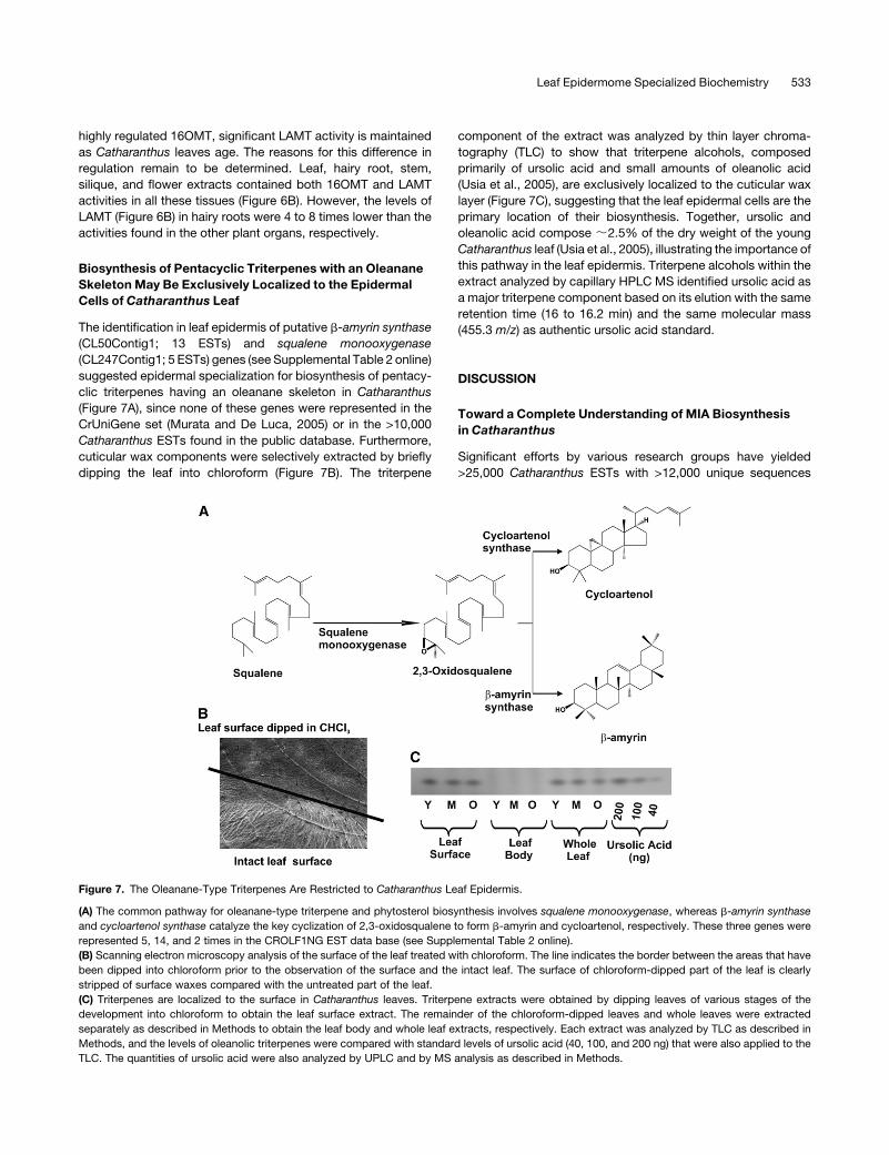
Biosynthesis of Pentacyclic Triterpenes with an Oleanane
Skeleton May Be Exclusively Localized to the Epidermal
Cells of Catharanthus Leaf
The identification in leaf epidermis of putative b-amyrin synthase
(CL50Contig1; 13 ESTs) and squalene monooxygenase
(CL247Contig1; 5 ESTs) genes (see Supplemental Table 2 online)
suggested epidermal specialization for biosynthesis of pentacy-
clic triterpenes having an oleanane skeleton in Catharanthus
(Figure 7A), since none of these genes were represented in the
CrUniGene set (Murata and De Luca, 2005) or in the >10,000
Catharanthus ESTs found in the public database. Furthermore,
cuticular wax components were selectively extracted by briefly
dipping the leaf into chloroform (Figure 7B). The triterpene
component of the extract was analyzed by thin layer chroma-
tography (TLC) to show that triterpene alcohols, composed
primarily of ursolic acid and small amounts of oleanolic acid
(Usia et al., 2005), are exclusively localized to the cuticular wax
layer (Figure 7C), suggesting that the leaf epidermal cells are the
primary location of their biosynthesis. Together, ursolic and
oleanolic acid compose ;2.5% of the dry weight of the young
Catharanthus leaf (Usia et al., 2005), illustrating the importance of
this pathway in the leaf epidermis. Triterpene alcohols within the
extract analyzed by capillary HPLC MS identified ursolic acid as
a major triterpene component based on its elution with the same
retention time (16 to 16.2 min) and the same molecular mass
(455.3 m/z) as authentic ursolic acid standard.
DISCUSSION
Toward a Complete Understanding of MIA Biosynthesis
in Catharanthus
Significant efforts by various research groups have yielded
>25,000 Catharanthus ESTs with >12,000 unique sequences
Figure 7. The Oleanane-Type Triterpenes Are Restricted to Catharanthus Leaf Epidermis.
(A) The common pathway for oleanane-type triterpene and phytosterol biosynthesis involves squalene monooxygenase, whereas b-amyrin synthase
and cycloartenol synthase catalyze the key cyclization of 2,3-oxidosqualene to form b-amyrin and cycloartenol, respectively. These three genes were
represented 5, 14, and 2 times in the CROLF1NG EST data base (see Supplemental Table 2 online).
(B) Scanning electron microscopy analysis of the surface of the leaf treated with chloroform. The line indicates the border between the areas that have
been dipped into chloroform prior to the observation of the surface and the intact leaf. The surface of chloroform-dipped part of the leaf is clearly
stripped of surface waxes compared with the untreated part of the leaf.
(C) Triterpenes are localized to the surface in Catharanthus leaves. Triterpene extracts were obtained by dipping leaves of various stages of the
development into chloroform to obtain the leaf surface extract. The remainder of the chloroform-dipped leaves and whole leaves were extracted
separately as described in Methods to obtain the leaf body and whole leaf extracts, respectively. Each extract was analyzed by TLC as described in
Methods, and the levels of oleanolic triterpenes were compared with standard levels of ursolic acid (40, 100, and 200 ng) that were also applied to the
TLC. The quantities of ursolic acid were also analyzed by UPLC and by MS analysis as described in Methods.
Leaf Epidermome Specialized Biochemistry 533
that have partly been submitted to the GenBank database
(Murata et al., 2006; Rischer et al., 2006; Shukla et al., 2006).
While these efforts identified only four known MIA biosynthesis
pathway genes (Table 2), this study, using a cDNA library
prepared from leaf epidermis–enriched mRNA, identified ESTs
for virtually all known MIA biosynthesis and regulatory genes up
to and including T16H (Figure 1) and 16OMT (Levac et al., 2008).
These results strongly support the biochemical specialization of
young leaf epidermis as the primary site for expression of most of
the pathway in vindoline biosynthesis from primary iridoid pre-
cursors and tryptamine to 16-methoxytabersonine (Murata and
De Luca, 2005). Most significantly, this data set is very likely to
contain the remaining uncharacterized biosynthesis and regula-
tory genes for the MIA pathway as shown by the CROLF1NG
(3655 unique sequences) guided identification and functional
characterization of LAMT (Figures 3 to 6) and 16OMT (Levac
et al., 2008). In this respect, it is relevant that the LAMT and
16OMT sequences were represented 12 and four times, respec-
tively, in the CROLF1NG data set, while only one EST for each
sequence was found in published Catharanthus EST data sets
(Table 2). These results highlight the remarkable value of random
sequencing of leaf epidermis–enriched cDNA libraries for inves-
tigating the specialized chemistry of this cell type. The results
also suggest that selection of candidate genes based on their
relative abundance in this library is a useful approach to identify
MIA biosynthesis enzyme candidates. In this context, further
sequencing of this epidermis-enriched cDNA library should be
considered to obtain more reliable data on the redundancies of
the ESTs (Table 1) and to reach saturation with respect to the
numbers of ESTs that encode enzymes that are related to MIA
biosynthesis. In addition, gene families with particular biochem-
ical functions could be selected for expression analysis by RT-
PCR using RNA harvested from different organs or cell types
(Murata and De Luca, 2005) to select for candidates to be
screened directly for biochemical function.
Use of the CROLF1NG Data Set to Elucidate the Pathway
for Secologanin Biosynthesis
Iridoids are cyclic monoterpenes, found in various plant species,
where they play important defensive roles in plant–herbivore
interactions. For example, aucubin and catalpol found in Plantago
lanceolata deter and retard the growth rate of generalist insect
herbivores (Harvey et al., 2005), whereas insect specialists are
attracted to and consume host plants to sequester iridoids for
defensive purposes or to use them as oviposition cues (Pereyra
and Bowers, 1988; Penuelas et al., 2006). In the case of MIA
biosynthesis, the iridoid secologanin is important as the source for
the terpene moiety of all the MIAs. While iridoids have also been
used in chemotaxonomy because of their structural diversity in
various plant species (Lopes et al., 2004), their localization within
the plant as well as their biochemistry and molecular biology
remain largely unknown.
Recent studies in Catharanthus have shown that while G10H is
preferentially expressed in IPAP cells (Burlat et al., 2004), SLS
(Figure 2) is preferentially expressed in leaf epidermal cells (Irmler
et al., 2000; Burlat et al., 2004; Murata and De Luca, 2005) and in
roots. This preferential expression of G10H and several steps in
the methyl erythritol phosphate (MEP) pathway has led to the
suggestion that the precursors for secoridoid biosynthesis may
be produced in IPAP cells and that an unknown intermediate is
transported to the leaf epidermis for elaboration into secologanin
(Burlat et al., 2004). Further investigations into this question have
been limited since other enzymes in this pathway remain to be
cloned. This report has identified and functionally characterized
LAMT, which catalyzes O-methylation of loganic acid (Figure 3)
and is strongly expressed in (Figure 6) but not restricted to leaf
epidermal cells (data not shown). In addition, the representation
of a single MEP pathway gene (4-diphosphocytidyl-2-C-methyl-D-
erythritol synthase, three ESTs; see Supplemental Table 1 online)
and the G10H (one EST; Table 1) gene in the CROLF1NG
data set, together with the abundance of 10-hydroxygeraniol
oxidoreductase–like transcripts and SLS transcripts (Table 1) in
epidermal cells, may mean that the complete secologanin bio-
synthesis pathway is expressed in the leaf epidermis (Murata and
De Luca, 2005). These results remain ambiguous and further
detailed studies are required to elucidate if an isoprenoid inter-
mediate produced in IPAP cells is required for the biosynthesis of
secologanin in Catharanthus leaf epidermal cells.
The epidermal localization of secologanin biosynthesis in
Catharanthus leaf fits their possible antiherbivory role since the
leaf surface is the first contact point between herbivores and the
plant. It would be interesting to know if other iridoid-producing
plants, including Plantago and Lonicera species (Pereyra and
Bowers, 1988; Harvey et al., 2005; Penuelas et al., 2006), also
preferentially produce their iridoids in leaf epidermal cells.
Two additional candidate ESTs in the CROLF1NG data set
could encode 7-deoxyloganetic acid 1-O-glucosyltransferase
(Yamamoto et al., 2002) (see Supplemental Table 8 online) and
7-deoxyloganin 7-hydroxylase (Yamamoto et al., 1999) (CYP72B
and CYP72C in Supplemental Table 9 online) involved in earlier
steps in the secologanin pathway (Figure 2). While these two
enzymes were partially purified and characterized from Lonicera
japonica cell cultures, their corresponding genes remain to be
cloned. Further experiments with these two candidate genes will
be performed to identify their possible role in these biochemical
functions.
Features of the Active Site of LAMT Based on the Crystal
Structure of SAMT
The SABATH family of carboxyl methyltransferases has distinct
characteristics that separates them from other OMTs. These
enzymes do not require any catalytic residues but rather provide
a hydrophobic pocket or reaction center where the desolvated
carboxylate moiety can attack the AdoMet (Zubieta et al., 2003).
SABATH family OMTs, such as JAMT, FAMT, and SAMT (Ross
et al., 1999; Seo et al., 2001; Yang et al., 2006), that O-methylate
fairly hydrophobic substrates require that the carboxyl moiety be
devoid of water to increase its reactivity. By contrast, loganic
acid with its glucose moiety and its higher water solubility may
require special conditions for eliminating the glucose-associated
water as loganic acid enters the active site of LAMT. The protein
alignment of LAMT to IAMT, SAMT, and JAMT in Figure 3B
reveals that active-site residues required for the hydrophobic
core (Y159, W163, I224, P227, L244, and I320) are conserved in
534 The Plant Cell
LAMT when compared with those based on the crystal structure
of SAMT (Zubieta et al., 2003). Two important differences involve
the substitution of SAMT amino acids W226 and F347 with H245
and D359, respectively, in LAMT.
Preliminary modeling studies of LAMT with loganic acid super-
imposed onto the salicylic acid binding site of SAMT (Figure 8)
showed that two different orientations of loganic acid had
favorable van der Waals and electrostatic interactions with the
protein and could be accommodated comfortably. The average
CHARMM interaction energy, >100 minimum energy poses, for
the first conformation within the LAMT comparative model was
�100.57 6 5.2 kcal/mol with an overall range of �113.32 kcal/
mol to �86.78 kcal/mol. For the second conformation, it was
�87.98 6 14 kcal/mol with an overall range of �139.29 kcal/mol
to �70.37 kcal/mol. Given that both conformers have similar
interaction energies with the protein, neither model can be ex-
cluded from consideration without further experimental evi-
dence.
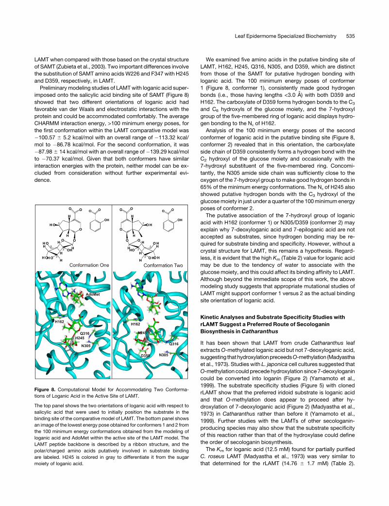
We examined five amino acids in the putative binding site of
LAMT, H162, H245, Q316, N305, and D359, which are distinct
from those of the SAMT for putative hydrogen bonding with
loganic acid. The 100 minimum energy poses of conformer
1 (Figure 8, conformer 1), consistently made good hydrogen
bonds (i.e., those having lengths <3.0 A) with both D359 and
H162. The carboxylate of D359 forms hydrogen bonds to the C3
and C6 hydroxyls of the glucose moiety, and the 7-hydroxyl
group of the five-membered ring of loganic acid displays hydro-
gen bonding to the Ne of H162.
Analysis of the 100 minimum energy poses of the second
conformer of loganic acid in the putative binding site (Figure 8,
conformer 2) revealed that in this orientation, the carboxylate
side chain of D359 consistently forms a hydrogen bond with the
C2 hydroxyl of the glucose moiety and occasionally with the
7-hydroxyl substituent of the five-membered ring. Concomi-
tantly, the N305 amide side chain was sufficiently close to the
oxygen of the 7-hydroxyl group to make good hydrogen bonds in
65% of the minimum energy conformations. The Ne of H245 also
showed putative hydrogen bonds with the C3 hydroxyl of the
glucose moiety in just under a quarter of the 100 minimum energy
poses of conformer 2.
The putative association of the 7-hydroxyl group of loganic
acid with H162 (conformer 1) or N305/D359 (conformer 2) may
explain why 7-deoxyloganic acid and 7-epiloganic acid are not
accepted as substrates, since hydrogen bonding may be re-
quired for substrate binding and specificity. However, without a
crystal structure for LAMT, this remains a hypothesis. Regard-
less, it is evident that the high Km (Table 2) value for loganic acid
may be due to the tendency of water to associate with the
glucose moiety, and this could affect its binding affinity to LAMT.
Although beyond the immediate scope of this work, the above
modeling study suggests that appropriate mutational studies of
LAMT might support conformer 1 versus 2 as the actual binding
site orientation of loganic acid.
Kinetic Analyses and Substrate Specificity Studies with
rLAMT Suggest a Preferred Route of Secologanin
Biosynthesis in Catharanthus
It has been shown that LAMT from crude Catharanthus leaf
extracts O-methylated loganic acid but not 7-deoxyloganic acid,
suggesting thathydroxylation preceeds O-methylation (Madyastha
et al., 1973). Studies with L. japonica cell cultures suggested that
O-methylation could precede hydroxylation since 7-deoxyloganin
could be converted into loganin (Figure 2) (Yamamoto et al.,
1999). The substrate specificity studies (Figure 5) with cloned
rLAMT show that the preferred iridoid substrate is loganic acid
and that O-methylation does appear to proceed after hy-
droxylation of 7-deoxyloganic acid (Figure 2) (Madyastha et al.,
1973) in Catharanthus rather than before it (Yamamoto et al.,
1999). Further studies with the LAMTs of other secologanin-
producing species may also show that the substrate specificity
of this reaction rather than that of the hydroxylase could define
the order of secologanin biosynthesis.
The Km for loganic acid (12.5 mM) found for partially purified
C. roseus LAMT (Madyastha et al., 1973) was very similar to
that determined for the rLAMT (14.76 6 1.7 mM) (Table 2).
Figure 8. Computational Model for Accommodating Two Conforma-
tions of Loganic Acid in the Active Site of LAMT.
The top panel shows the two orientations of loganic acid with respect to
salicylic acid that were used to initially position the substrate in the
binding site of the comparative model of LAMT. The bottom panel shows
an image of the lowest energy pose obtained for conformers 1 and 2 from
the 100 minimum energy conformations obtained from the modeling of
loganic acid and AdoMet within the active site of the LAMT model. The
LAMT peptide backbone is described by a ribbon structure, and the
polar/charged amino acids putatively involved in substrate binding
are labeled. H245 is colored in gray to differentiate it from the sugar
moiety of loganic acid.
Leaf Epidermome Specialized Biochemistry 535
Remarkably, rLAMT had a relatively high Km for AdoMet (742.1 6
37 mM) compared with other carboxyl methyltransferases from
the SABATH family (JAMT, Km 6.3 mM; SAMT, Km 9 mM) (Ross
et al., 1999; Seo et al., 2001). The high Km values for both loganic
acid and AdoMet suggest that the catalytic activity of LAMT may
be limiting the production of terpenoid component of MIA bio-
synthesis in C. roseus (Peebles et al., 2006).
Inhibition studies with the reaction products revealed that
loganin was a very good inhibitor (Ki of 215 6 30 mM) for LAMT,
with its 50 3 greater affinity for loganin than for loganic acid.
These results suggest that for LAMT to be active, loganin
concentrations would need to be kept very low in the cell, either
by rapid turnover of loganin into secologanin or by subcellular
compartmentation. Substrate specificity studies have suggested
that only loganin can serve as a substrate for SLS (Yamamoto
et al., 2000). The association of this cytochrome P450 with
endoplasmic reticulum membranes or related vesicles could
rapidly convert loganin into secologanin followed by its transfer
to the vacuole (Contin et al., 1999) together with tryptamine for
conversion into strictosidine by vacuole-associated STR (Stevens
et al., 1993). In this way, the SLS-mediated conversion of loganin
into secologanin, together with its subcellular compartmentali-
zation for MIA biosynthesis, could prevent loganin from inhibiting
LAMT.
Catharanthus Leaf Epidermis as the Exclusive Site of
Oleanane Triterpene Biosynthesis
The plant kingdom produces many thousands of triterpenes
exhibiting a broad range of pharmacological activities based on
>80 different carbon skeletons produced from (3S)-oxidosqualene
(Ebizuka et al., 2003). While the accumulation of high levels of
ursolic acid in C. roseus has been documented (Usia et al., 2005),
the sites of accumulation of these products have not been
elucidated. Studies with other plants showed that ursolic and
oleanolic acid accumulate on the surfaces of apple (Malus
domestica) (Bringe et al., 2005) and of grape berry (Vitis vinifera)
exocarp (Grncarevic and Radler, 1971) as well as in the epidermis
of grape leaves. In other studies with Avena strigosa, the ex-
pression of b-amyrin synthase was localized to epidermal cells of
root tips that accumulate triterpene avenacins (Qi et al., 2006).
The CROLF1NG data set suggests that the MVA pathway (see
Supplemental Table 1 online) and two key triterpene biosynthesis
genes (squalene monooxygenase [five ESTs] and b-amyrin syn-
thase [14 ESTs]) (see Supplemental Table 2 online) were prefer-
entially expressed in Catharanthus leaf epidermis. The selective
and quantitative extraction of surface leaf triterpenes with chlo-
roform (Figures 7B and 7C) suggests that once they are made
inside Catharanthus leaf epidermal cells, they are secreted where
they accumulate on the surface of leaf epidermal cells (Figure 9).
It is interesting that Catharanthus leaf epidermal cells are the
site of biosynthesis of triterpenes that are secreted into epi-
cuticular wax as well as of secologanin that is then incorpo-
rated into MIAs. The more frequent representation of five MVA
pathway–specific genes compared with the single representa-
tion of a MEP pathway gene (see Supplemental Table 1 online;
4-diphosphocytidyl-2-C-methyl-D-erythritol synthase) does sug-
gest that MVA pathway transcripts are selectively expressed in
leaf epidermal cells compared with those of the MEP pathway.
These differences might be attributed to the greater demand for
biosynthetic precursors for triterpene biosynthesis, since triter-
penes accumulate at much higher levels in Catharanthus than do
MIAs (Usia et al., 2005). Further biochemical and chemo-ecological
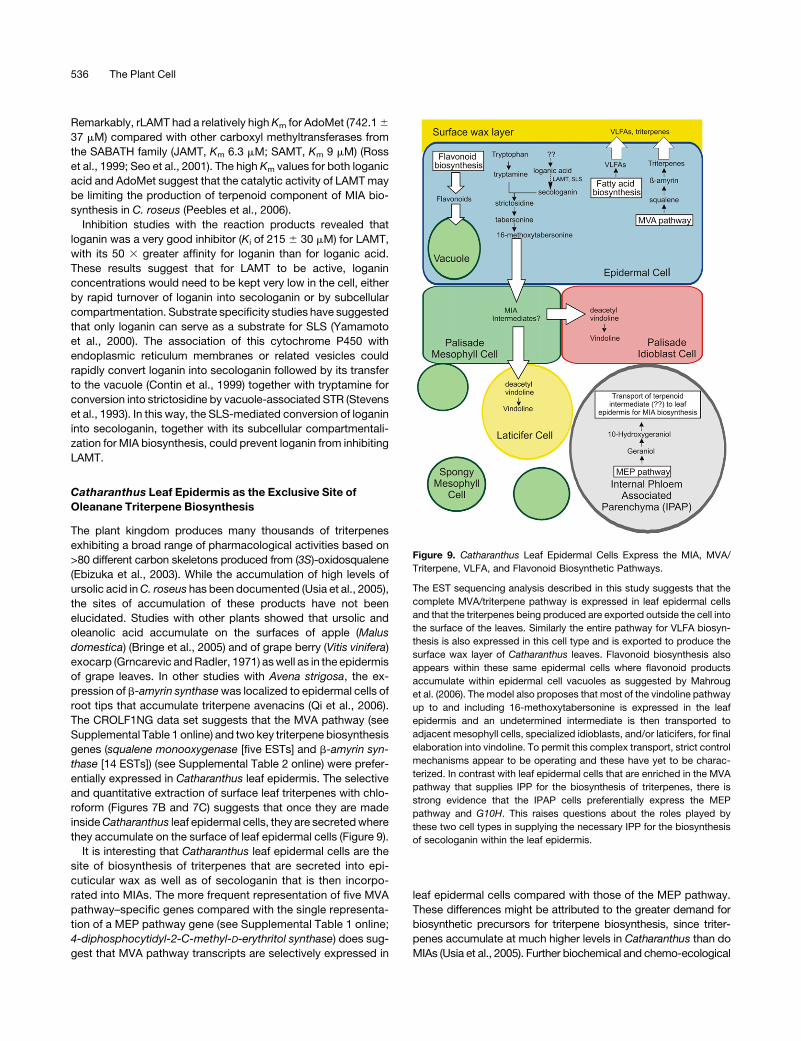
Figure 9. Catharanthus Leaf Epidermal Cells Express the MIA, MVA/
Triterpene, VLFA, and Flavonoid Biosynthetic Pathways.
The EST sequencing analysis described in this study suggests that the
complete MVA/triterpene pathway is expressed in leaf epidermal cells
and that the triterpenes being produced are exported outside the cell into
the surface of the leaves. Similarly the entire pathway for VLFA biosyn-
thesis is also expressed in this cell type and is exported to produce the
surface wax layer of Catharanthus leaves. Flavonoid biosynthesis also
appears within these same epidermal cells where flavonoid products
accumulate within epidermal cell vacuoles as suggested by Mahroug
et al. (2006). The model also proposes that most of the vindoline pathway
up to and including 16-methoxytabersonine is expressed in the leaf
epidermis and an undetermined intermediate is then transported to
adjacent mesophyll cells, specialized idioblasts, and/or laticifers, for final
elaboration into vindoline. To permit this complex transport, strict control
mechanisms appear to be operating and these have yet to be charac-
terized. In contrast with leaf epidermal cells that are enriched in the MVA
pathway that supplies IPP for the biosynthesis of triterpenes, there is
strong evidence that the IPAP cells preferentially express the MEP
pathway and G10H. This raises questions about the roles played by
these two cell types in supplying the necessary IPP for the biosynthesis
of secologanin within the leaf epidermis.
536 The Plant Cell
analyses will be required to elucidate the mechanism of triter-
pene accumulate in the leaf epicuticular waxes, their in vivo roles,
and the involvement of the MEP and MVA pathways in Cathar-
anthus leaf epidermal cells in relation to MIA and triterpene
biosynthesis.
The Possible Roles of the Plastidic MEP and Cytosolic MVA
Pathways in Secologanin Biosynthesis
Previous extensive analyses of the MEP pathway in MIA-
producing plants have suggested that the terpenoid moiety of
MIAs is predominantly, but not exclusively, derived from MEP
pathway in cell suspension and hairy root culture systems of
C. roseus and Rauvolfia serpentina cell cultures (Contin et al.,
1998; Eichinger et al., 1999; Hong et al., 2003). These results are
supported by studies in Arabidopsis that monoterpenes are
produced principally via the plastidic MEP pathway (Lichtenthaler,
1999). In the case of C. roseus, recent in situ RNA hybridization
studies showed that the MEP pathway genes DXS, DXR, MECS,
and G10H, the first committed enzyme in iridoid biosynthesis,
were expressed predominantly in IPAP cells (Burlat et al., 2004)
of the leaf vasculature. Recent immunocytolabeling studies pro-
vided more quantitative information to corroborate the high en-
richment of the MEP pathway in IPAP cells, but it was also found at
low levels in other cell types, including epidermal and mesophyll
cells (Oudin et al., 2007). The expression of G10H was used to
further suggest that MEP pathway–derived isopentenyl pyrophos-
phate was converted to 10-hydroxygeraniol or another derivative
for mobilization to the leaf epidermis for further elaboration into
secologanin and MIAs. While the baseline representation of MEP
pathway genes compared with those of the MVA pathway in the
CROLF1NG data set (see Supplemental Table 1 online) provides
support for the MEP pathway enrichment of phloem parenchyma
(Burlat et al., 2004), it also establishes the differential enrichment of
the MEP and MVA pathways in each cell type.
Since only a single G10H EST was detected in the CROLF1NG
data set, it does support in situ hybridization studies that show
preferential expression of G10H in phloem parenchyma (Burlat,
et al., 2004). Remarkably, it has been implied that the biosynthesis
of the similar iridoids in leaf beetles belonging to the Chrysome-
lidae family is divided between the fat body and a specialized
glandular reservoir that also accumulates these defensive secre-
tory compounds (Burse et al., 2007). The fat body expresses the
MVA pathway and a geraniol hydroxylase and glucosyltransferase
to produce 10-hydroxygeraniol-10-O-b-glucoside. This glucoside
is then transported to the glandular reservoir where a b-glucosidase
releases 10-hydroxygeraniol for enzyme-mediated oxidation and
cyclization/isomerization for the formation of toxic iridoids (Burse
et al., 2007). In the case of Catharanthus MIA biosynthesis, the
identification of a similar pathway step or intermediate in the
IPAP cells could be very helpful to completely prove that MEP
pathway–derived precursors are transported to the leaf epider-
mis from the internal phloem parenchyma for the biosynthesis of
secologanin (Burlat et al., 2004). Similarly, the hypothesis that
leaf epidermal cells alone are competent to supply sufficient
MEP pathway intermediates for secologanin biosynthesis and
MIA accumulation in Catharanthus leaves also needs to be fully
tested (Murata and De Luca, 2005).
The unique biochemical properties of distinct cells (i.e., ep-
idermal cells, mesophyll cells, and phloem parenchyma cells)
with respect to IPP biosynthesis are clearly related to their
complex roles to produce different metabolites. For example,
neither leaf epidermal cells nor IPAP cells, in contrast with those
of the palisade and spongy mesophyll, require IPP for biosyn-
thesis of chlorophyll and yet these two cell types require in-
creased expression of the MVA/MEP pathways for their unique
metabolic needs. The data presented here highlight the com-
plexity involved and present novel approaches to study these
questions.
The Versatility of the CA Technique and Its Possible Use
for Epidermome Analysis
Unlike LCM, CA can be used to harvest epidermis-enriched
materials from intact leaves, making the analysis of mRNA, active
protein, and metabolite analyses possible without significant
technical difficulties. The technical simplicity of CA technique
makes it valuable for (1) localizing enzyme activities to the leaf
epidermis (Murata and De Luca, 2005), (2) large-scale purifica-
tion of leaf epidermal proteins (Levac et al., 2008), and (3)
targeted random sequencing of cDNA libraries produced from
leaf epidermis–enriched mRNA, as shown in this study.
Our effort to establish leaf epidermis–specific ESTs not only
revealed the capability of the epidermis to manufacture mon-
oterpenes and triterpenes but indicated the value of the
CROLF1NG data set as a gene discovery tool with respect to
various pathways in the leaf epidermis. For example, numerous
putative candidate genes involved in fatty acid biosynthesis
were identified (see Supplemental Table 4 online) presumably
since fatty acid biosynthesis is elevated for the production of
cuticular waxes that are secreted to the surface of the epider-
mis. Similarly, Catharanthus leaf epidermis may express the
entire flavonoid pathway (Figure 9), as suggested previously
(Mahroug et al., 2006), since the majority of the genes encoding
the putative pathway for flavonoid biosynthesis, from PAL to
DFR, were also represented (see Supplemental Table 5 online).
It is noteworthy that the whole pathway for flavonoid biosyn-
thesis appears within the same cell type, in clear contrast with
the vindoline pathway that appears to require the involvement
of three or more leaf cell types (St. Pierre et al., 1999; Burlat
et al., 2004; Murata and De Luca, 2005). VLFA and triterpene
alcohols, on the other hand, are synthesized in the leaf epider-
mis and are secreted out to the cuticular wax layer (Figure 9).
The wide range of biochemical pathways described here re-
quire the movement of different end products within the cell,
outside to leaf epidermis or toward the mesophyll, and sug-
gests that the leaf epidermis also expresses the complex
transport mechanisms and controls that permit the proper
distribution of metabolites to appropriate destinations.
The discovery of entire pathways for the biosynthesis of
triterpenes, VLFAs, and flavonoids also promote the usefulness
of the CA technique as a very promising tool for global profiling of
gene expression, enzyme activities, and metabolites within the
leaf epidermal cells. The CA technique should in fact be consid-
ered to conduct analyses for any plant where the specialized
biological role of the leaf epidermis is of interest.
Leaf Epidermome Specialized Biochemistry 537

METHODS
Plant Material
The Catharanthus roseus (cv Little Delicata) plants were grown in a
greenhouse under a long-day photoperiod at 308C. Young leaves (1.5 cm
in length) were harvested for cDNA library construction, and more mature
leaves were also used for other experiments.
CA for cDNA Library Construction
CA was performed primarily as described previously (Murata and De
Luca, 2005), but with some modifications. The upper and lower epidermis
from 5 g of young Catharanthus leaves were selectively abraded with
carborundum (SiC) (Fisher Scientific) using a cotton swab to apply even
pressure. The epidermis was rubbed eight times per side (upper and lower
surface), and the leaf was then dipped in 4.0 mL of Trizol (Invitrogen) at 48C
and gently agitated for 5 s to release the epidermal cell content into the
solution to obtain 2.5 mL of extract from 5 g of abraded leaves. The extract
was then used for RNA isolation according to the manufacturer’s protocol.
Construction and Sequencing of Leaf Epidermis–Enriched
cDNA Library
The Catharanthus leaf epidermis–specific cDNA library was constructed
using the SMART cDNA library construction kit (Clontech Laboratories)
according to the manufacturer’s instructions. The cDNA was then ampli-
fied by PCR prior to the packaging to Gigapack III gold packaging extract
(Stratagene). The primary library (1.0 3 106 plaque-forming units) was
directly converted into plasmids by in vivo excision, and the Escherichia
coli colonies obtained were randomly picked for single-path sequencing
using primers from the 59 end of the inserts. The sequencing reactions
were performed using the Templiphi DNA sequencing template prepara-
tion kit (GE Healthcare), and the resulting DNA templates were sequenced
using ABI Prism Big Dye terminator sequencing kits and an ABI 3730
genetic analyzer (Applied Biosystems)
Sequence Analysis
The sequence files in ABI format were analyzed using the BLASTX al-
gorithm (Altschul et al., 1997). Multiple clones with the overlapping areas
of identical sequences were clustered and classified as ‘‘Clustered,’’ while
sequences that appeared only once in the ESTs were classified as
‘‘Singletons.’’ The threshold of the sequence similarity was set as E-values
at 10�6 and lower, while sequences that did not show significant homol-
ogy were named ‘‘No hits.’’ The sequences were archived in the FIESTA
software package (http://bioinfo.pbi.nrc.ca/napgen.beta//login.html) at
the Plant Biotechnology Institute of the National Research Council of
Canada. The functional categorization was first done automatically to
produce putative annotations, followed by the manual inspection to verify
the reasons for the annotation. All the sequences described in this report,
including those found in Tables 1 and 2, those found in Supplemental
Tables 1 to 10 online, those produced from the leaf base (Murata et al.,
2006), and those produced from the root tip cDNA (Murata et al., 2006)
libraries, are available in the GenBank (dbST) database.
Extraction and Analysis of Triterpenes in Catharanthus Leaves
Fresh leaves from different developmental stages (120 mg of young 1.5
cm long [Y], mid-aged 3.0 cm long [M], and older 4.5 cm long [O]) were
harvested. To obtain surface extracts, leaves were dipped in 3 mL of
chloroform (Gniwotta et al., 2005), vortexed for 2 min, and incubated for 5
min at room temperature. Surface-stripped leaves were then rinsed with
water to remove excess chloroform. To extract intact leaves or chloro-
form surface-stripped leaves, they were separately frozen in liquid nitro-
gen and homogenized with 3 mL of chloroform with a mortar and pestle.
The extracts were filtered through one layer of miracloth (VWR Canlab),
and the filtrates were transferred to 15-mL conical tubes together with 3
mL of water. The samples were mixed by vortex treatment (Genie 2 vortex
set at 10; Fisher Scientific) and then centrifuged at 5000g for 10 min to
separate the phases. The chloroform component was harvested and
evaporated to dryness by vacuum centrifugation in the SPD SpeedVac
(Fisher Scientific). The dry residues were resuspended in acetone
(300 mL); 2 mL were spotted on Machery Nagel Silica Gel G thin layer
chromatograms (Fisher Scientific) and submitted to TLC using ethyl
ether:petroleum ether (9:1) as solvent (Wagner et al., 1984). Chromato-
grams were sprayed with vanillin-phosphoric acid reagent (500 mg
vanillin dissolved in 50 mL of 50% phosphoric acid) and developed
over a hot plate for 5 min until triterpenes could be visualized. Ursolic acid
levels could be determined using an ursolic acid standard curve.
Capillary HPLC MS Analysis of Triterpenes
Ursolic acid standard (Mr 456) (Sigma Aldrich) and C. roseus triterpenoids
(crude extracts or samples partially purified by TLC) were submitted to
analysis using a Bruker HCTþ LC-MS system (Mode:ESI-Negative Inlet
Agilent 1100 LC; solvent system, acetonitrile/water; column, 50-mm C18
with a precolumn guard). Both crude and partially TLC purified triterpe-
noid samples displayed similar retention times (16.0 to 16.2 and 16.4 to
16.6 min) and identical masses (455.3 m/z) as authentic ursolic acid
standard. This verified that the major triterpene of Catharanthus surface
extracts was ursolic acid, as suggested in previous studies with whole
plants (Usia et al., 2005).
Scanning Electron Microscopy
The images were obtained using an AMRAY 1600 Turbo scanning elec-
tron microscope as described previously (Murata and De Luca, 2005).
LCM for the Isolation of RNA from Single Cells
C. roseus leaf was fixed and embedded in paraffin as described previ-
ously (Murata and De Luca, 2005). LCM, mRNA isolation, and amplifica-
tion were performed as described previously (Murata and De Luca, 2005).
LAMT Full-Length cDNA Cloning and Protein Expression in E. coli
The putative LAMT full-length cDNA sequence (accession number
EUO57974) was assembled in silico from 12 different ESTs to produce
CL57Contig1. PCR primers (LAMT 59 ATG, 59-CACCATGGTTGCCAC-
AATTGATTCC-39; LAMT 39 UTR, 59-TTAATTTCCCTTGCGTTTCAAG-39)
were designed to amplify the putative open reading frame based on its
similarity to known carboxylic acid methyltransferases. The PCR condi-
tions used were as follows: 20 mL reaction volume containing Takara- Ex
Taq Polymerase and a template composed of SMART cDNA from the leaf
base of C. roseus; denature, 15 s at 948C; re-anneal, 10 s at 558C;
elongation, 40 s at 728C; final extension of 5 min at 728C. Since the coding
sequence of LaMT had five restriction sites (EcoRI at 320 bp, EcoRI at 549
bp, SacI at 640 bp, XmnI at 969 bp, and BamHI at 1003 bp), the open
reading frame was inserted in the expression vector pET 30b (Novagen)
using NcoI and SacI. For this reason, the original PCR primers also
contained NcoI and SacI sites that were used for the subcloning.
The amplified PCR product was ligated into pGEM-TEasy (Fisher
Scientific), and the ligation mixture was used to transform heat shock
competent DE3-MRF’ E. coli cells. Cells were grown at 378C for 1 h and
then streaked onto Luria-Bertani agar plates with ampicillin as the select-
able marker. Plates were incubated overnight at 378C. Positive inserts
were confirmed with colony PCR using single colonies as a template and
538 The Plant Cell
the primers LAMT 59 ATG and LAMT 39 UTR. The LAMT insert was excised
from the pGEM-T EASY vector using the restriction endonuclease SalI
and NcoI (Fisher Scientific). The protein expression vector was digested
with the restriction endonuclease SalI, EcoRV, and NcoI (Fisher Scientific).
The vector and LAMT insert were ligated with Takara DNA Ligase (Fisher
Scientific). The ligation mixture was used to transform heat shock–
competent DE3-MRF’ E. coli cells. Cells were grown at 378C for 1 h and
then streaked onto Luria-Bertani agar plates with kanamycin as the
selectable marker. Positive inserts were verified with colony PCR using
individual colonies as a template and the primers LAMT 59 ATG and LAMT
39 UTR. The plasmids with positive inserts were sequenced (Robarts
Research Institute, London, Ontario, Canada). The original sequence from
CL57Contig1 contained a stop codon in the middle of the open reading
frame that turned out to be derived from sequencing error (data not
shown). The cloned 1116-bp LAMT coding sequence encoded a 371–
amino acid ORF with calculated molecular mass of 42,018 D.
Enzyme Assays for rLAMT
The standard radioactive enzyme assay (100 mL) contained 1 mg of
crude desalted rLAMT protein, 2.5 nCi S-Adenosyl-L-[14C]-methionine
(specific activity 58 mCi/mmol; GE Healthcare Canada), 0.5 mM loganic
acid, and LAMT enzyme assay buffer (100 mM Tris-HCl, pH 7.5, 14 mM
b-mercaptoethanol, and 25 mM KCl). Enzyme assays were incubated at
378C for 30 min. Assays were immediately frozen in liquid nitrogen and
lyophilized overnight. The dried material was resuspended into 15 mL
methanol and centrifuged at 10,400 rpm for 10 min in a ThermoIEC,
Micromass RF microcentrifuge (Fisher Scientific). The supernatant was
directly added to a 0.20-mm Polygram Sil G/UV254 Macherey-Nagel TLC
plate (Fisher Scientific), and the reaction products were developed with
chloroform:methanol (7:3). The radioactivity was detected by exposure of
the TLC to a storage phosphor screen (GE Healthcare Canada) for 16 h,
and the emissions were detected using the FLA-3000 PhosphorImager
(Fujifilm) with Multi Gauge version 3.0 software (Fujifilm).
The standard nonradioactive enzyme assay (100 mL) contained 4 mg of
crude desalted bacterial protein with rLAMT, 7.5 mM AdoMet, 1.3 mM
loganic acid, and LAMT enzyme assay buffer (100 mM Tris-HCl, pH 7.5,
14 mM b-mercaptoethanol, and 25 mM KCl). Enzyme assays were
incubated at 378C for 30 min. Assays were immediately frozen in liquid
nitrogen and lyophilized overnight. The dried material was resuspended
into 200 mL of 0.5% formic acid and 50 mL of acetonitrile. Samples were
centrifuged at 10,000g for 10 min. An aliquot of 200 mL was then filtered
through (0.22 mm) PALL filter (VWR Canada).
Reaction products were analyzed with UPLC (Waters). The analytes
were separated using an Aquity UPLC BEH C18 with a particle size of 1.7
mm and column dimensions of 1.0 3 50 mm. The solvent systems
containing A (0.5% formic acid) and B (100% acetonitrile) were used to
form a linear gradient (time 0 to 2.99 min [0.1 to 30% B], 2.99 to 3.50 [30%
B], 3.51 to 4.00 [30 to 50% B], and 4.01 to 5.00 [50 to 0.1% B]) at a flow
rate of 0.150 mL/min. The reaction product detected at 240 nm eluted
with an identical elution time and UV spectrum as authentic loganin
(retention time 2.2 min).
This reaction product and the loganin standard were analyzed using a
Bruker HCTþ LC-MS system (Mode:ESI-Negative Inlet Agilent 1100 LC)
(solvent system, acetonitrile/water [0.1% formic acid]; column, 50 mm
C18 with a precolumn guard). The reaction product displayed similar
retention times (8.6 to 8.8 min) and identical masses (445.1 m/z) as
authentic loganin standard.
Computational Methods for LAMT Sequence Alignment and
Modeling of the Active Site
ClustalW (http://align.genome.jp) was used to produce an alignment of
JAMT, SAMT, LAMT, and JAMT using a gap penalty of 10 and a gap
extension of 0.1. The protein alignment was then submitted to SWISS-
MODELLER using the alignment interface (http://swissmodel.expasy.
org//SWISS-MODEL.html) (Guex and Peitsch, 1997; Schwede et al.,
2003; Arnold et al., 2006). The software predicted the crystal structure of
SAMT (Protein Databank number 1M6E).
Using QUANTA 2005 (Accelrys), three-dimensional all-atom models of
both S-adenosyl-L-methionine and loganic acid were built. Partial charges
on the atoms were assigned using charge templates of QUANTA, and the
charge was smoothed over all atoms to give a total charge of þ1 for SAM
and �1 for loganic acid. The initial conformation of SAM in the model
binding site of LAMT was obtained by superimposing atoms onto the cor-
responding coordinates of the S-adenosylhomocysteine in the crystal
structure of salicylic acid carboxyl methyltransferase (1M6E) (Zubieta et al.,
2003). Two different orientations of loganic acid within the putative binding
pocket were obtained by superposition of the carboxylate, Ca and Cb of
loganic acid, onto the coordinates of the substrate salicylic acid in the 1M6E
structure. The hydrogen atoms on the LAMT protein were generated using
the H-build algorithm of CHARMM 31.1. (Accelrys; Brooks et al., 1983)
The CHARMM 31.1 algorithm was also used for all computer simulations
employed to locate low-energy conformations of loganic acid in the
putative binding site of the model for LAMT. Nonbonded interactions
were smoothed to zero beginning for atoms whose center-to-center dis-
tance was $11.0 A, with a final cutoff radius of 14.0 A. Nonbonded neighbor
lists were employed, for which the radius was set to 15.0 A; the neighbor
lists were updated using heuristic methods (Fincham and Ralston, 1981). All
energies were computed in vacuo. Covalent bonds to hydrogen atoms
were constrained using the SHAKE algorithm. (Ryckaert et al., 1977). During
molecular dynamics simulations, a time-step of 1 fs was used.
An initial energy minimization was done of both AdoMet and loganic acid
in the binding site. The atoms of the protein were fixed, and unfavorable
interactions between the protein and the cofactor and substrate were
relaxed by a sufficient number of steps of adopted-basis Newton-Raphson
(ABNR) energy minimization so that the final root mean square gradient
(RMSG) of the potential fell below 0.5 kcal/(mol A). Subsequently, con-
straints on the protein side chains were released and another round of
ABNR energy minimization was done until the RMSG was reduced to below
0.4 kcal/(mol A). To allow the system to overcome energy barriers and
explore nearby low-energy conformations, quenched molecular dynamics
runs were done starting from these energy-relaxed structures. The back-
bone atoms of the protein remained fixed during these simulations, and
amino acid side chains in the active site as well as AdoMet and loganic acid
were allowed to move freely. During the initial 5 ps of computer simulation,
the system was heated to 500K by rescaling the velocities of unconstrained
atoms and increasing the temperature by 5K every 0.05 ps. Subsequently,
the system was allowed to equilibrate at 500K for 100 ps, during which time
period the atomic velocities of unconstrained atoms were rescaled only if
the average temperature fell outside a temperature window of 500 6 10K.
Conformers were saved every 1 ps. Subsequent energy minimization of
these 100 conformations comprised the quenching stage: the ABNR mini-
mizations converged with all RMSG of 0.001 kcal/(mol A) satisfied. This
resulted in 100 low-energy conformations that were analyzed for both inter-
action energies and hydrogen bonds between loganic acid with the protein.
Accession Numbers
The EST sequences in Tables 1 and 2 as well as in Supplemental Tables
1 to 10 can be found in the GenBank (dbEST) database under accession
numbers FD660726 to FD661300.
Supplemental Data
The following materials are available in the online version of this article
Supplemental Table 1. CROLF1NG Genes Related to Isoprenoid
Biosynthesis.
Leaf Epidermome Specialized Biochemistry 539

Supplemental Table 2. CROLF1NG Genes Related to Downstream
Terpenoid Biosynthesis.
Supplemental Table 3. CROLF1NG Genes Related to Flavonoid
Biosynthesis.
Supplemental Table 4. CROLF1NG Genes Related to Lipid Biosyn-
thesis.
Supplemental Table 5. CROLF1NG Genes Related to Methyltrans-
ferases.
Supplemental Table 6. CROLF1NG Genes Related to Acyltransfer-
ases.
Supplemental Table 7. CROLF1NG Genes Related Cytochrome
P450–Dependent Monooxygenases.
Supplemental Table 8. CROLF1NG Genes Related to Glycosyltrans-
ferases
Supplemental Table 9. CROLF1NG Genes Related to 2-Oxogluta-
rate–Dependent Dioxygenases.
Supplemental Table 10. CROLF1NG Genes Related to Transcription
Factors.
ACKNOWLEDGMENTS
We thank Larry Pelcher and his team in the DNA Technologies Unit for
DNA sequencing and Jacek Nowak and Dustin Cram in the Bioinfor-
matics Unit (NAPGEN at the Plant Biotechnology Institute, National
Research Council of Canada, Saskatoon) for their help in sequencing
and in annotating the ESTs. We also thank Glenda Hooper (Brock
University) for technical assistance in scanning electron microscopy and
Tim Jones (Brock University) for mass spectrometric analysis. V.D.L.
holds a Canada Research Chair in Plant Biotechnology. This work was
supported by a grant from the Natural Sciences and Engineering
Research Council of Canada (NSERC) to V.D.L and by a joint NSERC
Strategic grant held together with Peter Facchini (University of Calgary,
Canada).
Received November 4, 2007; revised February 1, 2008; accepted
February 13, 2008; published March 7, 2008.
REFERENCES
Altschul, S.F., Madden, T.L., Schaffer, A.A., Zhang, J., Zhang, Z.,
Miller, W., and Lipman, D.J. (1997). Gapped BLAST and PSIBLAST:
A new generation of protein database search programs. Nucleic Acids
Res. 25: 3389–3402.
Arnold, K., Bordoli, L., Kopp, J., and Schwede, T. (2006). The SWISS-
MODEL workspace: A web-based environment for protein structure
homology modeling. Bioinformatics 22: 195–201.
Barthlott, W., Neinhuis, C., Cutler, D., Ditsch, F., Meusel, I., Theisen,
I., and Wilhelmi, H. (1998). Classification and terminology of plant
epicuticular waxes. Bot. J. Linn. Soc. 126: 237–260.
Bringe, K., Schumacher, C.F.A., Schmitz-Eiberger, M., Steiner, U.,
and Oerke, E.C. (2005). Ontogenetic variation in chemical and
physical characteristics of adaxial apple leaf surfaces. Phytochemistry
67: 161–170.
Brooks, B.R., Bruccoleri, R.E., Olafson, B.D., States, D.J.,
Swaminathan, S., and Karplus, M. (1983). CHARMM: A program
for macromolecular energy, minimization, and dynamics calculations.
J. Comput. Chem. 4: 187–217.
Broun, P. (2005). Transcriptional control of flavonoid biosynthesis: A
complex network of conserved regulators involved in multiple aspects
of differentiation in Arabidopsis. Curr. Opin. Plant Biol. 8: 272–279.
Burlat, V., Oudin, A., Courtois, M., Rideau, M., and St-Pierre, B.
(2004). Co-expression of three MEP pathway genes and geraniol
10-hydroxylase in internal phloem parenchyma of Catharanthus roseus
implicates multicellular translocation of intermediates during the bio-
synthesis of monoterpene indole alkaloids and isoprenoid-derived
primary metabolites. Plant J. 38: 131–141.
Burse, A., Schmidt, A., Frick, S., Kuhn, J., Gershenzon, J., and
Boland, W. (2007). Iridoid biosynthesis in Chrysomelina larvae: Fat
body produces early terpenoid precursors. Insect Biochem. Mol. Biol.
37: 255–265.
Cacace, S., Schroder, G., Wehinger, E., Strack, D., Schmidt, J., and
Schroder, J. (2003). A flavonol O-methyltransferase from Catharan-
thus roseus performing two sequential methylations. Phytochemistry
62: 127–137.
Coiner, H., Schroder, G., Wehinger, E., Liu, C.J., Noel, J.P., Schwab,
W., and Schroder, J. (2006). Methylation of sulfhydryl groups: A
new function in a family of plant O-methyltransferases. Plant J. 46:
193–205.
Collu, G., Unver, N., Peltenburg-Looman, A.M., van der Heijden, R.,
Verpoorte, R., and Memelink, J. (2001). Geraniol 10-hydroxylase, a
cytochrome P450 enzyme involved in terpenoid indole alkaloid bio-
synthesis. FEBS Lett. 508: 215–220.
Contin, A., van der Heijden, R., Lefeber, A.W.M., and Verpoorte, R.
(1998). The iridoid glucoside secologanin is derived from the novel
triose phosphate/pyruvate pathway in a Catharanthus roseus cell
culture. FEBS Lett. 434: 413–416.
Contin, A., van der Heijden, R., and Verpoorte, R. (1999). Accumu-
lation of loganin and secologanin in vacuoles from suspension cul-
tured Catharanthus roseus cells. Plant Sci. 147: 177–183.
De Luca, V., Balsevich, J., Tyler, R.T., and Kurz, W.G.W. (1987).
Characterization of a novel N-methyltransferase (NMT) from Cathar-
anthus roseus plants. Plant Cell Rep. 6: 458–461.
De Luca, V., and Cutler, A.J. (1987). Subcellular localization of en-
zymes involved in indole alkaloid biosynthesis in Catharanthus roseus.
Plant Physiol. 85: 1099–1102.
Dudareva, N., Andersson, S., Orlova, I., Gatto, N., Reichelt, M.,
Rhodes, D., Boland, W., and Gershenzon, J. (2005). The nonmeval-
onate pathway supports both monoterpene and sesquiterpene forma-
tion in snapdragon flowers. Proc. Natl. Acad. Sci. USA 102: 933–938.
Ebizuka, Y., Katsube, Y., Tsutsumi, T., Kushiro, T., and Shibuya, M.
(2003). Functional genomics approach to the study of triterpene
biosynthesis. Pure Appl. Chem. 75: 369–374.
Eichel, J., Gonzalez, J.C., Hotze, M., Matthews, R.G., and Schroder,
J. (1995). Vitamin-B12-independent methionine synthase from a
higher plant (Catharanthus roseus). Molecular characterization, regu-
lation, heterologous expression, and enzyme properties. Eur. J.
Biochem. 230: 1053–1058.
Eichinger, D., Bacher, A., Zenk, M.H., and Eisenreich, W. (1999).
Analysis of metabolic pathways via quantitative prediction of isotope
labeling patterns: a retrobiosynthetic 13C NMR study on the mono-
terpene loganin. Phytochemistry 51: 223–226.
Fincham, D., and Ralston, B.J. (1981). Molecular dynamics simulation
using the CRAY-1 vector processing computer. Comput. Phys.
Commun. 23: 127–134.
Fahn, W., Laussermair, E., Deus-Neumann, B., and Stockigt, J.
(1985). Late enzymes of vindoline biosynthesis. S-Adenosyl-L-methi-
onine: 11-O-demethyl-17-O-deacetylvindoline-11-O-methyltransfer-
ase and unspecific acetylesterase. Plant Cell Rep. 4: 337–340.
Gang, D.R., Beuerle, T., Ullmann, P., Werck-Reichhart, D., and
Pichersky, E. (2002). Differential production of meta-hydroxylated
540 The Plant Cell

phenylpropanoids in sweet basil peltate glandular trichomes and
leaves is controlled by the activities of specific acyltransferases and
hydroxylases. Plant Physiol. 130: 1536–1544.
Gniwotta, F., Vogg, G., Gartmann, V., Carver, T.L.W., Riederer, M.,
and Jetter, R. (2005). What do microbes encounter at the plant
surface? Chemical composition of pea leaf cuticular waxes. Plant
Physiol. 139: 519–530.
Grncarevic, M., and Radler, F. (1971). A review of the surface lipids of
grapes and their importance in the drying process. Am. J. Enol. Vitic.
22: 80–86.
Guex, N., and Peitsch, M.C. (1997). SWISS-MODEL and the Swiss-
PdbViewer: An environment for comparative protein modelling. Elec-
trophoresis 18: 2714–2723.
Harvey, J.A., van Nouhuys, S., and Biere, A. (2005). Effects of
quantitative variation in allelochemicals in Plantago lanceolata on
development of a generalist and a specialist herbivore and their
endoparasitoids. J. Chem. Ecol. 31: 287–302.
Hollenbach, B., Schreiber, L., Hartung, W., and Dietz, K.J. (1997).
Cadmium leads to stimulated expression of the lipid transfer protein
genes in barley: Implications for the involvement of lipid transfer
proteins in wax assembly. Planta 203: 9–19.
Hong, S.B., Hughes, E.H., Shanks, J.V., San, K.Y., and Gibson, S.I.
(2003). Role of the non-mevalonate pathway in indole alkaloid pro-
duction by Catharanthus roseus hairy roots. Biotechnol. Prog. 19:
1105–1108.
Hotze, M., Schroder, G., and Schroder, J. (1995). Cinnamate-
4-hydroxylase from Catharanthus roseus, and a strategy for the
functional expression of plant cytochrome P450 proteins as transla-
tional fusions with P450 reductase in Escherichia coli. FEBS Lett. 374:
345–350.
Irmler, S., Schroder, G., St-Pierre, B., Crouch, N.P., Hotze, M.,
Schmidt, J., Strack, D., Matern, U., and Schroder, J. (2000). Indole
alkaloid biosynthesis in Catharanthus roseus: New activities and
identification of cytochrome P450 CYP72A1 as secologanin synthase.
Plant J. 24: 797–804.
Kunst, L., and Samuels, A.L. (2003). Biosynthesis and secretion of
plant cuticular wax. Prog. Lipid Res. 42: 51–80.
Kutchan, T.M. (2005). A role for intra- and intercellular translocation in
natural product biosynthesis. Curr. Opin. Plant Biol. 8: 292–300.
Lange, B.M., Wildung, M.R., Stauber, E.J., Sanchez, C., Pouchnik,
D., and Croteau, R. (2000). Probing essential oil biosynthesis and
secretion by functional evaluation of expressed sequence tags from
mint glandular trichomes. Proc. Natl. Acad. Sci. USA 97: 2934–2939.
Levac, D., Murata, J., Kim, W.S., and De Luca, V. (2008). Application
of carborundum abrasion for investigating leaf epidermis: Molecular
cloning of Catharanthus roseus 16-hydroxytabersonine-16-O-methyl-
transferase. Plant J. 53: 225–236.
Lichtenthaler, H.K. (1999). The 1-deoxy-D-xylulose-5-phosphate path-
way of isoprenoid biosynthesis in plants. Annu. Rev. Plant Physiol.
Plant Mol. Biol. 50: 47–65.
Lopes, S., von Poser, G.L., Kerber, V.A., Farias, F.M., Konrath, E.L.,
Moreno, P., Sobral, M.E., Zuanazzi, J.A.S., and Henriques, A.T.
(2004). Taxonomic significance of alkaloids and iridoids glucosides in the
tribe Psychotrieae (Rubiaceae). Biochem. Syst. Ecol. 32: 1187–1195.
Madyastha, K.M., Guarnaccia, R., Baxter, C., and Coscia, C.J.
(1973). S-Adenosyl-L-methionine: Loganic acid methyltransferase. J.
Biol. Chem. 248: 2497–2501.
Mahroug, S., Courdavault, V., Thiersault, M., St-Pierre, B., and
Burlat, V. (2006). Epidermis is a pivotal site of at least four secondary
metabolic pathways in Catharanthus roseus aerial organs. Planta 223:
1191–1200.
Martin, C., and Glover, B.J. (2007). Functional aspects of cell pattern-
ing in aerial epidermis. Curr. Opin. Plant Biol. 10: 70–82.
Meijer, A.H., Souer, E., Verpoorte, R., and Hoge, J.H. (1993). Isolation
of cytochrome P-450 cDNA clones from the higher plant Catharanthus
roseus by a PCR strategy. Plant Mol. Biol. 22: 379–383.
Millar, A.A., Clemens, S., Zachgo, S., Giblin, E.M., Taylor, D.C., and
Kunst, L. (1999). CUT1, an Arabidopsis gene required for cuticular
wax biosynthesis and pollen fertility, encodes a very-long-chain fatty
acid condensing enzyme. Plant Cell 11: 825–838.
Murata, J., Bienzle, D., Brandle, J.E., Sensen, C.W., and De Luca, V.
(2006). Expressed sequence tags from Madagascar periwinkle
(Catharanthus roseus). FEBS Lett. 580: 4501–4507.
Murata, J., and De Luca, V. (2005). Localization of tabersonine
16-hydroxylase and 16-OH tabersonine-16-O-methyltransferase to
leaf epidermal cells defines them as a major site of precursor bio-
synthesis in the vindoline pathway in Catharanthus roseus. Plant J. 44:
581–594.
Oudin, A., Mahroug, S., Courdavault, V., Hervouet, N., Zelwer, C.,
Rodrıguez-Concepcion, M., St-Pierre, B., and Burlat, V. (2007).
Spatial distribution and hormonal regulation of gene products from
methyl erythritol phosphate and monoterpene-secoiridoid pathways
in Catharanthus roseus. Plant Mol. Biol. 65: 13–30.
Pauw, B., Hilliou, F.A.O., Sandonis, V., Chatel, M.G., de Wolf, C.J.F.,
Champion, A., Pre, M., van Duijn, B., Kijne, J.W., van der Fits, L.,
and Memelink, J. (2004). Zinc finger proteins act as transcriptional
repressors of alkaloid biosynthesis genes in Catharanthus roseus. J.
Biol. Chem. 279: 52940–52948.
Peebles, C.A.M., Hong, S.B., Gibson, S.I., Shanks, J.V., and San,
K.Y. (2006). Effects of terpenoid precursor feeding on Catharanthus
roseus hairy roots over-expressing the alpha or the alpha and beta
subunits of anthranilate synthase. Biotechnol. Bioeng. 93: 534–540.
Penuelas, J., Sardans, J., Stefanescu, C., Parella, T., and Filella, I.
(2006). Lonicera implexa leaves bearing naturally laid eggs of the
specialist herbivore Euphydryas aurinia have dramatically greater
concentrations of iridoid glycosides than other leaves. J. Chem.
Ecol. 32: 1925–1933.
Pereyra, P.C., and Bowers, M.D. (1988). Iridoid glycosides as ovipo-
sition stimulants for the buckeye butterfly, Junonia coenia (Nympha-
lidae). J. Chem. Ecol. 14: 917–928.
Pyee, J., Yu, H., and Kolattukudy, P.E. (1994). Identification of a lipid
transfer protein as the major protein in the surface wax of broccoli
(Brassica oleracea) leaves. Arch. Biochem. Biophys. 311: 460–468.
Qi, X., Bakht, S., Qin, B., Leggett, M., Hemmings, A., Mellon, F.,
Eagles, J., Werck-Reichhart, D., Schaller, H., Lesot, A., Melton, R.,
and Osbourn, A. (2006). A different function for a member of an
ancient and highly conserved cytochrome P450 family from essential
sterols to plant defense. Proc. Natl. Acad. Sci. USA 103: 18848–
18853.
Qin, G., Gu, H., Zhao, Y., Ma, Z., Shi, G., Yang, Y., Pichersky, E.,
Chen, H., Liu, M., Chen, Z., and Qu, L.J. (2005). An indole-3-acetic
acid carboxyl methyltransferase regulates Arabidopsis leaf develop-
ment. Plant Cell 17: 2693–2704.
Rischer, H., Oresic, M., Seppanen-Laakso, T., Katajamaa, M.,
Lammertyn, F., Ardiles-Diaz, W., Montagu, M.C.E., Inze, D.,
Oksman-Caldentey, K.M., and Goossens, A. (2006). Gene-to-metab-
olite networks for terpenoid indole alkaloid biosynthesis in Catharanthus
roseus cells. Proc. Natl. Acad. Sci. USA 103: 5614–5619.
Rohmer, M., Knani, M., Simonin, P., Sutter, B., and Sahm, H. (1993).
Isoprenoid biosynthesis in bacteria: A novel pathway for the early
steps leading to isopentenyl diphosphate. Biochem. J. 295: 517–524.
Ross, J., Nam, K., D’Auria, J., and Pichersky, E. (1999). S-Adenosyl-
L-methionine: Salicylic acid carboxyl methyltransferase, an enzyme in-
volved in floral scent production and plant defense, represents a
new class of plant methyltransferases. Arch. Biochem. Biophys.
367: 9–16.
Leaf Epidermome Specialized Biochemistry 541

Ryckaert, J.P., Ciccotti, G., and Berendsen, H.J.C. (1977). Numerical-
integration of cartesian equations of motion of a system with con-
straints – Molecular dynamics of N-alkanes. J. Comput. Phys. 23:
327–341.
Schmitz-Hoerner, R., and Weissenbock, G. (2003). Contribution of
phenolic compounds to the UV-B screening capacity of developing
barley primary leaves in relation to DNA damage and repair under
elevated UV-B levels. Phytochemistry 64: 243–255.
Schroder, G., Unterbusch, E., Kaltenbach, M., Schmidt, J., Strack,
D., De Luca, V., and Schroder, J. (1999). Light-induced cytochrome
P450-dependent enzyme in indole alkaloid biosynthesis: Tabersonine
16-hydroxylase. FEBS Lett. 458: 97–102.
Schroder, G., Wehinger, E., and Schroder, J. (2002). Predicting the
substrates of cloned plant O-methyltransferases. Phytochemistry 59:
1–8.
Schroder, G., Wehinger, E., Lukacin, R., Wellmann, F., Seefelder, W.,
Schwab, W., and Schroder, J. (2004). Flavonoid methylation: A novel
49-O-methyltransferase from Catharanthus roseus, and evidence that
partially methylated flavanones are substrates of four different flavo-
noid dioxygenases. Phytochemistry 65: 1085–1094.
Schwede, T., Kopp, J., Guex, N., and Peitsch, M.C. (2003). SWISS-
MODEL: An automated protein homology-modeling server. Nucleic
Acids Res. 31: 3381–3385.
Seo, H.S., Song, J.T., Cheong, J.J., Lee, Y.H., Lee, Y.W., Hwang, I.,
Lee, J.S., and Choi, Y.D. (2001). Jasmonic acid carboxyl methyl-
transferase: a key enzyme for jasmonate-regulated plant responses.
Proc. Natl. Acad. Sci. USA 98: 4788–4793.
Shepherd, T., and Griffiths, D.W. (2006). The effects of stress on plant
cuticular waxes. New Phytol. 171: 469–499.
Shukla, A.K., Shasany, A.K., Gupta, M.M., and Khanuja, S.P.S.
(2006). Transcriptome analysis in Catharanthus roseus leaves and
roots for comparative terpenoid indole alkaloid profiles. J. Exp. Bot.
57: 3921–3932.
Sterk, P., Booij, H., Schellekens, G.A., Van Kammen, A., De Vries,
S.C. (1991). Cell-specific expression of the carrot EP2 lipid transfer
protein gene. Plant Cell 3: 907–921.
Stevens, L.H., Blom, T.J.M., and Verpoorte, R. (1993). Subcellular
localization of tryptophan decarboxylase, strictosidine synthase and
strictosidine glucosidase in suspension cultured cells of Catharanthus
roseus and Tabernaemontana divaricata. Plant Cell Rep. 12: 573–576.
St-Pierre, B., Vazquez-Flota, F.A., and De Luca, V. (1999). Multicel-
lular compartmentation of Catharanthus roseus alkaloid biosynthesis
predicts intercellular translocation of a pathway intermediate. Plant
Cell 11: 887–900.
Thoma, S., Hecht, U., Kippers, A., Botella, J., de Vries, S., and
Somerville, C. (1994). Tissue-specific expression of a gene encoding
a cell wall-localized lipid transfer protein from Arabidopsis. Plant
Physiol. 105: 35–45.
Usia, T., Watabe, T., Kadota, S., and Tezuka, Y. (2005). Cytochrome
P450 2D6 (CYP2D6) inhibitory constituents of Catharanthus roseus.
Biol. Pharm. Bull. 28: 1021–1024.
van der Fits, L., and Memelink, J. (2000). ORCA3, a jasmonate-
responsive transcriptional regulator of plant primary and secondary
metabolism. Science 289: 295–297.
van der Fits, L., Zhang, H., Menke, F.L.H., Deneka, M., and Memelink,
J. (2000). Catharanthus roseus BPF-1 homologue interacts with an
elicitor-responsive region of the secondary metabolite biosynthetic
gene Str and is induced by elicitor via a JA-independent signal trans-
duction pathway. Plant Mol. Biol. 44: 675–685.
Wagner, G.J., Wang, E., and Shepherd, R.W. (2004). New approaches
for studying and exploiting an old protuberance, the plant trichome.
Ann. Bot. (Lond.) 93: 3–11.
Wagner, H., Bladt, S., and Zgainski, E. (1984). Plant Drug Analysis: A
Thin Layer Chromatography Atlas. (New York: Springer-Verlag).
Yamamoto, H., Katano, N., Ooi, A., and Inoue, K. (1999). Transfor-
mation of loganin and 7-deoxyloganin into secologanin by Lonicera
japonica cell suspension cultures. Phytochemistry 50: 417–422.
Yamamoto, H., Katano, N., Ooi, A., and Inoue, K. (2000). Secologanin
synthase which catalyzes the oxidative cleavage of loganin into
secologanin is a cytochrome P450. Phytochemistry 53: 7–12.
Yamamoto, H., Sha, M., Kitamura, Y., Yamaguchi, M., Kitano, N.,
and Inoue, K. (2002). Iridoid biosynthesis: 7-Deoxyloganetic acid
1-O-glucosyltransferase in cultured Lonicera japonica cells. Plant
Biotechnol. 19: 295–301.
Yang, Y., Yuan, J.S., Ross, J., Noel, J.P., Pichersky, E., and Chen, F.
(2006). An Arabidopsis thaliana methyltransferase capable of methyl-
ating farnesoic acid. Arch. Biochem. Biophys. 448: 123–132.
Zubieta, C., Ross, J.R., Koscheski, P., Yang, Y., Pichersky, E., and
Noel, J.P. (2003). Structural basis for substrate recognition in the salicylic
acid carboxyl methyltransferase family. Plant Cell 15: 1704–1716.
542 The Plant Cell

DOI 10.1105/tpc.107.056630; originally published online March 7, 2008; 2008;20;524-542Plant Cell
Jun Murata, Jonathon Roepke, Heather Gordon and Vincenzo De Luca Reveals Its Biochemical SpecializationCatharanthus roseusThe Leaf Epidermome of
This information is current as of February 1, 2018
Supplemental Data /content/suppl/2008/03/10/tpc.107.056630.DC1.html
References /content/20/3/524.full.html#ref-list-1
This article cites 76 articles, 19 of which can be accessed free at:
Permissions https://www.copyright.com/ccc/openurl.do?sid=pd_hw1532298X&issn=1532298X&WT.mc_id=pd_hw1532298X
eTOCs http://www.plantcell.org/cgi/alerts/ctmain
Sign up for eTOCs at:
CiteTrack Alerts http://www.plantcell.org/cgi/alerts/ctmain
Sign up for CiteTrack Alerts at:
Subscription Information http://www.aspb.org/publications/subscriptions.cfm
is available at:Plant Physiology and The Plant CellSubscription Information for
ADVANCING THE SCIENCE OF PLANT BIOLOGY © American Society of Plant Biologists