Embed Size (px)
Citation preview

The Latent Membrane Protein 1 (LMP1) Oncogene of Epstein-BarrVirus Can Simultaneously Induce and Inhibit Apoptosis in B Cells
Zachary L. Pratt,* Jingzhu Zhang,* and Bill Sugden
Department of Oncology, McArdle Laboratory for Cancer Research, University of Wisconsin-Madison, Madison, Wisconsin, USA
The latent membrane protein 1 (LMP1) of Epstein-Barr virus (EBV) regulates its own expression and the expression of humangenes via its two functional moieties; the transmembrane domains of LMP1 are required to regulate its expression via the un-folded protein response (UPR) and autophagy in B cells, and the carboxy-terminal domain of LMP1 activates cellular signalingpathways that affect cellular proliferation and survival. An apparent anomaly in the complex regulation of the UPR and au-tophagy by LMP1 is that the induction of either pathway can lead to cellular death, yet neither EBV-infected B cells nor B cellsexpressing only LMP1 die. Thus, we sought to understand how B cells that express LMP1 survive. The transmembrane domainsof LMP1 activated apoptosis in B cells, the apoptosis required the UPR, and the carboxy-terminal domain of LMP1 blocked thisapoptosis. The expression of the mRNA of Bcl2a1, encoding an antiapoptotic homolog of BCL2, correlated directly with the ex-pression of LMP1 in EBV-positive B-cell strains, and its expression inhibited the apoptosis induced by the transmembrane do-mains of LMP1. These findings illustrate how the carboxy-terminal domain of LMP1 supports survival of B cells in the presenceof the deleterious effects of the complex regulation of this viral oncogene.
Proto-oncogenes and viral oncogenes tend to be highly regu-lated in their expression. The latent membrane protein 1
(LMP1) of Epstein-Barr virus (EBV) is a viral oncogene that reg-ulates itself through a fascinating use of cellular pathways. Theamino-terminal six-transmembrane domains (6TM) of LMP1regulates its own synthesis and degradation cyclically in B cellsthrough its activation of both the unfolded protein response(UPR) and autophagy (38–40). In cells expressing low levels ofLMP1, the UPR induces the phosphorylation of eukaryotic trans-lation initiation factor 2 alpha (eIF2�) via protein kinase-like en-doplasmic reticulum kinase (PERK) and enhances the translationof activating transcription factor 4 (ATF4) (40). ATF4, in turn,drives the transcription of LMP1 by binding to an ATF/cyclicAMP response element (CRE) element in the promoter of LMP1(40, 62). Not only does the activation of the UPR via LMP1 blockprotein synthesis, but it also activates autophagy dose depend-ently, which facilitates the degradation of LMP1 (37, 39, 40). Au-tophagsomes are required for the degradation of LMP1, and lyso-some inhibitors prevent this degradation (38, 39).
While the 6TM of LMP1 activates the UPR and autophagy, itscarboxy-terminal domain drives the proliferation and survival ofEBV-infected B cells in vitro and in vivo (16, 29, 55, 80). LMP1activates the signaling pathways of nuclear factor-�B (NF-�B),activating protein 1 (AP-1), and signal transducer and activator oftranscription (STAT), a trait shared with the human cluster ofdifferentiation 40 (CD40) molecule (29). In fact, LMP1 can sub-stitute for the signaling of CD40 in B cells (29, 55, 68).
The UPR is activated after the endoplasmic reticulum (ER) isstressed, such as when the ER is overloaded with unfolded proteins(58). This response is characterized by the upregulation of thechaperone protein, heat shock 70-kDa protein 5 (BiP), and acti-vation of the signaling pathways of inositol-requiring enzyme 1alpha (IRE1�), PERK, and ATF6 (58). Proteases and chaperonesare activated to degrade misfolded proteins, or fold them prop-erly, respectively (58). However, the UPR induces apoptosis ifhomeostasis in the ER cannot be achieved (36, 64). For example,eIF2� is dephosphorylated during the late stages of the UPR and
can translate proapoptotic proteins whose transcription has beeninduced by the UPR, such as the proapoptotic, B-cell leukemialymphoma 2 (BCL2) homology 3 (BH3)-only proteins, BCL2 in-teracting mediator of cell death (BIM) and BH3 interacting deathdomain agonist (BID) (50, 64). The proapoptotic C/EBP homol-ogous protein (CHOP) is translated during the UPR, promotesapoptosis late in the UPR, and represses the transcription of theantiapoptotic protein, BCL2 (36, 43, 46, 53). The changes insteady-state levels of anti- and proapoptotic proteins affect theintegrity of the membrane of both the ER and mitochondria (31,64–66). For example, localization of BCL2-antagonist/killer(BAK) and BCL2-associated X protein (BAX) to mitochondria isrequired for ER stress-initiated apoptosis (14, 59, 78, 79). Both atthe ER and at the mitochondria, antiapoptotic BCL2 family mem-bers sequester BH3-only proteins and inhibit the activity of BAKand BAX (31, 64, 65). It therefore is the balance of proapoptotic(i.e., BCL2) and antiapoptotic (i.e., BAK, BH3-only proteins, andcaspases) factors at both the ER and mitochondria that determinethe fate of cells during ER stress.
Autophagy is mechanistically linked to the UPR and can coun-terbalance the expansion of the ER (5, 76). It is unclear whetherautophagy is cytoprotective or cytotoxic (33, 69). During the UPR,it appears autophagy is cytoprotective since disrupting autophagymakes some cells more susceptible to apoptosis induced by the
Received 30 November 2011 Accepted 27 January 2012
Published ahead of print 8 February 2012
Address correspondence to Bill Sugden, [email protected].
* Present address: Zachary L. Pratt, Department of Bacteriology, Microbial SciencesBuilding, University of Wisconsin-Madison, Madison, Wisconsin, USA, and JingzhuZhang, Department of Zoology, Zoology Research Building, University ofWisconsin-Madison, Madison, Wisconsin, USA.
Supplemental material for this article may be found at http://jvi.asm.org/.
Copyright © 2012, American Society for Microbiology. All Rights Reserved.
doi:10.1128/JVI.06966-11
4380 jvi.asm.org 0022-538X/12/$12.00 Journal of Virology p. 4380–4393
on July 14, 2018 by guesthttp://jvi.asm
.org/D
ownloaded from

UPR (51). However, autophagy induces cell death independentlyof caspases in BAK�/� and BAX�/� mouse embryonic fibroblastsafter the UPR is activated (60). Both Beclin1 and autophagy-re-lated 5 homolog (ATG5), components of the basic autophagicmachinery, affect apoptosis through autophagy-independentmechanisms (17, 77).
An apparent anomaly in the complex regulation of the expres-sion of LMP1 is that both the UPR and autophagy can lead toapoptosis, and yet neither EBV-infected B cells nor B cells express-ing only LMP1 at physiologic levels undergo apoptotic death. Weexamined how LMP1, in inducing the UPR and autophagy in Bcells, blocks apoptosis. We have found that the 6TM of LMP1 doesinduce apoptosis via its activation of the UPR and that its carboxy-terminal signaling blocks this apoptosis. mRNAs that were differ-entially expressed in EBV-positive B cells with differing levels ofLMP1 and encoding proteins that affect apoptosis were identified.One such transcript encodes an antiapoptotic homolog of BCL2,BCL2-related protein A1 (BCL2A1), whose expression contrib-utes to the survival of lymphocytes and lymphomas (48, 52, 70).The transcription of Bcl2a1 is activated by the signaling of bothCD40 and LMP1 in EBV-negative cells (7, 19, 20). We determinedthat the expression of BCL2A1 inhibited apoptosis induced by the6TM of LMP1.
MATERIALS AND METHODSCells and culturing conditions. 293T, HeLa, and H1299 cells were cul-tured in Dulbecco modified Eagle medium (DMEM; Invitrogen, Carls-bad, CA) supplemented with L-glutamine, 10% (vol/vol) fetal bovine se-rum (FBS; HyClone, Logan, UT), and antibiotics (200 U of penicillin/mland 200 mg of streptomycin/ml). HeLa are derived from a cervical carci-noma and H1299 from a non-small-cell lung carcinoma (25, 57). BJABcells are a B-cell line derived from an EBV-negative Burkitt’s lymphoma(47). BJAB-LMP1 and BJAB-6TM cells are B-cell clones derived fromBJAB cells engineered to conditionally express hemagglutinin (HA)-tagged intact LMP1 (HA-LMP1) or the 6TM derivative of LMP1 (HA-6TM) and were generated as described previously (37). BJAB-6TM/Con-trol (Ctl), BJAB-6TM/nerve growth factor receptor (NGFR)-LMP1,BJAB-6TM/BiP, and BJAB-6TM/BCL2A1 are all populations derivedfrom BJAB-6TM cells. BJAB/Ctl and BJAB/NGFR-LMP1 cells are popu-lations derived from BJAB cells. The EBV-positive B-cell strains 28-2 and22-5 have been previously described (39). The ability of LMP1 to induceautophagy in these cells has also been previously described (39). Themaxi-EBV used to generate both the 28-2 and 22-5 B-cell strains is derivedfrom the 2089 derivative of the B95-8 strain of EBV. The maxi-EBV ex-presses enhanced green fluorescent protein (eGFP) constitutively andmonomeric red fluorescent protein (mRFP) fused to the carboxy termi-nus of LMP1 (LMP1-mRFP) from the native promoter of LMP1. All B-cell lines were cultured in Roswell Park Memorial Institute 1640 (RPMI1640) medium (Invitrogen) supplemented with L-glutamine, 10% FBS,and antibiotics (200 U of penicillin/ml and 200 �g of streptomycin/ml)(R10F). All cell lines were incubated in air containing 5% carbon dioxide(CO2) at 37°C unless otherwise stated. BJAB-LMP1, BJAB-6TM, BJAB-6TM/Ctl, BJAB-6TM/BCL2A1, BJAB-6TM/BiP, and BJAB-6TM/NGFR-LMP1 were also supplemented with 1 �g of puromycin/ml for the main-tenance of the tetracycline repressor-Krüppel-associated box fusionprotein (tet-KRAB) and 1,000 �g of G418/ml to maintain the vectorsencoding HA-LMP1 or HA-6TM. BJAB, BJAB-LMP1, and BJAB-6TMcells and its derived populations were grown in the presence of doxycy-cline (Dox) to induce the expression of either HA-LMP1 or HA-6TM. Inthe experiments reflected in Fig. 1, 2, and 6, a maximum of 1 ng of Dox/mlwas used to induce either HA-LMP1 or HA-6TM. During the course ofour studies, the stock solution of Dox had degraded. A fresh stock wasprepared and 0.3 ng of Dox/ml induced the expression of the 6TM to
similar levels as was observed in Fig. 1, 2, and 6. This change is reflected inthe experiments described in Fig. 3 and 7, where a maximum of 0.3 ng ofDox/ml was used. Tunicamycin (TUN) diluted in dimethyl sulfoxide(DMSO) to 0.3, 1, and 2.5 �g/ml was used to induce the UPR chemicallyin BJAB, BJAB/Ctl, and BJAB/NGFR-LMP1 cells.
Transfection of HeLa and H1299. HeLa and H1299 were plated in10-cm dishes and grown to ca. 70% confluence. For each dish, 1 �g of avector encoding eGFP, up to 3 �g of vector, pSG5, encoding the cDNA ofLMP1, and pcDNA3.1 up to 4 �g of total DNA were diluted in 500 �l ofOpti-MEM (Invitrogen). A 12-�l portion of Lipofectamine (Invitrogen)diluted in 500 �l of Opti-MEM was incubated for 5 min at room temper-ature and combined with the DNA, and the DNA-Lipofectamine com-plexes were incubated for 25 min at room temperature. Then, 4 ml ofDMEM and 1 ml of transfection mixture were added to cells washed with1� phosphate-buffered saline (PBS), followed by incubation for 4 h in aircontaining 5% CO2 at 37°C. After the incubation, the medium was re-placed with 10 ml of fresh DMEM supplemented with 10% (vol/vol) FBS.The efficiency of transfection was determined 24 h later by countingeGFP-positive cells.
Plasmid construction. A retrovirus that encoded the NGFR-LMP1was constructed. The parent retrovirus (control [Ctl]), pCMMP-MCS-IRES-mRFP, p3313, is a modified Moloney murine leukemia virus plas-mid. It was digested with MluI and XhoI and then ligated to an insertcontaining the sequence encoding NGFR-LMP1 (26). The insert contain-ing NGFR-LMP1 was amplified via PCR with the forward primer (5=-AAG TAC GCG TTT CCA GAA GTA GTG AGG AGG C-3=) and thereverse primer (5=-AGT CCT GAC TCG AGA AGC CTA TGA CAT GGTAAT GCC-3=) from a vector kindly provided by Wolfgang Hammer-schmidt.
A retrovirus that encoded BiP was constructed. p3313 was digestedwith MfeI and XhoI and then ligated to an insert encoding BiP. Theinsert was amplified via PCR from vector, pOTB7, encoding BiP’scDNA (Open Biosystems, Huntsville, AL; clone 5020098, accessionnumber BC020235), with the forward primer (5=-AAT GCA ATT GTGGCA GGA TGA AGC TCT CCC-3=) and the reverse primer (5=-TGCTCT CGA GCC TAA CAA AAG TTC CTG AGT CC-3=). The ampliconwas subsequently digested. During the amplification of BiP, a Kozaksequence (CACC) was inserted upstream of BiP’s open reading frame.
To construct a retrovirus expressing BCL2A1 tagged with two copiesof the myc epitope (2�-myc-BCL2A1), the parent retrovirus, p3313, wasdigested with MfeI. This product was ligated to cDNA that encoded the2�-myc-BCL2A1, which was isolated by digestion with EcoRI from thevector, pcDNA3.1 plus 2�-myc-BCL2A1 (kindly provided by Celine Gé-linas) (61).
Generation of BJAB-6TM and BJAB populations. Retroviral particleswere generated by cotransfecting 293T cells (at 80% confluence) in a10-cm plate with 3 �g of a vector encoding the Gag and Pol elements of theMoloney murine leukemia virus, 1 �g of a vector encoding NF-�B, 0.5 �gof a vector encoding the vesicular stomatitis virus G protein, and 10 �g ofthe retroviral backbone vector using 40 �g of polyethyleneimine (25,000g/mol) in 5 ml of DMEM. Next, 5 ml of DMEM supplemented with 20%FBS was added after 4 h of incubation. After 24 h, 293T cells were irradi-ated with 30 Gy using a cesium-131 source. BJAB-6TM cells were trans-duced by plating cells at 106 cells/ml in DMEM supplemented with 10%FBS on top of the irradiated 293T cells for 24 h. Afterward, BJAB-6TMcells were plated in R10F for 2 days and then screened for their expressionof mRFP by fluorescence-activated cell sorting (FACS) on a FACS-Van-tage SE with FACS-Diva option (BD Biosciences, San Jose, CA). Thebrightest 10% of mRFP-positive cells were used to generate populations ofBJAB-6TM cells expressing the Ctl vector (p3313), 2�-myc-BCL2A1,BiP, or NGFR-LMP1. Also, BJAB/Ctl and BJAB/NGFR-LMP1 cells weregenerated in the same manner.
Activation of NGFR-LMP1 signaling. BJAB and BJAB-6TM cells en-gineered to express Ctl vector or NGFR-LMP1 were resuspended to 107
cells/ml in R10F. The cells were then treated with 0.5 �g of unconjugated
LMP1 Induces and Inhibits Apoptosis in B Cells
April 2012 Volume 86 Number 8 jvi.asm.org 4381
on July 14, 2018 by guesthttp://jvi.asm
.org/D
ownloaded from

mouse anti-NGFR antibody (Sigma-Aldrich, St. Louis, MO) per 106 cells.The cells were incubated at room temperature for 15 min, diluted to 5 �105 cells/ml in R10F, and then treated with 2 �g of goat anti-mouse IgG(KPL, Gaithersburg, MD) per 5 � 105 cells. Afterward, the cells wereincubated in air containing 5% CO2 at 37°C. Additional anti-NGFR anti-body and goat anti-mouse IgG were supplemented as needed daily toaccommodate proliferating cells.
Cell sorting of EBV-positive B cells for LMP1-mRFP. The EBV-pos-itive B-cell strains 28-2 and 22-5 were sorted for their levels of LMP1-mRFP by first selecting for all eGFP-positive cells and then isolating singlecells expressing the lowest 5% or highest 5% levels of mRFP or left un-sorted. Single cells were sorted on a FACS-Vantage SE with FACS-Divaoption.
RNA isolation. RNA was isolated using TRIzol reagent (Invitrogen) aspreviously described (13). Briefly, up to 107 cells were lysed in 1 ml ofTRIzol reagent. Chloroform was added, and the lysed cells were shaken todenature the proteins. The solution was centrifuged to clarify the aqueousand organic layers. Total RNA was collected from the aqueous layer andthen precipitated in isopropanol with the aid of linear acrylamide (Am-bion, Austin, TX) as previously described (23). The precipitated, totalRNA was washed twice with ice-cold 70% (vol/vol) ethanol. Afterward,the RNA was air-dried and resuspended in nuclease-free water.
Reverse transcription of mRNAs and microRNAs (miRNAs). Super-script II reverse transcriptase (Invitrogen) was used to reverse transcribe 1�g of total RNA into cDNA with an oligo(dT) primer at a final concen-tration of 5 �M. Briefly, RNA, oligo(dT), and water to a volume of 12 �lwere incubated at 70°C for 10 min. The samples were cooled on ice. Then,1� first strand buffer (250 nM Tris-HCl [pH 8.3], 375 mM KCl, 15 mM
MgCl2), 1 mM deoxynucleoside triphosphates (dNTP), 10 mM dithio-threitol, and 200 U of Superscript II reverse transcriptase were added tothe samples in a final volume of 20 �l. Samples were reverse transcribed at42°C for 60 min. The reverse transcription of 2�-myc-BCL2A1 was per-formed with gene-specific primers at a final concentration of 0.5 �M forexperiments that required its detection (see Table S1 in the supplementalmaterial).
Human miRNAs were specifically reverse transcribed using the Taq-Man MicroRNA reverse transcription kit (Applied Biosystems, FosterCity, CA). Stem-loop primers were obtained from Applied Biosystems.For each miRNA assayed, 200 ng of total RNA was reverse transcribed in1� Multiscribe reverse transcription buffer supplemented with 1 mMdNTPs, 3.8 U of RNase inhibitor, and 50 U of Multiscribe reverse trans-criptase (Applied Biosystems).
mRNA microarrays. Total RNA was hybridized to whole genome hu-man microarrays (whole human genome kit, 4 � 44K features, 60-mermicroarrays; Agilent, Foster City, CA) according to the manufacturer’sinstructions. A total of 150 ng of RNA was reverse transcribed with anoligo(dT)-T7 promoter primer into cDNA using an Agilent Quick-AmpTwo-Color kit according to the manufacturer’s protocol. The cDNA wasthen transcribed into cRNA containing CTP labeled with either Cy3 orCy5 using an Agilent T7 RNA polymerase transcription kit. Equal massesof Cy3- and Cy5-labeled samples were cohybridized to microarrays for thedetection of mRNAs and scanned with an Agilent G2505B DNA microar-ray scanner. Individual spots that were hybridized to mRNAs were eval-uated, and the background for each was subtracted using local back-ground subtraction. Arrays were normalized by locally weightedscatterplot smoothing (LOWESS) intraslide normalization (4). Microar-
FIG 1 6TM of LMP1 induced apoptosis in BJAB cells. (A) BJAB-6TM cells were treated for 2 days with increasing concentrations of doxycycline (Dox), and theexpression of HA-6TM (open arrowhead) in BJAB-6TM cells was compared to that of HA-LMP1 (closed arrowhead) in BJAB-LMP1 cells via Western blotting.(B) BJAB, BJAB-LMP1, and BJAB-6TM cells were induced (On) or not induced (Off) with Dox to express the two forms of LMP1 at equal levels for 2 days andthen assayed for cleaved PARP by Western blotting. Shown is a representative Western blot of three independent experiments. The expression of cleaved PARPwas normalized to the levels of �-tubulin and is represented as the amount of PARP (� the SD) relative to that in BJAB-6TM cells induced to express HA-6TM.(C) Caspase activity in induced or uninduced BJAB, BJAB-LMP1, and BJAB-6TM cells was measured as described in Materials and Methods. The activity ofcaspase-3 and -7 is represented as the RLU for each sample relative to that of BJAB cells cultured in the absence of Dox or uninduced BJAB-LMP1 or BJAB-6TMcells on day 1. Statistics were determined to identify significant changes in BJAB, BJAB-LMP1, and BJAB-6TM clones induced or not induced with Dox (*, P �0.05 [Wilcoxon rank sum, two sided]).
Pratt et al.
4382 jvi.asm.org Journal of Virology
on July 14, 2018 by guesthttp://jvi.asm
.org/D
ownloaded from
rays were analyzed with EDGE3 software (71). Statistics were determinedusing a Student t test and revised-false-discovery-rate (rFDR) correction(3, 24). The expression of mRNAs in 28-2 cells that expressed the lowest5% or highest 5% of LMP1-mRFP was compared to the expression of themRNAs in a pooled sample of unsorted 28-2 cells from three independentexperiments. The mRNA microarray data discussed here have been de-posited in NCBI’s Gene Expression Omnibus (1, 21) and are accessiblethrough GEO series accession number GSE33673 (http://www.ncbi.nlm.nih.gov/geo/query/acc.cgi?acc�GSE33673).
miRNA microarrays. miRNAs were isolated along with total RNA andhybridized to microarrays for the detection of miRNAs. Briefly, miRNAswere mixed with a set of Spike-In miRNAs (Exiqon, Woburn, MA),treated with calf intestinal phosphatase, and then labeled with the fluoro-phores Hy3 and Hy5 (Exiqon). miRNAs labeled with either Hy3 and Hy5were hybridized to the miRCURY locked nucleic acid microRNA arrays(v.11.0; Exiqon) and scanned on an Agilent G2505B DNA microarrayscanner. Individual spots that were hybridized to miRNAs were evaluated,and the background for each was subtracted using local background sub-traction (4). The arrays were normalized by LOWESS intraslide normal-ization. The expression of miRNAs in 28-2 cells that expressed the lowest5% or highest 5% of LMP1-mRFP was compared to the expression of themiRNAs in a pooled sample of unsorted 28-2 cells from three indepen-dent experiments. The miRNA microarray data discussed in this publica-tion has been deposited in NCBI’s Gene Expression Omnibus (1, 21) andare accessible through GEO series accession number GSE33972 (http://www.ncbi.nlm.nih.gov/geo/query/acc.cgi?acc�GSE33972).
Real-time PCR. Reverse-transcribed cDNA was amplified and de-tected by real-time PCR under the following conditions: 1� AmpliTaqGold PCR master mix (Applied Biosystems), 0.5 �M concentrations ofeach primer, 0.2 �M probe, 1� ROX reference dye (Invitrogen), andwater to 20 �l. The PCR cycling conditions were 50°C for 2 min and 95°Cfor 10 min and then 40 cycles of 95°C for 15 s and 60°C for 60 s. The probeswere labeled with fluorescein amidite (FAM) and either carboxytetram-ethylrhodamine (TAM) or Iowa Black FQ quencher (IABkFQ) at their 5=and 3= ends, respectively, and were purchased from IDT DNA (Coralville,
IA). Sequences for all primers and probes used are listed in Table S1 in thesupplemental material. Measurements were made with the ABI Prism7900 thermocycler and analyzed using SDS2.2.2 software (Applied Bio-systems). Standard curves to determine the amplification factor of glyc-eraldehyde-3-phosphate dehydrogenase (Gapdh), and Bcl2a1 were gener-ated by amplifying serial dilutions of their respective cDNA products(data not shown). cDNA of Gapdh and Bcl2a1 was amplified 2.12- and1.99-fold each cycle, respectively. All other cDNAs were assumed to am-plify 1.8-fold each cycle. The expression of the mRNAs was determined inthree steps. First, the cycle at which the amplification curve passed athreshold of detection (CT) was calculated. Second, the CT value of theamplified cDNA for each sample was compared to the CT value of a con-trol sample by the ��CT method (42). Finally, the expression of all re-verse-transcribed and amplified mRNAs was normalized to the expres-sion of the mRNA of Gapdh. The results are reported as the meanexpression of the mRNA relative to a control sample. Error bars representthe standard deviation (SD) of the mean from at least three independentexperiments.
The protocol to analyze the cDNA of miRNAs was similar to thatdescribed above with the following modifications: each 20-�l PCR con-tained 1 �l of 20� primer-probe mix for each miRNA assayed (AppliedBiosystems) and 1� TaqMan Universal Master Mix, no UNG AmpErase(Applied Biosystems). Individual reactions were performed for eachmiRNA as previously reported (10). Probes were labeled with FAM andTAM on the 5= and 3= ends, respectively. The levels of the miRNAs werenormalized to the human snoRNA, U38B.
Western blotting. The cells were harvested and resuspended in a 1:1volume of 1� NET (50 mM Tris-HCl [pH 7.4], 150 mM NaCl, 5 mMEDTA) and 2� sample buffer (100 mM Tris [pH 6.8], 10% [wt/vol] so-dium dodecyl sulfate [SDS], 10% [vol/vol] glycerol, 50 �l of �-mercap-toethanol/ml, 0.04% [wt/vol] bromophenol blue). Lysates were sonicatedto reduce the viscosity of the solution. Lysates were run on a 10% SDS-polyacrylamide gel electrophoresis (PAGE) gel, electrically transferred tonitrocellulose, and blocked overnight at 4°C in BLOTTO (5% [wt/vol]nonfat milk, 0.075% [vol/vol] Tween 20, 1� PBS) with the following
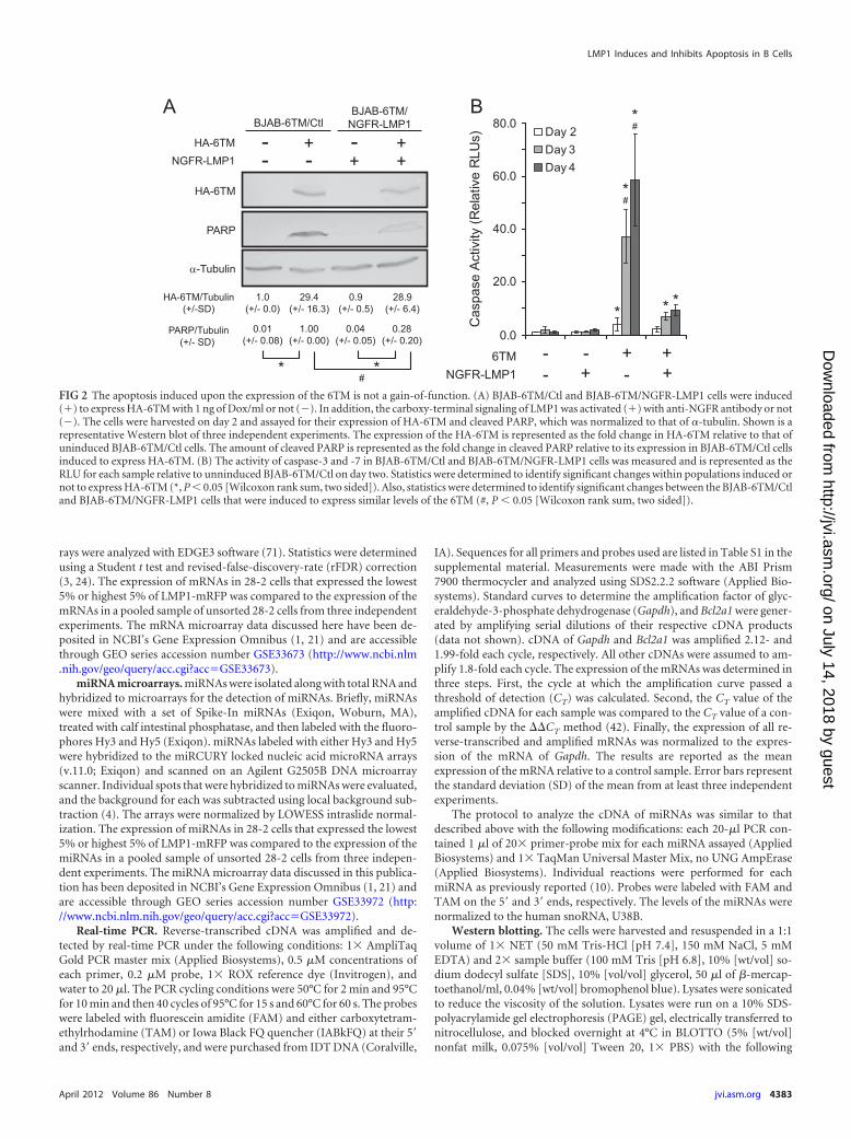
FIG 2 The apoptosis induced upon the expression of the 6TM is not a gain-of-function. (A) BJAB-6TM/Ctl and BJAB-6TM/NGFR-LMP1 cells were induced(�) to express HA-6TM with 1 ng of Dox/ml or not (�). In addition, the carboxy-terminal signaling of LMP1 was activated (�) with anti-NGFR antibody or not(�). The cells were harvested on day 2 and assayed for their expression of HA-6TM and cleaved PARP, which was normalized to that of �-tubulin. Shown is arepresentative Western blot of three independent experiments. The expression of the HA-6TM is represented as the fold change in HA-6TM relative to that ofuninduced BJAB-6TM/Ctl cells. The amount of cleaved PARP is represented as the fold change in cleaved PARP relative to its expression in BJAB-6TM/Ctl cellsinduced to express HA-6TM. (B) The activity of caspase-3 and -7 in BJAB-6TM/Ctl and BJAB-6TM/NGFR-LMP1 cells was measured and is represented as theRLU for each sample relative to unninduced BJAB-6TM/Ctl on day two. Statistics were determined to identify significant changes within populations induced ornot to express HA-6TM (*, P � 0.05 [Wilcoxon rank sum, two sided]). Also, statistics were determined to identify significant changes between the BJAB-6TM/Ctland BJAB-6TM/NGFR-LMP1 cells that were induced to express similar levels of the 6TM (#, P � 0.05 [Wilcoxon rank sum, two sided]).
LMP1 Induces and Inhibits Apoptosis in B Cells
April 2012 Volume 86 Number 8 jvi.asm.org 4383
on July 14, 2018 by guesthttp://jvi.asm
.org/D
ownloaded from

exceptions: (i) eIF2� and phospho-eIF2� were resolved by using SDS–12% PAGE, and (ii) Western blots used to detect 2�-myc-BCL2A1 wereincubated with the myc-antibody overnight rather than using BLOTTO.The blots were probed with primary antibody, followed by secondaryantibody conjugated to alkaline phosphatase. Secondary antibodies in-cluded donkey anti-mouse at 1:1,000 (Jackson Immunoresearch Labora-tories, West Grove, PA) and goat anti-rabbit at 1:1000 (Jackson Immu-noresearch Laboratories). The bands were visualized using a solution of0.13 mg of BCIP (5-bromo-4-chloro-3-indolylphosphate) p-toluidinesalt/ml and 0.33 mg of nitroblue tetrazolium chloride/ml. Western blotswere scanned and quantified using ImageQuant 5.2 software (GE Health-care, Piscataway, NJ). The expression of each protein was normalized tothe amount of �-tubulin. Experiments that were performed at least intriplicate were reported as the mean of the normalized expression �the SD.
The primary antibodies included mouse monoclonal anti-LMP1 an-tibody at 1:500 (Dako, Carpinteria, CA; clone CS1.4), mouse monoclonalanti-HA antibody at 1:2,000 (Sigma-Aldrich), rabbit polyclonal anti-poly-ADP-ribose polymerase (PARP; p85) antibody at 1:1,000 (Promega,Madison, WI), rabbit polyclonal anti-CREB-2 (ATF4) antibody at 1:100(Santa Cruz Biotechnology, Santa Cruz, CA; clone C-20), mouse mono-clonal anti-BiP antibody at 1:500 (BD Biosciences), mouse monoclonalanti-total eIF2� (Invitrogen) at 1:2,000, rabbit polyclonal anti-phosphor-ylated eIF2� (Invitrogen) at 1:500, unmodified mouse monoclonal anti-myc antibody (clone 9B11; Cell Signaling Technology, Beverly, MA) at1:1,000, and mouse monoclonal anti-�-tubulin antibody (Sigma-Al-drich) at 1:10,000.
CaspaseGLO 3/7 assay. Apoptosis was measured with the Caspase-GLO 3/7 assay (Promega). The CaspaseGLO 3/7 assay measures activity of
caspase-3 and -7 using light as a surrogate marker. Briefly, total cells werelysed and incubated for 1 h in the presence of luciferase, magnesium ion,and aminoluciferin covalently bound to the Asp-Glu-Val-Asp (DEVD)amino acid sequence. A total of 500 BJAB, BJAB-LMP1, and BJAB-6TMcells and their derivatives were assayed. Active caspase-3 and -7 cleaveafter the second aspartyl residue, releasing the aminoluciferin, a substrateof luciferase. Relative light units (RLU) were measured using a Monolight3010 luminometer (BD Biosciences). The results are reported as the meannormalized RLU from at least three independent experiments. Error barsrepresent the SDs of means.
Statistical analysis. Statistical tests (Wilcoxon rank sum) were per-formed using Mstat 5.01 provided by Norman Drinkwater (18).
RESULTS
The 6TM of LMP1, but not intact LMP1, induced apoptosis inthe EBV-negative B-cell line BJAB. The 6TM of LMP1 dose de-pendently induces the UPR and autophagy in B cells (37, 39, 40).LMP1 uses these pathways to regulate its synthesis and degrada-tion in EBV-positive B cells. However, B cells do not die when theyexpress intact LMP1, although their growth can be slowed (34,37). These observations are striking given that the UPR and au-tophagy can activate pathways of programmed cell death. Wesought to determine how LMP1 could regulate its expression inthis complex manner without causing B cells to die by apoptosis.BJAB cells were engineered to express conditionally either the6TM of LMP1 (BJAB-6TM) or intact LMP1 (BJAB-LMP1) tagged
FIG 3 BiP partially rescued BJAB cells expressing the 6TM from apoptosis. (A) BJAB cells were treated with either DMSO or 2.5 �g of tunicamycin (TUN)/mlfor 24 h, and then phospho-eIF2�, total eIF2�, and �-tubulin were each measured by Western blotting. Shown is a representative immunoblot of threeindependent experiments. The ratio of phospho-eIF2�-to-total eIF2� was calculated and normalized to the levels of �-tubulin and is represented as the foldchange in expression relative to that in DMSO-treated BJAB cells. (B) The activity of caspase-3 and -7 in DMSO- or TUN-treated BJAB cells was measured for2 days and is represented as the number of RLU relative to that in DMSO-treated BJAB cells. Statistics were determined in panels A and B to identify significantchanges between DMSO- and TUN-treated BJAB cells (*, P � 0.05 [Wilcoxon rank sum, two sided]). (C) BJAB-6TM/Ctl and BJAB-6TM/BiP cells were inducedto express HA-6TM with 0.3 ng of Dox/ml (�) or not (�). The expression of HA-6TM, BiP, ATF4, and cleaved PARP was assayed and normalized to the levelsof �-tubulin. The amount of HA-6TM, BiP, and ATF4 is represented as the fold change in expression relative to that in uninduced BJAB-6TM/Ctl cells. Theamount of cleaved PARP is represented as the fold change in expression relative to BJAB-6TM/Ctl cells in which the expression of 6TM was induced. (D) Also,the activity of caspase-3 and -7 was measured in BJAB-6TM/Ctl and BJAB-6TM/BiP cells. Caspase activity is represented as the RLU normalized to the RLU inuninduced BJAB-6TM/Ctl assayed on day 2. Statistics were determined for the data in panels C and D to identify significant differences within a population (*)or between populations of cells treated with similar amounts of Dox (#, P � 0.05 [Wilcoxon rank sum, two sided]).
Pratt et al.
4384 jvi.asm.org Journal of Virology
on July 14, 2018 by guesthttp://jvi.asm
.org/D
ownloaded from

with HA (37). HA-LMP1 and HA-6TM were expressed to similarlevels in these cell clones induced with Dox (Fig. 1A).
These cell clones were used to study the mechanisms by whichB cells survive the UPR and autophagy induced by LMP1. BJAB-LMP1 and BJAB-6TM were grown with or without Dox to induceor not induce, respectively, the expression of the variants of LMP1(Fig. 1A). Apoptosis, as reflected via caspase activity, was quanti-fied in two ways. First, cleaved PARP, a product of proteolysis bycaspases, was measured via Western blotting (Fig. 1B). Second, thecleavage of a luminescent substrate of activated caspase-3 and -7was measured (Fig. 1C). Apoptosis was detected as early as 2 daysafter inducing the expression of 6TM. The amount of cleavedPARP in induced BJAB-6TM cells was 6-fold greater than in un-induced cells (Fig. 1B). Similarly, induced BJAB-6TM cells had 8-and 33-fold more caspase activity after 2 and 3 days, respectively,than uninduced cells (Fig. 1C). In contrast, intact LMP1 did notcause apoptosis of BJAB cells. These findings indicated one ormore of the functions of the 6TM (aggregation for signaling, lo-calization to lipid rafts of Golgi, or the induction of the UPR andautophagy), but not intact LMP1, induced apoptosis (39–41).
The apoptosis induced by the 6TM is not a gain-of-functionbut is masked by the signaling of the carboxy-terminal domainof LMP1. Unlike the 6TM, the expression of intact LMP1 did notactivate apoptosis in BJAB cells (Fig. 1B and C). We hypothesizedthat signaling from the carboxy-terminal tail of LMP1 preventedapoptosis that was activated by the 6TM. Alternatively, it was pos-sible that the 6TM alone had gained an ability not present in intactLMP1. To determine whether the apoptosis caused by the 6TMwas a gain-of-function mutation, BJAB-6TM cells were engi-neered to express a derivative of LMP1, NGFR-LMP1, constitu-tively (BJAB-6TM/NGFR-LMP1). NGFR-LMP1 contains theamino terminus of NGFR fused to the carboxy terminus of LMP1,and the carboxy-terminal signaling of LMP1 is conditionally acti-vated when anti-NGFR antibody cross-links the NGFR moieties(26, 27). This construct can replace the signaling of intact LMP1,drives proliferation, and inhibits apoptosis in EBV-positive B-cellstrains (16, 26). In addition, we engineered BJAB-6TM cells toexpress the parental vector, pCMMP-MCS-IRES-mRFP, as a neg-ative control (BJAB-6TM/Ctl). The expression of the NGFR-
LMP1 in BJAB cells was confirmed to be similar to that of intactLMP1 by Western blotting (data not shown).
BJAB-6TM/Ctl and BJAB-6TM/NGFR-LMP1 cells were in-duced with Dox to express similar levels of HA-6TM (Fig. 2A).Activation of the carboxy-terminal signaling of LMP1 rescuedBJAB cells induced to express HA-6TM from apoptosis (Fig. 2).Similarly to BJAB-6TM cells, apoptosis was observed within 2days in BJAB-6TM/Ctl cells induced to express HA-6TM (Fig. 2).The apoptosis increased and on day 4 was 60-fold greater in in-duced BJAB-6TM/Ctl cells than in uninduced cells (Fig. 2B). Sig-naling from the carboxy terminus of LMP1 inhibited caspase ac-tivity by 84% each day and reduced the amount of detectablecleaved PARP by 72% (Fig. 2). Collectively, these data indicatedthat apoptosis activated by the 6TM of LMP1 was not a gain-of-function and that the carboxy-terminal signaling of LMP1 inhib-ited the apoptotic phenotype induced by the 6TM.
The apoptosis activated by the 6TM was induced, at least inpart, by the UPR. It seemed likely that the 6TM of LMP1 causedapoptosis via its activation of the UPR and of autophagy. In orderto test this likelihood, it was necessary to ascertain whether theUPR could cause apoptosis in BJAB cells or not. We sought toinduce the UPR chemically with tunicamycin, which causes ERstress (22). BJAB cells were treated with tunicamycin, or its carrier,DMSO, for 2 days. The increased ratio of phospho-eIF2� to totaleIF2� in BJAB cells treated with tunicamycin confirmed that theUPR was induced (Fig. 3A). After 1 and 2 days, caspase activity was12- and 28-fold greater, respectively, in BJAB cells treated withtunicamycin than with DMSO (Fig. 3B). This finding indicatedthat the UPR was a candidate for inducing apoptosis.
We next sought to determine whether inhibiting the UPRwould block the apoptosis induced by the 6TM of LMP1. To do so,the chaperone protein, BiP, was expressed exogenously in BJAB-6TM (BJAB-6TM/BiP) cells. The dissociation of BiP from PERK,IRE1�, and ATF6 activates the UPR (58). The level of the intro-duced BiP was �2-fold more than was endogenously expressed inBJAB-6TM/Ctl cells (Fig. 3C). The ability of BiP to repress theUPR was measured via the translation of ATF4, which was de-creased by 30% in the presence of exogenous BiP (Fig. 3C). After 2days of expressing the 6TM, the level of cleaved PARP was de-
FIG 4 The carboxy-terminal signaling of LMP1 inhibited apoptosis induced with tunicamycin (TUN). BJAB cells were transduced with a control vector(BJAB/Ctl) or a vector that expressed NGFR-LMP1 constitutively (BJAB/NGFR-LMP1). These cells were tested for their apoptotic response to increasingconcentrations of TUN. BJAB/NGFR-LMP1 cells were additionally treated with anti-NGFR antibody to activate the carboxy-terminal signaling of LMP1. (A) Theactivity of caspase-3 and -7 was measured via luminescence, and this activity is reported as the RLU relative to BJAB/Ctl cells cultured in the absence of TUN. (B)Cleaved PARP was measured in the two cell populations via Western blotting, and its levels were normalized to the expression of �-tubulin. The ratio of cleavedPARP to �-tubulin is represented as the fold change relative to BJAB/Ctl cells treated with 1 �g of TUN/ml. Statistics (P � 0.05 [Wilcoxon rank sum, two-sided])were determined to identify significant changes in caspase activity and cleaved PARP within the populations treated with differing concentrations of TUN (*) orbetween populations treated with the same concentration of TUN (#).
LMP1 Induces and Inhibits Apoptosis in B Cells
April 2012 Volume 86 Number 8 jvi.asm.org 4385
on July 14, 2018 by guesthttp://jvi.asm
.org/D
ownloaded from

FIG 5 The expression of LMP1-mRFP correlated with the differential expression of mRNAs encoding proteins that regulate apoptosis. (A) The EBV-positiveB-cell strain 28-2 was sorted by flow cytometry for the 5% of cells expressing the lowest (Low 5%) or highest (High 5%) levels of LMP1-mRFP. Monomers of
Pratt et al.
4386 jvi.asm.org Journal of Virology
on July 14, 2018 by guesthttp://jvi.asm
.org/D
ownloaded from
creased by 60% in the presence of exogenous BiP (Fig. 3C). Sim-ilarly, BiP inhibited the activity of caspase-3 and -7 by 25% after 2,3, and 4 days of inducing the expression of the 6TM (Fig. 3D).These data show that repressing the UPR blocked the apoptosisinduced by the 6TM of LMP1.
The carboxy-terminal signaling of LMP1 inhibited apoptosisinduced by the UPR in BJAB cells. That the carboxy-terminaldomain of LMP1 can inhibit apoptosis and its 6TM can activateapoptosis via the UPR led us to predict that the carboxy terminuscould inhibit apoptosis induced by the UPR when activated inde-pendently of the 6TM. To test this prediction, BJAB cells wereengineered to express NGFR-LMP1 (BJAB/NGFR-LMP1) or not(BJAB/Ctl), and apoptosis was measured during their treatmentwith increasing concentrations of tunicamycin. Tunicamycin in-duced the UPR to cause apoptosis in BJAB cells, and yet thecaspase activity was greatly inhibited by the carboxy-terminal sig-naling of LMP1 (Fig. 4A). Even when BJAB cells were treated withthe highest amount of tunicamycin (1 �g/ml), the carboxy-termi-nal signaling of LMP1 inhibited the UPR-induced apoptosis by90%. Similarly, the cleavage of PARP was inhibited by 90% whenthe carboxy-terminal signaling of LMP1 was activated in tunica-mycin-treated BJAB cells (Fig. 4B). These findings substantiatedthe prediction that the carboxy terminus of LMP1 can inhibitapoptosis induced by the UPR.
The signaling of LMP1 correlates with the expression of hu-man miRNAs and mRNAs. The carboxy-terminal signaling ofLMP1 strongly inhibited apoptosis induced by both 6TM and tu-nicamycin, supporting its role in overcoming UPR-activatedapoptosis in EBV-positive B cells. We sought to identify miRNAsand mRNAs that correlated with the expression of LMP1 in theEBV-positive B-cell strain, 28-2, and might contribute to the in-hibition of apoptosis, or regulate the UPR and autophagy.
28-2 cells contain EBV carrying a derivative of full-lengthLMP1 in which mRFP is fused to its carboxy terminus. The mRFPallows for sorting of single cells based on their levels of LMP1.Cells were sorted for those expressing the highest 5% or lowest 5%of LMP1-mRFP, which was confirmed by Western blotting forLMP1 (Fig. 5A). Thus, cells selected for the highest 5% levels ofLMP1-mRFP expressed 6-fold more of it than those sorted for thelowest 5% levels of this protein. The expression of LMP1-mRFPcorrelated dose dependently with markers of the UPR (Fig. 5B);the levels of BiP and ATF4 were 2- and 3.5-fold greater, respec-tively, in cells containing the highest levels of LMP1-mRFP than inthose containing the lowest levels. These results are consistentwith the dose-dependent induction of the UPR via LMP1 (40).
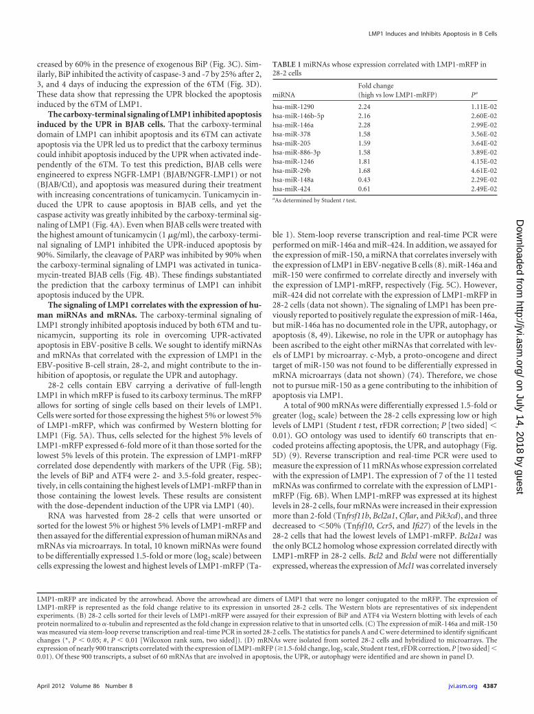
RNA was harvested from 28-2 cells that were unsorted orsorted for the lowest 5% or highest 5% levels of LMP1-mRFP andthen assayed for the differential expression of human miRNAs andmRNAs via microarrays. In total, 10 known miRNAs were foundto be differentially expressed 1.5-fold or more (log2 scale) betweencells expressing the lowest and highest levels of LMP1-mRFP (Ta-
ble 1). Stem-loop reverse transcription and real-time PCR wereperformed on miR-146a and miR-424. In addition, we assayed forthe expression of miR-150, a miRNA that correlates inversely withthe expression of LMP1 in EBV-negative B cells (8). miR-146a andmiR-150 were confirmed to correlate directly and inversely withthe expression of LMP1-mRFP, respectively (Fig. 5C). However,miR-424 did not correlate with the expression of LMP1-mRFP in28-2 cells (data not shown). The signaling of LMP1 has been pre-viously reported to positively regulate the expression of miR-146a,but miR-146a has no documented role in the UPR, autophagy, orapoptosis (8, 49). Likewise, no role in the UPR or autophagy hasbeen ascribed to the eight other miRNAs that correlated with lev-els of LMP1 by microarray. c-Myb, a proto-oncogene and directtarget of miR-150 was not found to be differentially expressed inmRNA microarrays (data not shown) (74). Therefore, we chosenot to pursue miR-150 as a gene contributing to the inhibition ofapoptosis via LMP1.
A total of 900 mRNAs were differentially expressed 1.5-fold orgreater (log2 scale) between the 28-2 cells expressing low or highlevels of LMP1 (Student t test, rFDR correction; P [two sided] �0.01). GO ontology was used to identify 60 transcripts that en-coded proteins affecting apoptosis, the UPR, and autophagy (Fig.5D) (9). Reverse transcription and real-time PCR were used tomeasure the expression of 11 mRNAs whose expression correlatedwith the expression of LMP1. The expression of 7 of the 11 testedmRNAs was confirmed to correlate with the expression of LMP1-mRFP (Fig. 6B). When LMP1-mRFP was expressed at its highestlevels in 28-2 cells, four mRNAs were increased in their expressionmore than 2-fold (Tnfrsf11b, Bcl2a1, Cflar, and Pik3cd), and threedecreased to �50% (Tnfsf10, Ccr5, and Ifi27) of the levels in the28-2 cells that had the lowest levels of LMP1-mRFP. Bcl2a1 wasthe only BCL2 homolog whose expression correlated directly withLMP1-mRFP in 28-2 cells. Bcl2 and Bclxl were not differentiallyexpressed, whereas the expression of Mcl1 was correlated inversely
LMP1-mRFP are indicated by the arrowhead. Above the arrowhead are dimers of LMP1 that were no longer conjugated to the mRFP. The expression ofLMP1-mRFP is represented as the fold change relative to its expression in unsorted 28-2 cells. The Western blots are representatives of six independentexperiments. (B) 28-2 cells sorted for their levels of LMP1-mRFP were assayed for their expression of BiP and ATF4 via Western blotting with levels of eachprotein normalized to �-tubulin and represented as the fold change in expression relative to that in unsorted cells. (C) The expression of miR-146a and miR-150was measured via stem-loop reverse transcription and real-time PCR in sorted 28-2 cells. The statistics for panels A and C were determined to identify significantchanges (*, P � 0.05; #, P � 0.01 [Wilcoxon rank sum, two sided]). (D) mRNAs were isolated from sorted 28-2 cells and hybridized to microarrays. Theexpression of nearly 900 transcripts correlated with the expression of LMP1-mRFP (�1.5-fold change, log2 scale, Student t test, rFDR correction, P [two sided] �0.01). Of these 900 transcripts, a subset of 60 mRNAs that are involved in apoptosis, the UPR, or autophagy were identified and are shown in panel D.
TABLE 1 miRNAs whose expression correlated with LMP1-mRFP in28-2 cells
miRNAFold change(high vs low LMP1-mRFP) Pa
hsa-miR-1290 2.24 1.11E-02hsa-miR-146b-5p 2.16 2.60E-02hsa-miR-146a 2.28 2.99E-02hsa-miR-378 1.58 3.56E-02hsa-miR-205 1.59 3.64E-02hsa-miR-886-3p 1.58 3.89E-02hsa-miR-1246 1.81 4.15E-02hsa-miR-29b 1.68 4.61E-02hsa-miR-148a 0.43 2.29E-02hsa-miR-424 0.61 2.49E-02aAs determined by Student t test.
LMP1 Induces and Inhibits Apoptosis in B Cells
April 2012 Volume 86 Number 8 jvi.asm.org 4387
on July 14, 2018 by guesthttp://jvi.asm
.org/D
ownloaded from

with LMP1 (Fig. 5D). In addition, reverse transcription and real-time PCR were used to assay for the differential expression of Pten,Bak1, Xbp1, and Pik3c3, but none was observed, and these findingswere not consistent with the mRNA microarrays (data notshown). The expression of LMP1 correlates with the splicing of themRNA of Xbp1 in EBV-positive and -negative B cells, but theanalysis we performed did not differentiate between the splicedand unspliced forms of the mRNA (40). Other mRNAs encodinggenes likely involved in UPR-induced apoptosis were identified,including Casp9, Birc2, Tp63l, and Scotin (Fig. 5D) (6, 11, 30, 32,45, 54, 56, 67, 72, 81).
BCL2A1 is a bona fide candidate for mediating the survival ofB cells that express LMP1. It was predicted that for candidategenes to contribute to the inhibition of apoptosis induced by the6TM, their expression would correlate with the expression of in-tact LMP1 in two additional models: (i) an independent EBV-
positive B-cell strain and (ii) BJAB-LMP1 cells, but not BJAB-6TM cells. These criteria were met by three of the seven mRNAsconfirmed by real-time PCR to correlate with the expression ofLMP1 in 28-2 cells (Fig. 6B). 22-5 cells, an EBV-positive B-cellclone similar to 28-2 cells, were sorted for levels of LMP1-mRFPvia flow cytometry (Fig. 6A). The expression of Bcl2a1, Pik3cd, andCflar correlated directly with the expression of LMP1-mRFP in22-5 cells (Fig. 6B). Furthermore, the expression of both Pik3cdand Cflar was 3-fold greater in BJAB cells induced to express HA-LMP1 than uninduced cells, and the expression of Bcl2a1 in-creased 8-fold in the same cells (Fig. 6C). However, when theexpression of HA-6TM was induced in BJAB cells, the levels of themRNAs of Bcl2a1 and Cflar remained unchanged, whereasthe amount of mRNAs of Pik3cd mRNA was reduced by 65%.
The mRNAs of Cflar and Pik3cd encode caspase-8 andFADD-like apoptosis regulator, abbreviated as c-FLIP, and
FIG 6 The expression of the mRNA of Bcl2a1 correlated with the signaling of LMP1 in EBV-positive and -negative B cells. (A) 22-5 cells were sorted by flowcytometry for the 5% of cells expressing the lowest (Low 5%) or highest (High 5%) levels of LMP1-mRFP. The expression of the LMP1-mRFP was normalizedto �-tubulin and is represented as the fold change relative to the expression in cells expressing the lowest levels of LMP1-mRFP. The Western blots shown arerepresentatives of three independent experiments. (B) mRNAs encoding Ifi27, Tnfsf10, Ccr5, Tnfrsf11b, Bcl2a1, Pik3cd, and Cflar were measured for theircorrelation with LMP1-mRFP in 28-2 and 22-5 cells. The expression of each mRNA is represented as the fold change in expression relative to that of unsorted 28-2cells or to that of 22-5 cells expressing the lowest 5% of mRFP. (C) The expression of the mRNA of Bcl2a1 (i), Cflar (ii), and Pik3cd (iii) was measured in BJAB,BJAB-LMP1, and BJAB-6TM cells that were treated with Dox (On) to induce the expression of the variants of LMP1 or left untreated (Off). (D) The expressionof the mRNA of Bcl2a1 was measured BJAB-6TM/Ctl and BJAB-6TM/NGFR-LMP1 cells treated with (�) or without (�) Dox and anti-NGFR antibody. Theexpression of the mRNA of Bcl2a1 is represented as the fold change relative to the expression in untreated BJAB, BJAB-LMP1, and BJAB-6TM cells in panel C andBJAB-6TM/Ctl cells in panel D. Statistics were determined to identify significant changes in expression (*, P � 0.05; #, P � 0.01 [Wilcoxon rank sum, two sided]).
Pratt et al.
4388 jvi.asm.org Journal of Virology
on July 14, 2018 by guesthttp://jvi.asm
.org/D
ownloaded from

phosphoinositide-3 kinase delta (PI3K�), respectively. Ifc-FLIP were to contribute to the LMP1-mediated inhibition ofapoptosis, its levels should increase in BJAB-LMP1 cells, butnot BJAB-6TM cells, upon the induction of the derivatives ofLMP1 with Dox. There was no change in the levels of c-FLIPwhen HA-LMP1 was induced in BJAB cells, as determined viaWestern blotting (data not shown). Western blotting was alsoused to quantify the phosphorylation of v-akt murine thy-moma viral oncogene homolog 1 (AKT), which is a reflectionof the activity of PI3K�. The phosphorylation of AKT was un-affected by HA-LMP1 in BJAB cells (data not shown). Thesefindings indicated LMP1 did not inhibit apoptosis via the in-creased expression of Pik3cd or Cflar.
In addition, we measured the expression of the mRNA ofBcl2a1 via reverse-transcription and real-time PCR in BJAB-6TMcells expressing NGFR-LMP1 or not (Ctl). When anti-NGFR an-tibody was added to cells expressing NGFR-LMP1 in the absenceof Dox, there was a 20-fold increase in the expression of themRNA of Bcl2a1 compared either to cells left untreated or toBJAB-6TM/Ctl cells regardless of their treatment (Fig. 6D). Whenboth anti-NGFR antibody and Dox were added, the levels of themRNA of Bcl2a1 increased 5-fold more in BJAB-6TM/NGFR-LMP1 cells than in the BJAB-6TM/Ctl cells. Altogether, the obser-vations in the B-cell strains 28-2 and 22-5 and in derivatives ofBJAB expressing variants of LMP1 indicated that Bcl2a1 was the
only candidate for contributing to the LMP1-mediated inhibitionof apoptosis among the genes tested.
The constitutive, exogenous expression of BCL2A1 inhibitedapoptosis in BJAB cells that also expressed the 6TM of LMP1.Bcl2a1 was an attractive candidate for inhibiting apoptosis in-duced by the 6TM of LMP1 because it binds and inhibits theactivities of both truncated BID (tBID) and BAK, proapoptoticproteins whose steady-state levels increase during the UPR (61).Thus, the ability of BCL2A1 to inhibit the activities of tBID andBAK could block UPR-induced apoptosis. Also, LMP1 has previ-ously been reported to activate the transcription of the mRNA ofBcl2a1 in B cells via NF-�B, and Bcl2a1 was the only mRNA en-coding an antiapoptotic member of the BCL2 family to correlatedirectly with the expression of LMP1 (Fig. 5D) (7, 19).
For these reasons, the ability of BCL2A1 to inhibit apoptosisinduced by the 6TM was tested. BJAB-6TM cells were transducedwith a vector that expresses 2�-myc-BCL2A1 (Fig. 7A and B). ThemRNA encoding the 2�-myc-BCL2A1 protein was expressed at35-fold-greater levels in this population of cells (BJAB-6TM/BCL2A1) than in uninduced BJAB-6TM/Ctl cells (Fig. 7B). BothBJAB-6TM/Ctl and BJAB-6TM/BCL2A1 cells were treated withincreasing concentrations of Dox to induce the expression of HA-6TM, which they did at similar levels (Fig. 7A).
We were unable to repress the expression of Bcl2a1 withshRNAs; however, the exogenous expression of BCL2A1 inhibited
FIG 7 The constitutive, exogenous expression of BCL2A1 inhibited apoptosis in BJAB cells that also expressed the 6TM. (A) BJAB-6TM/Ctl and BJAB-6TM/BCL2A1 cells were treated with 0 (�), 0.1 (�), or 0.3 (��) ng of Dox/ml to induce the expression of HA-6TM. Cells were harvested after 2 days to assay for theexpression of HA-6TM, 2�-myc-BCL2A1, and cleaved PARP. The expression of each protein was normalized to the levels of �-tubulin. The expression of theHA-6TM and 2�-myc-BCL2A1 is represented as the fold change in expression relative to that in uninduced BJAB-6TM/Ctl cells. The expression of cleaved PARP(� the SD) is represented as the fold change in expression relative to that in BJAB-6TM/Ctl cells induced to express HA-6TM with 0.3 ng of Dox/ml. (B) Theexpression of the mRNA encoding 2�-myc-BCL2A1 was measured via real-time PCR. The expression of the mRNA is represented as the fold change inexpression relative to that in uninduced BJAB-6TM/Ctl cells. (C) Caspase activity in BJAB-6TM/Ctl and BJAB-6TM/BCL2A1 cells induced to express HA-6TMdose dependently was measured. The caspase activity was normalized to the RLU of uninduced BJAB-6TM/Ctl cells on day 2. Statistics (P � 0.05 [Wilcoxon ranksum, two sided]) were determined to identify significant differences within populations treated with different concentrations of Dox (*) or between populationstreated with the same concentration of Dox (#).
LMP1 Induces and Inhibits Apoptosis in B Cells
April 2012 Volume 86 Number 8 jvi.asm.org 4389
on July 14, 2018 by guesthttp://jvi.asm
.org/D
ownloaded from
apoptosis induced by the 6TM. After 2 days of expressing the 6TMwith 0.1 or 0.3 ng of Dox/ml, only 50 and 40% of the PARP werecleaved, respectively, in BJAB cells expressing 2�-myc-BCL2A1compared to BJAB-6TM/Ctl cells (Fig. 7A). The activity ofcaspase-3 and -7 was similarly repressed by 2�-myc-BCL2A1 inthe presence of the 6TM of LMP1, where BCL2A1 inhibited apop-tosis by 35% each day in both Dox treatments (Fig. 7C). Thesefindings indicate that BCL2A1 inhibits apoptosis induced by the6TM of LMP1.
The expression of the mRNA of Bcl2a1 did not correlate withthe expression of LMP1 in epithelial cells in which LMP1 in-duces apoptosis. The signaling of LMP1 in epithelial cells may notparallel its signaling in B cells. For example, LMP1 does not inducethe UPR in the EBV-negative, NPC-derived cell line, HONE, as itdoes in B cells (33). We therefore sought to determine whetherLMP1 was capable of inducing apoptosis and activating the tran-scription of Bcl2a1 in two EBV-negative epithelial cell lines,H1299 and HeLa.
Both H1299 and HeLa were transfected with increasingamounts of a vector encoding intact LMP1. For both H1299 andHeLa, 60% of the cells were transfected after 24 h as assayed by theexpression of eGFP encoded by a cotransfected vector (data notshown). Western blots performed on lysates from cells harvestedat 24 h posttransfection showed LMP1 was expressed at increasinglevels in both cell lines (Fig. 8A). Intact LMP1 induced apoptosisin a dose-dependent manner in both H1299 and HeLa, as assayedby determining the activity of caspase-3 and -7 and the cleavage ofPARP (Fig. 8). The expression of the mRNA of Bcl2a1 was unde-tectable when assayed via real-time PCR (data not shown), a find-ing consistent with the notion that the capacity of LMP1 to inducethe mRNA of Bcl2a1 in B cells contributes to inhibiting its induc-tion of apoptosis in those cells.
DISCUSSION
The LMP1 oncogene is regulated through its effects on two cellu-lar pathways. The 6TM induces both the UPR and autophagy dosedependently in B cells and, in doing so, LMP1 can regulate its ownexpression (37–40). This regulation needs to be compatible withEBV’s successful, long-term infection of B cells and, accordingly,the activation of the UPR and autophagy via LMP1 does not causeB cells to die. We have now found in B cells that signaling from thecarboxy-terminal moiety of LMP1 inhibits the apoptosis that its
6TM induces as a consequence of its activation of the UPR andautophagy.
How does LMP1 inhibit its activation of apoptosis? The signal-ing of CD40 likely provides some clues. CD40 activates the tran-scription of mRNAs that encode antiapoptotic proteins (2). LMP1activates the same pathways as does CD40, activates the transcrip-tion of many of the same mRNAs as does CD40, and can replacethe signaling of CD40 in B cells in vivo (15, 28, 29, 35, 55, 68, 80).The analysis of microarrays showed that the expression of LMP1in EBV-positive B cells correlated with the expression of mRNAsthat affect apoptosis (Fig. 5D). The levels of seven of these mRNAsfound to correlate with the expression of LMP1 were validated viareverse transcription and real-time PCR (Fig. 6B). Both themRNAs encoding Pik3cd and Cflar correlated directly with theexpression of LMP1-mRFP (Fig. 6B). HA-LMP1 had no affect onthe phosphorylation of AKT in BJAB cells or on the levels ofCFLAR protein (Fig. 6B). Therefore, we concluded they could notgenerally contribute to the inhibition of apoptosis conferred to Bcells by the carboxy-terminal domain of LMP1. Among the mR-NAs that were affected by LMP1-mRFP, the one encodingBCL2A1, a homolog of BCL2, was the only candidate of the anti-apoptotic BCL2 family that correlated directly with levels ofLMP1-mRFP (Fig. 5D). These findings are supported by data thatLMP1 does not regulate the expression of BCL-2 nor BCL-xL in Bcells. The expression of mRNAs encoding Bcl2 and Bclxl, or theproteins, BCL2 and BCL-xL, do not change detectably when pri-mary B cells are infected with EBV (44; M. Sandburg, unpublisheddata). Similarly, the mRNA of Bcl2a1, but not other BCL2 ho-mologs, correlates with the conditional activation of the carboxy-terminal signaling of LMP1 in EBV-positive B-cell strains (16). Inaddition, the expression of the mRNA of Bcl2a1, but not that ofBclxl, is greater in lymphoblastoid cell lines (LCLs) than EBV-negative Burkitt’s lymphoma cell lines (BLs) (7).
We investigated Bcl2a1 further because its transcription is ac-tivated by both LMP1 and CD40 in EBV-negative B cells (7, 19,20). Activation of its transcription is dependent on NF-�B andsignaling from the carboxy-terminal transactivating region 2 ofLMP1 (20). BCL2A1 is also expressed at greater levels in B-cellchronic lymphocytic leukemia cells (B-CLLs) than in normal Bcells, helping to render B-CLLs resistant to apoptosis (48, 52, 70).Likewise, the exogenous expression of BCL2A1 in EBV-positiveMutuI cells inhibits apoptosis that is induced by serum starvation
FIG 8 The expression of LMP1 induced apoptosis in epithelial cells. (A) HeLa and H1299 cells were transfected with 0, 0.3, 1, or 3 �g of cDNA encoding LMP1.The expression of LMP1 and cleaved PARP was normalized to �-tubulin and is represented as the fold change in expression relative to HeLa or H1299 cellstransfected with 3 �g of cDNA encoding LMP1. (B) Caspase activity was measured 24 h posttransfection and is represented as the mean normalized RLU relativeto untransfected HeLa or H1299 cells. Statistics were determined to identify significant changes (*, P � 0.05 [Wilcoxon rank sum, two sided]).
Pratt et al.
4390 jvi.asm.org Journal of Virology
on July 14, 2018 by guesthttp://jvi.asm
.org/D
ownloaded from

(19). Our results show that the exogenous expression of BCL2A1inhibited apoptosis in BJAB cells induced to express HA-6TM,illustrating the induction of BCL2A1 by LMP1 is one way by whichLMP1 inhibits the apoptosis that it induces (Fig. 7 and 8).
The interplay between the 6TM of LMP1 in inducing apoptosisand its carboxy terminus in inhibiting apoptosis is similar to someof the combined functions of the B-cell receptor (BCR) and CD40in immature B cells (Fig. 9). The stimulation of the BCR in pri-mary B cells isolated from mice and in WEHI-231 B cells inducesthe UPR and autophagy (63, 73, 75). The activation of the UPR inimmature B cells also induces apoptosis and stimulated CD40 canrescue these B cells from the UPR-induced apoptosis (12, 63, 75).The induction of the UPR and autophagy by LMP1 can, undersome conditions, induce apoptosis, as can a stimulated BCR. Twofindings support this conclusion. First, EBV-negative BJAB cellsinduced to express the 6TM supported the UPR and died by apop-tosis (Fig. 1 and 3C) (38–40). Second, apoptosis induced by the6TM of LMP1 was partially inhibited when BiP, a chaperone pro-tein and mediator of the UPR, was expressed exogenously (Fig. 3Cand D). This partial inhibition likely reflects only a partial inhibi-tion of the UPR by BiP in these experiments. The exogenous ex-pression of BiP in WEHI-231 cells inhibits apoptosis after stimu-lation of the BCR (75). However, unlike the BCR, which requiresbinding to immunoglobulin to stimulate its signaling, the 6TMconstitutively induces the UPR, autophagy, and apoptosis in adose-dependent manner (Fig. 3C and 5B) (38, 40). Intact LMP1induces the UPR and autophagy, but also antiapoptotic genes,including Bcl2a1, to inhibit apoptosis and allow EBV-infected Bcells to survive to proliferate (Fig. 9).
Unlike its 6TM, the carboxy-terminal domain of LMP1 cannotfunction independently. Rather, the carboxy-terminal domainmust be oligomerized to signal, much like CD40 (28). Therefore,the carboxy-terminal signaling of intact LMP1 is directly linked tothe activities of its 6TM and is influenced by phenotypes in B cellsgenerated by the 6TM, including the UPR and autophagy. In con-trast, the signaling of CD40 occurs independently of the signalingof BCR. Thus, differences in the phenotypes of B cells signaling via
intact LMP1 or CD40 could be explained by the environments inwhich they signal.
ACKNOWLEDGMENTS
This study was supported by a Cancer Biology Predoctoral Training Grantfrom the National Institutes of Health (NIH; T32CA009135) and NIHgrants CA70723 and CA22443. B.S. is an American Cancer Society Re-search Professor.
The vector expressing NGFR-LMP1 was a gift from Wolfgang Ham-merschmidt and that expressing 2�-myc-BCL2A1 was a gift from CelineGélinas. We thank Dong Yun Lee for her helpful discussions throughoutthis work, Mark Sandburg for sharing unpublished data on the transcrip-tion of human genes upon the infection of primary B cells with EBV,Mitch Hayes for his help in preparing the microarray data for submissionto GEO, and both Danielle Westhoff Smith and Ngan Lam for their carefulreading of the manuscript.
REFERENCES1. Barrett T, et al. 2011. NCBI GEO: archive for functional genomics data
sets—10 years on. Nucleic Acids Res. 39:D1005–D1010.2. Basso K, et al. 2004. Tracking CD40 signaling during germinal center
development. Blood 104:4088 – 4096.3. Benjamini Y, Hochberg Y. 1995. Controlling the false discovery rate: a
practical and powerful approach to multiple testing. J. R. Stat. Soc. B57:289 –300.
4. Berger JA, et al. 2004. Optimized LOWESS normalization parameterselection for DNA microarray data. BMC Bioinform. 5:194.
5. Bernales S, McDonald KL, Walter P. 2006. Autophagy counterbalancesendoplasmic reticulum expansion during the unfolded protein response.PLoS Biol. 4:e423.
6. Bourdon JC, Renzing J, Robertson PL, Fernandes KN, Lane DP. 2002.Scotin, a novel p53-inducible proapoptotic protein located in the ER andthe nuclear membrane. J. Cell Biol. 158:235–246.
7. Cahir-McFarland ED, et al. 2004. Role of NF-�B in cell survival andtranscription of latent membrane protein 1-expressing or Epstein-Barrvirus latency III-infected cells. J. Virol. 78:4108 – 4119.
8. Cameron JE, et al. 2008. Epstein-Barr virus latent membrane protein 1induces cellular MicroRNA miR-146a, a modulator of lymphocyte signal-ing pathways. J. Virol. 82:1946 –1958.
9. Carbon S, et al. 2009. AmiGO: online access to ontology and annotationdata. Bioinformatics 25:288 –289.
10. Chen C, et al. 2005. Real-time quantification of microRNAs by stem-loopRT-PCR. Nucleic Acids Res. 33:e179.
11. Cheung HH, Lynn Kelly N, Liston P, Korneluk RG. 2006. Involvementof caspase-2 and caspase-9 in endoplasmic reticulum stress-inducedapoptosis: a role for the IAPs. Exp. Cell Res. 312:2347–2357.
12. Choi MS, et al. 1995. The role of bcl-XL in CD40-mediated rescue fromanti-mu-induced apoptosis in WEHI-231 B lymphoma cells. Eur. J. Im-munol. 25:1352–1357.
13. Chomczynski P, Sacchi N. 1987. Single-step method of RNA isolation byacid guanidinium thiocyanate-phenol-chloroform extraction. Anal.Biochem. 162:156 –159.
14. Deniaud A, Sharaf el Dein O, et al. 2008. Endoplasmic reticulum stressinduces calcium-dependent permeability transition, mitochondrial outermembrane permeabilization and apoptosis. Oncogene 27:285–299.
15. Devergne O, et al. 1996. Association of TRAF1, TRAF2, and TRAF3 withan Epstein-Barr virus LMP1 domain important for B-lymphocyte trans-formation: role in NF-�B activation. Mol. Cell. Biol. 16:7098 –7108.
16. Dirmeier U, et al. 2005. Latent membrane protein 1 of Epstein-Barr viruscoordinately regulates proliferation with control of apoptosis. Oncogene24:1711–1717.
17. Djavaheri-Mergny M, Maiuri MC, Kroemer G. 2010. Cross talk betweenapoptosis and autophagy by caspase-mediated cleavage of Beclin 1. Onco-gene 29:1717–1719.
18. Drinkwater N. 2008. Mstat. McArdle Laboratory for Cancer Research,University of Wisconsin-Madison, Madison, WI. http://mcardle.oncology.wisc.edu/mstat.
19. D’Souza B, Rowe M, Walls D. 2000. The bfl-1 gene is transcriptionallyupregulated by the Epstein-Barr virus LMP1, and its expression promotesthe survival of a Burkitt’s lymphoma cell line. J. Virol. 74:6652– 6658.
FIG 9 Comparison of how apoptosis is inhibited after the induction of theUPR by the BCR or LMP1. The independent signaling of the 6TM of LMP1 andits carboxy-terminal tail resembles the interaction between stimulated BCRand stimulated CD40 in immature B cells with anti-BCR and anti-CD40 anti-bodies, respectively. The stimulation of the BCR with anti-BCR antibody canactivate the UPR, which then induces apoptosis. Similarly, the 6TM of LMP1activates the UPR constitutively in the absence of a ligand, which also inducesapoptosis. Constitutive signaling from the carboxy-terminal domain of LMP1,like the conditional signaling of CD40, inhibits apoptosis induced by the UPR.Bcl2a1 is one gene both LMP1 and CD40 activate, and BCL2A1 inhibits theUPR-induced apoptosis activated by LMP1.
LMP1 Induces and Inhibits Apoptosis in B Cells
April 2012 Volume 86 Number 8 jvi.asm.org 4391
on July 14, 2018 by guesthttp://jvi.asm
.org/D
ownloaded from

20. D’Souza BN, et al. 2004. Nuclear factor �B-dependent activation of theantiapoptotic bfl-1 gene by the Epstein-Barr virus latent membrane pro-tein 1 and activated CD40 receptor. J. Virol. 78:1800 –1816.
21. Edgar R, Domrachev M, Lash AE. 2002. Gene Expr. Omnibus: NCBIgene expression and hybridization array data repository. Nucleic AcidsRes. 30:207–210.
22. Feige JJ, Scheffler IE. 1987. Analysis of the protein glycosylation defect ofa temperature-sensitive cell cycle mutant by the use of mutant cells over-expressing the human epidermal growth factor receptor after transfectionof the gene. J. Cell Physiol. 133:461– 470.
23. Gaillard C, Strauss F. 1990. Ethanol precipitation of DNA with linearpolyacrylamide as carrier. Nucleic Acids Res. 18:378.
24. Genovese C, Wasserman L. 2004. A stochastic approach to false discoverycontrol. Ann. Stat. 32:1035–1061.
25. Giaccone G, et al. 1992. Neuromedin B is present in lung cancer cell lines.Cancer Res. 52:2732s–2736s.
26. Gires O, et al. 1999. Latent membrane protein 1 of Epstein-Barr virusinteracts with JAK3 and activates STAT proteins. EMBO J. 18:3064 –3073.
27. Gires O, Ueffing M, Hammerschmidt W. 2001. Chimeric and mutatedvariants of LMP1. A helpful tool to analyze the structure-function rela-tionship of a pseudoreceptor. Methods Mol. Biol. 174:313–323.
28. Gires O, et al. 1997. Latent membrane protein 1 of Epstein-Barr virusmimics a constitutively active receptor molecule. EMBO J. 16:6131– 6140.
29. Graham JP, Arcipowski KM, Bishop GA. 2010. Differential B-lymphocyteregulation by CD40 and its viral mimic, latent membrane protein 1. Immu-nol. Rev. 237:226–248.
30. Hamanaka RB, Bobrovnikova-Marjon E, Ji X, Liebhaber SA, Diehl JA.2009. PERK-dependent regulation of IAP translation during ER stress.Oncogene 28:910 –920.
31. Heath-Engel HM, Chang NC, Shore GC. 2008. The endoplasmic retic-ulum in apoptosis and autophagy: role of the BCL-2 protein family. On-cogene 27:6419 – 6433.
32. Herold MJ, Kuss AW, Kraus C, Berberich I. 2002. Mitochondria-dependent caspase-9 activation is necessary for antigen receptor-mediatedeffector caspase activation and apoptosis in WEHI 231 lymphoma cells. J.Immunol. 168:3902–3909.
33. Hsiao JR, et al. 2009. Endoplasmic reticulum stress triggers XBP-1-mediated up-regulation of an EBV oncoprotein in nasopharyngeal carci-noma. Cancer Res. 69:4461– 4467.
34. Kaykas A, Sugden B. 2000. The amino terminus and membrane-spanning domains of LMP-1 inhibit cell proliferation. Oncogene 19:1400 –1410.
35. Kilger E, Kieser A, Baumann M, Hammerschmidt W. 1998. Epstein-Barr virus-mediated B-cell proliferation is dependent upon latent mem-brane protein 1, which simulates an activated CD40 receptor. EMBO J.17:1700 –1709.
36. Lai E, Teodoro T, Volchuk A. 2007. Endoplasmic reticulum stress:signaling the unfolded protein response. Physiology (Bethesda) 22:193–201.
37. Lam N, Sandberg ML, Sugden B. 2004. High physiological levels of LMP1result in phosphorylation of eIF2 alpha in Epstein-Barr virus-infectedcells. J. Virol. 78:1657–1664.
38. Lee DY, Lee J, Sugden B. 2009. The unfolded protein response andautophagy: herpesviruses rule! J. Virol. 83:1168 –1172.
39. Lee DY, Sugden B. 2008. The latent membrane protein 1 oncogene mod-ifies B-cell physiology by regulating autophagy. Oncogene 27:2833–2842.
40. Lee DY, Sugden B. 2008. The LMP1 oncogene of EBV activates PERK andthe unfolded protein response to drive its own synthesis. Blood 111:2280 –2289.
41. Lee J, Sugden B. 2007. A membrane leucine heptad contributes to traf-ficking, signaling, and transformation by latent membrane protein 1. J.Virol. 81:9121–9130.
42. Livak KJ, Schmittgen TD. 2001. Analysis of relative gene expression datausing real-time quantitative PCR and the 2���CT method. Methods 25:402– 408.
43. Marciniak SJ, et al. 2004. CHOP induces death by promoting proteinsynthesis and oxidation in the stressed endoplasmic reticulum. Genes Dev.18:3066 –3077.
44. Martin JM, Veis D, Korsmeyer SJ, Sugden B. 1993. Latent membraneprotein of Epstein-Barr virus induces cellular phenotypes independentlyof expression of Bcl-2. J. Virol. 67:5269 –5278.
45. Masud A, et al. 2007. Endoplasmic reticulum stress-induced death of
mouse embryonic fibroblasts requires the intrinsic pathway of apoptosis.J. Biol. Chem. 282:14132–14139.
46. McCullough KD, Martindale JL, Klotz LO, Aw TY, Holbrook NJ. 2001Gadd153 sensitizes cells to endoplasmic reticulum stress by down-regulating Bcl2 and perturbing the cellular redox state. Mol. Cell. Biol.21:1249 –1259.
47. Menezes J, Leibold W, Klein G, Clements G. 1975. Establishment andcharacterization of an Epstein-Barr virus (EBC)-negative lymphoblastoidB cell line (BJA-B) from an exceptional, EBV-genome-negative AfricanBurkitt’s lymphoma. Biomedicine. 22:276 –284.
48. Morales AA, et al. 2005. High expression of bfl-1 contributes to theapoptosis resistant phenotype in B-cell chronic lymphocytic leukemia.Int. J. Cancer 113:730 –737.
49. Motsch N, Pfuhl T, Mrazek J, Barth S, Grasser FA. 2007. Epstein-Barrvirus-encoded latent membrane protein 1 (LMP1) induces the expressionof the cellular microRNA miR-146a. RNA Biol. 4:131–137.
50. Novoa I, Zeng H, Harding HP, Ron D. 2001. Feedback inhibition of theunfolded protein response by GADD34-mediated dephosphorylation ofeIF2�. J. Cell Biol. 153:1011–1022.
51. Ogata M, et al. 2006. Autophagy is activated for cell survival after endo-plasmic reticulum stress. Mol. Cell. Biol. 26:9220 –9231.
52. Olsson A, et al. 2007. Upregulation of bfl-1 is a potential mechanism ofchemoresistance in B-cell chronic lymphocytic leukaemia. Br. J. Cancer97:769 –777.
53. Oyadomari S, et al. 2002. Targeted disruption of the Chop gene delaysendoplasmic reticulum stress-mediated diabetes. J. Clin. Invest. 109:525–532.
54. Pyati UJ, et al. 2011. p63 mediates an apoptotic response to pharmaco-logical and disease-related ER stress in the developing epidermis. Dev. Cell21:492–505.
55. Rastelli J, et al. 2008. LMP1 signaling can replace CD40 signaling in B cellsin vivo and has unique features of inducing class-switch recombination toIgG1. Blood 111:1448 –1455.
56. Rosati E, et al. 2010. Novel targets for endoplasmic reticulum stress-induced apoptosis in B-CLL. Blood 116:2713–2723.
57. Scherer WF, Syverton JT, Gey GO. 1953. Studies on the propagation invitro of poliomyelitis viruses. IV. Viral multiplication in a stable strain ofhuman malignant epithelial cells (strain HeLa) derived from an epider-moid carcinoma of the cervix. J. Exp. Med. 97:695–710.
58. Schroder M, Kaufman RJ. 2005. The mammalian unfolded protein re-sponse. Annu. Rev. Biochem. 74:739 –789.
59. Scorrano L, et al. 2003. BAX and BAK regulation of endoplasmic reticu-lum Ca2�: a control point for apoptosis. Science 300:135–139.
60. Shimizu S, et al. 2004. Role of Bcl-2 family proteins in a non-apoptoticprogrammed cell death dependent on autophagy genes. Nat. Cell Biol.6:1221–1228.
61. Simmons MJ, et al. 2008. Bfl-1/A1 functions, similar to Mcl-1, as aselective tBid and Bak antagonist. Oncogene 27:1421–1428.
62. Sjoblom A, Yang W, Palmqvist L, Jansson A, Rymo L. 1998. AnATF/CRE element mediates both EBNA2-dependent and EBNA2-independent activation of the Epstein-Barr virus LMP1 gene promoter. J.Virol. 72:1365–1376.
63. Skalet AH, et al. 2005. Rapid B cell receptor-induced unfolded proteinresponse in nonsecretory B cells correlates with pro- versus antiapoptoticcell fate. J. Biol. Chem. 280:39762–39771.
64. Szegezdi E, Logue SE, Gorman AM, Samali A. 2006. Mediators ofendoplasmic reticulum stress-induced apoptosis. EMBO Rep. 7:880 – 885.
65. Szegezdi E, Macdonald DC, Ni Chonghaile T, Gupta S, Samali A. 2009.Bcl-2 family on guard at the ER. Am. J. Physiol. Cell Physiol. 296:C941–C953.
66. Tabas I, Ron D. 2011. Integrating the mechanisms of apoptosis inducedby endoplasmic reticulum stress. Nat. Cell Biol. 13:184 –190.
67. Terrinoni A, et al. 2004. p73-alpha is capable of inducing scotin and ERstress. Oncogene 23:3721–3725.
68. Uchida J, et al. 1999. Mimicry of CD40 signals by Epstein-Barr virusLMP1 in B lymphocyte responses. Science 286:300 –303.
69. Ullman E, et al. 2008. Autophagy promotes necrosis in apoptosis-deficient cells in response to ER stress. Cell Death Differ. 15:422– 425.
70. Vogler M, et al. 2009. Concurrent up-regulation of BCL-XL and BCL2A1induces approximately 1000-fold resistance to ABT-737 in chronic lym-phocytic leukemia. Blood 113:4403– 4413.
71. Vollrath AL, Smith AA, Craven M, Bradfield CA. 2009. EDGE(3): aweb-based solution for management and analysis of Agilent two colormicroarray experiments. BMC Bioinform. 10:280.
Pratt et al.
4392 jvi.asm.org Journal of Virology
on July 14, 2018 by guesthttp://jvi.asm
.org/D
ownloaded from

72. Warnakulasuriyarachchi D, Cerquozzi S, Cheung HH, Holcik M. 2004.Translational induction of the inhibitor of apoptosis protein HIAP2 dur-ing endoplasmic reticulum stress attenuates cell death and is mediated viaan inducible internal ribosome entry site element. J. Biol. Chem. 279:17148 –17157.
73. Watanabe K, Ichinose S, Hayashizaki K, Tsubata T. 2008. Induction ofautophagy by B cell antigen receptor stimulation and its inhibition bycostimulation. Biochem. Biophys. Res. Commun. 374:274 –281.
74. Xiao C, et al. 2007. MiR-150 controls B cell differentiation by targetingthe transcription factor c-Myb. Cell 131:146 –159.
75. Yan BC, Adachi T, Tsubata T. 2008. ER stress is involved in B cell antigenreceptor ligation-induced apoptosis. Biochem. Biophys. Res. Commun.365:143–148.
76. Yorimitsu T, Nair U, Yang Z, Klionsky DJ. 2006. Endoplasmic reticulumstress triggers autophagy. J. Biol. Chem. 281:30299 –30304.
77. Yousefi S, et al. 2006. Calpain-mediated cleavage of Atg5 switches au-tophagy to apoptosis. Nat. Cell Biol. 8:1124 –1132.
78. Zhang D, Armstrong JS. 2007. Bax and the mitochondrial permeabilitytransition cooperate in the release of cytochrome c during endoplasmicreticulum-stress-induced apoptosis. Cell Death Differ. 14:703–715.
79. Zhang D, Lu C, Whiteman M, Chance B, Armstrong JS. 2008. Themitochondrial permeability transition regulates cytochrome c release forapoptosis during endoplasmic reticulum stress by remodeling the cristaejunction. J. Biol. Chem. 283:3476 –3486.
80. Zimber-Strobl U, et al. 1996. Epstein-Barr virus latent membrane protein(LMP1) is not sufficient to maintain proliferation of B cells but both it andactivated CD40 can prolong their survival. EMBO J. 15:7070 –7078.
81. Zocchi L, et al. 2008. Scotin: a new p63 target gene expressed duringepidermal differentiation. Biochem. Biophys. Res. Commun. 367:271–276.
LMP1 Induces and Inhibits Apoptosis in B Cells
April 2012 Volume 86 Number 8 jvi.asm.org 4393
on July 14, 2018 by guesthttp://jvi.asm
.org/D
ownloaded from


![Research Paper EBV(LMP1)-induced metabolic …EBV(LMP1) changes the cellular metabolic profile and plays an important part in cancer cell metabolic reprogramming [13, 16, 17]. Therefore,](https://img.pdfslide.us/doc/110x75/60dd05f4ec70eb601e176813/research-paper-ebvlmp1-induced-metabolic-ebvlmp1-changes-the-cellular-metabolic.jpg)