Embed Size (px)
Citation preview

The KidneyThe Kidney
Osmoregulation and ExcretionCrash course video link below:
https://www.youtube.com/watch?v=WtrYotjYvtU
Copyright © 2005 Pearson Education, Inc. publishing as Benjamin Cummings
IB Learning Objectives
• Define excretion.

Copyright © 2005 Pearson Education, Inc. publishing as Benjamin Cummings

Copyright © 2005 Pearson Education, Inc. publishing as Benjamin Cummings
Overview: A Balancing Act
• Physiological systems of animals operate in a fluid environment
• Relative concentrations of water and solutes must be maintained within fairly narrow limits

Copyright © 2005 Pearson Education, Inc. publishing as Benjamin Cummings
• Excretion definition –
– Chemical reactions of metabolism produce byproducts (waste).
– These byproduct can be toxic if the accumulate
– Excretion is the removal from the body the waste products of metabolism.

Copyright © 2005 Pearson Education, Inc. publishing as Benjamin Cummings
Osmoregulation, Homeostasis and Excretion
• Excretion plays an important role in maintaining homeostasis.
• Associated with both homeostasis and excretion is the process of osmoregulation.

Copyright © 2005 Pearson Education, Inc. publishing as Benjamin Cummings
IB Learning Objective
• Define osmoregulation

Copyright © 2005 Pearson Education, Inc. publishing as Benjamin Cummings
• Osmoregulation regulates solute concentrations and balances the gain and loss of water
• Excretion gets rid of metabolic wastes
Osmoregulation and Excretion

Copyright © 2005 Pearson Education, Inc. publishing as Benjamin Cummings
Osmosis
• Cells require a balance between osmotic gain and loss of water
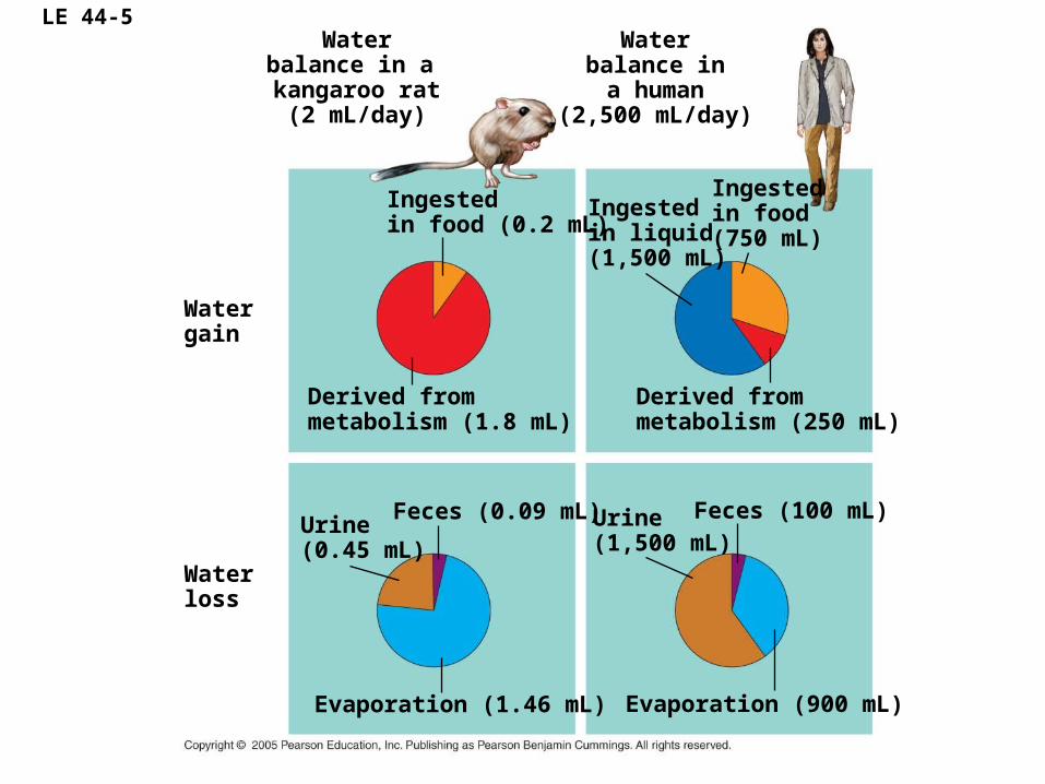
LE 44-5Water
balance in a kangaroo rat(2 mL/day)
Waterbalance ina human
(2,500 mL/day)
Watergain
Waterloss
Derived frommetabolism (1.8 mL)
Ingestedin food (0.2 mL)
Derived frommetabolism (250 mL)
Ingestedin food (750 mL)
Ingestedin liquid (1,500 mL)
Evaporation (900 mL)
Feces (100 mL)Urine(1,500 mL)
Evaporation (1.46 mL)
Feces (0.09 mL)Urine(0.45 mL)

LE 44-6
Control group(Unclipped fur)
Experimental group(Clipped fur)
Wat
er l
ost
per
day
(L/1
00 k
g b
od
y m
ass) 4
3
2
1
0

Copyright © 2005 Pearson Education, Inc. publishing as Benjamin Cummings
An animal’s nitrogenous wastes reflect its phylogeny and habitat
• The type and quantity of an animal’s waste products may greatly affect its water balance
• Among the most important wastes are nitrogenous breakdown products of proteins and nucleic acids

Copyright © 2005 Pearson Education, Inc. publishing as Benjamin Cummings
Forms of Nitrogenous Wastes
• Different animals excrete nitrogenous wastes in different forms: ammonia, urea, or uric acid

LE 44-8
Nitrogenous bases
Nucleic acids
Amino acids
Proteins
—NH2
Amino groups
Most aquatic animals, including most bony fishes
Mammals, most amphibians, sharks, some bony fishes
Many reptiles (including birds), insects, land snails
Ammonia Urea Uric acid

Copyright © 2005 Pearson Education, Inc. publishing as Benjamin Cummings
Urea
• The liver of mammals and most adult amphibians converts ammonia to less toxic urea
• The circulatory system carries urea to the kidneys, where it is excreted

Copyright © 2005 Pearson Education, Inc. publishing as Benjamin Cummings
IB Learning Objective
Draw and label a diagram of the kidney.
– Include the cortex, medulla, pelvis, ureter and renal blood vessels.

Copyright © 2005 Pearson Education, Inc. publishing as Benjamin Cummings

Copyright © 2005 Pearson Education, Inc. publishing as Benjamin Cummings

Copyright © 2005 Pearson Education, Inc. publishing as Benjamin Cummings
Excretory Processes
• Most excretory systems produce urine by refining a filtrate derived from body fluids
• Key functions of most excretory systems:
– Filtration: pressure-filtering of body fluids
– Reabsorption: reclaiming valuable solutes
– Secretion: adding toxins and other solutes from the body fluids to the filtrate
– Excretion: removing the filtrate from the system

LE 44-9
Filtration
Reabsorption
Secretion
Excretion
Excretorytubule
Capillary
Filtrate
Urin
e

The Kidney
• The kidneys regulate the amount of water, salts and other substances in the blood.
• The kidneys are fist-sized, bean shaped structures that remove nitrogenous wastes (urine) and excess salts from the blood
• Because the kidney regulates both salt and water concentration in the blood it is the central organ that controls osmoregulation.

Copyright © 2005 Pearson Education, Inc. publishing as Benjamin Cummings
The Kidney

The urinary system: The pathway of Urine to the outside the body.
• The ureters are tubes that carry urine from the pelvis of the kidneys to the urinary bladder.
• The urinary bladder temporarily stores urine until it is released from the body.
• The urethra is the tube that carries urine from the urinary bladder to the outside of the body.
• The outer end of the urethra is controlled by a circular muscle called a sphincter.
• These parts work together and are part of the urinary system.

Copyright © 2005 Pearson Education, Inc. publishing as Benjamin Cummings
Blood vessels of the mammalian kidney
Each kidney is supplied with blood by a renal artery and drained by a renal vein
Animation: Nephron IntroductionAnimation: Nephron Introduction

Copyright © 2005 Pearson Education, Inc. publishing as Benjamin Cummings
The Kidney

Copyright © 2005 Pearson Education, Inc. publishing as Benjamin Cummings
The kidney structure
Each kidney is composed of three sections:
• The cortex is where the blood is filtered.
• The medulla contains the collecting ducts which carry filtrate (filtered substances) to the pelvis.
• The pelvis is a hollow cavity where urine accumulates and drains into the ureter.

Copyright © 2005 Pearson Education, Inc. publishing as Benjamin Cummings
The Kidneys
Cortex
Medulla Renal artery
Renal vein
Ureter
To the bladder

Copyright © 2005 Pearson Education, Inc. publishing as Benjamin Cummings
The medulla and cortex
• The outer cortex and inner medulla are made up of a million or more tiny tubules called nephrons.
• Part of a nephron is in the medulla the other part is in the cortex.
• Nephrons is a thin walled tubules about (3 cm) long.

LE 44-13
Excretory organs and major associated blood vessels
RenalmedullaRenalcortex
Renalpelvis
Section of kidney from a ratKidney structure
Ureter
Kidney
Glomerulus
Bowman’s capsule
Proximal tubule
Peritubular capillaries
Afferentarteriolefrom renalartery
Efferentarteriole from glomerulus
Distaltubule
Collectingduct
SEM20 µm
Branch ofrenal vein
Filtrate and blood flow
Vasarecta
DescendinglimbAscendinglimb
LoopofHenle
Renalmedulla
Nephron
Torenalpelvis
Renalcortex
Collectingduct
Juxta-medullarynephron
Corticalnephron
Posterior vena cava
Renal artery and vein
Aorta
Ureter
Urinary bladder
Urethra

Copyright © 2005 Pearson Education, Inc. publishing as Benjamin Cummings
The Kidneys
• Structure of the KidneysKidney Nephron

Copyright © 2005 Pearson Education, Inc. publishing as Benjamin Cummings

Copyright © 2005 Pearson Education, Inc. publishing as Benjamin Cummings
IB Learning Objective
• Annotate a diagram of a glomerulus and associated nephron to show the function of each part.

Copyright © 2005 Pearson Education, Inc. publishing as Benjamin Cummings
Parts of the Nephron
• Each nephron consists of the following parts:
– 1) glomerulus ;
– 2) Bowman’s capsule ;
– 3) proximal tubule ;
– 4) loop of Henle ;
– 5) distal (convoluted) tubule ;
– 6) collecting duct.

Copyright © 2005 Pearson Education, Inc. publishing as Benjamin Cummings
Artery
Vein
Loop of Henle
Bowman’s capsule
Glomerulus
Capillaries
Collecting duct
To the ureter

Copyright © 2005 Pearson Education, Inc. publishing as Benjamin Cummings

Copyright © 2005 Pearson Education, Inc. publishing as Benjamin Cummings

Functions of the parts of the Kidney
• The glomerulus is a mass of thin-walled capillaries.
• The Bowman’s capsule is a double-walled, cup-shaped structure.
• The proximal tubule leads from the Bowman’s capsule to the Loop of Henle.
• The loop of Henle is a long loop which extends into the medulla.
• The distal tubule connects the loop of Henle to the collecting duct.

Copyright © 2005 Pearson Education, Inc. publishing as Benjamin Cummings
IB LEARNING OBJECTIVE
• Explain the process of ultrafiltration, including blood pressure, fenestrated blood capillaries and basement membrane

Copyright © 2005 Pearson Education, Inc. publishing as Benjamin Cummings
Five Steps in the Formation of Urine
1. Ultrafiltration in the renal capsule
2. Selective reabsorption in the proximal convoluted tubules
3. Water conservation in the loop of henle (osmoregulation)
4. Blood pH and ion concentration regulation in the distal convoluted tubule (osmoregulation)
5. Water reabsorption in the collecting ducts. (osmoregulation)

Copyright © 2005 Pearson Education, Inc. publishing as Benjamin Cummings

Copyright © 2005 Pearson Education, Inc. publishing as Benjamin Cummings
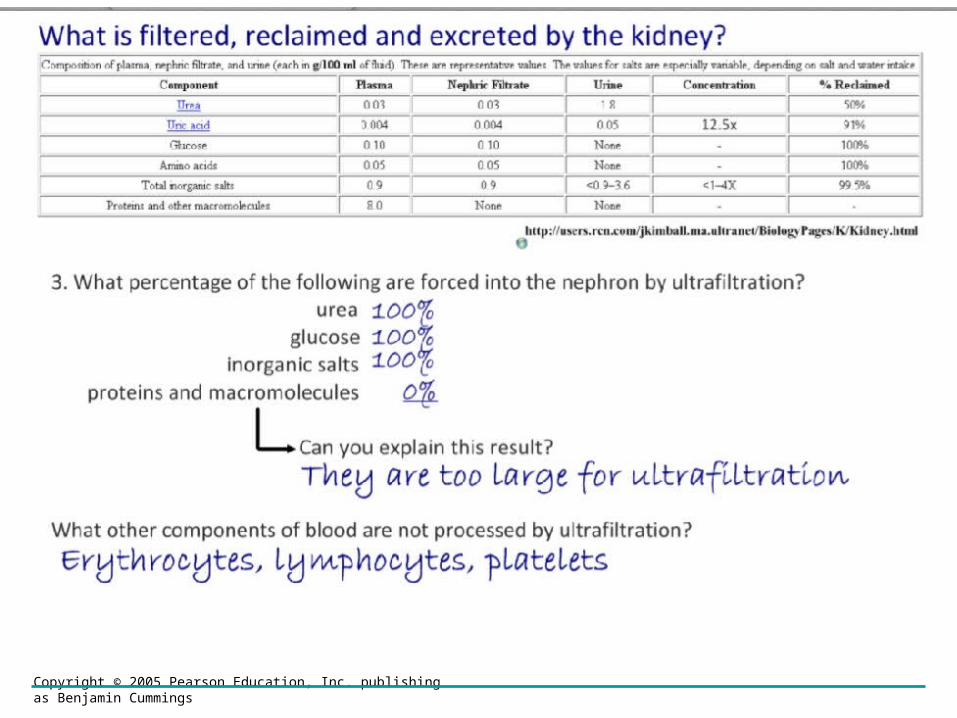
Step 1: Ultrafiltration in the renul capsule.
• Filtration occurs as blood pressure forces fluid from the blood in the glomerulus into the lumen of Bowman’s capsule
• This process is called Ultrafiltration because it is powered by pressure of the blood.
• The entire content of the blood is not forced out.
• The basement membrane of the of the capsule does not allow blood cells and proteins to enter the filtrate.

Copyright © 2005 Pearson Education, Inc. publishing as Benjamin Cummings

Copyright © 2005 Pearson Education, Inc. publishing as Benjamin Cummings
IB Learning Objective
• Explain the reabsorption of glucose, water and salts in the proximal convoluted tubule, including the roles of
– microvilli,
– osmosis
– and active transport.

Copyright © 2005 Pearson Education, Inc. publishing as Benjamin Cummings
Step 2 : Selective reabsorption in the proximal convoluted tubules
• The convoluted proximal tubules is the longest section of the nephron.
• The walls are one cell thick and they are packed with mitochondria.
• The cell membrane in contact with the filtrate is packed with microvilli to increase surface area for absorption.

Step 2 : Selective reabsorption in the proximal convoluted tubules (see figure 12.22 page 373)
• The proximal convoluted tubules absorb filtrate through the following mechanisms:
– Movement of water via osmosis
– Active transport of glucose and amino acids across membranes
– Movement of some minerals and ions via a combination of active transport, facilitated diffusion and some gas exchange of ions
– Diffusion of urea
– Movement of protein via pinocytosis (endocytosis)

Copyright © 2005 Pearson Education, Inc. publishing as Benjamin Cummings
Step 3 Water conservation in the loop of henle
• Urea is excreted from the body in solution, thus water loss is inevitable
• Water loss is minimized by having the solutes concentration in urine higher than the blood.
• The role of the loop of Henle is to maintain a high concentration of solutes in the medulla of the kidney
• The loop of henle has a descending and ascending limbs that parallels the blood supply.

Copyright © 2005 Pearson Education, Inc. publishing as Benjamin Cummings

LE 44-14
Filtrate
H2O
Salts (NaCl and others)
HCO3–
H+
Urea
Glucose; amino acids
Some drugs
Key
Active transport
Passive transportINNERMEDULLA
OUTERMEDULLA
NaCl
H2O
CORTEX
Descending limbof loop ofHenle
Proximal tubule
NaCl Nutrients
HCO3–
H+
K+
NH3
H2O
Distal tubule
NaCl HCO3–
H+K+
H2O
Thick segmentof ascendinglimb
NaCl
NaCl
Thin segmentof ascendinglimb
Collectingduct
Urea
H2O

Copyright © 2005 Pearson Education, Inc. publishing as Benjamin Cummings
IB LEARNING OBJECTIVE
• Explain the roles of the loop of Henle, medulla, collecting duct and ADH (vasopressin) in maintaining the water balance of the blood.

Copyright © 2005 Pearson Education, Inc. publishing as Benjamin Cummings

Copyright © 2005 Pearson Education, Inc. publishing as Benjamin Cummings
• The descending limb is permeable so salt diffuses into the loop of Henle and water diffuses out into the medulla tissue.
• At the hairpin zone (base of the loop) water and salt diffuse into the medulla tissue.
• In the ascending limb of the loop of Henle, salt diffuses from the permeable loop tubule into the interstitial fluid of the medulla, but water is retained
Step 3 Water conservation in the loop of henlefigure 12.23 page 374

Step 4: Blood pH and ion concentration regulation in the distal convoluted tubule
• The distal tubule cells are the same as in the proximal tubule (one cell thick, microvilli and lots of Mitochondria)
• The role of the distal tubule cells is to adjust the composition of the blood, in particular pH.
• Blood pH is initially buffered by blood proteins, but if it deviates from a pH of 7.4 the concentrations of Hydrogen ion (H+) and hydroxide (OH-) are adjusted
• Blood pH does not vary outside the range of pH 7.35 to 7.45, but urine pH ranges from 4.5 to 8.2.

Copyright © 2005 Pearson Education, Inc. publishing as Benjamin Cummings
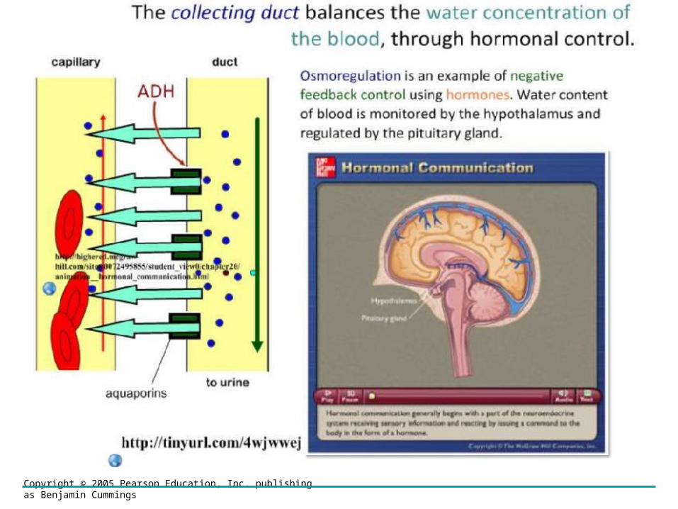
Step 5: Water reabsorption in the collecting ducts.
• The collecting ducts are where the water content is regulated.
• When the water content of the blood is low the antidiuretic hormone (ADH) is secreted from the posterior pituitary gland.
• When the water is the blood is high, NO ADH is released.

Copyright © 2005 Pearson Education, Inc. publishing as Benjamin Cummings

Copyright © 2005 Pearson Education, Inc. publishing as Benjamin Cummings
Step 5: Water reabsorption in the collecting ducts.
• The permeability of the walls of the collecting ducts are variable.
• If ADH is present the walls of the collecting tubules become fully permeable.
• This allows water to be withdrawn from the filtrate of the tubule in the medulla.
• The water will be taken up and redistributed throughout the body.
• ADH is remove from the body by the kidney
• When no ADH is present the walls of the collecting duct become less permeable.

Copyright © 2005 Pearson Education, Inc. publishing as Benjamin Cummings
Copyright © 2005 Pearson Education, Inc. publishing as Benjamin Cummings

Copyright © 2005 Pearson Education, Inc. publishing as Benjamin Cummings
Kidney Animations/ tutorials
• http://www.biologymad.com/resources/kidney.swf
• http://highered.mcgraw-hill.com/sites/0072507470/student_view0/chapter26/
• http://highered.mcgraw-hill.com/sites/0072507470/student_view0/chapter26/animation__micturition_reflex.html
• http://highered.mcgraw-hill.com/sites/0072507470/student_view0/chapter27/
• http://www.sumanasinc.com/webcontent/animations/content/kidney.html
• http://www.zerobio.com/target_practice_quiz/target_practice_quiz_kidney.swf
Hormonal Control Animation
http://highered.mcgraw-hill.com/sites/0072495855/student_view0/chapter20/animation__hormonal_communication.html
http://highered.mcgraw-hill.com/sites/0072495855/student_view0/chapter20/animation__blood_sugar_regulation_in_diabetics.html

LE 44-16a
Osmoreceptorsin hypothalamus
Hypothalamus
ADH
Pituitarygland
Increasedpermeability
Distaltubule
Thirst
Drinking reducesblood osmolarity
to set point
Collecting duct
H2O reab-sorption helpsprevent further
osmolarityincrease
Homeostasis:Blood osmolarity
STIMULUSThe release of ADH istriggered when osmo-receptor cells in the
hypothalamus detect anincrease in the osmolarity
of the blood

Copyright © 2005 Pearson Education, Inc. publishing as Benjamin Cummings
IB Learning Objective
• Explain the differences in the concentration of proteins, glucose and urea between blood plasma, glomerular filtrate and urine.

Copyright © 2005 Pearson Education, Inc. publishing as Benjamin Cummings
Differences in the composition of blood plasma, glomerular filtrate and urine
Urine
• The composition excrete from urine is variable.
• Greatly influenced by six factors
1. Diet (salt intake, protein consumed)
2. Physical Activity
3. Water intake
4. Amount of sweating
5. Environmental condition
6. State of health (i.e. Diabetics)

Copyright © 2005 Pearson Education, Inc. publishing as Benjamin Cummings
Differences in the composition of blood plasma, glomerular filtrate and urine
Blood plasma
• The composition of blood is constant
• Constancy is due to the efficiency of our homeostatic mechanisms

Copyright © 2005 Pearson Education, Inc. publishing as Benjamin Cummings
Differences in the composition of blood plasma, glomerular filtrate and urine
Glomerular Filtrate
• The composition of glomerular filtrate (ultrafiltration of the glomerus) is constant
• Constancy is due to the ultrafiltration, the pressure of the blood, and the size of blood proteins that are too large to filter.

Copyright © 2005 Pearson Education, Inc. publishing as Benjamin Cummings

Copyright © 2005 Pearson Education, Inc. publishing as Benjamin Cummings
IB Learning Objectives
• Explain the presence of glucose in the urine of untreated diabetic patients.

Copyright © 2005 Pearson Education, Inc. publishing as Benjamin Cummings

Copyright © 2005 Pearson Education, Inc. publishing as Benjamin Cummings

Copyright © 2005 Pearson Education, Inc. publishing as Benjamin Cummings

Copyright © 2005 Pearson Education, Inc. publishing as Benjamin Cummings
The composition of the urine of a diabetic patient
• The disease known as diabetes – blood glucose levels are erratic and frequently above normal.
• A consequence to this elevated blood glucose level is the failure of the kidney tubules to reabsorb all the glucose forced out of the blood.
• Thus the urine of a diabetic generally contains a lot of glucose.
• Raise glucose level in the urine is a symptom of a patient being a diabetic.
Copyright © 2005 Pearson Education, Inc. publishing as Benjamin Cummings
IB Practice Question

Copyright © 2005 Pearson Education, Inc. publishing as Benjamin Cummings

Copyright © 2005 Pearson Education, Inc. publishing as Benjamin Cummings
IB Practice Question

Copyright © 2005 Pearson Education, Inc. publishing as Benjamin Cummings
IB Practice Question
Copyright © 2005 Pearson Education, Inc. publishing as Benjamin Cummings
IB Practice Question

Copyright © 2005 Pearson Education, Inc. publishing as Benjamin Cummings
IB Practice Question

Copyright © 2005 Pearson Education, Inc. publishing as Benjamin Cummings
IB Practice Question

Copyright © 2005 Pearson Education, Inc. publishing as Benjamin Cummings

Copyright © 2005 Pearson Education, Inc. publishing as Benjamin Cummings
IB Practice Question
Copyright © 2005 Pearson Education, Inc. publishing as Benjamin Cummings

Copyright © 2005 Pearson Education, Inc. publishing as Benjamin Cummings
IB Practice Question

Copyright © 2005 Pearson Education, Inc. publishing as Benjamin Cummings

Copyright © 2005 Pearson Education, Inc. publishing as Benjamin Cummings
IB Practice Question

Copyright © 2005 Pearson Education, Inc. publishing as Benjamin Cummings


![Osmoregulation and Excretion [Important words are in bold]](https://img.pdfslide.us/doc/110x75/56649e895503460f94b8df70/osmoregulation-and-excretion-important-words-are-in-bold.jpg)