Embed Size (px)
Citation preview

THE JOURNAL OF BIOLOGICAL CHEMISTRY 0 1992 by The American Society for Biochemistry and Molecular Biology, Inc.
Vol. 267, No. 17, Issue of June 15, pp. 12142-12146,1992 Printed in U. S.A.
Initiation of Methyl-directed Mismatch Repair*
(Received for publication, November 22,1991)
Karin G. AuS, Katherine Welsh#, and Paul Modrich From the Department of Biochemistry, Duke Uniuersity Medical Center, Durham, North Carolina 27710
Escherichia coli MutH possesses an extremely weak d(GATC) endonuclease that responds to the state of methylation of the sequence (Welsh, K. M., Lu, A.-L., Clark, S. , and Modrich, P. (1987) J. Biol. Chem. 262, 16624-16629). MutH endonuclease is activated in a reaction that requires MutS, MutL, ATP, and Mg2+ and depends upon the presence of a mismatch within the DNA. The degree of activation correlates with the efficiency with which a particular mismatch is subject to methyl-directed repair (G-T > G-G > A-C > C-C), and activated MutH responds to the state of DNA ade- nine methylation. Incision of an unmethylated strand occurs immediately 5’ to a d(GATC) sequence, leaving 5’ phosphate and 3’ hydroxy termini (p NJ p Gp Ap- TpC). Unmethylated d(GATC) sites are subject to dou- ble strand cleavage by activated MutH, an effect that may account for the killing of dam- mutants by 2- aminopurine. The mechanism of activation apparently requires ATP hydrolysis since adenosine-6’-0-(3-thio- triphosphate) not only fails to support the reaction but also inhibits activation promoted by ATP. The process has no obligate polarity as d(GATC) site incision by the activated nuclease can occur either 3’ or 5’ to the mismatch on an unmethylated strand. However, acti- vation is sensitive to DNA topology. Circular hetero- duplexes are better substrates than linear molecules, and activity of DNAs of the latter class depends on placement of the mismatch and d(GATC) site within the molecule. MutH activation is supported by a 6- kilobase linear heteroduplex in which the mismatch and d(GATC) site are centrally located and separated by 1 kilobase, but a related molecule, in which the two sites are located near opposite ends of the DNA, is essentially inactive as substrate. We conclude that MutH activation represents the initiation stage of methyl-directed repair and suggest that interaction of a mismatch and a d(GATC) site is provoked by MutS binding to a mispair, with subsequent ATP-dependent translocation of one or more Mut proteins along the helix leading to cleavage at a d(GATC) sequence on either side of the mismatch.
The Escherichia coli methyl-directed, MutHLS-dependent mismatch repair system controls genetic variability by cor- recting DNA biosynthetic errors and ensuring the fidelity of homologous genetic recombination (reviewed in Refs. 1-4). The MutHLS pathway recognizes most base-base mispairs,
* This work was supported by Grant GM23719 from the National Institute of General Medical Sciences. The costs of publication of this article were defrayed in part by the payment of page charges. This article must therefore be hereby marked “aduertisement” in accordance with 18 U.S.C. Section 1734 solely to indicate this fact.
2 Syntex Fellow of the Life Sciences Research Foundation. 5 Present address: Agouron Pharmaceuticals Inc., San Diego, CA
92121.
as well as insertion-deletion mismatches involving a small number of nucleotides, but the efficiency of repair varies with the nature of the mismatch and can be influenced by sequence context. The system processes mispairs in a strand-specific manner, with discrimination of the two DNA strands being based on the state of adenine methylation of d(GATC) se- quences. Repair of a hemimethylated heteroduplex is re- stricted to the unmodified strand, symmetrically modified DNA is not processed, and a mismatch within an unmethyl- ated helix is corrected with little strand bias. One exception to the rule of methyl direction has been demonstrated the requirement for a hemimethylated/unmethylated d(GATC) site can be bypassed by a persistent strand break, in which case the associated DNA termini suffice to target correction to the incised strand (5 , 6).
The methyl-directed reaction has been reconstituted in a purified system comprised of E. coli MutH, MutL, MutS, DNA helicase 11, single strand DNA-binding protein, exonu- clease I, DNA polymerase I11 holoenzyme (preparations used possessed associated 5’ to 3’ exonuclease activity (7,8)’) DNA ligase, ATP, the four dNTPs, and the ligase cofactor NAD+ (6). Although the overall mechanism of the reaction remains to be established, analysis of MutH, MutL, and MutS has indicated that these three proteins are responsible for inter- action with the two DNA sites involved in a methyl-directed repair event. Thus, MutS binds to mismatched base pairs in a reaction that is independent of the presence of a d(GATC) sequence within the DNA (9-11). Welsh etal. (12) have shown that MutH possesses a M$+-dependent endonuclease that incises 5’ to d(GATC) sequences in a methyl-directed but mismatch-independent manner. Fully methylated sequences are resistant to attack, incision at hemimethylated sites is restricted to the unmodified strand, and unmethylated d(GATC) sequences are incised on either DNA strand. De- spite the mismatch independence and the extremely weak nature (turnover number < 1 h-’) of the MutH-associated endonuclease, Welsh et al. (12) proposed that this activity is indicative of MutH function in mismatch repair, with the protein undergoing mismatch-dependent activation during assembly of a repair complex. The function of MutH as a d(GATC) endonuclease in mismatch correction was con- firmed with the demonstration that a persistent strand break not only circumvents the requirement for the d(GATC) strand signal in mismatch repair, but bypasses the requirement for MutH as well (5 , 6).
While attempts to assign specific enzymatic or DNA bind- ing activities to MutL have yielded negative results, the protein has been shown to interact with the MutS-mismatch complex, provided that ATP or ATPyS is present (13,14). In this paper we report a second biochemical effect of MutL and demonstrate mismatch-dependent activation of the MutH- associated endonuclease in a reaction requiring MutL, MutS, and ATP.
D. Cooper and P. Modrich, manuscript in preparation.
12142

Initiation of Mismatch Repair 12143
HInCll
Eco47111
FIG. 1. Structure of f l M R heteroduplexes. Covalently closed, circular heteroduplex DNAs used in this study (10) contained a G-T, a n A-C, a C-C, or a G-G mismatch at position 5632 and a single d(GATC) sequence at coordinate 216. Sites of cleavage by restriction endonucleases used are also indicated, with the coordinates shown corresponding to the nucleotide 5' to the phosphodiester attacked. Outer and inner circles designate complementary and viral strands, respectively, with shaded regions corresponding to oligomer sequences used for indirect end labeling ("Materials and Methods").
MATERIALS AND METHODS
Proteins and DNA-E. coli MutH, MutL, and MutS were prepared a s described previously (9, 12, 13). E. coli topoisomerase I was a gift from Dr. James Wang (Harvard University). E. coli DNA ligase, restriction endonucleases, and T4 polynucleotide kinase were from commercial sources.
Covalently closed, circular heteroduplex DNAs were prepared in supercoiled form as described previously. Phages flMR8 and flMR9 were used to prepare C-C and G-G heteroduplexes and phages flMR1 and flMR3 to prepare G-T and A-C heteroduplexes (10). The struc- ture of these molecules is illustrated in Fig. 1. A control homoduplex containing a G.C base pair instead of a mismatch was prepared in an identical manner using flMR3 viral strand and RF2 DNA. Relaxed closed circular heteroduplex DNA was prepared by treating 4 pg of negatively supercoiled G-T heteroduplex with 1.6 pg of E. coli topoi- somerase I for 15 min at 37 "C in 0.05 M HEPES (potassium salt, pH 8.0), 0.02 M KC1,l mM dithiothreitol, 4 mM MgCl2, 0.05 mg/ml bovine serum albumin. The reaction was quenched by the addition of EDTA to 10 mM, followed by phenol and ether extraction and ethanol precipitation. The DNA was dissolved in 0.01 M Tris-HC1, pH 7.6, 1 mM EDTA. Linear RF or heteroduplex DNA was prepared by cleavage with the appropriate restriction endonuclease and DNA extracted and precipitated as above.
Oligonucleotides were synthesized using an Applied Biosystems 381A DNA synthesizer and were 5'-end-labeled using [y3'P]ATP (3000 Ci/mmol Du Pont-New England Nuclear) and T4 polynucleo- tide kinase. Labeling reactions were terminated by phenol extraction or by addition of EDTA to 10 mM and heating at 65 "C for 10 min. Unincorporated label was removed by chromatography over Sephadex G-25 (Pharmacia LKB Biotechnology Inc.) in 0.01 M Tris-HC1, pH 8.0, 1 mM EDTA, 0.1 M NaCl.
d(GATC) Endonuclease Assay-Endonuclease activity at d(GATC) sequences was assayed a t 37 "C in 10-pl reactions that were assembled a s follows: 7 pl of heteroduplex solution (containing 0.071 M HEPES (potassium salt, pH 8.0), 0.029 M KC1, 5.7 mM MgCI,, 1.4 mM dithiothreitol, 0.071 mg/ml bovine serum albumin, 2.9 mM ATP, and 0.10 pg (24 fmol) of covalently closed, circular duplex DNA) was prewarmed to 37 "C. Reactions were initiated by adding 3 pl of MutHLS diluent (0.02 M potassium phosphate, pH 7.4, 0.05 M KCl, 0.1 mM EDTA, 1 mM dithiothreitol, 1 mg/ml bovine serum albumin) containing, as indicated, 35 ng of MutS (360 fmol), 17 ng of MutL
' The abbreviations used are: RF, replicative form; HEPES, 4-(2- hydroxyethy1)-1-piperazineethanesulfonic acid; bp, base pair(s); 2- AP, 2-aminopurine.
(240 fmol), and 0.26 ng of MutH (10.4 fmol). After incubation at 37 "C for the appropriate time, reactions were quenched by transfer- ring the assay mixture to tubes preheated to 55 "C and incubating for 10 min. DNA samples were then digested with BsplO6, restriction reactions terminated by addition of one-third volume of 0.2 N NaOH, 0.04 M EDTA, 10% Ficoll 400, 0.1% bromcresol green, and samples electrophoresed at 1.5-4.0 V/cm through 1% agarose gels in 0.03 N NaOH, 2 mM EDTA.
DNA was transferred onto nylon or polysulfone membranes (Gel- man BioTrace R P or HP, ICN Biotrans, or Du Pont-New England Nuclear Genescreen Plus) as recommended by the manufacturer. Membranes were prehybridized at 37 "C for a t least 4 h in 2% sodium dodecyl sulfate, 0.5% polyvinyl pyrrolidone, 0.2% heparin, 1 mM EDTA, 1 M NaC1, 0.05 M Tris-HC1, pH 7.5, then hybridized at 37 "C overnight in the same solution containing 5'-"P-d(CTAATAGTAG- TAGCGTT) (oligomer I) to detect cleavage on the viral DNA strand or 5'-R'P-d(AACGCTACTACTATTAG) (oligomer 11) to detect cleav-
to base pairs 1-17 of the phages flMR1, flMR3, flMR8, and flMR9. age on the complementary DNA strand. These two probes correspond
In several cases d(GATC) cleavage occurring on the viral strand was scored using 5'-3'P-d(ACTTCAAATATCGCGTTTTA) (oligomer 111, nucleotides 342-361 of the complementary DNA strand).
After hybridization, membranes were washed twice at room tem- perature for 5 min in 1 X ssc (0.15 M NaC1, 15 mM sodium citrate, pH 7.0), 0.1% sodium dodecyl sulfate, then twice at room temperature for 5 min in 0.1 x SSC, 0.1% sodium dodecyl sulfate. Autoradiography of each membrane was carried out using Kodak XAR-5 film at several different exposure times. Endonuclease activity was quantitated by scanning densitometry. In some cases, membranes were stripped of probe by washing twice for 15 min at room temperature in 0.4 N NaOH. Membranes were then washed in 0.1 X SSC, 0.1% SDS at room temperature then prehybridized and hybridized as described above.
ATPase Assay-ATPase was assayedunder conditions of d(GATC) endonuclease except that the 10-pl reactions contained 50 mM [y- 32P]ATP (4 Ci/mmol), 0.78 ng of MutH, 26 ng of MutL, 105 ng of MutS, and 300 ng of G-T heteroduplex (methylated on the comple- mentary DNA strand). Reactions were quenched by the addition of 2 p1 of 60 mM EDTA, and 2 p1 was spotted onto polyethyleneimine- cellulose plates (EM Separations), which were developed in 0.5 M LiC1,0.3 M acetic acid. Spots were visualized by autoradiography, and radioactivity associated with ATP and Pi was determined by liquid scintillation counting of appropriate portions of the polyethylene- imine-cellulose plate.
RESULTS
MutL, MutS, and ATP Activate the MutH-associated d(GATC) Endonuclease-As discussed above, purified MutH possesses an extremely weak, mismatch-independent, but nevertheless tightly associated d(GATC) endonuclease activ- ity (12). The demonstration that a persistent strand break bypasses the requirements for MutH and for a d(GATC) strand signal in mismatch repair has confirmed a role for MutH as a d(GATC) endonuclease in this process (5, 6). MutH-specific endonuclease activity in the buffer used for mismatch repair assay corresponds to 0.0017 incision events/ min/monomer equivalent (12). However, the specific activity of MutH with respect to mismatch repair in the reconstituted methyl-directed system is 20-70-fold higher (-0.04-0.12 re- pair events/min/monomer for a G-T mismatch, the most efficiently processed base-base mispair (6)). We have there- fore tested the proposal (12) that MutH endonuclease may undergo activation during assembly of a repair complex on a heteroduplex.
As shown in Table I, the combination of MutH, MutL, MutS, and ATP results in the appearance of an endonuclease activity that incises the unmethylated strand of a hemimeth- ylated G-T heteroduplex in the vicinity of its single d(GATC) site. MutH, MutL, MutS, ATP, and a divalent cation are each required for this effect since activation was not detectable in reactions lacking one or more of the individual components. The intrinsic MutH activity is not detectable at the low concentration of the protein used in the experiments of Table

12144 Initiation of Mismatch Repair
TABLE I Requirements for the activation of MutH endonuclease
The hemimethylated flMRl/flMR3 heteroduplex contained a G-T mismatch and one d(GATC) site (methylation on complemen- tary strand; see Fig. 1). With the exception of omissions or additions indicated, endonuclease reactions and assay of site-specific cleavage by indirect end-labeling were performed as described under “Mate- rials and Methods.” Mapping relative to unique XbaI and BsplO6 restriction sites within the heteroduplex placed the site of viral strand cleavage at or near the d(GATC) sequence (not shown, see Fig. 2).
Reaction conditions Endonuclease frnol/ZO rnin
Complete 17 -H <0.5 -L <0.5 -S <0.5 -H - L C0.5 - H - S <0.5 -L-s <0.5 -M&L <0.5 -ATP <0.5 -ATP + ATPrS 0.8 ATP + ATP-yS (1 mM each) 1.3
TABLE I1 Methyl direction of the activated d(GATC) endonuclease
Endonuclease activity was measured using closed circular G-T flMRl/flMR3 heteroduplexes as described under “Materials and Methods.” Samples were removed periodically to obtain initial rates.
Methylated strand(s) Cleavage of C strand Cleavage of V strand frnollrnin frnollrnin
C C0.03 V 1.7 <0.03 C and V C0.03 C0.03 Neither 0.67 0.40
1.3
I (12), but it is evident that the degree of nuclease activation exceeds 30-fold when MutL, MutS, and ATP are also present. I t is pertinent that the concentrations of the four components used in these experiments (“Materials and Methods”) are identical to those used to study mismatch repair in a defined system (6) . Therefore, the rate of the endonuclease reaction can be compared with that for in Vitro mismatch correction. This point is considered below.
The intrinsic MutH activity incises hemimethylated or unmethylated duplexes immediately 5‘ to an unmethylated d(GATC) sequence (JpGpApTpC) to produce a strand break with 3’-OH and 5’-P termini (12). Table I1 demonstrates that the MutHLS-dependent, activated endonuclease responds to d(GATC) modification in a similar manner: each of the two hemimethylated configurations of a G-T heteroduplex was incised on the unmethylated strand, the symmetrically mod- ified heteroduplex was resistant to cleavage, and the unmeth- ylated substrate was subject to incision on either strand of the helix. In the case of the unmodified heteroduplex, d(GATC) cleavage during early stages of the reaction was largely limited to single strand incision, with prolonged in- cubation resulting in cleavage of the other strand to yield a double strand break (Table 111). Double strand cleavage of unmethylated DNA by the activated MutH endonuclease is of interest in view of the finding that 2-aminopurine killing of dam- mutants requires mutH, mutL, and mutS function (15). The possible relationship of these two observations will be considered below.
The specificity of phosphodiester bond hydrolysis by the MutHLS-dependent endonuclease is also identical to that of the intrinsic MutH activity. Strand breaks introduced by the activated species are efficiently closed by E. coli DNA ligase
TABLE 111 Single and double-strand cleavage of an unmethylated d(GATC) sequence
Endonuclease reactions containing unmethylated, closed circular G-T heteroduplex DNA were performed as described under “Mate- rials and Methods.” A portion of each reaction was electrophoresed through neutral agarose, and after staining with ethidium bromide (0.5 pg/ml for 1 h), products resulting from double strand cleavage a t the d(GATC) site were quantitated by virtue of their fluorescence using a photometric grade, cooled CCD imager equipped with a 600- nm band pass filter (Photometrics, Inc.). A second portion of each reaction was subjected to indirect end labeling (“Materials and Meth- ods’’) to determine the total yield of d(GATC) incision events in each DNA strand. The yield of single-cleaved molecules shown corresponds to the latter value after correction for double-strand events.
Time Single strand Double strand cleaved cleaved
min fmol 20 12 3.8 60 7 14
Hin cl I
FIG. 2. Specificity of cleavage by the activated MutHLS-
the circular flMRl/flMR3 G-T heteroduplex bearing d(GATC) dependent endonuclease. Endonuclease reactions, which utilized
methylation on the complementary strand, were performed as de- scribed under “Materials and Methods.” Products were hydrolyzed with HincII (Fig. 1) as indicated, and the DNA subjected to electro- phoresis on a 6% polyacrylamide sequencing gel. Dideoxy sequencing markers (lunes A, C, G, and T) were obtained using flMR8 RF DNA, Sequenase Version 2.0 (United States Biochemical), [a-”’PIdATP as the labeled nucleotide, and 5’ phosphorylated oligomer I1 (nucleotides 1-17 of the viral DNA strand) primer. An additional marker was prepared by digesting unmethylated flMR8 RF DNA with HincII and MboI (the latter activity cleaves immediately 5’ to the dG of the single d(GATC) site within the molecule). DNA was electroblotted from the sequencing gel onto a nylon membrane and probed with 5’- ”P-oligomer I.
demonstrating that, as in the case of MutH-associated activ- ity, hydrolysis produces 3’-OH and 5’-P termini (data not shown). Furthermore, as illustrated in Fig. 2, the MutHLS- dependent activity cleaves immediately 5‘ to the d(GATC) sequence (pNJpGpApTpC), a specificity identical to that previously established for the reaction mediated by MutH alone (12).
Activation Is Mismatch-dependent-While the activated form of the d(GATC) endonuclease parallels the weak MutH- associated activity with respect to site specificity, the two
Initiation of Mismatch Repair 12145
activities differ in their response to the presence of a mis- match. The rate of d(GATC) cleavage by the intrinsic MutH activity is unaffected by the presence of a mispair within the substrate (12), but the MutS requirement for endonuclease activation suggested that mismatch recognition might be in- volved in this process. Table IV shows that this is the case. The four different heteroduplexes tested were incised by the MutHLS-dependent activity at characteristic rates, with their relative rates of cleavage correlating with their differential sensitivity to methyl-directed repair (G-T > G-G > A-C > C- C) (6, 10).
Although the C-C heteroduplex used in these experiments is not subject to detectable methyl-directed repair (6, lo), it was subject to a low level of MutHLS-dependent cleavage at the hemimethylated d(GATC) site (Table IV, top), and a similar degree of cleavage was observed with a hemimethyl- ated G.C construct that was prepared in the same manner (“Materials and Methods”). In contrast to the behavior of the G-T, G-G, and A-C heteroduplexes, the rate of cleavage of hemimethylated G . C and C-C DNAs decreased rapidly after =IO% of the molecules had been incised, suggesting that only a subpopulation was sensitive to MutHLS cleavage. We have considered three potential explanations for the low level ac- tivity observed with the latter two DNAs: (i) sensitivity of the system to natural fluctuations in base pair conformation occurring within the helix; (ii) MutHLS recognizable damage incurred during heteroduplex preparation; (iii) presence of unknown base pair mismatches due to natural variation within the two DNA populations that are denatured and reannealed to produce the heteroduplex.
These possibilities were examined by testing activity of the system on unmethylated flMR3 RF DNA obtained from a dam- host. In contrast to the efficient incision observed on the two strands of unmethylated G-T heteroduplex (Table
TABLE IV Mismatch dependence of d(GATCI endonuclease activation
Top: endonuclease activity was measured using closed circular DNAs methylated on the complementary strand (“Materials and Methods”). Samples were removed periodically in order to obtain initial rates. The control G. C homoduplex was prepared in the same manner as the mismatch-containing heteroduplexes. Bottom: super- coiled, unmethylated flMR3 RF DNA was linearized with BsplO6. Half of this material was denatured with NaOH, neutralized, rean- nealed, and chromatographed over hydroxyapatite as previously de- scribed (17). The other half of the linear preparation (control) was treated in a similar manner, except that NaOH and the neutralizing solution were mixed prior to addition to the DNA. Endonuclease reactions were performed and d(GATC) cleavage quantitated by indirect end labeling as described under “Materials and Methods.” Bands corresponding to cleavage at the d(GATC) site were visually evident upon autoradiography only in the case of the denatured and reannealed sample. However, since these bands were only slightly above background in the densitometric profile, values are indicated as being approximate.
~
Substrate Methylated d(GATC) strand cleavage
Heteroduplex G-T G-G A-C c-c G.C
fmolfmin
C C C C C
1.3 1.1 0.41 0.15 0.14
RF DNA G . C (supercoiled) Neither <0.025 (C), <0.025 (V) G. C (linear control) Neither <0.025 (C), <0.025 (V) G.C (denatured and Neither -0.03 (C), -0.03 (V)
reannealed)
11), no detectable d(GATC) cleavage was observed on either strand of unmethylated circular or linear RF DNA (Table IV, bottom). However, alkaline denaturation and reannealing of linear RF DNA resulted in low, but detectable cleavage on each strand at the d(GATC) site of the reannealed species. In view of these findings and the subpopulation observation mentioned above, we attribute low level MutHLS cleavage of the hemimethylated G.C and C-C substrates to damage as- sociated with their preparation and/or natural variation within the DNAs used for their construction.
Activated MutH Turns Over in the d(GATC) Endonuclease Reaction-Turnover of the intrinsic endonuclease activity of purified MutH has not been demonstrated (12). In contrast, turnover of the activated species on heteroduplex DNA was observed during extended incubation at low MutH concentra- tions (Table V), with an apparent turnover number of 0.1 min” mol”. Since we have not attempted to saturate the system with heteroduplex DNA, this determination may well be an underestimate. This value can be compared with kcat values ranging from 0.1 to 100 min” mol” for Type I1 restric- tion endonucleases (16), activities that catalyze much simpler reactions.
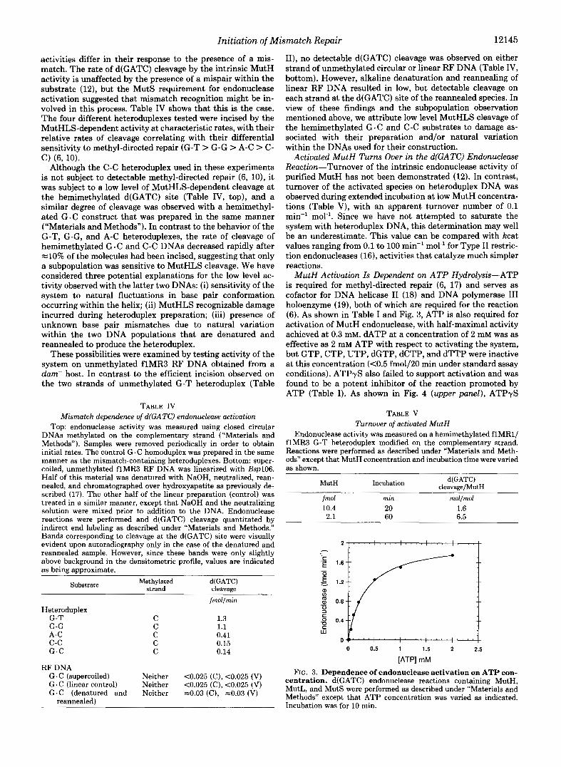
MutH Activation Is Dependent on ATP Hydrolysis-ATP is required for methyl-directed repair (6, 17) and serves as cofactor for DNA helicase I1 (18) and DNA polymerase I11 holoenzyme (19), both of which are required for the reaction (6). As shown in Table I and Fig. 3, ATP is also required for activation of MutH endonuclease, with half-maximal activity achieved at 0.3 mM. dATP at a concentration of 2 mM was as effective as 2 mM ATP with respect to activating the system, but GTP, CTP, UTP, dGTP, dCTP, and dTTP were inactive at this concentration (<0.5 fmol/20 min under standard assay conditions). ATPyS also failed to support activation and was found to be a potent inhibitor of the reaction promoted by ATP (Table I). As shown in Fig. 4 (upper panel), ATPyS
TABLE V Turnover of activated MutH
Endonuclease activity was measured on a hemimethylated flMR1/ flMR3 G-T heteroduplex modified on the complementary strand. Reactions were performed as described under “Materials and Meth- ods” except that MutH concentration and incubation time were varied as shown.
MutH Incubation d(GATC) cleavarre/MutH
fmol rnin mollno1 10.4 20 1.6 2.1 60 6.5
0 0.5 1 1.5 2 2.5
[ATP] rnM
FIG. 3. Dependence of endonuclease activation on ATP con- centration. d(GATC) endonuclease reactions containing MutH, MutL, and MutS were performed as described under “Materials and Methods” except that ATP concentration was varied as indicated. Incubation was for 10 min.

12146 Initiation of Mismatch Repair
O . ' r - " 0.6
0.4 0.5h
v I
0.3
0.2
0.1
1.2 n d
1.0 h 3
0.8 E
0.6 z 0.4
3 c 0.2 4 8 0
o 20 40 60 ao 100 120
FIG. 4. Inhibition of endonuclease activation and ATPase by ATP-yS. Upper panel, ATP hydrolysis occurring in reactions containing MutH, MutL, MutS, and heteroduplex DNA was deter- mined as described under "Materials and Methods" except that ATPyS was present as indicated. Values shown are corrected for triphosphate hydrolysis by a contaminating ATPase present in the MutL preparation (13). Cleavage at the d(GATC) site of the G-T heteroduplex (methylated on the complementary DNA strand) was determined under identical conditions except that unlabeled ATP was used. Measurements were corrected for a small amount of d(GATC) cleavage that was mediated by MutH alone and evident at the higher concentration of the protein used in these experiments. 0, ATPase; 0, d(GATC) endonuclease. Lower panel, ATP hydrolysis by MutS was determined as described under "Materials and Methods" except that MutH and MutL were omitted from the reaction. Hemi- methylated G-T heteroduplex (modified on complementary strand) and ATP-yS were present as indicated. 0, no DNA; 0, with hetero- duplex.
inhibited endonuclease activation as well as ATP hydrolysis that occurs in such reactions, with the coordinate inhibition of the two activities strongly suggesting that MutH activation requires hydrolysis of ATP.
Associated ATPase activity has not been identified in near homogeneous preparations of MutH or MutL (13); but MutS displays weak ATPase in the presence or absence of DNA (20). While genetic evidence suggests that MutS ATPase has a functional role in mismatch repair (20), it is premature to conclude that nucleotide hydrolysis associated with MutH activation is due solely to the mutS gene product. Neverthe- less, it is interesting to note that the MutS activity responds to ATPyS inhibition in a manner similar to that of the MutHLS-dependent endonuclease (Fig. 4, lower panel).
The Incised d(GATC) Sequence May Lie Either 3' or 5' to the Mismatch on the Unmethylated DNA Strand-An un- modified d(GATC) sequence can reside either 3' or 5' to the mismatch as viewed along the unmethylated strand. MutH activation might therefore depend on a particular orientation
:' K. G. Au and P. Modrich, unpublished observations.
of the two sites. Since the experiments described above used circular substrates and do not address this issue, we have tested linear heteroduplexes for their ability to support this reaction. The G-T heteroduplex was hydrolyzed with Eco47111, which cleaves the circular molecule within the longer path separating the two DNA sites (Fig. l) , to yield a blunt-ended, 6111-bp linear molecule in which the mispair and the d(GATC) site are separated by 1024 bp. As shown in Fig. 5, both hemimethylated configurations of the linear het- eroduplex were efficiently incised by the MutHLS-dependent activity. Since the d(GATC) sequence on the viral DNA strand of the molecule lies 3' to the mismatch while that on the complementary strand is located 5' to the mispair, these results indicate that activation lacks obligate polarity. An alternate interpretation is that polarity associated with inter- action of the two DNA sites can be bypassed by transient circle formation via interaction of the blunt ends of the linear molecule, but this possibility is excluded by experiments de- scribed immediately below.
Dependence of MutH Activation on Topology of the Hetero- duplex-During the course of these experiments, we noticed that the MutHLS system appeared to be less active on linear heteroduplex DNAs than on circular molecules. The activity of the system on several different substrate configurations is summarized in Table VI. Heteroduplexes that are routinely used to study in vitro mismatch correction are prepared in a negatively supertwisted form (10, 17). Cleavage of a hemi- methylated circular G-T heteroduplex with Eco47III yields a linear in which the mismatch and d(GATC) site are centrally located with a separation distance of 1 kilobase (Fig. 1). The activity of this linear DNA was consistently lower than that of the supercoiled species. Moreover, cleavage of the supercoil
- C strand indirectly labeled
m 0
"..~..* V strand indirectly labeled
0
FIG. 5. Incision of linear heteroduplexes by the MutHLS- dependent endonuclease. Linear G-T heteroduplexes in the two hemimethylated configurations were prepared by cleavage of circular DNAs with Eco47III. Endonuclease reactions were incubated for 20 min and terminated by addition of 5 pl of 0.2 N NaOH, 0.04 M EDTA, 10% Ficoll400,0.1% bromcresol green, without subsequent restriction endonuclease digestion. Marker DNA in the left lane was prepared by digestion of unmethylated flMR3 RF with Eco47III and MboI. Upper panel, the membrane was probed for cleavage on the complementary strand using 5'-"'PP-oligomer 11. Lowerpanel, the same membrane was probed with 5'-"P-oIigomer I to detect cleavage on the viral DNA strand.

Initiation of Mismatch Repair 12147
TABLE VI Dependence of MutH activation on DNA topology
Experiment 1: linear substrates were prepared by digesting a hemi- methylated G-T heteroduplex (modified on the complementary strand) with Eco47III or HincII (Fig. 1). d(GATC) endonuclease reactions were performed as described under “Materials and Meth- ods,” except that Bspl06 digestion was omitted for the Em47111 linear heteroduplex, and products derived from the HincII linear were detected by indirect labeling using oligomer 111. Samples were re- moved periodically in order to obtain initial rates. Experiment 2: relaxed and linear G-T hemimethylated heteroduplexes used in these experiments bore d(GATC) modification on the complementary strand, while the supercoiled DNA was methylated on the viral strand. Endonuclease assays were performed as described under “Materials and Methods” with an incubation time of 10 min except that reactions with two substrates contained 50 ng (12 fmol) of each DNA. In those cases where substrate was relaxed heteroduplex, Eco47III linear het- eroduplex, or a mixture of these two DNAs, restriction cleavage subsequent to d(GATC) endonuclease assay utilized XbaI, and prod- ucts were quantitated by indirect labeling with 5’-32P-oligomer 111. Products derived from d(GATC) cleavage of supercoiled heterodu- plex, HincII linear heteroduplex, or a mixture of both DNAs were measured using 5’-end-labeled oligomers I1 and 111 to score cleavage on complementary and viral strands, respectively.
Heteroduplex topolow d(GATC) cleavage
Experiment 1 Supercoiled Relaxed Eco47III linear HincII linear
fmollmin
1.3 2.5 0.64
co.10
fmolllO min
Experiment 2 Relaxed Eco477II linear Supercoiled HincII linear
Relaxed + Eco47III linear
18
16 <1
6.5
8.8 2.3
Supercoiled + 8.5 HincII linear C0.50
with HincII, which places the two sites near opposite ends of a blunt-ended linear at a separation distance of 5.4 kilobases (Fig. I), resulted in virtual loss of substrate activity. The reduced activity of the two linear molecules is not due to the absence of superhelical stress as the corresponding relaxed circular heteroduplex was an excellent substrate? The pres- ence of a diffusable inhibitor in the linear DNA preparations was also excluded since circles were preferentially incised by the activated endonuclease in reactions containing equimolar concentrations of circular and linear DNAs (Table VI). The MutHLS system thus responds to placement of the mismatch and the d(GATC) site within the heteroduplex. The mecha- nistic basis of this effect has not been defined, but it is seemingly dependent on the proximity of one or both sites to a DNA end and/or the distance by which the two sites are separated along the helix contour.
DISCUSSION
Provocation of a methyl-directed repair event requires a mismatch and one or more hemimethylated or unmethylated
Although competition experiments have not been done, the re- laxed circular G-T heteroduplex has consistently proven to be a better substrate than negatively supertwisted DNA (Table VI). Since the MutHLS system probably recognizes base pair mismatches in their intrahelical conformations (21), this effect may be due to destabili- zation of the intrahelical G-T conformation in underwound mole- cules.
d(GATC) sites (17, 22-24), and extract experiments have shown that repair is associated with mismatch- and MutHLS- dependent incision of an unmethylated strand in the vicinity of a d(GATC) sequence (22). Since a persistent strand break bypasses both the d(GATC) and the MutH requirements of the reaction, d(GATC) endonuclease activity has been attrib- uted to the mutH gene product (5, 6). This conclusion is consistent with the finding that MutH possesses a weak d(GATC) endonuclease, but only if this latent activity under- goes mismatch-dependent activation (12). We have shown that this is the case.
Several observations lead us to infer that the mismatch- and MutHLS-dependent partial reaction described here rep- resents the initiation step in methyl-directed mismatch cor- rection (3). The excision-repair mode of the reaction (25) implies a requirement for at least one strand break (or DNA end). In fact, analysis of the fate of closed circular heterodu- plex DNA in the purified system has failed to reveal signifi- cant covalent alteration of the substrate if MutS, MutL, or MutH is omitted from the reaction (6).3 Each of these proteins is therefore necessary for introduction of the initial strand break that occurs during the course of repair. As shown in this paper, MutH, MutL, and MutS are also sufficient to mediate a cleavage event that has been previously implicated in methyl-directed repair, with the substrate specificity of the MutHLS endonuclease being similar to that of the overall reaction.
In order to conclude that MutHLS-dependent cleavage represents the initial step in methyl-directed mismatch cor- rection, it is also necessary to show that d(GATC)-incised heteroduplex is produced rapidly enough to be a kinetically significant intermediate in the overall reaction. To permit this sort of comparison, most of our experiments utilized protein and DNA concentrations identical to those used pre- viously to characterize the reconstituted methyl-directed re- action (6). The rate of MutHLS cleavage under these condi- tions compares favorably with the rate of mismatch repair. For example, cleavage of the two hemimethylated configura- tions of the G-T heteroduplex used in the experiments of Tables I1 and IV occurred at rates of 1.3 and 1.7 fmol min” (viral and complementary strands, respectively) as compared to 1.2 and 0.4 fmol min-’ for mismatch repair of these mole- cules (6). A similar case holds for the other mismatches in Table IV, and we conclude that incision by the activated MutH endonuclease is sufficiently fast for the product to be an intermediate in the overall repair reaction. It is pertinent to note that although MutHLS cleavage is somewhat more efficient on the complementary DNA strand in the case of both hemimethylated and unmethylated heteroduplexes (Table II), an opposite preference has been observed for repair occurring on these DNAs (6, 10). However, since repair di- rected by a strand break also occurs more efficiently on the viral strand (6), the strand preference evident in the overall reaction presumably reflects events subsequent to incision.
Double strand cleavage by the intrinsic MutH endonuclease has not been observed (12), but the activated form of the protein can cleave both DNA strands at a d(GATC) site in an unmethylated heteroduplex (Table 111). This finding is of interest in view of the controversy concerning the mechanism of killing of dam- mutants by 2-aminopurine (2-AP). Since mutH-, mutL-, and nuts- mutations that block mismatch repair also alleviate 2-AP lethality in methylase mutants, Glickman and Radman (26) have attributed killing to undi- rected repair of mismatches involving the base analogue. This proposal presumes that such mispairs occur with a sufficiently high frequency so that a lethal double strand break may ensue

12148 Initiation of Mismatch Repair
when nearby mismatches provoke excision on opposite DNA strands. Grafstrom et al. (27) have questioned this view with the argument that the 2-AP-C mismatch, a likely candidate for provocation of the MutHLS system, occurs too infre- quently to result in overlap of excision tracts. Furthermore, Grafstrom and colleagues (27) have identified mutH muta- tions that, while capable of suppressing the lethal effect of 2- AP in dam-' strains, are not associated with a major defect in methyl-directed repair. The nature of these mutations thus suggests that killing by the base analogue does not result from the normal course of methyl-directed correction. These diver- gent observations can be rationalized by our finding that activated MutH endonuclease can cleave both strands of an unmethylated d(GATC) site, assuming that double strand cleavage in this manner is associated with a significant lethal cross-section. Such a mechanism would account for the mutH, mutL, and mutS dependence observed by Glickman and Rad- man (26), with a single mismatch being sufficient to promote a lethal event. The phenotype of the mutH mutations men- tioned above can also be accommodated within this scheme if their effect is to simply reduce the efficiency of second strand cleavage by activated MutH nuclease.
The finding that d(GATC) incision by the MutHLS activity can occur either 3' or 5' to the mismatch on the unmethylated DNA strand implies that the mechanism of activation of MutH endonuclease lacks obligate directionality. This sug- gests that the methyl-directed system may possess a bidirec- tional capability, with a d(GATC) site located on either side of the mismatch being sufficient to target repair to the un- modified strand. This possibility is consistent with several in uiuo observations (28, 29) and, in fact, has recently been confirmed by the finding that both hemimethylated configu- rations of a linear G-T heteroduplex with a single d(GATC) site (molecules similar to those used in Fig. 5) are subject to methyl-directed repair in E. coli extracts and in the purified reconstituted system.'
Since a strand break is sufficient to determine strand specificity of mismatch repair (5 , 6), the simplest mechanism invokes initiation of excision at the site of MutH cleavage (3, 4). Such a mechanism requires that the repair system assess the absolute orientation of the heteroduplex so that excision from the incised d(GATC) site proceeds with appropriate directionality. This requirement can be met only if interaction between the mismatch and the d(GATC) site is mediated along the helix contour. Two possibilities have been proposed in this respect (21): directional protein-DNA translocation or polymerization of an asymmetric protein along the helix. Several results reported here are consistent with potential involvement of translocation in the MutHLS-dependent cleavage reaction. Directed translocation requires energy in- put, and as described above, ATP hydrolysis is apparently necessary for MutH activation. The unusual response of the MutHLS endonuclease to substrate topology can also be interpreted in this manner. Our finding that linear substrates are consistently less active than circular molecules is reminis- cent of similar observations with Type I restriction endonu-
cleases (30), activities in which DNA translocation is a key element of mechanism (31,32). Possible involvement of trans- location in the MutHLS reaction is currently being examined using more direct methods.
REFERENCES 1. Meselson, M. (1988) in Recombination of the Genetic Material
(Low, K. B., ed) pp. 91-113, Academic Press, San Diego 2. Radman, M. (1988) in Genetic Recombination (Kucherlapati, R.,
and Smith, G. R., eds) pp. 169-192, American Society for Microbiology, Washington, D. C.
3. Modrich, P. (1989) J. Bwl. Chem. 264,6597-6600 4. Modrich, P. (1991) Annu. Reu. Genet. 25, 229-253 5. Lande-Rouault, F., Maenhaut, M. G., and Radman, M. (1987)
EMBO J. 6, i12i-1127 6. Lahue, R. S., Au, K. G., and Modrich, P. (1989) Science 245,
160-164 7. Livingston, D. M., and Richardson, C. C. (1975) J. Biol. Chem.
8. McHenry, C. S., and Crow, W. (1979) J. Biol. Chem. 254, 1748-
9. Su. S.-S., and Modrich. P. (1986) Proc. Natl. Acad. Sci. U. S. A.
250,470-478
1753
83,5057-5061 10. Su. S.-S.. Lahue. R. S.. Au. K. G.. and Modrich. P. (1988) J. Biol.
Chern. '263,6829-6835 '
Acids Res. 16, 7843-7854
Biol. Chem. 262,15624-15629
Biol. Chem. 264,1000-1004
1549-1555
, . .
11. Jiricny, J., Su, S.-S., Wood, S. G., and Modrich, P. (1988) Nucleic
12. Welsh, K. M., Lu, A.-L., Clark, S., and Modrich, P. (1987) J.
13. Grilley, M., Welsh, K. M., Su, S.-S., and Modrich, P. (1989) J.
14. Bende, S. M., and Grafstrom, R. H. (1991) Nucleic Acids Res. 19,
15. Glickman, B. W., and Radman, M. (1980) PFOC. Natl. Acad. Sci.
16. Bennett, S. P., and Halford, S. E. (1989) Curr. Top. Cell. Regul.
17. Lu, A.-L., Clark, S., and Modrich, P. (1983) Proc. Natl. Acad. Sci.
18. Matson. S. W.. and Kaiser-Rogers. K. A. (1990) Annu. Rev.
U. S. A . 77, 1063-1067
30,57-104
U. S. A. 80,4639-4643 - .
Biochem. 59,'289-329 19. Kornberc. A.. and Baker. T. A. (1991) DNA Replication, W. H.
Freeman and Co., New'York 20. Haber, L. T., and Walker, G. C. (1991) EMBO J. 10, 2707-2715 21. Modrich, P. (1987) Annu. Reu. Biochem. 56, 435-466 22. Lu, A.-L., Welsh, K., Clark, S., Su, S.-S., and Modrich, P. (1984)
Cold Spring Harbor Symp. Quant. Biol. 49,589-596 23. Langle-Rouault, F., Maenhaut-Michel, G., and Radman, M.
24. Lahue, R. S., Su, S. S., and Modrich, P. (1987) Proc. Natl. Acad. Sci. U. S. A. 84, 1482-1486
25. Su, S.-S., Grilley, M., Thresher, R., Griffith, J., and Modrich, P. (1989) Genome 31, 104-111
26. Glickman, B. W. (1982) in Molecular and Cellular Mechanisms of Mutagenesis (Lemontt, J. F., and Generoso, W. M., eds) pp. 65-87, Plenum Publishing Corp., New York
27. Grafstrom, R. H., Amsterdam, A., and Zachariasewycz, K. (1988) J. Bacteriol. 170, 3485-3492
28. Lu, A.-L. (1987) J. Bacteriol. 169, 1254-1259 29. Bruni, R., Martin, D., and Jiricny, J. (1988) Nucleic Acids Res
30. Horiuchi, K., and Zinder, N. D. (1972) Proc. Natl. Acad. Sci.
31. Yuan, R. (1981) Annu. Rev. Biochem. 50, 285-315 32. Endlich, B., and Linn, S. (1985) J. Biol. Chem. 260, 5720-5728
(1986) EMBO J. 5, 2009-2013
16,4875-4890
U. S. A. 69, 3220-3224