Embed Size (px)
Citation preview

J Physiol 591.4 (2013) pp 787–797 787
The
Jou
rnal
of
Phys
iolo
gy
Neuroscience
SYMPOS IUM R EV IEW
Synchronization and desynchronization in epilepsy:controversies and hypotheses
Premysl Jiruska1,2,3, Marco de Curtis4, John G. R. Jefferys3, Catherine A. Schevon5, Steven J. Schiff6
and Kaspar Schindler7
1Department of Developmental Epileptology, Institute of Physiology, Academy of Sciences of Czech Republic, Prague, CZ-14220, Czech Republic2Department of Neurology, Charles University, 2nd School of Medicine, University Hospital Motol Prague, CZ-15006, Czech Republic3Neuronal Networks Group, School of Clinical and Experimental Medicine, University of Birmingham, Birmingham B15 2TT, UK4Unit of Experimental Neurophysiology and Epileptology, Fondazione Istituto Neurologico Carlo Besta, Milan, Italy5Department of Neurology, Columbia University, New York, NY, USA6 Center for Neural Engineering, Departments of Engineering Science and Mechanics, Neurosurgery, and Physics, The Pennsylvania State University,University Park, PA 16802 USA7 Department of Neurology, Bern University Hospital, Bern, Switzerland
Abstract Epilepsy has been historically seen as a functional brain disorder associated withexcessive synchronization of large neuronal populations leading to a hypersynchronous state.Recent evidence showed that epileptiform phenomena, particularly seizures, result from complexinteractions between neuronal networks characterized by heterogeneity of neuronal firing anddynamical evolution of synchronization. Desynchronization is often observed preceding seizuresor during their early stages; in contrast, high levels of synchronization observed towards theend of seizures may facilitate termination. In this review we discuss cellular and networkmechanisms responsible for such complex changes in synchronization. Recent work has identifiedcell-type-specific inhibitory and excitatory interactions, the dichotomy between neuronal firingand the non-local measurement of local field potentials distant to that firing, and the reflectionof the neuronal dark matter problem in non-firing neurons active in seizures. These recentadvances have challenged long-established views and are leading to a more rigorous and realisticunderstanding of the pathophysiology of epilepsy.
(Received 17 July 2012; accepted after revision 21 November 2012; first published online 26 November 2012)Corresponding author P. Jiruska: Department of Developmental Epileptology, Institute of Physiology,Academy of Sciences of Czech Republic, Videnska 1083, Prague 4-Krc, Czech Republic, CZ-14220.Email: [email protected]
Premysl Jiruska is former Epilepsy Research UK fellow. Currently, he works at Institute of Physiology,Academy of Sciences of Czech Republic. In his research he focuses on mechanisms of ictogenesis andfunctional organization of epileptic foci. Marco de Curtis is Head of Pre-clinical Neuroscience Laboratoriesat Fondazione Istituto Neurologico Carlo Besta and John G. R. Jefferys is Professor of Neuroscience atUniversity of Birmingham. Both share common interest in function of neuronal networks in health anddisease. Catherine A. Schevon is a clinical neurophysiologist and her major research interests are the cellularand network mechanisms of seizure generation and spread, and how these relate to clinical diagnostic studies.Steven J. Schiff is Director of the Penn State Center for Neural Engineering and focuses on mechanisms ofcontrol of epilepsy, migraines, and Parkinson’s disease. Kaspar Schindler is a clinical neurologist and worksat Bern University Hospital and his main research interest is ictal dynamics and synchronization in humanepilepsy.
The report was presented at the symposium Why do some brains seize? Molecular, cellular and network mechanisms, which took place at the EpilepsyResearch UK Expert International Workshop, Oxford, UK on 15–16 March 2012.
C© 2013 The Authors. The Journal of Physiology C© 2013 The Physiological Society DOI: 10.1113/jphysiol.2012.239590

788 P. Jiruska and others J Physiol 591.4
Introduction
Coordinated neuronal activity and interactions betweenneurons and neuronal populations are the basic featuresof brain function. In the healthy brain, cognitive processesrequire precise integration of neural activity at specificspatiotemporal scales (Varela et al. 2001; Uhlhaas & Singer2006). Such synchronization occurs at time scales rangingfrom milliseconds to hours, and over short and long intra-cerebral distances. Alteration of synchronization can beobserved in several neurological and psychiatric disorders(Uhlhaas & Singer 2006).
One of the main disorders in which altered neuronalinteractions play a crucial role is epilepsy. Historically, ithas been seen as a disorder whose hallmark, spontaneousseizures, consists of high-amplitude, often rhythmic,activity. This has been interpreted as abnormally enhancedneuronal excitability and synchronization. Indeed, theterm hypersynchrony was coined to describe this distinctivehigh-amplitude pathological behaviour (Penfield & Jasper1954; Westbrook 1991; Margineanu 2010). In thisreview, we discuss the existing literature on seizuresynchronization in light of our emerging knowledgeof interactions between neuronal populations duringseizures.
Synchronization and how it is measured
Synchrony implies that two systems, x(t) and y(t), willasymptotically converge so that their trajectories becomeidentical, x(t) = y(t) (Brown & Rulkov 1997), and thatsuch dynamics are structurally stable (Pecora & Carroll1990). In practice, one admits the interposition of afunction, f , that transforms the values from one systemto the other x(t) = f (y(t)) (Rulkov et al. 1995), andtypically this function f is the identity – a straight linewith slope equal to 1. For non-identical systems, they cancome close, but never exactly synchronize, and neuronsand their populations are never identical (for review seeSchiff 2012). Synchrony is traditionally inferred fromlinear correlation (in the time domain) or coherence (inthe frequency domain; Fig. 1). Since synchrony reflects astable functional relationship, perturbing neural systemsto probe stability can be very useful when pushing thethreshold of subtle synchronization (Francis et al. 2003),although this is rarely done in testing whether measuresof correlation in epilepsy reflect true synchronization.
Synchronization in epilepsy and seizures is conceptuallycomplex and both decreases and increases in synchronyare integral features. As there are several valid methodsof computation, and measurements can be taken ondifferent types of signals and across different spatialscales, the results should be always interpreted withappropriate attention to context (Varela et al. 2001;Pikovsky et al. 2003). At the cellular level, synchronization
is typically inferred from a linear measure of correlatedfiring between neurons and can be examined betweenvarious neuronal subtypes, e.g. between principal neuronsor distinct interneuronal subclasses (Salinas & Sejnowski2001; Ziburkus et al. 2006). Interactions between neuro-nal populations are often examined indirectly using fieldpotentials, which are presumed to reflect the synapticcurrents arising locally from the collective activity of eachof these neuronal populations. During seizures importantexceptions to this assumption can occur due, for instance,to the distance between the neuronal somata and theirsynaptic terminals. Spatially, synchronization can beinvestigated on a local (micro) scale, between adjacentneurons or cortical columns, or across many centimetersof cortex. Synchronization can also be measured ondifferent time scales. Neural firing and high-frequencyoscillations are best examined on a millisecond timescale, while low-frequency synchronization requires datasamples extending over tens of seconds, hours or even days(Welsh et al. 1995).
When interpreting synchronization data, it is essentialto know if the synchronization is expressed as a singleabsolute value, or as a relative value demonstrating arate of change. In the first case, synchronization (ordesynchronization) can be determined by a measureexceeding (or falling below, respectively) a definedthreshold. In the second case, synchronization is examinedin terms of how it changes in time: an increasing trend incoupling can be interpreted as synchronization, while adecreasing trend can be described as desynchronization.
Rulkov et al. (1995) defined the term generalizedsynchronization for non-identical coupled non-linearsystems. While linear correlation assesses a linearfunctional relationship, generalized synchronizationdetects a non-linear functional relationship between thedynamics of two systems. It was readily apparent thatmethods to detect non-linear synchronization could revealrelationships in neuronal systems to which linear methodswere blind (Schiff et al. 1996). But how much functionalimportance lies within the hard-to-detect non-linearregime remains an open question (Netoff et al. 2006).There are a range of metrics of neuronal systems thatare used to assess the degree of synchrony – time lags,phase relationships between oscillating signals, mutualinformation, mathematical continuity – and none hasproved a clear winner for different systems and differenttypes of coupling. The non-linear techniques tend to bemore sensitive to noise than the linear ones, and noiseappears to represent an important factor along with thedegree of non-linearity in choosing a detection technique(Netoff et al. 2004). The role of non-linear techniques todetect synchronization in epilepsy remains an active andopen area for investigation.
Finally, dynamical synchronization in noisy systemsis a statistical phenomenon, and employing strict
C© 2013 The Authors. The Journal of Physiology C© 2013 The Physiological Society

J Physiol 591.4 Synchronization in epilepsy 789
statistical null hypotheses to establish the significanceof synchronization should be applied to any of theabove strategies. Even determining whether systems arenot coupled, given noisy electrode data with sharedfrequencies, can be a difficult albeit necessary questionto address (Netoff et al. 2006). Recent work invokingmethods from random matrix theory (Fig. 2; Li et al.2007; Muller et al. 2008) and random networks (Zhao et al.2011) demonstrate that our conceptual understanding ofstatistical synchrony continues to advance, and will furtherchange our concept of synchrony in epileptic seizures.
Cellular activity during seizures
Penfield and Jasper hypothesized that seizures are anextreme form of synchronous brain activity, characterizedby decreased inhibition and enhanced excitation, leadingto a transient condition of intense, hypersynchronousneuronal activity (Penfield & Jasper 1954). Electro-encephalographic (EEG) recordings during such episodesreveal high-amplitude ictal (seizure) discharges in theBerger (1–25 Hz) frequency bands (Penfield & Jasper,1954).
Figure 1. Application of linear correlation and coherence to measure synchronizationA, synchronization between the hippocampus (Hippo) and entorhinal cortex (EC) was examined before and duringseizure in a tetanus toxin model of temporal lobe epilepsy (Jiruska et al. unpublished results). B, correlation of0.5 was observed between signals from hippocampus and entorhinal cortex recorded before onset of the seizure.Coherence, often interpreted as a correlation at each frequency, shows an obvious peak in coherence at frequency9 Hz. C, cross-correlation and coherence during early and final parts of seizure (D) (Jiruska P & Jefferys JGR;unpublished results).
C© 2013 The Authors. The Journal of Physiology C© 2013 The Physiological Society

790 P. Jiruska and others J Physiol 591.4
It has long been assumed that sites where the EEGshows high amplitude ictal discharges are participatingin the seizure; i.e. that high-amplitude waveforms andintense neural firing are spatially tightly coupled. Thisconcept was first advanced in the 1930s (Bishop 1932),and was further supported by simultaneous intracellularand extracellular recordings from in vitro models (Jefferys& Haas 1982; Konnerth et al. 1984; Jensen & Yaari 1997).
However, coupling between neuronal firing and fieldpotentials is more complex than this. In early studies, itbecame obvious that not all sampled neurons participatedin the tonic and clonic ictal patterns. Matsumoto &Ajmone-Marsan (1964) reported in a cat model of focalepilepsy the observed increase in neuronal firing inonly one third of recorded neurons, which they termed‘active’, describing the remaining neurons as ‘passive’. An
Figure 2. Synchronization profile ofseizure activity in the low-calcium modelin hippocampal slices in vitroA, signals recorded from CA1 area with ninemicroelectrodes separated by ∼300 μm. B,wavelet phase synchronization was used tocalculate the temporal profile of a globalsynchronization index and shows aprogressive increase in synchronization whichreaches maximal values towards the end ofthe seizure. C, random matrix analysis wasapplied to determine the temporal profile ofthe first participation index. This indexidentifies the largest synchronization clusterand its components. Colours indicate howmuch each channel contributes to the clusterat each time. Cold colours indicate a lowcontribution while hot colours mean a highcontribution. D, the second largest cluster ofsynchronization. The drop in globalsynchronization index (thin arrow) is due tothe development of two independent clustersof synchrony. During the final part of theseizure, when synchronization reaches itsmaximal value, ictal activity is generated by asingle large cluster of synchronous activity(thick arrow) to which nearly all channelscontribute (Jiruska P & Jefferys JGR;unpublished results; random matrix analysiswas described in detail in Li et al. 2007).
C© 2013 The Authors. The Journal of Physiology C© 2013 The Physiological Society

J Physiol 591.4 Synchronization in epilepsy 791
even higher proportion of passive neurons was foundin recordings from humans, using depth electrodes withmicrowire bundles (Wyler et al. 1982; Wyler & Ward,1986, Babb et al. 1987), or the 96-electrode Utah array(Truccolo et al. 2011). The latter study found that neuro-nal firing rates across the territory sampled by the arraybecame much more heterogeneous at the start of seizuresthan in the preictal period, probably contributed to bypassive neurons. Using similar recording techniques buta different site selection strategy, Schevon et al. (2012)showed that core areas within the seizure onset zonedemonstrate the tonic/clonic ictal patterns predicted bythe animal studies. Very highly synchronous firing wasdetected in the clonic phase, with action potential spiketiming that was tightly locked to the phase of ictaldischarges in the Berger bands (Fig. 3). Yet ahead of theictal wavefront, firing appeared heterogeneous, similar tothat reported by the Truccolo et al. study (Fig. 3).
Calcium imaging studies permitted detailed analysis ofseizure propagation in vitro. Working with a zero-Mg2+
mouse model, Trevelyan et al. found that the tonicfiring phase is compressed into a narrow ictal wavefrontthat spreads outward from the origin and progressivelyrecruits additional sites to the seizure (Trevelyan et al.2006, 2007). Behind the wavefront, neuronal burst firingbecomes nearly perfectly aligned across sites, due to rapidmultidirectional synaptic propagation within the recruitedarea (Trevelyan et al. 2007). Strong postsynaptic barragesemanating from recruited sites invade the non-recruitedregions ahead of the wavefront, triggering a strong, rapidfeedforward inhibitory response. This response, termed‘surround inhibition’, has previously been documented inboth in vitro and in vivo seizure models (Prince & Wilder1967, Schwartz & Bonhoeffer 2001). Because of surroundinhibition, pyramidal cell firing ahead of the ictal wave-front is restrained, and a mismatch arises between firingand the postsynaptic potentials that contribute to EEG.
Because of the rapid and extensive distribution of thepowerful synaptic currents generated during a seizure, thesize of the penumbra (region in which local field potentials
and neural firing are dissociated) can be arbitrarilylarge and even multilobar (Schevon et al. 2012). Spatialextension of penumbras that are large relative to the seizurefocus can explain why many microelectrode studies inhumans (Wyler et al. 1982; Babb et al. 1987; Truccoloet al. 2011; Bower et al. 2012) failed to record the expectedneural signature of a seizure.
These studies highlight that EEG and local fieldpotential synchronization (reflecting non-local sub-threshold inputs) may be dissociated from spikesynchronization (reflecting local suprathreshold neuro-nal outputs). This context is important to keep in mindwhen investigating the spatial heterogeneity of seizureactivity, and for interpreting observed synchronization ofEEG signals between sites at differing spatial scales. Thisphenomenon is analogous to the neuronal dark matterproblem, as eloquently discussed by Shoham et al. (2006).In seizures, however, we now have evidence that suchdark matter regions, where neurons are receiving synapticinputs but not necessarily producing output spikes, is aspatiotemporal process. Seizing neurons spend much oftheir time listening and the level of neuronal participationduring seizures is regulated by the level of inhibitory toneand surround inhibition. Metrics of synchrony, dependingupon whether they measure spikes or local field potentials,will bear complex spatiotemporal relationships to suchseizure evolution, as we now discuss.
Synchronization and seizure onset
The large-scale spatial structure of seizure-generatingsites is complex. Some evidence suggests that the seizureonset zone is functionally organized into small neuro-nal clusters. Microelectrode studies of in vivo and in vitromodels of epilepsy and seizures have revealed synchronousfiring clustered in tiny (<1 mm diameter) micro-domains. Activity in these microdomains was presentduring epileptogenesis and ictogenesis, and manifested inextracellular recordings as high-frequency signals (Braginet al. 2000; Jiruska et al. 2010a). In human surface and
Figure 3. Early parts of a human seizure recording from two Utah array microelectrodes (3 mm apart)The figure shows simultaneous multi-unit activity (MUA; 300 Hz–3 kHz, 500th order FIR bandpass filter, blacktraces) and ‘micro’ EEG (uEEG; <50 Hz low-pass filter, grey trace). The activity in the bottom channel joins theseizure several seconds after the top channel, and shows MUA during the penumbral, tonic and clonic phases ofthe seizure. Note that the two MUA recordings are highly synchronized during the clonic phase, but at no othertime. The penumbral phase clearly demonstrates dissociation between MUA and EEG (Schevon CA, McKhann G,Goodman RR, Yuste R, Emerson RG, Trevelyan AJ unpublished results; for details see Schevon et al. 2012).
C© 2013 The Authors. The Journal of Physiology C© 2013 The Physiological Society
792 P. Jiruska and others J Physiol 591.4
depth microelectrode recordings, highly localized EEGevents termed microseizures have been detected, mostprominently in the seizure onset zone (Schevon et al.2008, 2010; Stead et al. 2010). While microseizures aregood candidates for being characteristic markers of theseizure onset zone, their precise role in seizure genesisand propagation has yet to be established. For example,it is possible that instead of expanding outward froma single constrained cortical site, seizures develop fromcoalescing of spatially separate epileptic microdomains(Bragin et al. 2000; Bikson et al. 2003; Jiruska et al.2010a). The coalescing of distributed cortical micro-domains has been postulated to underlie the transitionto higher-amplitude ictal discharges and the emergence ofa macroseizure from microseizures (Jiruska et al. 2010a;Stead et al. 2010). Several mechanisms for this transitionhave been proposed, including: synchronous excitatorysynaptic input, decreased volume of the extracellularspace, increased extracellular potassium and loss ofinhibitory restraint (Fox et al. 2007; Jiruska et al.2010a; Trevelyan et al. 2007). Inhibition can promotesynchronization through paradoxical mechanisms: byGABAergic depolarization resulting from imbalance of
chloride (reviewed by Pavlov et al. 2013, in this issue)or by synchronous recovery from inhibition of a largerpopulation of principal neurons (Klaassen et al. 2006).
Desynchronization during seizures
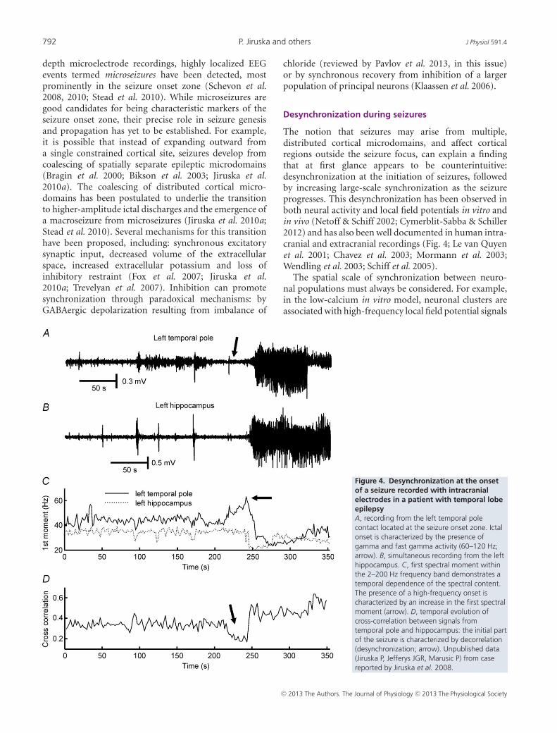
The notion that seizures may arise from multiple,distributed cortical microdomains, and affect corticalregions outside the seizure focus, can explain a findingthat at first glance appears to be counterintuitive:desynchronization at the initiation of seizures, followedby increasing large-scale synchronization as the seizureprogresses. This desynchronization has been observed inboth neural activity and local field potentials in vitro andin vivo (Netoff & Schiff 2002; Cymerblit-Sabba & Schiller2012) and has also been well documented in human intra-cranial and extracranial recordings (Fig. 4; Le van Quyenet al. 2001; Chavez et al. 2003; Mormann et al. 2003;Wendling et al. 2003; Schiff et al. 2005).
The spatial scale of synchronization between neuro-nal populations must always be considered. For example,in the low-calcium in vitro model, neuronal clusters areassociated with high-frequency local field potential signals
Figure 4. Desynchronization at the onsetof a seizure recorded with intracranialelectrodes in a patient with temporal lobeepilepsyA, recording from the left temporal polecontact located at the seizure onset zone. Ictalonset is characterized by the presence ofgamma and fast gamma activity (60–120 Hz;arrow). B, simultaneous recording from the lefthippocampus. C, first spectral moment withinthe 2–200 Hz frequency band demonstrates atemporal dependence of the spectral content.The presence of a high-frequency onset ischaracterized by an increase in the first spectralmoment (arrow). D, temporal evolution ofcross-correlation between signals fromtemporal pole and hippocampus: the initial partof the seizure is characterized by decorrelation(desynchronization; arrow). Unpublished data(Jiruska P, Jefferys JGR, Marusic P) from casereported by Jiruska et al. 2008.
C© 2013 The Authors. The Journal of Physiology C© 2013 The Physiological Society
J Physiol 591.4 Synchronization in epilepsy 793
at the onset of seizures. As the seizure progresses, adjacentclusters (∼150 μm diameter separation) demonstrateincreased synchronization (Jiruska et al. 2010a). Incontrast, synchronization between more widely separatedclusters (>300 μm) was low interictally and strengthenedand expanded with the progression of each seizure(Jiruska et al. 2010a). The interpretation of early ictaldesynchronization in human recordings depends stronglyon electrode location relative to the recruited seizureterritory. If synchrony is measured between two regionsat a time when one is generating seizure activity while thesecond remains unaffected, then the measure will indicatedesynchronization (Le van Quyen et al. 2001).
Desynchronization has been described during seizureonsets characterized by the presence of signals inthe beta and gamma ranges (15–40 Hz) classified aslow-amplitude fast activity (Wendling et al. 2003; de Curtis& Gnatkovsky 2009) recorded in seizures originating fromboth neocortex and entorhinal cortex in humans and in anin vitro guinea pig model of seizures (Wendling et al. 2003;Gnatkovsky et al. 2008; de Curtis & Gnatkovsky 2009).They found suppressed action potential firing at seizureinitiation, while local (mainly somatic-projecting) inter-neurons demonstrate sustained and intensive dischargesresulting in prominent and fast inhibitory postsynapticpotentials in principal cells (Wendling et al. 2002; de Curtis& Gnatkovsky 2009). The most plausible explanation forthese findings is the ‘inhibitory veto’ in the area ahead ofthe ictal wavefront, described above (Trevelyan et al. 2006,2007), i.e. the recordings were penumbral and the seizurefocus was located elsewhere.
The concept of spike synchronization needs to be under-stood within a framework where synaptic conductionpathways have finite conduction velocity and each pathis of a different length. Synchrony, the concept of a stablefunctional relationship where elements do similar thingsat the same time, needs to be broadened to that ofpolychronicity in neural networks, when time lags mustbe incorporated into the firing time of each neuron aspopulation activity is considered (Izhikevich 2006). Theepileptic brain possesses myriad of mechanisms that maysynchronize the action potential firing on a millisecondtime scale, involving both synaptic and non-synapticmechanisms. There is a large amount of literaturereviewing these mechanisms, which are beyond the scopeof the present review (Dudek et al. 1986; Jefferys, 1995;Carlen et al. 2000; Jefferys et al. 2012a,b; Zorec et al. 2012).
Caution must, however, be observed in extrapolatingthese results to all types of seizures. Differences existbetween experimental models and between epilepsysyndromes. Several distinct and region-specific seizureonset patterns are known and may have different cellular,network and synchronization mechanisms (Bragin et al.2005, 2009; Bertram 2009; de Curtis & Gnatkovsky2009). Human seizures, as reflected in the complex
and varied clinical semiological findings, coupled withthe distinctive electrographic patterns of various seizuretypes, all bespeak to the likelihood of different dynamicalmechanisms in these ictal events.
The impact of spatial scale on synchronization has beenwell demonstrated in the genesis of interictal pathologicalhigh-frequency oscillations (Jefferys et al. 2012). In normalbrain exposed to pro-epileptic conditions, populationsof bursting cells can synchronize their action potentialfiring (Foffani et al. 2007). This collective firing manifestsin local field potentials as high-frequency oscillations,with spectral peaks corresponding to the inter-spikefrequency of individual neurons firing at 200–300 Hz(Dzhala & Staley 2004). In contrast, in the chronicepileptic brain, interictal high-frequency oscillationswith frequencies up to 800 Hz (fast ripples) have beenrecorded (Staba et al. 2002; Bragin et al. 2004; Jiruskaet al. 2010b). It has been suggested that oscillations atfrequencies far higher than the maximum firing rate of apyramidal cell can be explained by an emergent networkphenomenon generated by independent (out-of-phase)neuronal populations (Bikson et al. 2003; Foffani et al.2007; Ibarz et al. 2010). This epileptic phenomenondemonstrates the importance of spatial scale andthe nuances of synchronization measures. Althoughtemporally uncorrelated, the out-of-phase firing may stillbe regarded as synchronized, or at least have a poly-chronous relationship, in terms of having a stable phase(Rosenblum et al. 1996) or temporal lags (Rosenblumet al. 1997). Demont-Guignard et al. (2012) examinedin detail the role of pyramidal cell population sizes, thespatial distribution of hyperexcitable pyramidal cells andthe level of synchronization between action potentialsin forming interictal high-frequency oscillations. In theircombined computational and in vitro study they showedthat a weak level of synchronization between afferentaction potentials can manifest as extracellular fast ripples,while the progressive increase in the synchronizationbetween action potentials resulted in a shift from fast rippleoscillations to an interictal EEG spike discharge.
Synchronization and seizure termination
Synchronization reaches its peak during the final stagesof the seizure (Fig. 2). A consistent observation acrossdifferent in vitro and in vivo models and human seizuresis that they terminate simultaneously over large areas ofthe brain (Topolnik et al. 2003; Timofeev & Steriade 2004;Schindler et al. 2007b; Truccolo et al. 2011). Therefore, ithas been suggested that either enhancing or disruptingsynchronization may promote seizure termination andmay potentially be used as abortive therapy.
Fires stop when there is nothing left to ignite; epilepticseizures might stop when there is nothing left to excite.Thus, one possible way in which seizures terminate is
C© 2013 The Authors. The Journal of Physiology C© 2013 The Physiological Society

794 P. Jiruska and others J Physiol 591.4
the creation of an extended area of hypoexcitable tissue,or a ‘zone of relative refractoriness’. An extensive seriesof in vivo experiments used multi-site intracellular andextracellular neuronal recordings and local cortical fieldpotential recordings to investigate initiation, developmentand cessation of spontaneous spike-wave seizures incats under ketamine–xylazine anaesthesia (Timofeev &Steriade 2004). Early after seizure onset, the propagationtime between field potentials recorded at multiple sitesof neocortex progressively decreased while long-rangesynchrony increased and reached a maximum towardsthe end of the seizure (Topolnik et al. 2003). Whenall the affected neuronal pools are drawn into highlysynchronous paroxysmal activity the seizure terminates.Increasing synchronization towards the end of seizureshas not only been noted in animal models, but alsoin several studies of intracranially recorded seizures inhumans (Schiff et al. 2005; Schindler et al. 2007a,b,c;Kramer et al. 2010). In the cat model of spontaneousspike-wave seizures, the synchronous activity towards theend of the seizures was in the form of ∼1 Hz oscillationssynchronized by EPSPs, but paced by intrinsic neuro-nal oscillations. Each cycle of the oscillation is initiatedby hyperpolarization-activated depolarizing current (Ih)and then enhanced by Na+ and Ca2+ currents. Thesedepolarizing currents then activate potassium currents(IK), which have a hyperpolarizing effect. The idea isthat Ca2+-dependent activation of the hyperpolarizingcurrents eventually overcomes the depolarizing effect ofthe Ih component of the oscillation. As this effect occurssimultaneously it results in seizure termination across theentire synchronized territory (Timofeev & Steriade, 2004).
Computer modelling of neuronal networks supportsthe counterintuitive theory that ongoing networkactivity can be both initiated and terminated bysynchronous excitatory synaptic input. Membraneimpedance shunting, due to massively increasedmembrane conductance, disrupts synaptic integration andresults in decreased efficacy of excitatory transmission thatcan promote termination of synchronized neural bursting(Gutkin et al. 2001). Another candidate mechanism forseizure termination is inhibitory transmission. Inhibitionmediated by interneurons, their mutual interconnectivityand their connectivity with principal cells may providea mechanism by which synchronized excitation maycause potent synchronization of inhibition (Pavlovet al. 2013, in this issue). This phenomenon hasbeen reported during interictal spikes (Lebovitz 1979;de Curtis et al. 2001) and ictal discharges (Lado &Moshe 2008), possibly contributing to their synchronouspost-excitatory depression. Most recently, a frameworkto unify the dynamics of seizure termination has beenoffered characterizing termination as a discontinuouscritical transition or bifurcation (Kramer et al. 2012).
Post-excitatory depression is also mediated bynon-synaptic mechanisms, particularly by changes in theextracellular environment that may outlast the recurrentsynaptic inhibition. In the piriform cortex of the isolatedguinea pig brain preparation, rhythmic interictal spikes arefollowed by a prolonged extracellular alkaline shift thatclosely correlates with inter-spike periodicity (de Curtiset al. 1998). Such pH changes induced by synchronousdischarging of neurons during an interictal spike maypromote synchronous post-spike depression of principalcell activity via decoupling of gap junctions. A similarphenomenon may, in principle, occur during the peri-odic bursting typical of the clonic (late) seizure phase,and could eventually contribute to seizure termination viasynchronization of inhibition.
These diverse mechanisms all share the involvement ofprogressively greater large-scale synchronization of neuro-nal activity as seizures progress, which may be consideredas an emergent self-regulatory mechanism for seizuretermination.
Epilepsy and hypersynchrony
The term ‘hypersynchrony’ appeared to originate fromearly studies on human electroencephalogram andepilepsy, and was initially used to describe the diffusespatial distribution of a normal monomorphic 3–5 Hzrhythm occurring (hypnagogic hypersynchrony) inchildren during drowsiness (Kellaway & Fox 1952). Theterm hypersynchrony seemed to be first applied toseizures by Penfield & Jasper (1954), and referred to anunderlying mechanism of excessive synchronization ofactivity in a large population of neurons manifesting ashigh-amplitude rhythmic epileptic discharges. It has sincebeen extrapolated to indicate synchronization of both fieldpotentials and neuronal activity at larger spatial scales.This review demonstrates that synchronization in epilepsyis very complex, and may appear different depending onthe spatial scale, the definition of synchrony and the signalsbeing measured (i.e. neuronal spikes vs. low-frequencyfield potentials). As this review demonstrates, seizuresare far more complex, dynamical phenomena thanthey were postulated to be when hypersynchrony wasfirst suggested to be an adequate organizing principleto account for seizures. Although the term hyper-synchrony has embedded itself within the acceptedprofessional knowledge base of epilepsy, the accumulatingevidence now suggests that a simple metric of increasedsynchronization is inadequate to account for seizures.
Conclusions
Synchronization in epilepsy is a much more complexphenomenon than often presented in the literature.The study of ictal synchronization is a rapidly evolving
C© 2013 The Authors. The Journal of Physiology C© 2013 The Physiological Society

J Physiol 591.4 Synchronization in epilepsy 795
field, which benefited from introduction of moresophisticated statistical methods of synchronizationcharacterization. Recent advances and observationson ictal synchronizations/desynchronizations have sub-stantially contributed to our understanding of itspathophysiology, creating novel insights into theorganization of epileptic networks and ictogenesis.Further, these observations may open new therapeuticmethods for promoting seizure termination.
References
Babb TL, Wilson CL & Isokawa-Akesson M (1987). Firingpatterns of human limbic neurons duringstereoencephalography (SEEG) and clinical temporal lobeseizures. Electroencephalogr Clin Neurophysiol 66, 467–482.
Bertram EH (2009). Temporal lobe epilepsy: where do theseizures really begin? Epilepsy Behav 14, 32–37.
Bikson M, Fox JE & Jefferys JGR (2003). Neuronal aggregateformation underlies spatiotemporal dynamics ofnonsynaptic seizure initiation. J Neurophysiol 89, 2330–2333.
Bishop G (1932). Cyclic changes in excitability of the opticpathway of the rabbit. American Journal of Physiology-Legacy103, 213–224.
Bower MR, Stead M, Meyer FB, Marsh WR & Worrell GA(2012). Spatiotemporal neuronal correlates of seizuregeneration in focal epilepsy. Epilepsia 53, 807–816.
Bragin A, Azizyan A, Almajano J & Engel J Jr (2009). The causeof the imbalance in the neuronal network leading to seizureactivity can be predicted by the electrographic pattern of theseizure onset. J Neurosci 29, 3660–3671.
Bragin A, Wilson CL, Almajano J, Mody I & Engel J Jr (2004).High-frequency oscillations after status epilepticus:epileptogenesis and seizure genesis. Epilepsia 45, 1017–1023.
Bragin A, Wilson CL & Engel J Jr (2000). Chronicepileptogenesis requires development of a network ofpathologically interconnected neuron clusters: a hypothesis.Epilepsia 41, S144–S152.
Bragin A, Wilson CL, Fields T, Fried I & Engel J Jr (2005).Analysis of seizure onset on the basis of wideband EEGrecordings. Epilepsia 46, 59–63.
Brown R & Rulkov NF (1997). Designing a coupling thatguarantees synchronization between identical chaoticsystems. Phys Rev Lett 78, 4189–4192.
Carlen PL, Skinner F, Zhang L, Naus C, Kushnir M & PerezVelazquez JL (2000). The role of gap junctions in seizures.Brain Res Brain Res Rev 32, 235–241.
Chavez M, Le Van Quyen M, Navarro V, Baulac M &Martinerie J (2003). Spatio-temporal dynamics prior toneocortical seizures: amplitude versus phase couplings. IEEETrans Biomed Eng 50, 571–583.
Cymerblit-Sabba A & Schiller Y (2012). Development ofhypersynchrony in the cortical network duringchemoconvulsant-induced epileptic seizures in vivo.J Neurophysiol 107, 1718–1730.
de Curtis M & Gnatkovsky V (2009). Reevaluating themechanisms of focal ictogenesis: the role of low-voltage fastactivity. Epilepsia 50, 2514–2525.
de Curtis M, Librizzi L & Biella G (2001). Dischargethreshold is enhanced for several seconds after a singleinterictal spike in a model of focal epileptogenesis. Eur JNeurosci 14, 174–178.
de Curtis M, Manfridi A & Biella G (1998). Activity-dependentpH shifts and periodic recurrence of spontaneous interictalspikes in a model of focal epileptogenesis. J Neurosci 18,7543–7551.
Demont-Guignard S, Benquet P, Gerber U, Biraben A, MartinB & Wendling F (2012). Distinct hyperexcitabilitymechanisms underlie fast ripples and epileptic spikes. AnnNeurol 71, 342–352.
Dudek FE, Snow RW & Taylor CP (1986). Role of electricalinteractions in synchronization of epileptiform bursts. AdvNeurol 44, 593–617.
Dzhala VI & Staley KJ (2004). Mechanisms of fast ripples in thehippocampus. J Neurosci 24, 8896–8906.
Foffani G, Uzcategui YG, Gal B & Menendez de la PL (2007).Reduced spike-timing reliability correlates with theemergence of fast ripples in the rat epileptic hippocampus.Neuron 55, 930–941.
Fox JE, Bikson M & Jefferys JGR (2007). The effect of neuronalpopulation size on the development of epileptiformdischarges in the low calcium model of epilepsy. NeurosciLett 411, 158–161.
Francis, JT, Gluckman, BJ & Schiff, SJ (2003). Sensitivityof Neurons to Weak Electric Fields. J Neurosci 23, 7255–7261.
Gnatkovsky V, Librizzi L, Trombin F & de Curtis M (2008).Fast activity at seizure onset is mediated by inhibitorycircuits in the entorhinal cortex in vitro. Ann Neurol 64, 674–686.
Gutkin BS, Laing CR, Colby CL, Chow CC & Ermentrout GB(2001). Turning on and off with excitation: the role ofspike-timing asynchrony and synchrony in sustained neuralactivity. J Comput Neurosci 11, 121–134.
Ibarz JM, Foffani G, Cid E, Inostroza M & Menendez de la PL(2010). Emergent dynamics of fast ripples in the epileptichippocampus. J Neurosci 30, 16249–16261.
Izhikevich EM (2006). Polychronization: computation withspikes. Neural Comput 18, 245–282.
Jefferys JGR (1995). Nonsynaptic modulation of neuronalactivity in the brain: electric currents and extracellular ions.Physiol Rev 75, 689–723.
Jefferys JGR, de la Prida LM, Wendling F, Bragin A, Avoli M,Timofeev I & Lopes da Silva FH (2012). Mechanisms ofphysiological and epileptic HFO generation. Prog Neurobiol98, 250–264.
Jefferys JGR & Haas HL (1982). Synchronized bursting of CA1hippocampal pyramidal cells in the absence of synaptictransmission. Nature 300, 448–450.
Jefferys JGR, Jiruska P, de Curtis M & Avoli M (2012). Limbicnetwork synchronization and temporal lobe epilepsy. InJasper’s Basic Mechanisms of the Epilepsies, eds. Noebels JL,Avoli M, Rogawski MA, Olsen RW & Delgado-Escueta AV,pp. 176–189. Oxford University Press.
Jensen MS & Yaari Y (1997). Role of intrinsic burst firing,potassium accumulation, and electrical coupling in theelevated potassium model of hippocampal epilepsy. JNeurophysiol 77, 1224–1233.
C© 2013 The Authors. The Journal of Physiology C© 2013 The Physiological Society

796 P. Jiruska and others J Physiol 591.4
Jiruska P, Csicsvari J, Powell AD, Fox JE, Chang WC,Vreugdenhil M, Li X, Palus M, Bujan AF, Dearden RW &Jefferys JGR (2010a). High-frequency network activity,global increase in neuronal activity, and synchrony expansionprecede epileptic seizures in vitro. J Neurosci 30, 5690–5701.
Jiruska P, Finnerty GT, Powell AD, Lofti N, Cmejla R & JefferysJG (2010b). Epileptic high-frequency network activity in amodel of non-lesional temporal lobe epilepsy. Brain 133,1380–1390.
Jiruska P, Tomasek M, Netuka D, Otahal J, Jefferys JG, Li X &Marusic P (2008). Clinical impact of a high-frequencyseizure onset zone in a case of bitemporal epilepsy. EpilepticDisord 10, 231–238.
Kellaway P & Fox BJ (1952). Electroencephalographic diagnosisof cerebral pathology in infants during sleep. I. Rationale,technique, and the characteristics of normal sleep in infants.J Pediatr 41, 262–287.
Klaassen A, Glykys J, Maguire J, Labarca C, Mody I & Boulter J(2006). Seizures and enhanced cortical GABAergic inhibitionin two mouse models of human autosomal dominantnocturnal frontal lobe epilepsy. Proc Natl Acad Sci U S A 103,19152–19157.
Konnerth A, Heinemann U & Yaari Y (1984). Slowtransmission of neural activity in hippocampal area CA1 inabsence of active chemical synapses. Nature 307, 69–71.
Kramer MA, Eden UT, Kolaczyk ED, Zepeda R, EskandarEN & Cash SS (2010). Coalescence and fragmentation ofcortical networks during focal seizures. J Neurosci 30, 10076–10085.
Kramer MA, Truccolo W, Eden UT, Lepage KQ, Hochberg LR,Eskandar EN, Madsen JR, Lee JW, Maheshwari A, Halgren E,Chu CJ & Cash SS (2012). Human seizures self-terminateacross spatial scales via a critical transition. Proc Natl AcadSci U S A. in press.
Lado FA & Moshe SL (2008). How do seizures stop? Epilepsia49, 1651–1664.
Le Van Quyen M, Martinerie J, Navarro V, Baulac And M &Varela FJ (2001). Characterizing neurodynamic changesbefore seizures. J Clin Neurophysiol 18, 191–208.
Lebovitz RM (1979). Autorhythmicity of spontaneousinterictal spike discharge at hippocampal penicillin foci.Brain Res 172, 35–55.
Li X, Cui D, Jiruska P, Fox JE, Yao X & Jefferys JG (2007).Synchronization measurement of multiple neuronalpopulations. J Neurophysiol 98, 3341–3348.
Margineanu DG (2010). Epileptic hypersynchrony revisited.Neuroreport 21, 963–967.
Matsumoto H & Ajmone-Marsan C (1964). Cortical cellularphenomena in experimental epilepsy: ictal manifestations.Exp Neurol 9, 305–326.
Mormann F, Kreuz T, Andrzejak RG, David P, LehnertzK & Elger CE (2003). Epileptic seizures are precededby a decrease in synchronization. Epilepsy Res 53, 173–185.
Muller M, Baier G, Rummel C & Schindler K (2008).Estimating the strength of genuine and random correlationsin non-stationary multivariate time series. Europhys Lett 84,10009.
Netoff TI, Carroll TL, Pecora LM & Schiff SJ (2006). Detectingcoupling in the presence of noise and nonlinearity, InHandbook of Time Series Analysis, eds. Schelter BI,Winterhalder M & Timmer J, pp. 265–282. Wiley-VCHVerlag GmbH & Co. KGaA
Netoff TI, Pecora, LM & Schiff SJ (2004). Analytical couplingdetection in the presence of noise and nonlinearity. Phys RevE Stat Nonlin Soft Matter Phys 69, 017201.
Netoff TI & Schiff SJ (2002). Decreased neuronalsynchronization during experimental seizures. J Neurosci 22,7297–7307.
Pavlov I, Kaila K, Kullmann DM & Miles R (2013). Corticalinhibition, pH and cell excitability in epilepsy: what areoptimal targets for antiepileptic interventions? J Physiol 591,765–774.
Pecora L & Carroll T (1990). Synchronization in chaoticsystems. Phys Rev Lett 64, 821–824.
Penfield W & Jasper H (1954). Epilepsy and the FunctionalAnatomy of the Human Brain. Little Brown, Boston.
Pikovsky A, Rosenblum M & Kurths J (2003). Synchronization:A Universal Concept in Nonlinear Sciences. CambridgeUniversity Press.
Prince DA & Wilder BJ (1967). Control mechanisms in corticalepileptogenic foci. “Surround” inhibition. Arch Neurol 16,194–202.
Rosenblum MG, Pikovsky AS & Kurths J (1996). Phasesynchronization of chaotic oscillators. Phys Rev Lett 76,1804–1807.
Rosenblum MG, Pikovsky AS & Kurths J (1997). From phase tolag synchronization in coupled chaotic oscillators. Phys RevLett 78, 4193–4196.
Rulkov NF, Sushchik MM, Tsimring LS & Abarbanel HDI(1995). Generalized synchronization of chaos indirectionally coupled chaotic systems. Phys Rev E Stat PhysPlasmas Fluids Relat Interdiscip Topics 51, 980–994.
Salinas E & Sejnowski TJ (2001). Correlated neuronal activityand the flow of neural information. Nat Rev Neurosci 2,539–550.
Schevon CA, Goodman RR, McKhann G Jr & Emerson RG(2010). Propagation of epileptiform activity on asubmillimeter scale. J Clin Neurophysiol 27, 406–411.
Schevon CA, Ng SK, Cappell J, Goodman RR, McKhann G Jr,Waziri A, Branner A, Sosunov A, Schroeder CE & EmersonRG (2008). Microphysiology of epileptiform activity inhuman neocortex. J Clin Neurophysiol 25, 321–330.
Schevon CA, Weiss SA, McKhann G Jr, Goodman RR, Yuste R,Emerson RG & Trevelyan AJ (2012). Evidence of aninhibitory restraint of seizure activity in humans. NatCommun 3, 1060.
Schiff SJ (2012). Neural Control Engineering: The EmergingIntersection between Control Theory and Neuroscience. MITPress, Cambridge.
Schiff SJ, Sauer T, Kumar R & Weinstein SL (2005). Neuronalspatiotemporal pattern discrimination: the dynamicalevolution of seizures. NeuroImage 28, 1043–1055.
Schiff SJ, So P, Chang T, Burke RE & Sauer T (1996). Detectingdynamical interdependence and generalized synchronythrough mutual prediction in a neural ensemble. Phys Rev E54, 6708–6724.
C© 2013 The Authors. The Journal of Physiology C© 2013 The Physiological Society
J Physiol 591.4 Synchronization in epilepsy 797
Schindler K, Elger CE & Lehnertz K (2007a). Changes of EEGsynchronization during low-frequency electric stimulationof the seizure onset zone. Epilepsy Res 77, 108–119.
Schindler K, Elger CE & Lehnertz K (2007b). Increasingsynchronization may promote seizure termination: evidencefrom status epilepticus. Clin Neurophysiol 118, 1955–1968.
Schindler K, Leung H, Elger CE & Lehnertz K (2007c).Assessing seizure dynamics by analysing the correlationstructure of multichannel intracranial EEG. Brain 130,65–77.
Schwartz TH & Bonhoeffer T (2001). In vivo optical mappingof epileptic foci and surround inhibition in ferret cerebralcortex. Nat Med 7 , 1063–1067.
Shoham S, O’Connor DH & Segev R (2006). How silent is thebrain: is there a “darkmatter” problem in neuroscience? JComp Physiol A Neuroethol Sens Neural Behav Physiol 192,777–784.
Staba RJ, Wilson CL, Bragin A, Fried I & Engel J Jr (2002).Quantitative analysis of high-frequency oscillations (80–500Hz) recorded in human epileptic hippocampus andentorhinal cortex. J Neurophysiol 88, 1743–1752.
Stead M, Bower M, Brinkmann BH, Lee K, Marsh WR, MeyerFB, Litt B, Van GJ & Worrell GA (2010). Microseizures andthe spatiotemporal scales of human partial epilepsy. Brain133, 2789–2797.
Timofeev I & Steriade M (2004). Neocortical seizures:initiation, development and cessation. Neuroscience 123,299–336.
Topolnik L, Steriade M & Timofeev I (2003). Partial corticaldeafferentation promotes development of paroxysmalactivity. Cereb Cortex 13, 883–893.
Trevelyan AJ, Sussillo D, Watson BO & Yuste R (2006).Modular propagation of epileptiform activity: evidence foran inhibitory veto in neocortex. J Neurosci 26, 12447–12455.
Trevelyan AJ, Sussillo D & Yuste R (2007). Feedforwardinhibition contributes to the control of epileptiformpropagation speed. J Neurosci 27, 3383–3387.
Truccolo W, Donoghue JA, Hochberg LR, Eskandar EN,Madsen JR, Anderson WS, Brown EN, Halgren E & Cash SS(2011). Single-neuron dynamics in human focal epilepsy.Nat Neurosci 14, 635–641.
Uhlhaas PJ & Singer W (2006). Neural synchrony in braindisorders: relevance for cognitive dysfunctions andpathophysiology. Neuron 52, 155–168.
Varela F, Lachaux JP, Rodriguez E & Martinerie J (2001). Thebrainweb: phase synchronization and large-scale integration.Nat Rev Neurosci 2, 229–239.
Welsh DK, Logothetis DE, Meister M & Reppert SM (1995).Individual neurons dissociated from rat suprachiasmaticnucleus express independently phased circadian firingrhythms. Neuron 14, 697–706.
Wendling F, Bartolomei F, Bellanger JJ, Bourien J & Chauvel P(2003). Epileptic fast intracerebral EEG activity: evidence forspatial decorrelation at seizure onset. Brain 126,1449–1459.
Wendling F, Bartolomei F, Bellanger JJ & Chauvel P (2002).Epileptic fast activity can be explained by a model ofimpaired GABAergic dendritic inhibition. Eur J Neurosci 15,1499–1508.
Westbrook GL (1991). Seizures and epilepsy. In Principles ofNeural Science, eds. Kandel ER, Jessel TM & Schwartz JH.McGraw-Hill, New York.
Wyler AR, Ojemann GA & Ward AA Jr (1982). Neurons inhuman epileptic cortex: correlation between unit and EEGactivity. Ann Neurol 11, 301–308.
Wyler AR & Ward AA Jr (1986). Neuronal firing patterns fromepileptogenic foci of monkey and human. Adv Neurol 44,967–989.
Zhao L, Beverlin B, Netoff T & Nykamp DQ (2011).Synchronization from second order network connectivitystatistics. Front Comput Neurosci 5, 28.
Ziburkus J, Cressman JR, Barreto E & Schiff SJ (2006).Interneuron and pyramidal cell interplay during in vitroseizure-like events. J Neurophysiol 95, 3948–3954.
Zorec R, Araque A, Carmignoto G, Haydon PG, Verkhratsky A& Parpura V (2012). Astroglial excitability andgliotransmission: an appraisal of Ca2+ as a signalling route.ASN Neuro 4, e00080.
Acknowledgement
This work was supported by a Czech Ministry of HealthGrant IGA NT/11460-4, Epilepsy Research UK grant (P1102),MRC Grant G0802162, Swiss National Science Foundation(project no. SNF 320030 122010), NIH grant 1R01EB014641-01US-German Collaborative Research in Computational Neuro-science (CRCNS), and Medical Research Council grant(G0802162). The authors declare no conflict of interest.
C© 2013 The Authors. The Journal of Physiology C© 2013 The Physiological Society