Embed Size (px)
Citation preview
The Journal of Pathology, INVITED REVIEW
The path to a better biomarker: application of a risk management framework for the
implementation of PD-L1 and TILs as immuno-oncology biomarkers into breast cancer
clinical trials and daily practice
Paula I Gonzalez-Ericsson1*
, Elisabeth S Stovgaard2, Luz F Sua
3, Emily Reisenbichler
4, Zuzana
Kos 5 Jodi M Carter
6, Stefan Michiels
7, John Le Quesne
8,9, Torsten O Nielsen
10, Anne-Vibeke
Lænkholm11
, Stephen B Fox12,13
, Julien Adam14
, John MS Bartlett15,16
, David L Rimm4, Cecily
Quinn17
, Dieter Peeters18,19
, Maria V Dieci20,21
, Anne Vincent-Salomon22
Ian Cree23
, Akira I
Hida24
, Justin M Balko1,25,26
, Harry R Haynes27,28
, Isabel Frahm29
, Gabriela Acosta-Haab30
,
Marcelo Balancin31
, Enrique Bellolio32
, Wentao Yang33
, Pawan Kirtani34
, Tomoharu Sugie35
,
Anna Ehinger36
, Carlos A Castaneda37
, Marleen Kok38
, Heather McArthur39
, Kalliopi
Siziopikou40
, Sunil Badve41
, Susan Fineberg42
, Allen Gown43
, Giuseppe Viale44,45
, Stuart J
Schnitt46,47
, Giancarlo Pruneri45,48
, Frederique Penault-Llorca49,50
, Stephen Hewitt51
, E Aubrey
Thompson52
, Kimberly H Allison53
, William F Symmans54
, Andrew M Bellizzi55
Edi Brogi56
,
David A Moore57
, Denis Larsimont58
, Deborah A Dillon46
, Alexander Lazar54
, Huangchun
Lien59
, Matthew P Goetz60
, Glenn Broeckx61
, Khalid El Bairi62
, Nadia Harbeck63
, Ashley
Cimino-Mathews64
, Christos Sotiriou65
, Sylvia Adams66
, Shi-wei Liu67
, Sibylle Loibl68
, I-Chun
Chen69
, Sunil R Lakhani70
, Jonathan W Juco71
, Carsten Denkert72
, Elizabeth F Blackley73
,
Sandra Demaria74
, Roberto Leon-Ferre60
, Oleg Gluz75
, Dimitrios Zardavas76
, Kenneth
Emancipator71
, Scott Ely77
, Sherene Loi13,78
,
Roberto Salgado78,79
, Melinda Sanders1,26
* on
behalf of the International Immuno-Oncology Biomarker Working Group.
Acc
epte
d A
rticl
e
This article is protected by copyright. All rights reserved.
This article has been accepted for publication and undergone full peer review but has not been through the copyediting, typesetting, pagination and proofreading process which may lead to differences between this version and the Version of Record. Please cite this article as doi:10.1002/path.5406

2
1Breast Cancer Research Program, Vanderbilt University Medical Center, Nashville, USA.
2Department of Pathology, Herlev and Gentofte Hospital, University of Copenhagen, Herlev,
Denmark.
3Department of Pathology and Laboratory Medicine, Fundación Valle del Lili, and Faculty of
Health Sciences, Universidad ICESI, Cali, Colombia.
4Department of Pathology, Yale School of Medicine, New Haven, USA.
5Department of Pathology, BC Cancer Agency, Vancouver, Canada
6Department of Laboratory Medicine and Pathology, Mayo Clinic, Rochester, USA.
7Biostatistics and Epidemiology Service, Centre de Recherche en Epidémiologie et Santé des
Populations, Gustave Roussy, Université Paris-Sud, Villejuif, France.
8Leicester Cancer Research Centre, University of Leicester, Leicester, UK.
9MRC Toxicology Unit, University of Cambridge, Leicester, UK
10Department of Pathology and Laboratory Medicine, University of British Columbia,
Vancouver, Canada.
11Department of Surgical Pathology, Zealand University Hospital, Denmark
12Department of Pathology, Peter MacCallum Cancer Centre, Melbourne, Australia.
13Sir Peter MacCallum Department of Oncology, University of Melbourne, Parkville, Australia.
14Department of Pathology, Gustave Roussy, Grand Paris, France.
15Ontario Institute for Cancer Research, Toronto, Canada.
16 Edinburgh Cancer Research Centre, Institute of Genetics and Molecular Medicine, Edinburgh,
UK.
17Department of Pathology, St Vincent's University Hospital and University College Dublin,
Dublin, Ireland.
Acc
epte
d A
rticl
e
This article is protected by copyright. All rights reserved.

3
18HistoGeneX NV, Antwerp, Belgium
19AZ Sint-Maarten Hospital, Mechelen, Belgium.
20Department of Surgery, Oncology and Gastroenterology, University of Padova, Padova, Italy.
21Medical Oncology 2, Istituto Oncologico Veneto - IRCCS, Padova, Italy.
22Department of Pathology, Insitut Curie, Paris, France.
23International Agency for Research on Cancer (IARC), World Health Organization, Lyon,
France.
24Department of Pathology, Matsuyama Shimin Hospital, Matsuyama, Japan.
25Department of Medicine, Vanderbilt University Medical Center, Nashville, USA.
26Department of Pathology, Microbiology and Immunology, Vanderbilt University Medical
Center, Nashville, USA.
27Department of Cellular Pathology, North Bristol NHS Trust, Bristol UK
28Translational Health Sciences, University of Bristol, UK.
29Department of Pathology, Sanatorio Mater Dei, Buenos Aires, Argentina.
30Department of Pathology, Hospital de Oncología Maria Curie, Buenos Aires, Argentina
31Department of Pathology, Faculty of Medicine, University of São Paulo, São Paulo, Brazil.
32Department of Pathology, Universidad de La Frontera, Temuco, Chile.
33Department of Pathology, Fudan University Shanghai Cancer Centre, Shanghai, China.
34Department of Histopathology, Manipal Hospitals Dwarka, New Delhi.
35Breast Surgery, Kansai Medical University Hospital, Hirakata, Japan.
36Department of Clinical Genetics and Pathology, Skane University Hospital, Lund University,
Lund, Sweden.
Acc
epte
d A
rticl
e
This article is protected by copyright. All rights reserved.

4
37Department of Medical Oncology, Instituto Nacional de Enfermedades Neoplásicas, Lima,
Perú.
38Divisions of Medical Oncology, Tumor Biology & Immunology, The Netherlands Cancer
Institute, Amsterdam, The Netherlands.
39Medical Oncology, Department of Medicine, Cedars-Sinai Medical Center, Los Angeles, USA.
40Department of Pathology, Breast Pathology Section, Northwestern University, Chicago, USA.
41Department of Pathology and Laboratory Medicine, Indiana University, Indianapolis, USA.
42Department of Pathology, Montefiore Medical Center and the Albert Einstein College of
Medicine, Bronx, USA.
43PhenoPath Laboratories, Seattle, USA.
44Department of Pathology, Istituto Europeo di Oncologia IRCCS, Milan, Italy.
45University of Milan, Milan, Italy.
46Department of Pathology, Dana-Farber Cancer Institute, Boston, USA.
47Department of Pathology, Brigham and Women’s Hospital and Harvard Medical School,
Boston, USA.
48Department of Pathology, IRCCS Fondazione Instituto Nazionale Tumori, Milan, Italy.
49Department of Biology and Pathology, Centre Jean Perrin, Clermont Ferrand, France.
50UMR INSERM 1240, Université Clermont Auvergne, Clermont Ferrand, France.
51Experimental Pathology Laboratory, Laboratory of Pathology, Center for Cancer Research,
National Cancer Institute, National Institutes of Health, Bethesda, USA.
52Department of Cancer Biology, Mayo Clinic, Jacksonville, USA.
53Department of Pathology, Stanford University, Stanford, USA.
Acc
epte
d A
rticl
e
This article is protected by copyright. All rights reserved.
5
54Department of Pathology, Division of Pathology and Laboratory Medicine, The University of
Texas, MD Anderson Cancer Center, Houston, United States.
55Department of Pathology, University of Iowa Hospitals and Clinics, Iowa City, USA.
56Department of Pathology, Memorial Sloan Kettering Cancer Center, New York, USA.
57CRUK
Lung Cancer Centre of Excellence, UCL Cancer Institute, and Department of Cellular Pathology,
UCLH, London, UK.
58Department of Pathology, Institut Jules Bordet, Université Libre de Bruxelles, Brussels,
Belgium.
59Graduate Institute of Pathology, National Taiwan University, Taipei, Taiwan.
60 Department of Oncology, Mayo Clinic, Rochester, USA.
61Department of Pathology, University Hospital Antwerp, Belgium.
62Cancer Biomarkers Working Group, Faculty of Medicine and Pharmacy, Mohamed Ist
University, Oujida, Morocco.
63Breast Center, Department of OB&GYN and CCC (LMU), University of Munich, Munich,
Germany.
64Departments of Pathology and Oncology, The Johns Hopkins Hospital, Baltimore, USA.
65Department of Medical Oncology, Institut Jules Bordet, Université Libre de Bruxelles,
Brussels.
66Perlmutter Cancer Center, New York University Medical School, New York, USA.
67Sichuan Cancer Hospital, Chengdu, China.
68German Breast Group, Neu-Isenburg, Germany.
69Department of Medical Oncology, National Taiwan University Cancer Center, Taipei, Taiwan.
Acc
epte
d A
rticl
e
This article is protected by copyright. All rights reserved.

6
70The University of Queensland, Centre for Clinical Research, and Pathology Queensland, Royal
Brisbane and Women's Hospital, Herston, Australia.
71Translational Medicine, Merck & Co, Inc, Kenilworth, USA.
72Institute of Pathology, Universitätsklinikum Gießen und Marburg GmbH, Standort Marburg
and Philipps-Universität Marburg, Marburg, Germany.
73Department of Medical Oncology, Peter MacCallum Cancer Centre, Melbourne, VIC,
Australia.
74Department of Radiation Oncology, Department of Pathology and Laboratory Medicine, Weill
Cornell Medicine, New York, USA.
75Johanniter GmbH - Evangelisches Krankenhaus Bethesda Mönchengladbach, West German
Study Group, Mönchengladbach, Germany.
76Oncology Clinical Development, Bristol-Myers Squibb, Princeton, USA.
77Translational Medicine, Bristol-Myers Squibb, Princeton, USA.
78Division of Research, Peter MacCallum Cancer Centre, Melbourne, Australia.
79Department of Pathology, GZA-ZNA Hospitals, Antwerp, Belgium.
*Correspondence to: Paula Gonzalez Ericsson, Breast Cancer Research Program, Vanderbilt
University Medical Center, Nashville, USA. E-mail: [email protected]
Melinda Sanders, Department of Pathology, Microbiology and Immunology, and Breast Cancer
Research Program, Vanderbilt University Medical Center, Nashville, USA. E-mail:
Acc
epte
d A
rticl
e
This article is protected by copyright. All rights reserved.
7
Short running title: Implementation of PD-L1 and TILs as immuno-oncology biomarkers for
breast cancer.
Conflicts of interest: ACM is a consultant for and reports research grant support from Bristol-
Myers Squibb. AE is on the Roche advisory board and is a lecturer paid by Roche, Amgen and
Novartis. AIH reports honoraria from Chugai Pharmaceutical, Taiho Pharmaceutical and
Novartis Pharma. CQ is chair of the European Working Group for Breast Screening Pathology
(EWGBSP), which has received funding from various companies for group meetings and also
reports honoraria from Roche and Exact Sciences. DAM reports speaker fees from AstraZeneca.
DAD is on the advisory board of Oncology Analytics, Inc., and consults for Novartis. DLR is on
the advisory board for Amgen, AstraZeneca, Cell Signaling Technology, Cepheid, Daiichi
Sankyo, GlacoSmithKline, Konica Minolta, Merck, Nanostrings, Perkin Elmer, Roche, Ventana
and Ultivue.He is a consultant for Biocept, NextCure, Odonate and Sanofi and he is a founder
and equity holder of PixelGear. He reports research support from AstraZeneca, Cepheid,
Navdigate BioPharma, NextCure, Lilly and Ultivue and instrument support from Ventana,
Akoya perkin Elmer and Nanostrings. He reports travel honorarium from Bristol-Myers Squibb
and royalties from Rarecyte. DZ is an employee of Bristol-Myers Squibb with stock ownership.
FPL is an advisor for AstraZeneca, Bayer, Bristol-Myers Squibb, Diaceutics, Elli-Lilly, Illumina,
MSD and Roche, and reports research funding form AstraZeneca, Bayer, Bristol-Myers Squibb,
MSD and Roche. GV reports honoraria from Roche, Ventana, Agilent, MSD, Bristol-Myers
Squibb and AstraZeneca. HM is a consultant for Amgen, Bristol-Myers Squibb, Celgene, Eli
Lilly, Genentech/Roche, Immunomedics, Merck, OBI Pharma, Pfizer, Puma, Spectrum
Pharmaceuticals, Syndax Pharmaceuticals, Peregrine, Calithera, Daiichi-Sankyo and
Acc
epte
d A
rticl
e
This article is protected by copyright. All rights reserved.

8
TapImmune, and has research supported by Bristol-Myers Squibb, MedImmune, LLC
AstraZeneca, BTG and Merck. HRH has received travel and educational support from Roche
Ventana. JA is an advisor for AstraZeneca, Bayer, Bristol-Myers Squibb, MSD, Roche and
Diaceutics, and reports research funding form MSD, Bayer and Pierre Fabre. JB is a consultant
for Insight Genetics Inc., BioNTech AG, Biotheranostics Inc., Pfizer, Rna Diagnostics Inc. and
oncoXchange, and reports honoraria form NanoString Technologies, Oncology Education and
Biotheranostics Inc. He reports research funding from Thermo Fisher Scientific, Genoptix,
Agendia, NanoString Technologies Inc., Stratifyer GmbH and Biotheranostics Inc. and travel
expenses from Biotheranostics and NanoString Technologies. He possesses patents regarding
biomarker evaluation and gene signatures to predict response to treatment. JMB reports research
support from Genentech/Roche, Bristol-Myers Squibb and Incyte Corporation, has received
consulting expert witness fees from Novartis and is an inventor on provisional patents regarding
immunotherapy targets and biomarkers in cancer. JJ is a full-time employee of Merck & Co.,
Inc. and owns stock in Merck & Co., Inc., Regeneron and Illumina. KE is a full-time employee
of Merck & Co., Inc. and owns stock in that company as well as Bayer AG and Johnson &
Johnson. His wife is a full-time employee of Bristol-Myers Squibb and owns stock in that
company. KS reports one-time honorarium from Roche Ventana. MK is on the advisory board
for Bristol-Myers Squibb and Daiichi (uncompensated) and her institute receives funding from
BMS, Roche and AZ. OG reports travel support from Roche, Celgene, Daiichi and honoraria
advisory boards form Roche, Celgene, Novartis, Pfizer, Eli Lilly, GHI, Nanostring, Amgen and
MSD. RLF reports travel support from Immunomedics. RS reports research funding from Roche
Genentech, Puma Biotechnology and Merck, honoraria for consulting for Bristol-Myers Squibb
and travel funding from Roche Genentech, Merck and AstraZeneca. SD reports past research
Acc
epte
d A
rticl
e
This article is protected by copyright. All rights reserved.

9
grant support from Lytix Biopharma and Nanobiotix, and honoriaria for consulting from EMD
Serono and Mersana Therapeutics. SE is an employee of Bristol-Myers Squibb with stock
ownership. SF served on an expert panel for Genomic Health. SL receives research funding to
her institution from Novartis, Bristol-Myers Squibb, Merck, Roche-Genentech, Puma
Biotechnology, Pfizer, Astra Zeneca, Eli Lilly and Seattle Genetics. She has acted as consultant
(not compensated) to Seattle Genetics, Pfizer, Novartis, Bristol-Myers Squibb, Merck,
AstraZeneca and Roche-Genentech. She has acted as consultant (paid to her institution) to Aduro
Biotech, Novartis and G1 Therapeutics. TON has a proprietary interest in the PAM50 subtype
classifier form Bioclassifer LLC, Nanostring Technologies. TS reports speaker fees from
AstraZeneca, Novartis, Chugai, Pfizer, Eisai, Takeda, Kyowa Kirin, Lilly, MSD, Genomic
Health and a research grant from Chugai, Pfizer and Eisai.
Word count: 5598
Acc
epte
d A
rticl
e
This article is protected by copyright. All rights reserved.

10
Abstract
Immune checkpoint inhibitor therapies targeting PD-1/PD-L1 are now standard of care in
oncology across several hematologic and solid tumor types, including triple negative breast
cancer (TNBC). Patients with metastatic or locally advanced TNBC with PD-L1 expression on
immune cells occupying ≥1% of tumor area demonstrated survival benefit with the addition of
atezolizumab to nab-paclitaxel. However, concerns regarding variability between
immunohistochemical PD-L1 assay performance and inter-reader reproducibility have been
raised. High tumor-infiltrating lymphocytes (TILs) have also been associated with response to
PD-1/PD-L1 inhibitors in patients with breast cancer. TILs can be easily assessed on
hematoxylin and eosin stained slides and have shown reliable inter-reader reproducibility. As an
established prognostic factor in early stage TNBC, TILs are soon anticipated to be reported in
daily practice in many pathology laboratories worldwide. Since TILs and PD-L1 are parts of an
immunological spectrum in breast cancer, we propose the systematic implementation of
combined PD-L1 and TIL analyses as a more comprehensive immune-oncological biomarker for
patient selection for PD-1/PD-L1 inhibition-based therapy in patients with breast cancer.
Although practical and regulatory considerations differ by jurisdiction, the pathology community
has the responsibility to patients to implement assays that lead to optimal patient selection. We
propose herewith a risk-management framework that may help mitigate the risks of suboptimal
patient selection for immuno-therapeutic approaches in clinical trials and daily practice based on
combined TILs/PD-L1 assessment in breast cancer.
Keywords: PD-L1, TILs, breast cancer, biomarker risk-management, immunotherapy Acc
epte
d A
rticl
e
This article is protected by copyright. All rights reserved.

11
Introduction
Immune checkpoint inhibitor (ICI) therapies targeting programmed cell death 1 (PD-1) and
programmed death ligand 1 (PD-L1) are now standard of care in oncology. Anti-PD-1
pembrolizumab (Keytruda®, Merck & Co. Inc., Kenilworth, NJ, USA) and nivolumab
(Opdivo®, Bristol-Myers Squibb Company, New York, NY, USA), and anti-PD-L1
atezolizumab (Tecentriq®, Genentech Inc, South San Francisco, CA, USA), durvalumab
(Imfinzi®, AstraZeneca plc, Cambridge, UK) and avelumab (Bavencio®, Merck KGA,
Darmstadt, Germany) have been approved to treat multiple tumor types, in many countries. To
date, atezolizumab specifically has been approved for triple negative breast cancer (TNBC). At
the same time, immunohistochemistry (IHC) based detection of PD-L1 expression has been
proposed as the predictive biomarker to select patients that may benefit from these therapies.
Five primary antibody clones have been developed in the form of assays paired with a specific
staining platform. PD-L1 22C3 (Agilent Technologies Inc., Santa Clara, CA, USA), 28-8
(Agilent Technologies Inc.), SP142 (Roche Tissue Diagnostics, Tucson, AZ, USA), SP263
(Roche Tissue Diagnostics) and 73-10 (Agilent Technologies Inc.) have been used in clinical
trials of the above-mentioned drugs, respectively. In addition, laboratory-developed tests (LDTs)
using any of the above-mentioned primary antibodies or the E1L3N clone with different staining
platforms are in use in research and clinical scenarios. Parallel to the multiple assays, multiple
scoring systems exist. Table 1 shows technical details and defines scoring methods used for each
antibody. Furthermore, different cut-offs are used to define PD-L1 positivity for different tumor
types, while for certain indications PD-L1 testing is not required for PD-1/PD-L1 inhibition-
based therapy, from now on referred to as ICI.
Acc
epte
d A
rticl
e
This article is protected by copyright. All rights reserved.

12
For several years the oncology and pathology communities have raised concerns about
the reliability of IHC based detection of PD-L1 to appropriately select patients for ICI. To date,
while PD-L1 is currently the only approved biomarker for these agents, it remains controversial
given the complexities of its clinical use due to variability in assay performance of the PD-L1
IHC antibodies, spatial and temporal heterogeneity, absence of a unified scoring system and
concerns about inter-reader reproducibility for scoring PD-L1 on immune cell (ICs). Due to these
inconsistencies, some patients who could benefit might not receive treatment while others may
be treated based on erroneous test results, exposing them to potential adverse side effects with no
drug benefit. Additionally, since PD-1/PD-L1 interaction is only one of many factors that may
determine the clinical response to immunotherapeutics, it is unlikely that a single biomarker will
sufficiently predict clinical outcomes in response to ICI. The use of composite biomarkers can
provide biologically relevant information on multiple factors that determine response. In a meta-
analysis, combined biomarker approaches such as PD-L1 IHC and tumor mutational burden
(TMB) and multiplex fluorescent IHC evaluating protein co-expression and spatial relationships,
demonstrated an improved performance over PD-L1 or TMB alone [1]. As guardians of patient’s
samples, pathologists partnered with clinicians, industry and regulators must guide evidence-
based inclusion of biomarkers in clinical trials and daily practice to ensure the best patient
outcomes possible. Stromal tumor-infiltrating lymphocytes (TILs) have also been studied as a
predictive biomarker of response to ICI for a variety of cancers including breast cancer (BC).
TILs can be assessed on a simple hematoxylin and eosin (H&E) slide with reliable
reproducibility among pathologists when they adhered to the standardized method [2, 3]. We
propose PD-L1 and TILs as a more comprehensive composite biomarker.
Acc
epte
d A
rticl
e
This article is protected by copyright. All rights reserved.
13
A good biomarker should be analytically valid, robust, reproducible and clinically useful.
To be incorporated into daily practice, it must also be affordable and accessible to pathologists in
both academic and community-hospital practices worldwide [4]. In this review, we propose a
systematic implementation of combined PD-L1 and TIL analysis as a comprehensive immune-
oncological biomarker for patient selection for ICI in both clinical trials and daily practice. In
support of this position, we will outline the evolution of PD-L1 and TILs as biomarkers, from the
analytical and clinical validation phases through clinical implementation, review the challenges
we have encountered and propose mitigation approaches within a risk-management framework
as previously published [5]. The collective of available evidence anticipates enhancement of
patient selection and safety by the systematic implementation of combined PD-L1 and TIL
analysis.
Technical validation phase: analytical validity of PD-L1 immunohistochemistry
Biomarker development starts with an initial discovery in pre-clinical studies, which we will not
cover in this review, followed by a validation phase in which the biomarker is adapted to
clinically applicable assay platforms and subjected to analytical and clinical validation [6]. For
PD-L1 IHC, analytical validity refers to the accuracy and consistency of the technique to detect
the presence of PD-L1 protein. To be able to analyse the accuracy and consistency of the test we
must first define the presence of PD-L1 protein. PD-L1 can be expressed on solid and
hematologic tumor cells (TCs) and on ICs, including macrophages, dendritic cells, lymphocytes
and granulocytes [7, 8]. PD-L1 is expressed in the cytoplasm and/or on the cell membrane. A
PD-L1 positive (PD-L1+) TC has been defined as showing partial or complete membranous
staining of any intensity [8-13]. Accompanying cytoplasmic staining is often observed but
Acc
epte
d A
rticl
e
This article is protected by copyright. All rights reserved.

14
ignored in TC. On the other hand, a PD-L1+ IC is one that shows membranous or cytoplasmic
staining of any intensity. Cytoplasmic staining may show a punctate or granular pattern, most
commonly observed with SP142 [11, 12, 14]. IC can be observed in aggregates or as single cells
dispersed in the intra-tumoral or peritumoral stroma as well as admixed with TC [8, 14].
Chromogenic IHC based detection of PD-L1 has been largely concordant with other
methods to detect PD-L1 expression, such as immunofluorescence, mass spectrometry and RNA
in situ hybridization [9, 15-18]. Each PD-L1 diagnostic kit has shown precision, reproducibility
and robustness when standard operating procedures and optimization of conditions are followed
[8, 14, 19-21]. Studies comparing PD-L1 assays performance on archival, routine clinical
practice and clinical trial TNBC samples have shown discrepancies among SP142, SP263 and
22C3 assays. PD-L1 positivity defined as the proportion of tumor area occupied by PD-L1
positive immune cells (ICA) ≥1% with SP142 showed between 20-38%, 10-35%, and 7-19%
fewer number of PD-L1+ cases compared to SP263 ICA≥1% and 22C3 combined positive score
(CPS) ≥1 and ICA≥1% respectively [22-26]. Prevalence with each assay is shown in Table 2.
Similar findings were observed in previous multi-institutional studies on archival clinical non-
small cell lung cancer (NSCLC) and urothelial carcinoma specimens, in which results between
22C3, 28-8, SP263, 73-3 and E1L3N assays were broadly comparable, while SP142 has shown
lower PD-L1 expression on both TC and IC [9, 10, 12, 13, 16, 38-43].
To investigate this discordance, a study mapped the antibody binding sites for each
antibody [44]. SP142, SP263 and E1L3N bind amino-acid residues in the cytoplasmic tail of PD-
L1 [14, 44, 45], while 22C3 and 28-8 target the extracellular domain [44, 46]. 22C3 and 28-8
binding sites contain N-linked glycosylation sites, which may lead to variability in antigen
retrieval. N-glycosylation may also affect binding efficacy of antibodies with cytoplasmic
Acc
epte
d A
rticl
e
This article is protected by copyright. All rights reserved.
15
binding, differences between mass spectrometry and E1L3N IHC were reported on melanoma
samples with high glycan modifications, suggesting that posttranslational modifications could
interfere with recognition of binding sites [17]. SP142 and SP263 bind to the same epitope [44],
hence the above described discordance between these assays may be due to differences in assay
protocol leading to insufficient antibody saturation. The visualization and amplification methods
have been shown to affect the extent and pattern of expression of PD-L1 on IC and TC [47], at
least partly explaining the discordance among assays.
Inter-observer reproducibility represents a major challenge to the reliable assessment of
any IHC assay; this is especially true for PD-L1. While inter-pathologist reproducibility for the
assessment of PD-L1 on TC is high, concordance has been lower for IC evaluation across
multiple tumor types [10, 13, 39], irrespective of the assay. Scoring IC is harder from a
methodological standpoint. Identification of IC may be straightforward in some cases, but
complex in others, especially when trying to differentiate between TC and intra-tumoral
monocytic (macrophages/dendritic) cells, which cannot easily be distinguished on H&E.
Additionally, the four kits reportedly show different IC staining patterns: 22C3, 28-8 and SP263
assays mainly stain macrophages and dendritic cells, whereas the SP142 assay, while staining a
lower number of ICs, also identifies some lymphocyte-like cells [47]. Using SP142, the majority
of non-neoplastic cells were CD68+, while 5% were CD8+ [48]. Two multi-institutional studies,
including up to 19 pathologists show moderate agreement (interclass correlation coefficient
(ICC) 0.560-0.805) between pathologists for SP142 assay on triple negative breast cancer
(TNBC) samples [23, 25]. Pathologists were trained on the evaluation of PD-L1 IHC and were
required to pass a proficiency test in one of these studies [23]. Agreement for other assays was
slightly lower. Table 3 shows details of studies evaluating inter-observer reproducibility on BC
Acc
epte
d A
rticl
e
This article is protected by copyright. All rights reserved.

16
samples. Of interest, SP142 has been shown to have the highest concordance among readers for
PD-L1 IC≥1% in studies including other tumor types [10-12], though the differences are not
statistically significant. This may be because SP142 stains TC with lower prevalence, allowing
the IC staining to be more easily identified.
Overall percent agreement (OPA) is the proportion of samples that are classified the same
by all observers. The Food and Drug Administration summary of safety and effectiveness data
showed an OPA of 91.1% with SP142, however, this study only included 3 pathologists [14]. In
contrast, the study including 19 pathologists found an OPA of 41% with SP142. Recently,
Reisenbichler et al. [25] showed a new method for analysis of OPA as a function of the number
of observers. If there is high concordance, then the plot will plateau at a high OPA with a small
number of observers. They showed a decrease in OPA for PD-L1 ICA≥1% as the number of
observers increased, reaching a plateau of 40% at 9 observers. Results of real-world training
conducted by Roche demonstrated an OPA of 98% between 903 pathologists from 75 countries
assessing 28 TNBC cases in a proficiency test, however the methodology for calculating OPA
was not disclosed on the abstract [49]. On re-analysis of the National Comprehensive Cancer
Network (NCCN) study with lung cancer samples, OPA between 13 pathologists increased from
0% with a three-category score to 18% using a two-category scale (IC≥1% and <1%), or even
67% if an outlier pathologist is excluded [38], showing that two categories are more
reproducible. Moreover, low values, such as 1%, show lower inter-reader reproducibility [51].
Clinical validation phase: clinical validity and utility of PD-L1 IHC and TILs as predictive
biomarkers of response to PD-1/PD-L1 inhibitors
Acc
epte
d A
rticl
e
This article is protected by copyright. All rights reserved.

17
Clinical validation refers to how reliably the biomarker correlates with response to ICI and
divides the patient population into groups with divergent expected outcomes. Clinical utility is a
measure of whether clinical use of a test improves clinical outcome and assists clinical decision
making [52]. The gold standard for evaluating biomarker clinical utility is the outcome of
prospective randomized trials which include biomarker evaluation in the study design, such that
it is powered to specifically evaluate the benefit derived from the new drug according to
biomarker status [52-54]. However, most randomized trials adopt a primary endpoint of drug
efficacy and do not employ a biomarker design. Table 4 shows the characteristics and results of
clinical trials utilizing PD-L1 IHC and TILs as predictive biomarkers of response to ICI in BC.
Patients with newly diagnosed metastatic or locally advanced PD-L1 ICA≥1% TNBC
demonstrated survival benefit with the addition of the PD-L1 inhibitor atezolizumab to nab-
paclitaxel in the randomized phase III IMpassion130 trial in which all patients were
prospectively tested for PD-L1 with SP142 [28]. Evaluation of progression-free (PFS) and
overall survival (OS) in the PD-L1+ subgroup was one of the primary efficacy endpoints. Even
though primary endpoint of OS for the intention-to-treat (ITT) population was not reached, and
although pre-specified statistical testing hierarchy prevented further formal analysis, OS was
improved within the PD-L1+ subgroup with the addition of atezolizumab [28, 63].
No improved outcome was observed for pre-treated metastatic TNBC patients with PD-1
inhibitor pembrolizumab as monotherapy or compared to chemotherapy (treatment per physician
choice: vinorelbine, capecitabine or gemcitabine) in the ITT population nor PD-L1+ populations
(PD-L1 combined positive score (CPS)≥1 or CSP≥10 with 22C3) on the randomized phase III
KEYNOTE-119 study [31]. Large randomized trials with survival endpoints, like the
aforementioned, are generally required to establish medical utility of a predictive biomarker.
Acc
epte
d A
rticl
e
This article is protected by copyright. All rights reserved.

18
Nevertheless, retrospective analysis of specimens collected from prospective trials may also
establish biomarker clinical utility if appropriately designed and if archival tissue is available
from enough patients to have adequate statistical power [64]. An exploratory analysis with a cut-
off of CPS≥20 did show a longer benefit in OS with the addition of pembrolizumab to
chemotherapy [31]. To further reliably establish clinical utility, these results should be validated
in similar, but separate cohorts [64]. Likewise, response to pembrolizumab monotherapy or in
combination with chemotherapy was independent of PD-L1 status (CPS≥1) on single arm phase
II KEYNOTE-086 and KEYNOTE-150 trials respectively [33, 34]. Of note, patients
participating in these studies were pre-treated. TNBC patients with PD-L1 IC≥1% and IC≥5%
showed improved survival outcomes with nivolumab after induction treatment on the phase II
TONIC trial [36].
For patients with metastatic trastuzumab-resistant HER2-positive (HER2+) BC, PD-L1
CPS≥1 was predictive of response to the pembrolizumab plus trastuzumab combination in the
single arm phase II PANACEA trial [58]. Conversely, on the phase II randomized KATE-2 trial,
although response was numerically higher in patients with PD-L1 ICA≥1% tumors, no significant
benefit was observed with the addition of atezolizumab to T-DM1 [59]. Notably, in an
exploratory biomarker-analysis, the hazard ratio (HR) for OS was similar for PD-L1 as for TILs
in this trial, suggesting that both predict benefit from the addition of atezolizumab to T-DM1.
In the neoadjuvant setting, increase in pathological complete response (pCR) rate
observed with the addition of pembrolizumab to chemotherapy was independent of PD-L1 status
(CPS≥1) on the randomized phase III KEYNOTE-552 trial [60]. Similarly, PD-L1 ICIC%≥1% not
only failed to predict pCR after the addition of durvalumab to chemotherapy, but in fact, was
Acc
epte
d A
rticl
e
This article is protected by copyright. All rights reserved.

19
predictive of response in the chemotherapy only arm on the phase II randomized GeparNuevo
trial [37].
Exploratory analysis of randomized phase III KEYNOTE-119 trial showed that patients
with TILs higher than the median (5%) had better OS in the pembrolizumab monotherapy arm
but not in the chemotherapy arm [55]. TILs greater than the median were also shown to be
predictive of response to single agent pembrolizumab regardless of PD-L1 status on retrospective
biomarker analysis of the previously treated PD-L1 unselected cohort A of KEYNOTE-086
(median TILs 5%), but even more so within PD-L1+ treatment naïve cases on cohort B (median
TILs 17.5%) [57]. Furthermore, patients with TNBC and HER2+ BC that responded to treatment
with pembrolizumab alone and in combination with trastuzumab showed higher median TILs on
the single arm phase II KEYNOTE-086 and PANACEA trials [57, 58] and on the TONIC phase
II trial evaluating nivolumab after induction treatment [36].
In the neoadjuvant setting, baseline TILs evaluated as a continuous variable and stratified
(<10%, 11-59%, ≥60%) were predictive of pCR in both the durvalumab plus chemotherapy and
chemotherapy plus placebo arms of GeparNuevo [37]. Additionally, overall T cell density was
associated with pCR in response to pembrolizumab in the randomized phase II I-SPY 2 trial [65].
It is important to keep in mind that TILs have also proven predictive of response to
neoadjuvant chemotherapy (NAC) in patients with TNBC and HER2+ BC [66, 67] and strongly
prognostic of outcome in patients with early TNBC treated with standard anthracycline-based
adjuvant chemotherapy [68-70] on phase III and pooled trials. In addition, in early stage
treatment naïve TNBC patients, high TIL-counts predict >98% 5-year survival, suggesting that
the benefit of chemotherapy is probably very limited in this group [71, 72]. PD-L1 baseline
expression has also been positively associated with response to anthracycline-based NAC in
Acc
epte
d A
rticl
e
This article is protected by copyright. All rights reserved.

20
hormone receptor positive BC [73] and TNBC [74]. However, both PD-L1 and TILs are
predictive of response to monotherapy ICI, proving predictive capacity beyond chemotherapy
treatment.
Clinical implementation: inclusion of PD-L1 and TILs in clinical trials.
Given the existing evidence, we propose systematic implementation of combined PD-L1
and TIL analyses as a comprehensive immune-oncological integral biomarker for patient
selection for ICI in BC clinical trials. Since both have proven to be influential determinants of
response to ICI, the use of both markers as stratification factors on randomized clinical trial
designs could improve balance of baseline characteristics among arms. Trial design should
include PD-L1 and TIL analyses in real time, pre-specifying the inclusion of both biomarkers in
the protocol and ensuring well-powered biomarker clinical utility data that can be used for
regulatory submissions of both TILs and PDL1 as markers of efficacy for immunotherapy.
Additionally, new protocols can be written to conduct prospective-retrospective biomarker
analysis on archival tissues from completed trials. All studies must be conducted and analyzed in
a standardized manner per Reporting Recommendations for Tumor Marker Prognostic Studies
(REMARK) criteria [75, 76]. TILs should be scored as recommended by the International
Immuno-oncology Biomarker Working Group (TIL-WG) [2, 3] as a continuous variable with
clinically relevant cut-offs in mind. A recent publication demonstrated the feasibility of the
application of a web-based TILs scoring platform to enable the use of TILs as a stratification
factor in an immunotherapy clinical trial for TNBC within a risk-management framework [77].
This pilot study proposes a standardized workflow that can be used in future clinical trials.
Acc
epte
d A
rticl
e
This article is protected by copyright. All rights reserved.
21
In BC, both PD-L1 and TILs have shown higher expression in primary tumors than in
metastases [2, 24, 57]. Nonetheless, PD-L1 expression on either primary breast (HR PFS:
0.61[0.47-0.81]) or metastatic lesions (HR OS: 0.55[0.32-0.93]) were both predictive of response
to atezolizumab and nab-paclitaxel combination [24]. Although the most recent sample may be
more representative of the current immunologic status, evaluating all available samples on
clinical trials would provide useful data to define the most appropriate time point for testing. Pre
and on-treatment TILs have been associated with response to ICI [61, 62]. On-treatment biopsies
could be included in protocols, since they may provide real-time information to help guide future
treatment choices.
Furthermore, the existence of multiple scoring systems for PD-L1 assays precludes the
harmonization of assays and complicates reproducibility of scoring among pathologists. A single
scoring system would allow a more accurate and direct comparison among assays and simplify
scoring, likely facilitating adoption into clinical practice. For BC patients, clinical benefit has
been correlated with PD-L1 expression on IC [7, 27, 35, 36]. Moreover, PD-L1 expression on
macrophages was associated with outcome in response to neoadjuvant durvalumab [78]. While
PD-L1 expression on TCs with SP263 was predictive of response to durvalumab in the
neoadjuvant setting [37], in the advanced setting, expression on TCs evaluated by SP142 [7, 24,
27], 22C3 [36] and 73-10 [35] was not predictive. We therefore encourage reporting PD-L1
expression as IC, TC%/tumor positive score (TPS) and CPS separately for all assays in clinical
trials to assess which scoring system is most clinically relevant for each setting. Note that IC
scored as proportion of tumor area occupied by PD-L1 expressing IC is not equivalent to IC as a
percent of TC, given that most BC contain distinct stromal areas in-between tumor areas; a score
Acc
epte
d A
rticl
e
This article is protected by copyright. All rights reserved.

22
normalized by cross-sectional area produces lower scores than a score normalized by number of
TCs.
We believe that the application of systematic criteria for combined PD-L1 and TIL
analyses to future clinical trial designs will produce reliable data to better understand which
patients will benefit the most from ICI. The resultant data could ultimately allow the conduction
of a meta-analysis to provide clinically impactful data. Nevertheless, PD-L1 expression and IC
presence are subject to dynamic regulation processes that are biologically incompletely
understood. Additionally, several other factors also influence responses to ICI, including tumor
neoantigen load, IC composition, and expression of other costimulatory and inhibitory
molecules. Additional biomarkers may help further refine patient selection. These potential
biomarkers will likely be predictive in a tumor type specific dependent manner. For instance,
TMB has been showed to be a predictive biomarker of response to ICI across multiple cancers in
retrospective studies [79]. However, mutational load is relatively low in BC. Additionally, in
TMB estimates are variable across laboratories [80], with slower turnaround and higher cost
compared to IHC. HLA-DR tumor expression has been associated with response to ICI in breast
[81] and other tumor types. Further investigation of these and other biomarkers in correlative
studies in clinical trials is warranted, such as those evaluated by multiplex fluorescence IHC or
gene expression profiling.
Clinical implementation: inclusion of PD-L1 and TILs in daily practice.
An analytically and clinically validated biomarker assay can be implemented into clinical care,
but level 1 evidence is needed to change clinical practice. Results from randomized phase III
Acc
epte
d A
rticl
e
This article is protected by copyright. All rights reserved.

23
IMpassion130 [28] led to the accelerated approval of atezolizumab and nab-paclitaxel as the
standard treatment regimen for PD-L1+ (ICA≥1%) metastatic TNBC in many countries. Clinical
implementation of a biomarker requires three key elements: regulatory approval, reimbursement
by health systems and incorporation into clinical practice guidelines [6]. Regulatory approval is
different in every country. Only the SP142 assay has been approved by regulatory agencies as
the companion diagnostic test for the administration of atezolizumab and nab-paclitaxel in
countries such as the United States of America (USA), Japan, Sweden, Peru and Argentina.
Whereas in certain counties in the European Union (EU), China and Brazil, any PD-L1 assay can
be used as long as it has been validated. In the EU, drugs are generally not regulatorily linked to
a companion diagnostic test. The NCCN and other guidelines [82] include PD-L1 diagnostic
testing as part of the workup for recurrent or metastatic TNBC as well as other tumor types.
However, to date, in most countries, PD-L1 testing is not performed routinely on metastatic
TNBC, but mainly upon oncologist request.
Following regulatory approval and incorporation into clinical practice guidelines, a
biomarker must also be affordable and accessible to pathologists in both academic and
community-hospital practices worldwide to be successfully incorporated into daily practice. In
Japan, where the SP142 assay is the approved companion diagnostic test for TNBC, only this
assay is covered by the health system. In the USA, the SP142 assay and LDTs are covered by
health insurance. In Peru, PD-L1 testing is covered by prepaid health insurance but it is not yet
covered by the public health system. In Argentina, Australia, Brazil, Chile, India, Morocco and
some countries in the EU, the test is not yet covered by the health system. In the UK, the
National Institute for Health and Care Excellence (NICE), the UK regulatory agency that
evaluates drug efficacy, reported: ―Atezolizumab with nab-paclitaxel [...] does not meet NICE’s
Acc
epte
d A
rticl
e
This article is protected by copyright. All rights reserved.

24
criteria for inclusion in the Cancer Drugs Fund. This is because it does not have the potential to
be cost effective at the current price, and there is no clear evidence that further trial data would
resolve the uncertainties‖ [83].
Subsequently, each pathology laboratory faces challenges including sample selection,
sample processing, choice of assay, quality assurance, and interpretation to ensure correct
implementation and consequent accurate patient selection. Table 5 summarizes these and
previously stated risks along with proposed mitigation approaches to ease the implementation of
PD-L1 testing into clinical practice. It has been suggested that labs should test as many time
points as are available such as to maximize patient eligibility for treatment. However, such an
approach will be costly without proven benefit to the patient. It is also unclear whether insurance
companies will pay for testing of multiple samples.
From a clinical perspective, it is imperative that an assay identifies patients likely to
respond to ICI, rather than identifying a greater proportion of PD-L1+ patients. The lower
prevalence of PD-L1+ cases detected by the SP142 assay could potentially lead to fewer patients
selected for therapy (false negative tests), while use of SP263 or 22C3 could lead to greater
patient eligibility at the expense of false positive tests, unnecessarily subjecting a subset of these
patients to toxicity and financial costs without clinical benefit. In an exploratory post hoc
analysis of IMpassion130, the PD-L1+ population identified by each assay independently
showed clinical benefit with similar HR (HR [95% CI]: SP142 ICA≥1%: PFS:0.60 [0.47-0.78],
OS: 0.74 [0.54-1.101]), 22C3 CPS≥1: PFS: 0.68 [0.56-0.82], OS: 0.78 [0.62-0.99], SP263
IC≥1%: PFS: 0.64 [0.53-0.79], OS: 0.75 [0.59-0.96]) [24]. 22C3 and SP263 identified a larger
PD-L1+ population, of which the SP142 positive cases are a subgroup. Of note, the biomarker
evaluable population (BEP) only included 68% of the original ITT population, and while it may
Acc
epte
d A
rticl
e
This article is protected by copyright. All rights reserved.
25
be adequately sized to reliably identify a larger treatment effect in the two-category test positive
patients, it could be underpowered to analyse a tripartite population of dual assay analysis. OPA
for analytical concordance with SP142 (ICA≥1%) were 64% (22C3 CPS≥1) and 69% (SP263
IC≥1%), demonstrating that the assays are not equivalent [24]. Nevertheless, even if mostly
driven by the SP142 positive subpopulation, SP263 and 22C3 identified patients that showed
improved PFS and OS, making them clinically interchangeable since they identify populations
with near similar clinical outcomes [9]. Further studies such as this, done in partnership between
academia, industry and regulatory entities, need to be encouraged, preferably before formal
regulatory approval of an assay as a companion diagnostic linked to a specific drug. In a meta-
analysis including samples from various tumor types, each diagnostic kit was found to better
match with properly validated corresponding LDTs than to other diagnostic kit assays [43].
Although further studies are warranted, the use of LDTs is a reality in daily practice.
From a practical point of view, a single pathology laboratory cannot have all assays
available. Labs performing PD-L1 IHC testing for NSCLC already use other assays, most
commonly 22C3 and SP263 assays or an LDT [38, 40]. Developing and validating the SP142
assay could be an unwarranted burden for some laboratories. SP142 and 22C3 commercial
diagnostic assays are performed on different platforms, each a large capital expenditure. In
countries where regulatory agencies permit, PD-L1 could be performed as an LDT, if
analytically validated. For the SP142 antibody, similar PD-L1 expression was observed with
different platforms [15], although using a different detection method has proven to impact assay
performance [47]. In countries where the regulatory agencies mandate the use of the SP142
assay, smaller hospitals will likely need to outsource testing to a reference laboratory. To date, in
most countries, only a handful of large academic hospitals and reference labs are performing PD-
Acc
epte
d A
rticl
e
This article is protected by copyright. All rights reserved.

26
L1 testing for TNBC. The choice of assay should be an agreement between pathologists,
oncologist and patients, and be directed by good laboratory practices and common sense. Patient
advocates need to be aware of how the choice of an assay can influence treatment decisions.
For quality assurance purposes, tonsil control tissue must be included as positive and
negative controls alongside the clinical case to accept or reject the assay run. Tonsil tissue is
recommended since it demonstrates granular punctate staining on lymphocytes arranged in
aggregates and dispersed single cell patterns, diffuse staining in the reticulated crypt epithelium,
and absence of staining on superficial squamous epithelium [8]. A control sample staining close
to the cut-off point is also recommended [84]. Unlike HER2, PD-L1 has no reflex alternative
testing method that can be employed to ascertain accuracy. Additionally, since the different PD-
L1 assays are not equivalent, they cannot be tested against each other for accuracy. Pathology
laboratories must audit their PD-L1 positivity rates as part of internal quality assurance.
Prevalence of PD-L1+ (ICA>1%) TNBC with SP142 was 41% (44% on primary and 36% on
metastatic samples) on IMpassion130 [24, 28]. Other studies have shown a similar range of
prevalence 32-58% on TNBC samples using SP142 ICA≥1% [14, 22-25, 28-30]; one study had
an outlier prevalence of 78%, in which the first 25 patients were selected only if PD-L1+, then
enrolment was extended to all patients [7]. However, PD-L1+ prevalence reaches 54-87% and
46-86% when using SP263 ICA≥1% and 22C3 CPS≥1 respectively [22-25, 31-34]. Prevalence of
PD-L1+ on each of the cited studies is shown on table 2. As part of an external quality
assessment and validation, samples with known PD-L1 expression should be tested and
compared on proficiency tests. A validated standardized PD-L1 Index Tissue Microarray [16]
containing cell line samples with known varying PD-L1 expression levels could be used for this
purpose. For LDTs, laboratories must show results comparable to those obtained in clinical trials
Acc
epte
d A
rticl
e
This article is protected by copyright. All rights reserved.

27
with a diagnostic assay validated to predict potential response to a particular drug in a particular
disease as a gold standard [85]. The Canadian Association of Pathologists has published a guide
to ensure the quality of PD-L1 testing [86].
As previously discussed, inter-observer reproducibility is one of the main pitfalls
regarding PD-L1 validity as a viable prognostic or predictive marker. These errors in patient
selection not only put patients at risk, but also generate extra costs for health systems, generating
issues at the national regulatory level regarding reimbursement-criteria. Pathologists must be
trained to interpret and score PD-L1 assays. Training material developed by assay manufactures,
including a digital training platform with a proficiency test, can be accessed freely [87, 88]. The
value of training should be established in statistically rigorous studies that include post-training
evaluation with proper decay time. Additionally, pathologists must participate in external quality
assurance programs. A guideline for the interpretation of PD-L1 IHC developed by pathologists
for pathologists, like those for TILs [2, 3, 89], ER [90] and HER2 [91], is needed. Such a
guideline developed by the International Association for the Study of Lung Cancer is available [92].
Even though reproducibility among pathologists has been shown to be higher with a two-
category scoring [38], we believe the percentage of PD-L1+ ICA should be incorporated into the
pathology report in addition to a positive or negative PD-L1 deliberation.
Another tool available for pathologists that can improve reproducibility is digital image
analysis of whole-slide images. Evaluation of TILs in solid tumors is a highly suitable
application for computational assessment, automated quantification by computer-based image
analysis provides accurate and reproducible results that can aid pathologists, especially for
borderline cases surrounding the clinically relevant 1% cut-off that are challenging to distinguish
by eye. In the basic retrospective research realm, image analysis algorithms have shown better or
Acc
epte
d A
rticl
e
This article is protected by copyright. All rights reserved.
28
comparable concordance between the automated algorithm score and the mean pathologist score
than between pathologists [9, 93]. Like any biomarker, computer-based image analysis
algorithms would need to be analytically and clinically validated with demonstrated clinical
utility such that results are consistent with trial materials used to established cut points for
clinical decision-making and approved by corresponding regulatory agencies before they can be
implanted into daily practice. A recent publication outlines possible workflows and challenges
for analytical and clinical validation of computational TILs assessment [94], paving the path for
its incorporation into clinical trials and daily practice.
In view of the considerable level Ib evidence for the prognostic value of TILs, the expert
panels at St Gallen 2019 [95] and authors of the 2019 edition of the World Health Organization
Classification of Tumors of the Breast recommended quantification of TILs in TNBC.
Internationally, some institutions have already begun incorporating TILs into pathology reports,
paving the way for TIL counts to inform BC therapies. Going forward, a standardized format for
reporting TIL counts, similar to those used to report hormone receptors, will need to be adopted.
Given the inherent variability in TIL distribution and heterogeneity of sampling, we propose that
TIL counts should be scored in treatment naïve and advanced setting BC specimens, while in the
clinical post-treatment setting TILs should be scored only on clinical trial samples according to
established guidelines [96]. TILs should be scored as recommend by the TIL-WG [2, 3] as a
continuous variable, with clinically relevant cut-offs in mind.
Even though TILs will require validation in accordance with regulatory standards prior to
be clinically recommended as a predictive biomarker for response to ICI, TILs≥5% have been
shown to be predictive of response to pembrolizumab on the exploratory analysis of the
randomized phase III KEYNOTE-119 clinical trial [57]. Additionally, TILs have been
Acc
epte
d A
rticl
e
This article is protected by copyright. All rights reserved.

29
analytically validated, with three ring studies showing reliable inter-reader reproducibility [97-
99], and have the advantage of being easily assessed on a simple H&E slide with an existing
standardized method that is available to the pathology community though numerous publications
and at the TIL-WG website [2, 89]. In a recent publication, an analysis of the most discordant
cases on the ring studies identified possible pitfalls for scoring TILs, including technical factors,
sample heterogeneity, variability in defining tumor boundaries, differentiating lymphocytes from
mimics, and limited stroma for evaluation. Approaches to avoid these pitfalls have been covered
in [97] and associated educational resources are available at the TIL-WG website [89]. Once
pathologists score TILs in their daily practice for prognostic purposes, this information will
already be present in the report. As shown by Liu et al using SP142 LDT, a significant
proportion of PD-L1+ ICs are macrophages [48], while TILs are composed of lymphocytes and
plasma cells. In addition to providing this biologically relevant predictive information, TILs can
also serve as a starting point. It is improbable that a tumor with no TILs will be PD-L1+.
Similarly, PD-L1 borderline cases are likely to have low TILs. At the same time, cases with high
TILs are highly likely to be PD-L1+, as evidenced on the BEP of IMpassion130 exploratory
analysis, in which virtually all cases with TILs>20% were PD-L1+ [24]. Therefore, used in
combination with TILs it may conceptually not matter which PD-L1 assay is used, as long as it is
validated according to international standards. TILs are highly likely to be the backbone of
predictive and prognostic information.
In conclusion, pathologists have a responsibility to patients to implement assays that lead
to the most optimal selection of patients for immunotherapies. Solving the current issues in
implementation of PD-L1 assays in clinical trials and daily practice require a partnership
between industry, academia and regulating agencies, involving patient advocates. Since TILs and
Acc
epte
d A
rticl
e
This article is protected by copyright. All rights reserved.

30
PD-L1 are part of an immunological spectrum in BC, and PD1-PD-L1 interaction is only one of
many factors that may determine the clinical outcome of immunotherapeutic therapies, assessing
both as a composite biomarker may be the best way to identify patients most likely to respond to
ICI. However, reality and regulatory implementations dictate that practices will vary across
different jurisdictions. We propose herewith a risk-management framework that may help
mitigate the risks of suboptimal patient selection for immune-therapeutic approaches in BC.
Acknowledgements: The authors recognize the members of the International Immuno-Oncology
Biomarker Working Group for reviewing and providing critical feedback on the manuscript. RS
is supported by the Breast Cancer Research Foundation, New York, US. SL is supported by the
National Breast Cancer Foundation of Australia Endowed Chair and the Breast Cancer Research
Foundation, New York. EAT is supported by the Breast Cancer Research Foundation.
Author contribution statement: RS conceived the presented idea, PGE did the literature search
and took the lead in writing the manuscript, with the guidance of RS and MS. All authors
provided critical feedback and helped shape the manuscript.
Disclaimer: This work includes contributions from, and was reviewed by, individuals who are
employed by Bristol-Myers Squibb and Merck & Co, Inc. The content is solely the responsibility
of the authors and does not necessarily represent the official views of Bristol-Myers Squibb or
Merck & Co, Inc. Where authors are identified as personnel of the International Agency for
Research on Cancer / World Health Organization, the authors alone are responsible for the views
Acc
epte
d A
rticl
e
This article is protected by copyright. All rights reserved.

31
expressed in this article and they do not necessarily represent the decisions, policy or views of
the International Agency for Research on Cancer / World Health Organization.
List of abbreviations
BC, breast cancer; CPS, combined positive score; BEP, biomarker evaluable population; EU,
European Union; HR, hazard ratio; IC, immune cell; ICA, PD-L1 positive immune cell area; ICC,
interclass correlation coefficient; ICI, immune checkpoint inhibitor (used in this manuscript
specifically for PD-1/PD-L1 inhibition-based therapy); IHC, immunohistochemistry; ITT,
intention-to-treat; LDT, laboratory-developed test; NAC, neoadjuvant chemotherapy; NICE,
National Institute for Health and Care Excellence; NCCN, National Comprehensive Cancer
Network; NSCLC, non-small cell lung cancer; OPA, overall percent agreement; OS, overall
survival; pCR, pathological complete response; PFS, progression-free survival; TC, tumor cell;
TILs, tumor-infiltrating lymphocytes; TMB, tumor mutational burden; TNBC, triple negative
breast cancer; TPS, tumor positive score
References
1. Lu S, Stein JE, Rimm DL, et al. Comparison of biomarker modalities for predicting
response to PD-1/PD-L1 checkpoint blockade: a systematic review and meta-analysis. JAMA
Oncol 2019; 5: 1195-1204.
2. Hendry S, Salgado R, Gevaert T, et al. Assessing Tumor-infiltrating Lymphocytes in
Solid Tumors: A Practical Review for Pathologists and Proposal for a Standardized Method
From the International Immunooncology Biomarkers Working Group: Part 1: Assessing the Host
Acc
epte
d A
rticl
e
This article is protected by copyright. All rights reserved.
32
Immune Response, TILs in Invasive Breast Carcinoma and Ductal Carcinoma In Situ, Metastatic
Tumor Deposits and Areas for Further Research. Adv Anat Pathol. 2017; 24: 235-251.
3. Hendry S, Salgado R, Gevaert T, et al. Assessing tumor infiltrating lymphocytes in solid
tumors: a practical review for pathologists and proposal for a standardized method from the
International Immuno-Oncology Biomarkers Working Group: Part 2: TILs in melanoma,
gastrointestinal tract carcinomas, non-small cell lung carcinoma and mesothelioma, endometrial
and ovarian carcinomas, squamous cell carcinoma of the head and neck, genitourinary
carcinomas, and primary brain tumors. Adv Anat Pathol 2017; 24: 311-335.
4. Salgado R, Solit DB, Rimm DL, et al. Addressing the dichotomy between individual and
societal approaches to personalised medicine in oncology. Eur J Cancer 2019; 114: 128-136.
5. Hall JA, Salgado R, Lively T, et al. A risk-management approach for effective
integration of biomarkers in clinical trials: perspectives of an NCI, NCRI, and EORTC working
group. Lancet Oncol 2014; 15: 184-193.
6. Goossens N, Nakagawa S, Sun X, et al. Cancer biomarker discovery and validation.
Transl Cancer Res 2015; 4: 256-269.
7. Emens LA, Cruz C, Eder JP, et al. Long-term clinical outcomes and biomarker analyses
of atezolizumab therapy for patients with metastatic triple-negative breast cancer: a phase 1
study. JAMA Oncol 2019; 5: 74-82.
8. Vennapusa B, Baker B, Kowanetz M, et al. Development of a PD-L1 complementary
diagnostic immunohistochemistry assay (SP142) for atezolizumab. Appl Immunohistochem Mol
Morphol 2019; 27: 92-100. Acc
epte
d A
rticl
e
This article is protected by copyright. All rights reserved.

33
9. Tretiakova M, Fulton R, Kocherginsky M, et al. Concordance study of PD-L1 expression
in primary and metastatic bladder carcinomas: comparison of four commonly used antibodies
and RNA expression. Mod Pathol 2018; 31: 623-632.
10. Tsao MS, Kerr KM, Kockx M, et al. PD-L1 immunohistochemistry comparability study
in real-life clinical samples: results of blueprint phase 2 project. J Thorac Oncol 2018; 3: 1302-
1311.
11. Scheel AH, Dietel M, Heukamp LC, et al. Harmonized PD-L1 immunohistochemistry for
pulmonary squamous-cell and adenocarcinomas. Mod Pathol 2016; 29: 1165-1172.
12. Schwamborn K, Ammann JU, Knüchel R, et al. Multicentric analytical comparability
study of programmed death-ligand 1 expression on tumor-infiltrating immune cells and tumor
cells in urothelial bladder cancer using four clinically developed immunohistochemistry assays.
Virchows Arch 2019; 475: 1-10.
13. Hirsch FR, McElhinny A, Stanforth D, et al. PD-L1 immunohistochemistry assays for
lung cancer: results from phase 1 of the blueprint PD-L1 IHC assay comparison project. J
Thorac Oncol 2017; 12: 208-222.
14. Summary of Saftey and Effectiveness Data VENTANA PD-L1 (SP142) Assay. [cited
2019]. Available from https://www.accessdata.fda.gov/cdrh_docs/pdf16/p160002s009b.pdf
15. Humphries MP, Hynes S, Bingham V, et al. Automated tumour recognition and digital
pathology scoring unravels new role for PD-L1 in predicting good outcome in ER-/HER2+
breast cancer. J Oncol 2018; 2018: 2937012.
16. Martinez-Morilla S, McGuire J, Gaule P, et al. Quantative assessment of PD-L1 as an
analyte in immunohistochemistry diagnostic assays using a standardized cell line tissue
microarray. Lab Invest. 2019; 100: 4-15.
Acc
epte
d A
rticl
e
This article is protected by copyright. All rights reserved.
34
17. Morales-Betanzos CA, Lee H, Gonzalez-Ericsson PI, et al. Quantitative mass
spectrometry analysis of PD-L1 protein expression, N-glycosylation and expression
stoichiometry with PD-1 and PD-L2 in human melanoma. Mol Cell Proteomics 2017; 16: 1705-
1717.
18. Rehman JA, Han G, Carvajal-Hausdorf DE, et al. Quantitative and pathologist-read
comparison of the heterogeneity of programmed death-ligand 1 (PD-L1) expression in non-small
cell lung cancer. Mod Pathol 2017; 30: 340-349.
19. Rebelatto MC, Midha A, Mistry A, et al. Development of a programmed cell death
ligand-1 immunohistochemical assay validated for analysis of non-small cell lung cancer and
head and neck squamous cell carcinoma. Diagn Pathol 2016; 11: 95.
20. Roach C, Zhang N, Corigliano E, et al. Development of a companion diagnostic PD-L1
immunohistochemistry assay for pembrolizumab therapy in non–small-cell lung cancer. Appl
Immunohistochem Mol Morphol 2016; 24: 392-397.
21. Phillips T, Millett MM, Zhang X, et al. Development of a diagnostic programmed cell
death 1-ligand 1 immunohistochemistry assay for nivolumab therapy in melanoma. Appl
Immunohistochem Mol Morphol 2018; 26: 6-12.
22. Scott M, Scorer P, Barker C, Al-Masri H. Comparison of patient populations identified
by different PD-L1 assays in in triple-negative breast cancer (TNBC). Ann Oncol 2019;
30(Suppl_3): 1-26.
23. Noske A, Ammann J, Wagner D, et al. Reproducibility and concordance of 4 clinically
developed programmed death-ligand 1 (PD-L1) immunohistochemistry (IHC) assays in triple
negative breast cancer (TNBC). Ann Oncol 2019; 30(Suppl_5): v104-142.
Acc
epte
d A
rticl
e
This article is protected by copyright. All rights reserved.

35
24. Rugo H, Loi S, Adams S, et al. Performance of PD-L1 immunohistochemistry (IHC)
assays in unresectable locally advanced or metastatic triple-negative breast cancer (mTNBC):
Post-hoc analysis of IMpassion130. Ann Oncol 2019; 30(Suppl_5): v851-934.
25. Reisenbichler ES, Pelekanou V, Yaghoobi V, et al. Prospective multi-institutional
evaluation of pathologist assessment of PD-L1 assays in triple negative breast cancer. SABCS
2019.
26. Downes MR, Slodkowska E, Katabi N, et al. Inter‐and intraobserver agreement of
programmed death ligand 1 scoring in head and neck squamous cell carcinoma, urothelial
carcinoma and breast carcinoma. 2020; 76: 191-200.
27. Emens LA, Loi S, Rugo HS, et al. IMpassion130: efficacy in immune biomarker
subgroups from the global, randomized, double-blind, placebo-controlled, phase III study of
atezolizumab+ nab-paclitaxel in patients with treatment-naïve, locally advanced or metastatic
triple-negative breast cancer. San Antonio Breast Cancer Symposium; 2018; Abstract GS1-04.
Available at https://www.sabcs.org/SABCS/2018/AllAbstracts_2018-12-03_Updated.pdf.
28. Schmid P, Adams S, Rugo HS, et al. Atezolizumab and nab-paclitaxel in advanced triple-
negative breast cancer. N Engl J Med 2018; 379: 2108-2121.
29. Carter JM Polley MYC, Sinnwell JP, et al. Frequency, characteristics and prognostic
factors of PD-L1+ triple negative breast cancer using the PD-L1 SP142 companion assay.
Cancer Res 2020; 80(Suppl): PD1-08.
30. Adams S, Diamond JR, Hamilton E, et al. Atezolizumab plus nab-paclitaxel in the
treatment of metastatic triple-negative breast cancer with 2-year survival follow-up: a phase 1b
clinical trial. JAMA Oncol 2019;5(3):334-342.
Acc
epte
d A
rticl
e
This article is protected by copyright. All rights reserved.

36
31. Cortés J, Lipatov O, Im S, et al. KEYNOTE-119: Phase 3 study of pembrolizumab
(pembro) versus single-agent chemotherapy (chemo) for metastatic triple negative breast cancer
(MTNBC). Ann Oncol 2019; 30(Suppl_5): v851-934.
32. Nanda R, Chow LQ, Dees EC, et al. Pembrolizumab in patients with advanced triple-
negative breast cancer: phase Ib KEYNOTE-012 study. J Clin Oncol. 2016; 34: 2460-2467.
33. Adams S, Schmid P, Rugo HS, et al. Phase 2 study of pembrolizumab (pembro)
monotherapy for previously treated metastatic triple-negative breast cancer (mTNBC):
KEYNOTE-086 cohort A. J Clin Oncol 2017; 35(Suppl): 1008-1008.
34. Tolaney S KK, Kaklamani V et al. Phase 1b/2 study to evaluate eribulin mesylate in
combination with pembrolizumab in patients with metastatic triple negative breast
cancer. Cancer Res 2018; 78(Suppl): PD6-13.
35. Dirix LY, Takacs I, Jerusalem G, et al. Avelumab, an anti-PD-L1 antibody, in patients
with locally advanced or metastatic breast cancer: a phase 1b JAVELIN solid tumor study.
Breast Cancer Res. 2018; 167: 671-686.
36. Voorwerk L, Slagter M, Horlings HM, et al. Immune induction strategies in metastatic
triple-negative breast cancer to enhance the sensitivity to PD-1 blockade: the TONIC trial. Nat
Med 2019; 25: 920-928.
37. Loibl S, Untch M, Burchardi N, et al. A randomised phase II study investigating
durvalumab in addition to an anthracycline taxane-based neoadjuvant therapy in early triple
negative breast cancer: clinical results and biomarker analysis of GeparNuevo study. Ann Oncol
2019; 30: 1279-1288. Acc
epte
d A
rticl
e
This article is protected by copyright. All rights reserved.

37
38. Rimm DL, Han G, Taube JM, et al. Reanalysis of the NCCN PD-L1 companion
diagnostic assay study for lung cancer in the context of PD-L1 expression findings in triple-
negative breast cancer. Breast Cancer Res 2019; 21: 72.
39. Rimm DL, Han G, Taube JM, et al. A prospective, multi-institutional, pathologist-based
assessment of 4 immunohistochemistry assays for PD-L1 expression in non–small cell lung
cancer. JAMA Oncol 2017; 3: 1051-1058.
40. Velcheti V, Patwardhan PD, Liu FX, et al. Real-world PD-L1 testing and distribution of
PD-L1 tumor expression by immunohistochemistry assay type among patients with metastatic
non-small cell lung cancer in the United States. PLoS One 2018; 13: e0206370.
41. Eckstein M, Erben P, Kriegmair MC, et al. Performance of the Food and Drug
Administration/EMA-approved programmed cell death ligand-1 assays in urothelial carcinoma
with emphasis on therapy stratification for first-line use of atezolizumab and pembrolizumab.
Eur J Cancer 2019; 106: 234-243.
42. Zavalishina L, Tsimafeyeu I, Povilaitite P, et al. RUSSCO-RSP comparative study of
immunohistochemistry diagnostic assays for PD-L1 expression in urothelial bladder cancer.
Virchows Arch 2018; 473: 719-724.
43. Torlakovic E, Lim HJ, Adam J, et al. ―Interchangeability‖ of PD-L1
immunohistochemistry assays: a meta-analysis of diagnostic accuracy. Mod Pathol 2020; 33: 4-
17.
44. Lawson NL, Dix CI, Scorer PW, et al. Mapping the binding sites of antibodies utilized in
programmed cell death ligand-1 predictive immunohistochemical assays for use with immuno-
oncology therapies. Mod Pathol 2019; doi: 10.1038/s41379-019-0372-z. [Epub ahead of print].
Acc
epte
d A
rticl
e
This article is protected by copyright. All rights reserved.
38
45. SUMMARY OF SAFETY AND EFFECTIVENESS DATA: VENTANA PD-L1
(SP263) Assay. [cited 2019]. Available from:
https://www.accessdata.fda.gov/cdrh_docs/pdf16/P160046B.pdf.
46. SUMMARY OF SAFETY AND EFFECTIVENESS DATA: PD-L1 IHC 28-8 pharmDx.
[cited 2019]. Available from: https://www.accessdata.fda.gov/cdrh_docs/pdf15/P150025b.pdf.
47. Schats KA, Van Vré EA, Boeckx C, et al. Optimal evaluation of programmed death
ligand-1 on tumor cells versus immune cells requires different detection methods. Archiv Pathol
Lab Med 2018; 142: 982-991.
48. Liu Y, Zugazagoitia J, Ahmed FS, et al. Immune cell PD-L1 co-localizes with
macrophages and is associated with outcome in PD-1 pathway blockade therapy. Clin Cancer
Res 2020; 26: 970-977.
49. Dennis E, Kockx M, Harlow G, et al. Effective and globally reproducible digital
pathologist training program on PD-L1 immunohistochemistry scoring on immune cells as a
predictive biomarker for cancer immunotherapy in triple negative breast cancer. Cancer Res
2020; 80(Suppl): PD5-02.
50. Solinas C, Van den Eynden G, De Wind A, et al. Reliability of immune biomarker
assessment in breast cancer: A report on interobserver variability from studies at a single
institution. Cancer Res 2018; 78(Suppl): 1624.
51. Marchetti A, Barberis M, Franco R, et al. Multicenter comparison of 22C3 PharmDx
(Agilent) and SP263 (Ventana) assays to test PD-L1 expression for NSCLC patients to be treated
with immune checkpoint inhibitors. J Thorac Oncol 2017; 12: 1654-1663. Acc
epte
d A
rticl
e
This article is protected by copyright. All rights reserved.

39
52. Dobbin KK, Cesano A, Alvarez J, et al. Validation of biomarkers to predict response to
immunotherapy in cancer: volume II—clinical validation and regulatory considerations. J
Immunother Cancer 2016; 4: 77.
53. Freidlin B, McShane LM, Korn EL. Randomized clinical trials with biomarkers: design
issues. J Natl Cancer Inst 2010; 102: 152-160.
54. Sargent DJ, Conley BA, Allegra C, et al. Clinical trial designs for predictive marker
validation in cancer treatment trials. J Clin Oncol 2005; 23: 2020-2027.
55. Loi S, Winer E, Lipatov O, et al. Relationship between tumor-infiltrating lymphocytes
(TILs) and outcomes in the KEYNOTE-119 study of pembrolizumab vs chemotherapy for
previously treated metastatic triple-negative breast cancer (mTNBC). Cancer Res 2020;
80(Suppl): PD5-03.
56. Adams S, Loi S, Toppmeyer D, et al. Phase 2 study of pembrolizumab as first-line
therapy for PD-L1–positive metastatic triple-negative breast cancer (mTNBC): preliminary data
from KEYNOTE-086 cohort B. J Clin Oncol 2017; 35(Suppl): 1088-1088.
57. Loi S, Adams S, Schmid P, et al. Relationship between tumor infiltrating lymphocyte
(TIL) levels and response to pembrolizumab (pembro) in metastatic triple-negative breast cancer
(mTNBC): Results from KEYNOTE-086. Ann Oncol 2017; 28(Suppl5): v605-649.
58. Loi S, Giobbie-Hurder A, Gombos A, et al. Pembrolizumab plus trastuzumab in
trastuzumab-resistant, advanced, HER2-positive breast cancer (PANACEA): a single-arm,
multicentre, phase 1b–2 trial. Lancet Oncol 2019; 20: 371-382.
59. Emens L, Esteva F, Beresford M, et al. Overall survival (OS) in KATE2, a phase II study
of programmed death ligand 1 (PD-L1) inhibitor atezolizumab (atezo)+ trastuzumab emtansine
Acc
epte
d A
rticl
e
This article is protected by copyright. All rights reserved.
40
(T-DM1) vs placebo (pbo)+ T-DM1 in previously treated HER2+ advanced breast cancer (BC).
Ann Oncol 2019; 30(Suppl5): v104-142.
60. Schmid P, Cortés J, Dent R, et al. KEYNOTE-522: Phase 3 study of pembrolizumab
(pembro)+ chemotherapy (chemo) vs placebo (pbo)+ chemo as neoadjuvant treatment, followed
by pembro vs pbo as adjuvant treatment for early triple-negative breast cancer (TNBC). Ann
Oncol 2019; 30(Suppl5): v851-934.
61. Loi S, Schmid P, Cotres J et al. Relationship between tumor infiltrating lymphocytes
(TILs) and response to pembrolizumab (pembro) + chemotherapy (Chemo) as neoadjuvant
treatment (NAT) for triple negative breast cancer (TNBC): phase Ib KEYNOTE-173 trial.
Cancer Res 2019; 79(Suppl): P3-10-09.
62. Loi S, Schmid P, Aktan G, Karantza V, Salgado R. Relationship between tumor
infiltrating lymphocytes (TILs) and response to pembrolizumab (pembro)+ chemotherapy (CT)
as neoadjuvant treatment (NAT) for triple-negative breast cancer (TNBC): Phase Ib KEYNOTE-
173 trial. Ann Oncol 2019; 30(Suppl3): iii1-26.
63. Schmid P, Rugo HS, Adams S, et al. Atezolizumab plus nab-paclitaxel as first-line
treatment for unresectable, locally advanced or metastatic triple-negative breast cancer
(IMpassion130): updated efficacy results from a randomised, double-blind, placebo-controlled,
phase 3 trial. Lancet Oncol 2020; 21: 44-59.
64. Simon RM, Paik S, Hayes DF. Use of archived specimens in evaluation of prognostic and
predictive biomarkers. J Natl Cancer Inst 2009; 101: 1446-1452.
65. Campbell MJ, Yau C, Bolen J, et al. Analysis of immune cell infiltrates as predictors of
response to the checkpoint inhibitor pembrolizumab in the neoadjuvant I-SPY 2 TRIAL. Cancer
Res 2019; 79(Suppl): Abstract CT003
Acc
epte
d A
rticl
e
This article is protected by copyright. All rights reserved.
41
66. Denkert C, Von Minckwitz G, Brase JC, et al. Tumor-infiltrating lymphocytes and
response to neoadjuvant chemotherapy with or without carboplatin in human epidermal growth
factor receptor 2-positive and triple-negative primary breast cancers. J Clin Oncol 2015; 33: 983-
991.
67. Denkert C, von Minckwitz G, Darb-Esfahani S, et al. Tumour-infiltrating lymphocytes
and prognosis in different subtypes of breast cancer: a pooled analysis of 3771 patients treated
with neoadjuvant therapy. Lancet Oncol 2018; 19: 40-50.
68. Adams S, Gray RJ, Demaria S, et al. Prognostic value of tumor-infiltrating lymphocytes
in triple-negative breast cancers from two phase III randomized adjuvant breast cancer trials:
ECOG 2197 and ECOG 1199. J Clin Oncol 2014; 32: 2959-2966.
69. Loi S, Drubay D, Adams S, et al. Tumor-infiltrating lymphocytes and prognosis: a
pooled individual patient analysis of early-stage triple-negative breast cancers. J Clin Oncol
2019; 37: 559-569.
70. Loi S, Michiels S, Salgado R, et al. Tumor infiltrating lymphocytes are prognostic in
triple negative breast cancer and predictive for trastuzumab benefit in early breast cancer: results
from the FinHER trial. Ann Oncol 2014; 25: 1544-1550.
71. Park JH, Jonas SF, Bataillon G, et al. Intrinsic prognostic value of tumor infiltrating
lymphocytes (TILs) in early-stage triple negative breast cancer (TNBC) not treated with adjuvant
chemotherapy. A pooled analysis of 4 individual cohorts. Ann Oncol 2019; 30:1941-1949.
72. Leon-Ferre RA, Polley M-Y, Liu H, et al. Impact of histopathology, tumor-infiltrating
lymphocytes, and adjuvant chemotherapy on prognosis of triple-negative breast cancer. Breast
Cancer Res Treat 2018; 167: 89-99.
Acc
epte
d A
rticl
e
This article is protected by copyright. All rights reserved.
42
73. Wimberly H, Brown JR, Schalper K, et al. PD-L1 expression correlates with tumor-
infiltrating lymphocytes and response to neoadjuvant chemotherapy in breast cancer. Cancer
Immunology Res 2015; 3: 326-332.
74. Cerbelli B, Pernazza A, Botticelli A, et al. PD-L1 expression in TNBC: A predictive
biomarker of response to neoadjuvant chemotherapy? BioMed Res Int 2017; 2017: 1750925.
75. McShane LM, Altman DG, Sauerbrei W, et al. Reporting recommendations for tumor
marker prognostic studies (REMARK). J Natl Cancer Inst 2005; 97: 1180-1184.
76. Sauerbrei W, Taube SE, McShane LM, et al. Reporting recommendations for tumor
marker prognostic studies (REMARK): an abridged explanation and elaboration. J Natl Cancer
Inst 2018; 110: 803-811.
77. Jan Hudecek LV, van Seijen M, Nederlof I, et al. Application of a risk-management
framework for integration of stromal Tumor Infiltrating Lymphocytes in clinical trials. NPJ
Breast Cancer 2020; https://doi.org/10.1038/XXXXX.
78. Ahmed FS, McGuire J, Gaule P, et al. Quantitative assessment of PD-L1 protein
expression on macrophages and tumor cells as predictive markers of response to neoadjuvant
durvalumab and chemotherapy in triple negative breast cancer (TNBC). Cancer Res 2020;
80(Suppl): P4-10-14.
79. Samstein RM, Lee C-H, Shoushtari AN, et al. Tumor mutational load predicts survival
after immunotherapy across multiple cancer types. Nat Genet 2019; 51: 202-206.
80. Merino DM, McShane L, Butler M, et al. TMB standardization by alignment to reference
standards: Phase II of the Friends of Cancer Research TMB Harmonization Project. J Clin Oncol
2019; 37(Suppl): 2624-2624.
Acc
epte
d A
rticl
e
This article is protected by copyright. All rights reserved.
43
81. Wulfkuhle JD, Yau C, Wolf DM, et al. Quantitative MHC II protein expression levels in
tumor epithelium to predict response to the PD1 inhibitor pembrolizumab in the I-SPY 2 Trial. J
Clin Oncol 2019; 37(Suppl): 2631-2631.
82. Network NCC. National Comprehensive Cancer Network guidelines [cited 2019].
Available from: https://www.nccn.org/professionals/physician_gls/default.aspx.
83. NICE: National Institute for Health and Care. Atezolizumab with nab-paclitaxel for
treating PD L1-positive, triple-negative, advanced breast cancer. [cited 2019] Avilable from:
www.nice.org.uk › guidance › GID-TA10433 › documents (2019).
84. Cree IA, Booton R, Cane P, et al. PD‐L1 testing for lung cancer in the UK: recognizing
the challenges for implementation. Histopathology 2016; 69: 177-186.
85. Cheung CC, Lim HJ, Garratt J, et al. Diagnostic accuracy in fit-for-purpose PD-L1
testing. Appl Immunohistochem Mol Morphol 2019; 27: 251-257.
86. Cheung CC, Barnes P, Bigras G, et al. Fit-for-purpose PD-L1 biomarker testing for
patient selection in immuno-oncology: Guidelines for clinical laboratories from the Canadian
Association of Pathologists-Association Canadienne Des Pathologistes (CAP-ACP). Appl
Immunohistochem Mol Morphol 2019; 27: 699-714.
87. Diagnostics RT. Pathology Education Portal. Available from: education.ventana.com.
88. Aligent. PD-L1 IHC 22C3 Training Program. Available from:
https://www.agilent.com/en-us/products/pharmdx/pd-l1-ihc-22c3-training-program.
89. International Immuno-Oncology Biomarker Working Group on Breast Cancer. Available
from: https://www.tilsinbreastcancer.org/.
90. Hammond MEH, Hayes DF, Dowsett M, et al. American Society of Clinical
Oncology/College of American Pathologists guideline recommendations for
Acc
epte
d A
rticl
e
This article is protected by copyright. All rights reserved.

44
immunohistochemical testing of estrogen and progesterone receptors in breast cancer
(unabridged version). Arch Pathol Lab Med 2010; 134: 48-72.
91. Wolff AC, Hammond MEH, Allison KH, et al. Human epidermal growth factor receptor
2 testing in breast cancer: American Society of Clinical Oncology/College of American
Pathologists clinical practice guideline focused update. Arch Pathol Lab Med 2018; 142: 1364-
1382.
92. International Association for the Study of Lung Cancer. IASLC Atlas of PD-L1
immunohistochemistry testing in lung cancer. [cited 2019]. Available from:
https://www.iaslc.org/Portals/0/iaslc_pd-l1_atlas_mar2018_lo-res.pdf?ver=2019-06-06-153849-
143.
93. Taylor CR, Jadhav AP, Gholap A, et al. A multi-institutional study to evaluate automated
whole slide scoring of immunohistochemistry for assessment of Programmed Death-Ligand 1
(PD-L1) expression in non–small cell lung cancer. Appl Immunohistochem Mol Morphol 2019;
27: 263-269.
94. Amgad M, Stovgaard ES, Balslev E, et al. Report on computational assessment of Tumor
Infiltrating Lymphocytes from the International Immuno-Oncology Biomarker Working group.
NPJ Breast Cancer 2020; https://doi.org/10.1038/XXXXX.
95. Balic M, Thomssen C, Würstlein R, et al. St. Gallen/Vienna 2019: a brief summary of the
consensus discussion on the optimal primary breast cancer treatment. Breast Care 2019; 14: 103-
110.
96. Dieci MV, Radosevic-Robin N, Fineberg S, et al. Update on tumor-infiltrating
lymphocytes (TILs) in breast cancer, including recommendations to assess TILs in residual
Acc
epte
d A
rticl
e
This article is protected by copyright. All rights reserved.
45
disease after neoadjuvant therapy and in carcinoma in situ: a report of the International Immuno-
Oncology Biomarker Working Group on Breast Cancer. Semin Cancer Biol 2018; 52: 16-25.
97. Kos Z, Roblin E, Kim R, et al. Pitfalls in Assessing Stromal Tumour Infiltrating
Lymphocytes (sTILs) in Breast Cancer. NPJ Breast Cancer 2020;
https://doi.org/10.1038/XXXXX.
98. Denkert C, Wienert S, Poterie A, et al. Standardized evaluation of tumor-infiltrating
lymphocytes in breast cancer: results of the ring studies of the international immuno-oncology
biomarker working group. Mod Pathol 2016; 29: 1155-1164.
99. Kim RS, Song N, Gavin PG, et al. Stromal tumor-infiltrating lymphocytes in NRG
Oncology/NSABP B-31 adjuvant trial for early-Stage HER2-positive breast cancer. J Natl
Cancer Inst 2019; 111: 867-871.
Acc
epte
d A
rticl
e
This article is protected by copyright. All rights reserved.

46
Table 1: Technical details, scoring system and use on completed breast cancer clinical trials
for each PD-L1 antibody.
Commercial diagnostic assays used in clinical trials Biosimilar
diagnostic
antibodies
used in clinical
practice
Assay SP142 22C3 SP263 73-3 28-8 E1L3N (Cell
Signalling),
CAL10
(Zytomed),
QR1 (Quartett),
ZR3 (Cell
Mark)
Binding
epitope
C-terminus
cytoplasmic
domain
Discontinuous
segments on the
extracellular domain
C-terminus
cytoplasmic
domain
C-terminus
cytoplasmic
domain
Discontinuous
segments on
the
extracellular
domain
E1L3N:
C-terminus
cytoplasmic
domain
Platform Ventana
BenchMark
ULTRA
Agilent Link 48 Ventana
BenchMark
ULTRA
Agilent Link
48
Agilent Link
48
any
Scored
cell type
IC IC and TC IC or TC IC or TC TC depending on
score
Scoring
system
ICA:
PD-L1+IC
tumor area
CPS:
PD-L1+IC + PD-L1+TC
TC
TPS:
PD-L1+TC
TC
T%:
PD-L1+TC
TC
ICIC%:
PD-L1+IC
IC
T%:
PD-L1+TC
TC
ICIC%:
PD-L1+IC
IC
T%:
PD-L1+TC
TC
depending
Partner
drug
Atezolizumab Pembrolizumab Durvalumab Avelumab Nivolumab any
Breast
cancer
clinical
trials
IMpassion130 NCT01375842
NCT01633970
KATE-2
KEYNOTE-119 KEYNOTE-150
KEYNOTE-086
KEYNOTE-012
PANACEA
KEYNOTE-173
KEYNOTE-552
TONIC (Nivolumab)
GeparNuevo
JAVELIN
none
CPS: combined positive score; IC: immune cells; TC: tumor cells; TPS: tumor positive score.
Acc
epte
d A
rticl
e
This article is protected by copyright. All rights reserved.
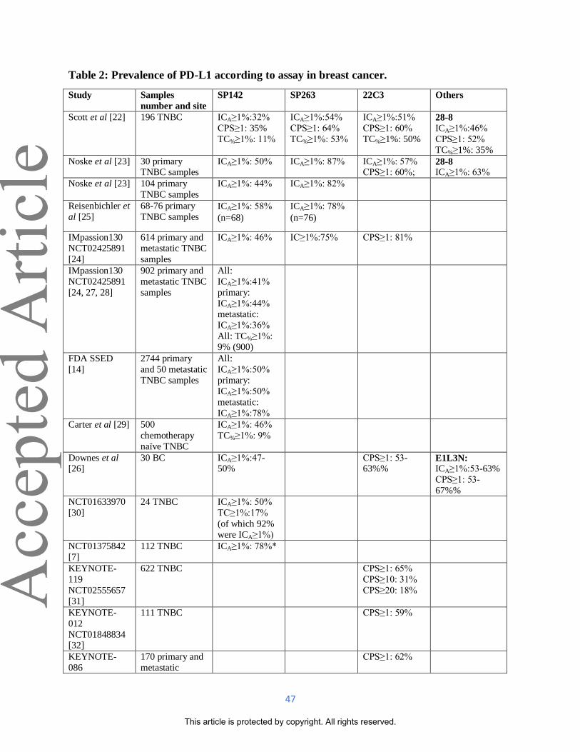
47
Table 2: Prevalence of PD-L1 according to assay in breast cancer.
Study Samples
number and site
SP142 SP263 22C3 Others
Scott et al [22] 196 TNBC ICA≥1%:32%
CPS≥1: 35%
TC%≥1%: 11%
ICA≥1%:54%
CPS≥1: 64%
TC%≥1%: 53%
ICA≥1%:51%
CPS≥1: 60%
TC%≥1%: 50%
28-8 ICA≥1%:46%
CPS≥1: 52%
TC%≥1%: 35%
Noske et al [23] 30 primary TNBC samples
ICA≥1%: 50%
ICA≥1%: 87%
ICA≥1%: 57% CPS≥1: 60%;
28-8 ICA≥1%: 63%
Noske et al [23] 104 primary
TNBC samples
ICA≥1%: 44%
ICA≥1%: 82%
Reisenbichler et
al [25]
68-76 primary
TNBC samples ICA≥1%: 58%
(n=68)
ICA≥1%: 78%
(n=76)
IMpassion130
NCT02425891
[24]
614 primary and
metastatic TNBC
samples
ICA≥1%: 46%
IC≥1%:75%
CPS≥1: 81%
IMpassion130
NCT02425891
[24, 27, 28]
902 primary and
metastatic TNBC
samples
All:
ICA≥1%:41%
primary:
ICA≥1%:44% metastatic:
ICA≥1%:36%
All: TC%≥1%:
9% (900)
FDA SSED
[14]
2744 primary
and 50 metastatic
TNBC samples
All:
ICA≥1%:50%
primary:
ICA≥1%:50%
metastatic:
ICA≥1%:78%
Carter et al [29] 500
chemotherapy
naïve TNBC
ICA≥1%: 46%
TC%≥1%: 9%
Downes et al [26]
30 BC ICA≥1%:47-50%
CPS≥1: 53-63%%
E1L3N: ICA≥1%:53-63%
CPS≥1: 53-
67%%
NCT01633970
[30]
24 TNBC ICA≥1%: 50%
TC≥1%:17%
(of which 92%
were ICA≥1%)
NCT01375842
[7]
112 TNBC ICA≥1%: 78%*
KEYNOTE-
119
NCT02555657
[31]
622 TNBC CPS≥1: 65%
CPS≥10: 31%
CPS≥20: 18%
KEYNOTE-
012
NCT01848834 [32]
111 TNBC CPS≥1: 59%
KEYNOTE-
086
170 primary and
metastatic
CPS≥1: 62%
Acc
epte
d A
rticl
e
This article is protected by copyright. All rights reserved.

48
NCT02447003
[33]
samples TNBC
KEYNOTE-
150
NCT02513472
[34]
107 TNBC CPS≥1: 46%
JAVELIN
NCT01772004
[35]
136 BC, 48
TNBC
73-3 All: IC≥10%:
9% TNBC: IC≥10%:
19%
TONIC
NCT02499367
[36]
70 metastatic
TNBC samples
IC≥1%: 86%
IC≥5%: 67%
GeparNuevo
NCT02685059
[37]
158 TNBC ICIC% and/or
TC%≥1%: 87%
CPS: combined positive score; FDA SSED: Food and Drug Administration summary of safety and effectiveness
data; IC: immune cells; ICA: PD-L1-positive immune cell area; met: metastatic or non-primary sample; n: number of
patients included in the analysis; prim: primary sample; TC: tumour cells; TNBC: triple negative breast cancer.
* The first 25 patients were selected only if PD-L1+, then enrolment was extended to all patients, explaining the
higher PD-L1 prevalence.
Acc
epte
d A
rticl
e
This article is protected by copyright. All rights reserved.

49
Table 3: Studies evaluating inter-reader reproducibility on breast cancer samples.
Study Assay and
scoring
participating
pathologists
Samples
evaluated
Training Concordance
Reisenbichler et al
[25]
SP142 CDA
ICA≥1%
19
68 primary
TNBC
No specific training
for the study.
ICC 0.560, OPA
41%
SP263 CDA
ICA≥1%
ICC 0.513
Noske et al [23]
SP142 CDA
ICA≥1%
7 30 primary
TNBC
Trained on digital
platform for the evaluation of PD-L1
IC with SP142 and
had to pass a
proficiency exam.
ICC 0.805
SP263 CDA
ICA≥1%
ICC 0.616
22C3 CDA
ICA≥1%
ICC 0.605
28-8 CDA
ICA≥1%
ICC 0.460
FDA SSED [14] SP142 CDA
ICA≥1%
3 60 TNBC Not specified. OPA 91.1%
Dennis et al [49] SP142 CDA
ICA≥1%
903 28 TNBC Regional trainer
lead sessions and
digital platform
training conducted by Roche
International
Pathologist Training
program. A
proficiency test was
evaluated.
OPA 98%
Downes et al [26] SP142 CDA
ICA≥1%
3 30 BC Not specified. ICC 0.956, OPA
98%
22C3 CDA
CPS≥1
ICC 0.862, OPA
93%
E1L3N LDT
IC≥1%
ICC 0.862, OPA
93%
E1L3N LDT
CPS≥1
ICC 0.815, OPA
91%
Solinas et al [50] E1L3N LDT
IC≥1%
2 441 BC Not specified. ICC 0.10-0.58 for
primary treatment
naïve tumours,
ICC 0.94[0.84-
0.97] for NAC
treated,
ICC 0.00 [-0.54-
0.35] for relapses
Acc
epte
d A
rticl
e
This article is protected by copyright. All rights reserved.

50
CDA: commercial diagnostic assay; FDA SSED: Food and Drug Administration summary of safety and
effectiveness data; ICC: Interclass correlation coefficient; LDT: laboratory developed test; OPA: overall percent
agreement.
Note: Definition of methodology for OPA, i.e. whether all individual pathologists were compared or if the mean test
score for all pathologists was compared to the consensus, was not available for all the studies in Table 3 precluding
fair comparison among studies.
Acc
epte
d A
rticl
e
This article is protected by copyright. All rights reserved.

51
Table 4: Studies evaluating clinical validity and utility of PD-L1 IHC and/or TILs as a
predictive biomarker of response to PD1/PD-L1 inhibitors in breast cancer.
Clinical trial Drug
Tumor
type (n)
Biomarker
details (n)
Predictive capacity of PD-L1/TILs
Advanced setting
IMpassion130 NCT02425891
[24, 27, 28]
nab paclitaxel +/-
atezolizumab
randomized
phase III
UnTx LAdv or
mTNBC
(902)
PD-L1 (SP142) was
prospectively
tested at BTx
(902) and used
as a stratification
factor for
randomization.
TILs were
evaluated
retrospectively
(460 and 614). PD-L1 SP263
and 22C3 were
performed
retrospectively
on BEP (614)
post-hoc
exploratory
analysis.
Improved PFS (HR 0.62 [0.49-0.78]) and OS (HR 0.62 [0.45-0.86]) with the addition
of atezolizumab in PD-L1+ tumors (SP142
ICA≥1%). ORR 56 vs 46% in the ITT
population and 59 vs 43% in PD-L1+
tumors (p=0.002).
Better PFS (0.53 [0.38-0.74]) and OS (0.57
[0.35-0.92]) for TIL>10%PD-L1≥1%
population (n=460).
PD-L1+ cases showed higher median TILs
(10% [IQR:5-20]) on BEP.
Improved PFS and OS (0.64 [0.53-0.79]; 0.75 [0.59-0.96]) with the addition of
atezolizumab in SP263 (IC≥1%) and (0.68
[0.56-0.82]; 0.78 [0.62-0.99]) 22C3
(CPS≥1) on BEP. Median PFS SP142 4.2
months, 22C3 2.1 months, SP263 2.2
months, and median OS SP142 9.4 months,
22C3 2.4 months, SP263 3.3 months.
NCT01375842
[7]
atezolizumab
single arm
phase Ib
PreTx
mTNBC
(116)
PD-L1 (SP142)
tested
prospectively
(116).
TICs (116).
PD-L1 ICA≥1% (ORR:12 vs 0%; HR: 0.55
[0.33-0.92]) and TICs>10% (HR:0.54
[0.35-0.83]) were associated with better
outcome. TICs>10% was independently
associated with ORR, PFS and OS in multivariate analysis. PD-L1 TC≥1% was
not associated with response.
NCT01633970
[30]
atezolizumab +
nab paclitaxel
single arm
phase Ib
UnTx (13)
and PreTx
(20)
mTNBC
(33)
PD-L1 (SP142)
and TILs tested
retrospectively at
BTx (23 and 20)
and PostTx (11
and 15,
respectively).
No statistically significant association of
baseline PD-L1 or TILs with response.
Numerically higher ORR (41.7 vs 33.3%)
and longer PFS (6.9 vs 5.1 mo) and OS
(21.9 vs 11.4 mo) in PD-L1+ tumor
(ICA≥1%). Numerically higher OS in
TILs>5%.
Changes in PD-L1 or TILs were not
associated with clinical response.
KEYNOTE-
119 NCT02555657
[31, 55]
physician’s
choice chemo +/-
pembrolizumab
randomized
phase III
PreTx
mTNBC (622)
PD-L1 (22C3)
was prospectively
tested at BTx
(622) and used
as a stratification
factor for
randomization.
No improved outcome in ITT population or
PD-L1+ tumors (CPS≥10 p=0.057; CPS≥1 p=0.073). For CPS≥20, HR OS: 0.58 [0.38-
0.88].
Better OS for TILs≥5% (0.75 [0.59-0.96])
in the pembrolizumab arm but not the
chemotherapy arm (1.46 [1.11-1.92]).
TILs and PD-L1 CPS moderately correlated
(0.45). TILs (p=0.004) and CPS (p=0.09)
were independently predictive.
KEYNOTE-
150
NCT02513472
pembrolizumab
+ eribulin single
arm
PreTx and
UnTx
mTNBC
PD-L1 (22C3) ORR independent (30.6 vs 22.4%) of PD-
L1 status (CPS≥1).5
Acc
epte
d A
rticl
e
This article is protected by copyright. All rights reserved.

52
[34] phase Ib/II (107)
KEYNOTE-
086
NCT02447003
[33, 56, 57]
pembrolizumab
single arm
phase II
A: PreTx
mTNBC
(170)
B: UnTx
PD-L1+
mTNBC
(84)
PD-L1 (22C3)
was
prospectively
tested at BTx
(254).
TILs were
evaluated retrospectively
(193).
ORR independent of PD-L1 status (CPS≥1)
on cohort A (5.7 vs 4.7%). No difference in
PFS or OS between PD-L1+ and PD-L1-.
21.4% ORR cohort B.
Better ORR in pts with TILs>median in
cohort A (6 vs 2%) and B (39 vs 9%) and
combined cohorts (OR: 1.26 [1.03-1.55]). Higher median Higher TILs in responders
vs non-responders in cohort A (10% vs 5%)
and cohort B (50% vs 15%).
KEYNOTE-
012
NCT01848834
[32]
pembrolizumab
single arm
phase Ib
PreTx PD-
L1+
mTNBC
(32)
PD-L1 (22C3)
was
prospectively
tested at BTx
(32)
Increasing ORR (p=0.028) and reduction in
HR (p=0.012) with increasing PD-L1
expression.
TONIC
NCT02499367
[36]
nivolumab
+previous
induction
therapy
(Rx/chemo) randomized
phase II
PreTx and
UnTx
mTNBC
(67)
PD-L1 (22C3)
and TILS at
BTx, after
induction and
PostTx.
Higher BTx TILs (median 12.5 vs 6%,
p=0.004) and PD-L1 on IC (median 15 vs
5%) on responders vs non-responders.
Better PFS and OS was observed in PD-L1
IC≥5% patients. No difference was observed between PD-L1 TC≥1% and <1%
populations.
PANACEA
NCT02129556
[58]
pembrolizumab
+ trastuzumab
single arm
phase Ib/II
PreTx
LAdv or
mHER2+
BC
Ib: PD-
L1+ (6)
II: PD-
L1+ &
PD-L1-
(52)
PD-L1
(QualTek/ 22C3)
tested
prospectively at
BTx (58). TILs
were evaluated
retrospectively
(48).
II: Higher ORR (15 vs 0%) in PD-L1+
(CPS≥1). Longer OS for PD-L1+
population.
Higher TILs levels in objective responders
(median ~25 vs 1.5% p=0.006) and in PD-
L1+ population (p=0.0004).
KATE-2
NCT02924883
[59]
T-DM1+/-
atezolizumab
randomized
phase II
PreTx
LAdv or
mHER2+
BC (202)
PD-L1 (SP142)
tested
prospectively
(202).
PFS survival benefit (HR 0.60[0.32-1.11])
and numerically higher ORR (54 vs 33%) in
PD-L1+ tumors (ICA≥1%) with the addition
of atezolizumab.
JAVELIN
NCT01772004
[35]
avelumab single
arm
phase Ib
PreTx
LAdv or
mBC
(168)
PD-L1 (73-10)
evaluated
prospectively on
IC and TC (168).
Better ORR in PD-L1+ (IC≥10%) BC (16.7
vs 1.6% p=0.039, 22.2 vs 2.6% in TNBC).
No association between outcome and PD-
L1+ (HR PFS: 0.66[0.34-1.26], OS: 0.62
[0.25-1.54]). PD-L1 on TC showed no
association with response.
Neoadjuvant setting
KEYNOTE-
552
NCT03036488 [60]
Neoadjuvant
paclitaxel +
carboplatin + AC/EC +/-
pembrolizumab
randomized
phase III
UnTx
TNBC
(602)
22C3 pCR achieved irrespective PD-L1 status
(CPS≥1) with the addition of
pembrolizumab (68.9 vs 45.3%).
KEYNOTE-
173
NCT02622074
Neoadjuvant
pembrolizumab
+ nab-paclitaxel
UnTx
LAdv
TNBC
PD-L1 (22C3)
retrospectively
tested on BTx
Higher BTx (p=0.028) and OnTx TILs
(p=0.005) and BTxPD-L1 CPS (p=0.021)
were associated with pCR. Responders had
Acc
epte
d A
rticl
e
This article is protected by copyright. All rights reserved.

53
AC: doxorubicin + cyclophosphamide; BEP: biomarker evaluable population; BTx: baseline or pre-treatment; CPS:
combined positive score; Δ: change between baseline and after treatment; EC: epirubicin + cyclophosphamide; HR:
hazard ratio; IC: immune cells; IQR: interquartile range; LAdv: unresectable locally advanced; ITT: intention-to-
treat population; mBC: metastatic breast cancer, all subtypes; mTNBC: metastatic TNBC; n: number of patients
included in the analysis; OnTx: on treatment; OR: odds ratio; ORR: objective response rates; OS: overall survival;
PostTX: post-treatment; PreTx: previously treated; PFS: progression free survival; Rx: radiation; TIC: tumor
infiltrating immune cells (lymphocytes, macrophages, dendritic cells and granulocytes) scored as a percentage of
tumor area; TC: tumor cells; UnTx: untreated.
[61, 62] +/- carboplatin
+/-AC
randomized
phase Ib
(60) (52). TILs were
retrospectively
evaluated on
BTx (53) and
OnTx (50)
samples.
higher median pre (40% vs 10%) and OnTx
(65% vs 22.5%) TILs.
Acc
epte
d A
rticl
e
This article is protected by copyright. All rights reserved.

54
Table 5: Risks associated with the integration of PD-L1 as immuno-oncological biomarkers
for clinical trials and the daily practice.
Risk Description of risk Mitigation approach/Recommendation
Risks to patient safety
Provision of
inappropriate treatment
because of false-
positive or false-
negative test results
Inter-pathologist variability and use of
different assays with different sensibilities may mislead
categorization of PD-L1 status.
Incorrect results lead to inappropriate
treatment allocation and put patient safety at risk.
See below.
Physical harm
or inconvenience
associated with
tissue biopsy
Heterogeneity of PD-L1 expression
between primary and metastatic lesions in TNBC [14] can lead to misleading
categorization depending on the
sample tested.
Define optimal sample for PD-L1 testing
from data of future clinical trials. When both primary and metastatic samples
available, test both if possible.
Operational risks
Failure of
sample
collection, processing and
quality
Poor quality samples can result in
unreliable test results.
Ensure correct sample fixation for 6 to 72hs
and processing. Determine sample adequacy
on H&E: presence of TC and tumor-associated IC. Cut 4um sections for PD-L1
IHC testing along with sections for other
IHC to preserve tissue in biopsy samples.
Use within 2 months of cutting [14].
Within
laboratory assay
variability
Drifts in assay results over time can
result in unreliable test results.
Follow staining protocol with optimized
conditions. Include control tissue (tonsil) to
test acceptance criteria [14]. Internal and
external quality assurance. Audit positivity rates [85].
Risks to biomarker development
Difference in PD-L1
expression
prevalence
among assays
SP142 has shown PD-L1 expression on a lower number of TC and IC
compared to the other assays [9, 10,
12, 13, 16, 22-24, 38-42].
It is more important that an assay identifies the patients who will most likely respond,
than identifying a greater proportion of PD-
L1 positive patients. Even though assays are
not analytically equivalent, clinical utility interchangeability must be further studied.
Use of multiple
scoring systems
The existence of multiple scoring
systems for the PD-L1 assays preclude the homologation of assays and
complicate reproducibility.
For BC, PD-L1 expressed in IC and not in
TC has been shown to be predictive of
response [7, 27, 35, 36]. Future clinical
trials should evaluate the most effective PD-
L1 scoring system. Cut-points must be reproducible.
Inter pathologist
variability to
read assay
Quantification of PD-L1 on IC has
been shown not be reproducible to
expected standards [10-13, 18, 23, 38, 39].
Ensure training on expected staining profile
and cut-off for pathologist participating in
clinical trials. Use of a single scoring system. Automated quantification by
computer-based image analysis. Evaluate
interobserver variability with a sufficiently large and statistically powered number of
Acc
epte
d A
rticl
e
This article is protected by copyright. All rights reserved.

55
pathologists to ensure reproducibility.
Temporal and
Spatial heterogeneity
Both PD-L1 and TILs have
demonstrated higher expression in primary tumors than in metastases [2,
24, 57].
Evaluating all available samples on clinical
trials would provide useful data, since the most appropriate time point for testing has
not yet been clearly established.
Unique biomarker as
companion
diagnostic test
Due to the complexity of immune response it is unlikely a single
biomarker will sufficiently predict
response to ICI.
Since both PD-L1 and TILs have shown to be predictive of response to ICI [28,57], the
use of both as stratification factors and for
composite biomarker analysis in future
clinical trials may help further optimize patient selection. Enough samples should be
secured to further investigate other
biomarkers on exploratory analysis.
Risks to biomarker implementation into daily practice
Regulatory
approval differs
per country
Implementation into daily practice is
dependent on regulatory approval.
Thorough and timely scientific interaction
between the pathology community, industry
and regulatory and national reimbursement agencies is needed.
Biomarker
accessibility and
affordability
PD-L1 testing is not yet covered by
health insurance in many countries.
Thorough and timely scientific interaction
between the pathology community, industry
and regulatory and national reimbursement agencies is needed.
Use of multiple
PD-L1 assays for a single
analyte
With multiple PD-L1 assays available,
pathology labs cannot be expected to have all tests available, causing
variability in test results between
laboratories.
Choice of assay will depend on regional
regulations, availability of antibody, automated staining platform and optimized
assay in currently in use. Consider LDTs.
Outsource to reference laboratories.
Difference in PD-L1
expression
prevalence among assays
SP142 has shown PD-L1 expression on a lower number of TC and IC
compared to the other assays [9, 10,
12, 13, 16, 22-24, 38-42].
It is more important that an assay identifies the patients who will most likely respond,
than identifying a greater proportion of PD-
L1+ patients. For BC SP142, SP263 and 22C3 have shown to identify patients that
derive better outcome in response to
atezolizumab and nab-paclitaxel [24].
Inter pathologist variability to
read assay
Quantification of PD-L1 on IC has been shown not be reproducible to
expected standards [10-13, 18, 23, 38,
39].
Training on expected staining profile and cut-off. Interpretation guideline. Use of a
single scoring system. Automated
quantification by computer-based image analysis.
Unique
biomarker as
companion diagnostic test
Due to the complexity of immune
response it is unlikely a single
biomarker will sufficiently predict response to ICI. The use of PD-L1+ IC
score as a unique biomarker test maybe
suboptimal in real world conditions.
Since TILs and PD-L1 are part of an
immunological spectrum and PD-1/PD-L1
interaction is only one of many factors that may determine the clinical outcome of
immunotherapeutic therapies, assessing
both as a composite biomarker could be a
better way to identify patients most likely to respond to ICI.
H&E: hematoxylin and eosin; IC: immune cells; IHC: immunohistochemistry; LDTs: laboratory developed test; PD-
L1+ IC: proportion of tumor area covered by IC with discernible PD-L1 staining of any intensity expressed as a
Acc
epte
d A
rticl
e
This article is protected by copyright. All rights reserved.

56
percentage; ICI: PD-1/PD-L1 inhibition based therapy; TC: tumor cell; TILs: tumor infiltrating lymphocytes;
TNBC: triple negative breast cancer.
Acc
epte
d A
rticl
e
This article is protected by copyright. All rights reserved.