Embed Size (px)
Citation preview

The
Journ
al o
f Exp
erim
enta
l M
edic
ine
ARTICLE
JEM © The Rockefeller University Press $8.00Vol. 203, No. 2, February 20, 2006 401–412 www.jem.org/cgi/doi/10.1084/jem.20051129
401
<doi>10.1084/jem.20051129</doi><aid>20051129</aid>Isoprenoids determine Th1/Th2 fate in pathogenic T cells, providing a mechanism of modulation of autoimmunity by atorvastatin
Shannon E. Dunn,1 Sawsan Youssef,1 Matthew J. Goldstein,1 Thomas Prod’homme,2 Martin S. Weber,2 Scott S. Zamvil,2 and Lawrence Steinman1
1Department of Neurology and Neurological Sciences, Stanford University, Stanford, CA 943052Department of Neurology, University of California San Francisco, CA 94143
3-hydroxy-3-methylglutaryl–coenzyme A (HMG-CoA) reductase is a critical enzyme in the mevalonate pathway that regulates the biosynthesis of cholesterol as well as isoprenoids that mediate the membrane association of certain GTPases. Blockade of this enzyme by atorvastatin (AT) inhibits the destructive proinfl ammatory T helper cell (Th)1 response during experimental autoimmune encephalomyelitis and may be benefi cial in the treatment of multiple sclerosis and other Th1-mediated autoimmune diseases. Here we present evi-dence linking specifi c isoprenoid intermediates of the mevalonate pathway to signaling pathways that regulate T cell autoimmunity. We demonstrate that the isoprenoid geranyl-geranyl-pyrophosphate (GGPP) mediates proliferation, whereas both GGPP and its precursor, farnesyl-PP, regulate the Th1 differentiation of myelin-reactive T cells. Depletion of these isoprenoid intermediates in vivo via oral AT administration hindered these T cell responses by decreasing geranylgeranylated RhoA and farnesylated Ras at the plasma membrane. This was associated with reduced extracellular signal–regulated kinase (ERK) and p38 phosphorylation and DNA binding of their cotarget c-fos in response to T cell receptor activation. Inhibition of ERK and p38 mimicked the effects of AT and induced a Th2 cytokine shift. Thus, by connecting isoprenoid availability to regulation of Th1/Th2 fate, we have elucidated a mechanism by which AT may suppress Th1-mediated central nervous system autoimmune disease.
CORRESPONDENCEScott S. Zamvil:[email protected]
Abbreviations used: AT,
atorvastatin; CNS, central ner-
vous system; EAE, experimental
autoimmune encephalomyelitis;
ERK, extracellular signal–
regulated kinase; FTI, farensyl-
transferase inhibitor; GGTI,
geranylgeranyltransferase inhibi-
tor; HMG-CoA, 3-hydroxy-
3-methylglutaryl–coenzyme A;
MS, multiple sclerosis; PP,
pyrophosphate; Tg, transgenic.
Recently, it was reported that oral administra-tion of simvastatin (Zocor) reduced the ex-pected number of new gadolinium-enhancing lesions in relapsing-remitting multiple sclerosis (MS) patients by 44% in a small 6-mo open- label trial (1). The related drug atorvastatin (AT) (Lipitor) has also shown promising results in lowering disease scores and vascular risk fac-tors in patients with rheumatoid arthritis (2) and is currently being tested in MS in a multi-center, placebo-controlled trial (http://www.immunetolerance.org/trials) (3). Studies in experimental autoimmune encephalomyelitis (EAE) (4–8) and other animal models of Th1-mediated disease (9, 10) have revealed that statin drugs interfere with the autoimmune de-
S.E. Dunn and S. Youssef contributed equally to this work.
S.S. Zamvil and L. Steinman contributed equally to this work.
The online version of this article contains supplemental material.
S.E. Dunn and S. Youssef contributed equally to this work.
S.S. Zamvil and L. Steinman contributed equally to this work.
The online version of this article contains supplemental material.
struction of target tissues by inhibiting multiple arms of the immune response.
In EAE, statins prevent the production of infl ammatory mediators TNF-α, IFN-γ, and iNOS by astrocytes and microglia in the central nervous system (CNS) (4) and blunt inducible expression of MHCII and costimulatory mol-ecules by these cells (5). These drugs also aff ect T cells, the initiators of the autoimmune pro-cess, by preventing their expansion, Th1 diff er-entiation, and migration across the blood–brain barrier (5–8). When myelin-reactive CD4+ T cells are primed with antigen in the presence of statins, they instead secrete lower amounts of TNF-α and the Th1 cytokine IFN-γ and higher amounts of protective Th2 cytokines such as IL-4 (5, 6, 8). Adoptive transfer studies have de-monstrated that AT-generated, myelin-reactive Th2 cells have a limited capacity to migrate into the CNS (unpublished data) and provide sustained
on April 2, 2006
ww
w.jem
.orgD
ownloaded from
http://www.jem.org/cgi/content/full/jem.20051129/DC1Supplemental Material can be found at:

402 ATORVASTATIN INDUCES TH2 BIAS BY INHIBITING RAS AND RHOA | Dunn et al.
protection from further attacks of paralysis (5). Interestingly, a similar bias toward a higher ratio of Th2/Th1 CD4+ cells in peripheral blood has been observed in human cardiac patients receiving low-dose (20 mg/d) AT therapy (11).
Despite these advances, the molecular mechanism by which statins modulate T cell immunity is unknown. This is important given the broad clinical administration of these agents (�25 million people world-wide) (12). Identifi ed
Figure 1. The mevalonate pathway intermediate farnesyl-PP and its alcohol precursor farnesol reverse the Th2 bias promoted by atorvastatin. (A) The mevalonate pathway. Metabolites and enzymes in the pathway are shown in black, drug inhibitors are shown in red, alcohol precursors to metabolites are shown in green, and HMG-CoA reductase and pathway end-products are shown in blue. CoQ, coeznyme Q10/ubi-quinone. (B) Purifi ed naive B10.Pl CD4+ cells stimulated with 5 μg/ml αCD3/αCD28 were cultured in the presence or absence of 10 μM atorva-statin (AT), 100 μM mevalonate, or 5 μM farnesyl-PP. Proliferation (top) was measured by [3H]-thymidine incorporation, and IFN-γ (middle) and IL-4 (bottom) accumulation in culture supernatants were measured by ELISA. These metabolite doses were optimal in reversal of AT effects.
Values are mean ± SE of three cultures. Results are representative of three independent experiments. *, denotes a signifi cant (P < 0.05) differ-ence from αCD3/αCD28 stimulation group. (C and D) Female SJL mice (n = 10/group) were immunized with PLP p139-151 in CFA and daily were fed PBS or AT (10 mg/kg) and/or were injected with farnesol (FOH) (5 mg/kg, i.p.). At day 10 after immunization, two spleens were taken from representative mice in each group and pooled, and isolated spleno-cytes were cultured ex vivo with PLP p139-151 peptide. C shows the mean ± SE clinical scores of these mice, whereas D shows the prolifera-tion of [3H] incorporation (left), IFN-γ (middle), and IL-4 (right) produc-tion by these cells. *, denotes a signifi cant difference from (P < 0.05) PBS control.
on April 2, 2006
ww
w.jem
.orgD
ownloaded from

JEM VOL. 203, February 20, 2006 403
ARTICLE
protein targets of these drugs include 3-hydroxy-3- methylglutaryl–coenzyme A (HMG-CoA) reductase, a rate-limiting enzyme in the mevalonate pathway that generates cholesterol and isoprenoid derivatives (Fig.1 A) (13, 14) and the integrin, leukocyte function antigen-1 (15). In the present study, we provide novel evidence that the Th2 bias promoted by AT is the result of a reduction of the mevalo-nate pathway-derived isoprenoids, farnesyl-pyrophosphate (PP) and all-trans geranylgeranyl-PP in T cells. These lipid derivatives are known to covalently attach to and mediate the membrane association of certain signaling proteins and metabolites in cells (13, 14). We show that in vivo deple-tion of these lipids in EAE mice via oral AT administration prevented the membrane association of farnesylated Ras and geranylgeranylated RhoA, but not other GTPases in lymph node cells. This was associated with compromised TCR- induced activation of Ras and RhoA eff ectors such as extracellular signal–regulated kinase (ERK), p38, and their cotarget c-fos. Because c-fos (as part of AP-1) transac-tivates the IFN-γ promoter and represses the IL-4 pro-moter (16), our results explain how AT can bias naive CD4+ T cells to produce relatively higher amounts of IL-4 in the early period of antigen signaling and thus trigger the Th2 program of diff erentiation. By connecting specifi c isoprenoids in the mevalonate pathway to T cell receptor signaling pathways that regulate cytokine production, we have elucidated a mechanism of how statins may inhibit Th1-mediated autoimmunity.
RESULTSAT prevents Th1 differentiation of CD4+ cells by targeting HMG-CoA reductasePrevious reports have shown that the eff ect of AT in T cells is reversed by mevalonate (5, 6), suggesting that the actions of this drug may be mediated through inhibition of HMG-CoA reductase (Fig. 1 A). However, because statins are structurally similar to mevalonate and have other ligands on immune cells (i.e., leukocyte function antigen-1) (15), it is also possible that such a reversal is a result of competition between meval-onate and AT for binding on protein targets other than HMG-CoA reductase. We thus tested whether the eff ects of AT on T cells could also be reversed by farnesyl-PP, the last common intermediate in the mevalonate pathway that has a diff erent structure than mevalonate. In response to αCD3/αCD28 costimulation, purifi ed naive (CD62L+/CD44low) CD4+ T cells proliferated and diff erentiated to secrete high amounts of the Th1 cytokine IFN-γ (Fig. 1 B). As shown previously (5, 6, 8), AT blunted these T cell responses and enhanced their production of IL-4 (Fig. 1 B). Farnesyl-PP, like mevalonate, fully reversed the eff ects of AT and, when administered alone, enhanced the TCR-induced prolifera-tion of CD4+ cells (Fig. 1 B).
Paralleling these in vitro fi ndings, administration of farnesol, a lipophilic, alcohol precursor to farnesyl-PP (17), reversed the clinical and immune modulatory eff ects of AT in vivo during EAE (Fig. 1, C and D, and Table S1, available
at http://www.jem.org/cgi/content/full/jem.20051129/DC1). Interestingly, myelin (PLP p139-151)-reactive cells taken from mice treated with farnesol alone also proliferated more vigorously and secreted higher amounts of IFN-γ ex vivo in response to stimulation with specifi c peptide (Fig. 1 D). Collectively, these fi ndings confi rm that AT modu-lates the Th diff erentiation of CD4+ cells by inhibiting the production of an intermediate in the mevalonate pathway. They also suggest that accumulation of farnesyl-PP in re-sponse to farnesol treatment may enhance the expansion of Th1 eff ector cells.
Figure 2. Oral atorvastatin treatment does not affect circulating or T cell cholesterol. (A) B10.PL mice (n = 4/group) were treated with either PBS or 10 mg/kg atorvastatin (AT) for 30 d after which time the liver was removed and HMG-CoA reductase (HMG-CoAR) transcript lev-els were assessed using real-time RT-PCR. *, denotes a signifi cant differ-ence (P < 0.05) from PBS liver. (B) Serum levels of total cholesterol (mg/dL) in mice treated orally with PBS or AT (10 mg/kg). Serum was taken from mice at 5 d after treatment in the morning after an overnight fast. Values are mean ± SE of fi ve mice. (C) T cells were isolated from the spleens of mice treated as in B and total cholesterol (μg/mg protein) was measured in lipid extracts of these cells using the Amplex Red Choles-terol Assay. (D) Flow cytometric analysis of fi lipin staining in purifi ed T cells taken from mice treated as in B (left) or in T cells that were incu-bated in vitro in serum-free media for 2 h with the cholesterol-depleting agent β-methyl-cyclodextrin (BMCD) (right). The latter experiment served as a positive control to demonstrate the sensitivity of this assay to detect cholesterol changes.
on April 2, 2006
ww
w.jem
.orgD
ownloaded from

404 ATORVASTATIN INDUCES TH2 BIAS BY INHIBITING RAS AND RHOA | Dunn et al.
Effects of AT on T cell growth and differentiation are independent of cholesterol reductionCholesterol is a major product of farnesyl-PP (Fig. 1 A). Although it has been reported that depletion of this lipid in T cells can attenuate TCR signaling by disruption of choles-terol-rich lipid rafts (18), a recent in vitro study of malig-nant T cells showed lipid raft disruption by statins only in the complete absence of exogenous cholesterol in the me-dia (19). We found that in the context of the whole organ-ism, AT treatment (10 mg/kg) reduced the production of this metabolite by the liver as indicated by an elevation in the mRNA expression of HMG-CoA reductase (this gene is negatively regulated by sterols in this tissue; reference 13) (Fig. 2 A). However, the total amount of cholesterol in the serum or in T cells was unchanged by this treatment (Fig. 2, B and C). The level of membrane-associated cholesterol was also not altered in T cells in response to oral administration of this drug as assessed via fl ow cytometric analysis of fi lipin staining (Fig. 2 D, left). The lack of detectable change in membrane cholesterol with AT was not a result of limits in the sensitivity of this assay, because decreased fi lipin fl uores-cence was observed in T cells incubated in serum-free media with the cholesterol-depleting agent β-methyl cyclodextrin (Fig. 2 D, right).
To more specifi cally address the role of the sterol branch of the mevalonate pathway in immunomodulation, we examined the eff ects of the squalene synthetase inhibi-tor Zaragozic Acid A (10 mg/kg) on encephalitogenic T cell responses during EAE. At this dose, Zaragozic Acid A inhibits liver sterol synthesis (20), reduces circulating cho-lesterol (Fig. 3 A) and at the same time causes an accumula-tion of farnesol in mice (20). Daily subcutaneous injection of mice with Zaragozic Acid had opposite eff ects to AT and caused PLP p139-151–reactive cells to proliferate more vigorously (Fig. 3 B) and secrete higher amounts of IFN-γ (Fig. 3 C) and lower amounts of IL-4 (Fig. 3 D) compared with PBS-injected counterparts. Zaragozic Acid A treat-ment also exacerbated EAE at several time-points in disease (Fig. 3 E and Table S2, available at http://www.jem.org/
cgi/content/full/jem.20051129/DC1). To examine whether the enhanced proliferation of Th1
cells triggered by Zaragozic Acid A was a result of enhanced fl ux of farnesyl-PP into isoprenoid branches of the pathway, we investigated the eff ect of this drug on the de novo incor-poration [1-3H] farnesyl-PP into T cell proteins in vitro. We found that in comparison to media control, Zaragozic Acid A–treated T cells displayed an increased abundance of tritium-labeled proteins in the 21–29 kD range, which is compatible
Figure 3. Specifi c inhibition of the sterol branch of the mevalo-nate pathway enhances T cell proliferation and Th1 differentiation. SJL mice (n = 10/group) were immunized with PLP p139-151 in CFA and were injected once daily with either PBS or Zaragozic Acid A (10 mg/kg, i.p.). At day 10 after immunization, axillary, brachial, and inguinal lymph nodes were taken from representative mice in each group and pooled, and isolated cells were cultured ex vivo with PLP p139-151 peptide. (A) Total cholesterol (μM) in serum taken at the peak of disease activity. Values are mean ± SE of fi ve mice. (B–D) Proliferation of PLP p139-151
reactive cells (B) was measured by [3H]-thymidine incorporation, and IFN-γ (C) and IL-4 (D) accumulation in culture media was measured by ELISA. Values are mean ± SE of three cultures. (E) Mean ± SE clinical scores of mice in the various groups over the time-course of disease are shown. *, denotes a difference (P < 0.05) from PBS control. (F) De novo incorpo-ration of [1-3H]-farnesyl-PP into cellular proteins in the absence or pres-ence of Zaragozic Acid A (200 μM) in T cell cultures. The protein marker is in lane 3 (top). Gels were stained with Coomassie blue to ensure that equivalent amounts of each protein sample were loaded.
on April 2, 2006
ww
w.jem
.orgD
ownloaded from

JEM VOL. 203, February 20, 2006 405
ARTICLE
with the size of small G proteins (Fig. 3 F). Collectively, these results suggest that the major eff ects of AT on Th1 diff eren-tiation and clinical disease are cholesterol independent and instead the result of reductions in isoprenoid biosynthesis.
AT inhibits T cell growth and Th1 differentiation by preventing protein isoprenylationFarnesyl-PP serves as a metabolic precursor to a variety of isoprenoid derivatives such as decaprenyl-PP (isoprenoid moiety of ubiquinone), 2-cis-geranylgeranyl-PP (important for N-glycoprotein synthesis), and all-trans geranylgeranyl-PP (13, 14) (Fig. 1 A). The latter isoprenoid along with
farnesyl-PP are, respectively, attached to the COOH termi-nus of certain signaling proteins via the action of geranylger-anyl transferases and farnesyltransferase (21). These isoprenoid modifi cations mediate the membrane association of many GTPases (21). We thus tested whether specifi c isoprenoid derivatives could reverse the eff ects of AT on T cells cultured with specifi c peptide. Splenocytes from MBP Ac1-11 TCR transgenic (Tg) mice were used as a source of T cells for these assays.
Of all the metabolites tested, only all-trans geranylgeranyl-PP, the isoprenoid moiety of geranylgeranylated proteins, and its precursor, farnesyl-PP, restored the antigen-driven
Figure 4. Prenylated proteins have a regulatory function in T cell growth and differentiation. (A and B) Splenocytes taken from MBP Ac1-11 TCR Tg mice were stimulated with 5 μg/ml Ac1-11 peptide in the presence or absence of 10 μM atorvastatin (AT), 5 μM farnesyl-PP (FPP), 5 μM all-trans geranylgeranyl-PP (all-trans GGPP), 1 μM 2-cis geranyl-geraniol (2-cis GGOH), or 1 μM ubiquinone (all in A) or DMSO vehicle, 5 μM of the farnesyltransferase inhibitor FTI-277 or 5 μM of the geranyl-geranyltransferase-I inhibitor GGTI-298 (all in B). Proliferation (top) was measured by [3H]-thymidine incorporation, and IFN-γ (middle), and IL-4 (bottom) accumulation in culture supernatants was measured via ELISA. These results are representative of data from at least three independent experiments. *, denotes a difference (P < 0.05) from peptide stimulation alone. Note that the additive effects of FTI and GGTI were not examined in
these experiments because this drug combination compromised T cell viability (not depicted). Values are mean ± SE of triplicate cultures. (C) 5 μM farnesyl-PP is effective at restoring the prenylation of Ras, a representative farnesylated protein and 5 μM all-trans GGPP is effective at restoring the prenylation of Rap, a representative geranylgeranylated protein. Because the carboxy-termini of these proteins are cleaved upon prenylation, posttranslationally modifi ed forms (single arrows) of these proteins can be distinguished from their unprenylated forms (double arrows) via altered mobility on Western blots of SDS-PAGE gels (reference 27). (D) 1 and 5 μM of FTI-277 and 5 μM GGTI-298, respectively, inhibit Ras farnesylation and Rap1 geranylgeranylation. (E) Protein samples from cells treated with FTI, GGTI, or vehicle control (as in B) were subjected to Western blot analysis of T-bet and GATA-3 expression.
on April 2, 2006
ww
w.jem
.orgD
ownloaded from
406 ATORVASTATIN INDUCES TH2 BIAS BY INHIBITING RAS AND RHOA | Dunn et al.
proliferation of AT-treated T cells and reversed the Th2 bias promoted by this drug (Fig.4 A). The doses of farnesyl-PP and geranylgeranyl-PP used in these assays were eff ective at restoring the prenylation of protein targets (Fig. 4 C). We also tested the eff ects of these isoprenoids in AT-treated T cells stimulated with α-CD3 and α-CD28 (Fig. S1, available at http://www.jem.org/cgi/content/full/jem.20051129/DC1) and found that both farnesyl-PP and geranylgeranyl-PP con-tributed to the reversal of AT eff ects on T cell proliferation and Th1 diff erentiation. Collectively, these data strongly suggest that AT modulates Th diff erentiation by inhibiting isoprenoid production in T cells.
To confi rm the involvement of isoprenylated proteins in T cell growth and Th1 diff erentiation, we investigated the eff ects of inhibitors of farnesyltransferase (FTI-277) and gera-nylgeranyltransferase-I (GGTI-298) on antigen-driven T cell responses (Fig. 4, B and D). Treatment of MBP Ac1-11 TCR Tg T cells with an eff ective dose (Fig. 4 D) of the geranyl-geranyltransferase inhibitor (GGTI) signifi cantly inhibited T cell proliferation (Fig. 4 B). However, the growth of these cells was not altered by treatment with the farnesyltransferase inhibitor (FTI) (Fig. 4 B). Consistent with the prominent in-volvement of protein farnesylation in regulating Th1 diff er-entiation, FTI-277 inhibited the production of IFN-γ and induced a subtle increase in IL-4 by T cells (Fig. 4 B). In par-allel, this drug decreased T-bet protein and caused a subtle increase in GATA-3 expression (Fig. 4 E). GGTI-298, though not infl uencing IFN-γ secretion or T-bet expression, induced low-level IL-4 production and a subtle increase in GATA-3 expression (Fig. 4, B and E). Similar eff ects of FTI-277 and GGTI-298-treatment on T proliferation and cytokine production were observed in cultures of purifi ed T cells stimulated with anti-CD3 and anti-CD28 (Fig. S1). Note that in both of these T cell assays, the amount of IL-4 triggered by each of these prenyltransferase inhibitors did not approach the level induced by AT (Fig. 4 B and Fig. S1), sug-gesting that geranylgeranylated proteins cooperate with farnesylated proteins to fully repress the production of this Th2 cytokine in T cells in response to antigen signals. Col-lectively, these results suggest that AT prevents T cell prolif-eration and Th1 diff erentiation by inhibiting the biosynthesis of farnesyl-PP and all-trans geranylgeranyl-PP, leading to reduced prenylation of protein targets.
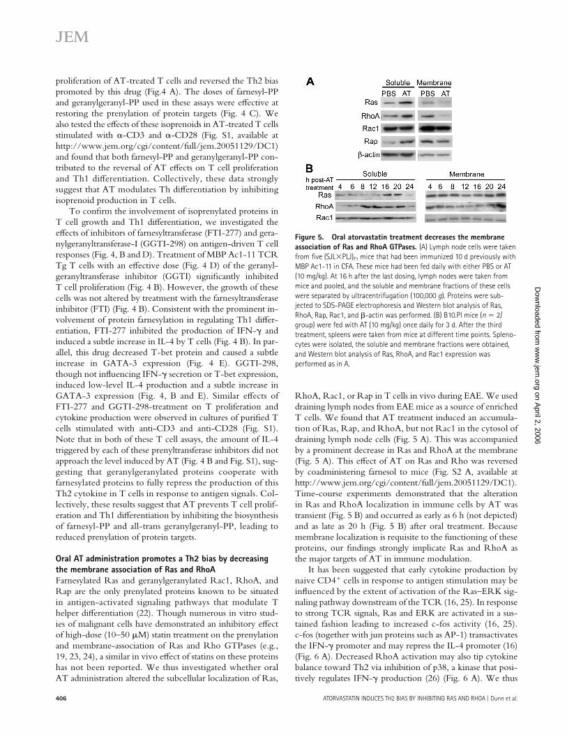
Oral AT administration promotes a Th2 bias by decreasing the membrane association of Ras and RhoAFarnesylated Ras and geranylgeranylated Rac1, RhoA, and Rap are the only prenylated proteins known to be situated in antigen-activated signaling pathways that modulate T helper diff erentiation (22). Though numerous in vitro stud-ies of malignant cells have demonstrated an inhibitory eff ect of high-dose (10–50 μM) statin treatment on the prenylation and membrane-association of Ras and Rho GTPases (e.g., 19, 23, 24), a similar in vivo eff ect of statins on these proteins has not been reported. We thus investigated whether oral AT administration altered the subcellular localization of Ras,
RhoA, Rac1, or Rap in T cells in vivo during EAE. We used draining lymph nodes from EAE mice as a source of enriched T cells. We found that AT treatment induced an accumula-tion of Ras, Rap, and RhoA, but not Rac1 in the cytosol of draining lymph node cells (Fig. 5 A). This was accompanied by a prominent decrease in Ras and RhoA at the membrane (Fig. 5 A). This eff ect of AT on Ras and Rho was reversed by coadministering farnesol to mice (Fig. S2 A, available at http://www.jem.org/cgi/content/full/jem.20051129/DC1). Time-course experiments demonstrated that the alteration in Ras and RhoA localization in immune cells by AT was transient (Fig. 5 B) and occurred as early as 6 h (not depicted) and as late as 20 h (Fig. 5 B) after oral treatment. Because membrane localization is requisite to the functioning of these proteins, our fi ndings strongly implicate Ras and RhoA as the major targets of AT in immune modulation.
It has been suggested that early cytokine production by naive CD4+ cells in response to antigen stimulation may be infl uenced by the extent of activation of the Ras–ERK sig-naling pathway downstream of the TCR (16, 25). In response to strong TCR signals, Ras and ERK are activated in a sus-tained fashion leading to increased c-fos activity (16, 25). c-fos (together with jun proteins such as AP-1) transactivates the IFN-γ promoter and may repress the IL-4 promoter (16) (Fig. 6 A). Decreased RhoA activation may also tip cytokine balance toward Th2 via inhibition of p38, a kinase that posi-tively regulates IFN-γ production (26) (Fig. 6 A). We thus
Figure 5. Oral atorvastatin treatment decreases the membrane association of Ras and RhoA GTPases. (A) Lymph node cells were taken from fi ve (SJL×PLJ)F1 mice that had been immunized 10 d previously with MBP Ac1-11 in CFA. These mice had been fed daily with either PBS or AT (10 mg/kg). At 16 h after the last dosing, lymph nodes were taken from mice and pooled, and the soluble and membrane fractions of these cells were separated by ultracentrifugation (100,000 g). Proteins were sub-jected to SDS-PAGE electrophoresis and Western blot analysis of Ras, RhoA, Rap, Rac1, and β-actin was performed. (B) B10.Pl mice (n = 2/group) were fed with AT (10 mg/kg) once daily for 3 d. After the third treatment, spleens were taken from mice at different time points. Spleno-cytes were isolated, the soluble and membrane fractions were obtained, and Western blot analysis of Ras, RhoA, and Rac1 expression was performed as in A.
on April 2, 2006
ww
w.jem
.orgD
ownloaded from

JEM VOL. 203, February 20, 2006 407
ARTICLE
tested whether AT treatment compromised the TCR- induced activation of ERK and p38 kinases in lymph nodes during EAE. Consistent with eff ects on Ras and RhoA, in vivo AT treatment blunted the TCR-induced activation of ERK and p38 as well as decreased the DNA-binding of c-fos (a target of ERK and p38) and ATF-2 (a target of p38 and JNK) (Fig. 6, C and D). This eff ect of AT treatment on T cell ERK and p38 phosphorylation was reversed by cotreatment of mice with farnesol (Fig. S2 B). Consistent with the lack of a major eff ect of AT on the subcellular localization of Rac1, this drug did not modulate JNK phosphorylation nor the ac-tivity of its primary target, c-jun (Fig. 6, C and D). Oral AT treatment also did not inhibit other signaling events triggered by anti-CD3 or anti-CD28 cross-linking including IκB deg-radation (Fig. 6 C), Zap-70 phosphorylation (Fig. 6 E), and calcium fl ux (Fig. 6 F).
To test the hypothesis that this inhibition of Ras-ERK and RhoA-p38 signaling by AT was responsible for the Th2 bias induced by this drug, we explored the eff ects of specifi c inhibitors of MEK (PD98059), an upstream eff ector of ERK, and p38 (SB203580) on the antigen-driven diff erentiation of
naive CD4+ cells. Exposure of CD4+ cells to 10 μM of the MEK inhibitor (a dose that does not aff ect the proliferation or viability of T cells; unpublished data) decreased the produc-tion of IFN-γ and increased the secretion of IL-4 by these cells in response to specifi c peptide (Fig. 7 A). This concentra-tion of PD98059 also reduced the phosphorylation of ERK (Fig. 7 A, bottom). Similarly, exposure of T cells to the p38 kinase inhibitor at doses that inhibit p38 phosphorylation (Fig. 7 B, bottom) and activity (26) induced IL-4 production (only at 0.1 μM) and inhibited IFN-γ production by these cells (Fig. 7 B). The p38 kinase inhibitor also had a more profound eff ect than the MEK inhibitor in inhibiting T cell prolifera-tion through higher doses tested (10–100 μM) (unpublished data). Collectively, these data support the hypothesis that Ras-ERK and RhoA-p38 signaling pathways are the targets of AT in Th2 diff erentiation and inhibition of T cell growth.
In vivo treatment with an FTI prevents Th1 differentiation during EAEIn light of the more prominent involvement of protein farnesyl-ation and ERK signaling in regulating the Th1 diff erentiation of
Figure 6. Oral atorvastatin treatment inhibits ERK and p38 sig-naling pathways in T cells. (A and B) Cartoon summarizing the location of prenylated proteins (in green) in signaling pathways downstream of TCR stimulation and the transcription factors that act at IFN-γ and IL-4 promoters in PBS- (A) and AT-dosed (B) mice. Not all pathway intermedi-ates are shown. (C–E) Lymph node cells (C and D) or purifi ed CD4+ cells (E) taken from mice treated as in Fig. 5 were cultured (2 × 10⁶/ml) with 5 μg/ml αCD3 and 5 μg/ml αCD28 antibodies for various durations. (C) Western blot analysis of phospho-ERK, ERK, phospho-JNK, JNK,
phospho-p38, p38 MAPK, and IκB using whole cell lysates prepared from these cells. (D) shows results of an ELISA-based DNA binding assay of c-fos, ATF-2, and c-jun using nuclear extracts prepared from parallel cultures that were stimulated with 5 μg/ml αCD3/αCD28 antibodies for 10 h. (E) Western blot analysis of phospho-Zap 70 and Zap 70 in lysates of these cells. (F) FACS analysis of calcium fl ux in CD4+ T cells in response to anti-CD3 cross-linking. The arrow denotes the time when the stimulus was added to cells.
on April 2, 2006
ww
w.jem
.orgD
ownloaded from

408 ATORVASTATIN INDUCES TH2 BIAS BY INHIBITING RAS AND RHOA | Dunn et al.
MBP-reactive cells in vitro and reports that FTIs (but not GGTIs) are well tolerated in mice (27), we investigated the clinical benefi ts of the FTI, L-744,832 (30 mg/kg) on the de-velopment of EAE (Fig. 8, A–D). We administered the maxi-mum tolerated dose of this drug, which is reported to cause regression of Ras-related tumors in mice (28). Similar to in vitro fi ndings using FTI-277 (Fig. 4 B), IFN-γ production by PLP p139-151–reactive cells from L-744,832–treated mice was markedly reduced compared with control counterparts (Fig. 8 C). Despite this, FTI-treated mice still developed EAE, albeit the day of onset of clinical symptoms for grade one or higher (12.7 ± 0.6 in FTI vs. 11.3 ± 0.6 in vehicle, P < 0.01) and or grade two or higher (19.4 ± 2.0 in FTI vs. 16.0 ± 1.8 in ve-hicle, P < 0.01) was signifi cantly delayed compared with ve-hicle controls (Fig. 8 A). The development of infl ammation in FTI-treated mice may relate to the fact that this drug caused PLP p139-151–reactive cells to secrete higher amounts of IL-2 (Fig. 8 B), yet did not promote IL-4 secretion (not depicted) nor dampen the production of the proinfl ammatory cytokine TNF-α by these cells (Fig. 8 D). Collectively, these results
suggest that dual inhibition of protein farnesylation and gera-nylgeranylation is required to fully promote a Th2 bias of CD4+ cells and prevent the development of CNS infl amma-tion during EAE.
D I S C U S S I O N Several studies have demonstrated that in vivo statin treatment can cause a protective Th2 bias in animal models of Th1-mediated autoimmune disease (for review see reference 3). These fi ndings have created considerable enthusiasm for test-ing these agents in patients with MS and rheumatoid arthritis (1, 2). The purpose of the present study was to defi ne the mechanism of immune modulation by this drug. Here, we show that the eff ect of AT in causing a Th2 bias is the result of a reduction in the biosynthesis of isoprenoids that serve as membrane attachments for Ras and RhoA. Farnesyl-PP, which mediates Ras farnesylation, restored IFN-γ production by AT-treated myelin-reactive T cells, whereas all-trans-geranylgeranyl-PP, the precursor to RhoA geranylgera-nylation, reversed the AT block in T cell proliferation and
Figure 7. MEK and p38 inhibitors induce Th2 bias of naive CD4+ cells. Naive CD4+ T cells were isolated from spleens of MBP Ac1-11 TCR Tg mice and were cultured with specifi c peptide in the presence or ab-sence of 5 μg/ml MBP Ac1-11, 1, or 10 μM of the MEK inhibitor PD98059 (A) or 0.1–1 μM of the p38 inhibitor SB203580 (B). Note that these par-ticular doses of MEK or p38 inhibitors did not affect the proliferation or viability of T cells (data not depicted), but did inhibit the phosphorylation of ERK and p38 (bottom). *, denotes a signifi cant (P < 0.05) difference from MBP Ac1-11 stimulated group. β-actin served as a loading control. Values are mean ± SE of triplicate cultures.
Figure 8. In vivo administration of the FTI L-744,832 inhibits Th1 differentiation. Female SJL mice were immunized with PLP p139-151 in CFA and daily were injected s.c. with either vehicle or L-744,832 (30 mg/kg). At day 10 after immunization, spleens were taken from representative mice in each group and isolated splenocytes were cultured ex vivo with PLP p139-151 peptide. (A) The mean ± SE clinical scores of these mice. (B–D) PLP p139-151-stimulated production of IL-2 (B), IFN-γ (C), and TNF-α (D) by isolated splenocytes. Values are mean ± SE of triplicate cultures. *, denotes a signifi cant difference from vehicle control.
on April 2, 2006
ww
w.jem
.orgD
ownloaded from

JEM VOL. 203, February 20, 2006 409
ARTICLE
coordinated with farnesyl-PP to fully reverse the increase in IL-4 promoted by this drug. We also show for the fi rst-time that oral dosing with AT compromised the membrane asso-ciation of Ras and RhoA GTPases and the TCR-induced activation of targets ERK and p38 in T cells. Inactivation of ERK and p38 signaling mimicked the eff ects of AT, strongly implicating Ras-ERK and RhoA-p38 pathways as the targets of this drug. Thus, by linking isoprenoid intermediates to two arms of the TCR signaling cascade that modulate T cell cytokine production, we have elucidated a mechanism of how statins may cause a Th2 bias in vivo during EAE.
Our fi nding that oral dosing with AT caused Ras, RhoA, and Rap GTPases to be preferentially diverted to the cytosol instead of the membrane of lymph node cells in vivo is the fi rst demonstration of its kind. These results thus expand upon previous in vitro reports of lovastatin in tumor cell lines (19, 23, 24) as well as lend concrete support to the prevailing notion that the pleiotrophic eff ects of statins in treating ath-erosclerosis and cancer in animal models are the result of re-ductions in isoprenoid biosynthesis (29–32). Though oral AT administration signifi cantly depleted Ras and RhoA at the membrane of lymph node cells, in comparison, Rap or Rac1 were only marginally aff ected. The reason for this diff erence is unclear, but may relate to variations in the half-lives of these proteins (24). Nonetheless, because membrane-associa-tion is necessary for the functioning of GTPases, we conclude that Ras and RhoA are the major targets of statin in immune modulation in vivo.
Previous reports have demonstrated that the strength of ERK activation in response to TCR stimulation may infl u-ence cytokine production and alter the course of T helper diff erentiation by altering the binding of c-fos to AP-1 con-sensus elements (16). Peptides that bind the TCR with strong avidity induce Th1 diff erentiation of CD4+ cells and cause a sustained activation of Ras and ERK that triggers c-fos ac-tivity (16, 25). c-fos (with jun as AP-1) appears to repress the IL-4 promoter (16) and coordinates with other transcription factors such as ATF-2 and NFAT to mediate the transactiva-tion of the IFN-γ promoter (26) (Fig. 6 A). However, under conditions when ERK activity is low, such as in the presence of a weak-binding altered peptide ligand (16), or weakened Ras (33), or MEK signaling (16, 34), IFN-γ production is minimal and IL-4 is induced. In this regard, our fi ndings that AT decreased the DNA binding of c-fos and inhibited the activation or Ras-ERK and RhoA-p38 signaling pathways that target this transcription factor provide a mechanistic ex-planation for the Th2 bias promoted by statins. Interference of p38 activity by AT may have also contributed to this Th2 deviation by reducing the activity of ATF-2 at the IFN-γ promoter (26). In light of this evidence, we propose a model for AT action on T cells in vivo (Fig. 6, compare A and B), whereby this drug selectively inhibits the membrane associa-tion of Ras and RhoA leading to decreased activation of ERK and p38 signaling pathways that target AP-1 and ATF-2.
Despite the established role for Ras and ERK in the reg-ulation of IFN-γ production (in this paper and references
16, 33, 34), in vivo FTI treatment only delayed the onset of EAE. These fi ndings emphasize the involvement of geranyl-geranylated proteins in mediating the development of CNS infl ammation. Indeed, our data indicate a role for geranyl-geranylated Rho proteins in mediating the proliferation of encephalitogenic T cells. These results thus correspond with previous reports that statins cause G1 arrest (35) and that geranylgeranylated proteins regulate the G1 to S transition (36). We also found that inhibition of protein geranylgera-nylation in myelin-reactive cells was requisite for full induc-tion of IL-4 and was likely required for the attenuation of TNF-α production by AT (i.e., AT [reference 5], but not FTI treatment diminishes T cell production of this cyto-kine). These fi ndings, coupled with the established involve-ment of geranylgeranylated Rho proteins in regulating the transmigration of T cells across brain endothelium (7), emphasize that dual inhibition of farnesylation and geranyl-geranylation by statins is necessary to prevent the develop-ment of CNS autoimmunity.
Although reducing the production of farnesyl-PP by AT hindered T cell growth and Th1 diff erentiation, in vivo treat-ments that cause increased incorporation of this metabolite into prenylated proteins, such as farnesol (17) or Zaragozic Acid A (18, 37), induced pathogenic myelin-reactive T cells to proliferate more vigorously, secrete higher amounts of IFN-γ, and precipitate worse EAE. Thus, accumulation of farnesyl-PP may enhance Th1-mediated infl ammation in vivo. Though we observed in vitro Zaragozic Acid A treat-ment to trigger an increased accumulation of farnesyl-PP in T cells, it remains unclear whether this also occurred in mice in response to in vivo treatment or whether it was the in-creased liver production of farnesyl-PP–derived lipids (18, 37) that was responsible for the immune modulatory eff ects of this drug. The latter possibility (i.e., that the liver produc-tion of isoprenoids can boost adaptive immune responses) is provocative and provides an interesting correlate to reports that metabolic precursors in the mevalonate pathway are preferentially diverted toward isoprenoid as opposed to sterol biosynthesis in the liver during the acute phase response to infl ammatory stimuli (38, 39).
In conclusion, we show that farnesylated Ras and gera-nylgeranylated RhoA proteins are the targets of AT in im-mune modulation in vivo. Our results also underscore that inhibition of both protein farnesylation and geranylgeranyl-ation in T cells is key to promoting a Th2 bias and preventing the development of EAE. Because oral AT administration is well tolerated, whereas continuous and combined FTI and GGTI therapy is toxic in vivo (27), we maintain that of these, statin treatment is a more promising approach than selective inhibition of individual prenyltransferases for treatment of MS and other Th1-mediated autoimmune diseases.
MATERIALS AND METHODSReagents. Mevalonate, farnesol, farnesyl-PP, all-trans geranylgeranyl-PP,
ubiquinone, Zaragozic Acid A, β-methyl cyclodextrin, and fi lipin were ob-
tained from Sigma-Aldrich. Cold 2-cis geranylgeraniol, [acetyl-3H] acetyl
CoA (20 Ci/mmol), and [1-3H] farnesyl-PP (60 Ci/mmol) were obtained
on April 2, 2006
ww
w.jem
.orgD
ownloaded from
410 ATORVASTATIN INDUCES TH2 BIAS BY INHIBITING RAS AND RHOA | Dunn et al.
from American Radiochemicals. Purifi ed AT was a gift from R. Laskey
(Pfi zer, New York, NY). FTI-277, GGTI-298, L-744,832, SB203580,
and PD98059 were obtained from Calbiochem. Fluo-4 was obtained
from Invitrogen. Peptides encoding myelin basic protein (MBP), Ac1–11
(Ac-A S Q K R P S Q R H G ), or proteolipid protein (PLP) p139–151 (H C L G K-
W L G H P D K F ) were synthesized by the Stanford Pan Facility and purifi ed by
HPLC. Pan Ras (clone 18) and Rac1 (clone 102) antibodies were obtained
from BD Transduction Labs. RhoA (clone 26C4), Rap1 (clone sc-65),
T-bet (clone sc-21749), and GATA-3 (clone sc-268) antibodies were obtained
from Santa Cruz Biotechnology, Inc. Phospho(p42/44)-ERK (no. 9101S),
p42/p44 MAPK (no. 9102), phospho-JNK (no. 9251S), JNK (no. 9252),
phospho-p38 (no. 9216S), p38 (no. 9212), phospho-Zap-70 (no. 2701),
and IκBα (no. 9242) antibodies were all obtained from Cell Signaling. The
pan-Zap-70 antibody (clone 1E7.2) was obtained from Upstate Biotechnol-
ogy. Goat anti–mouse IgG antibody was obtained from Jackson Immuno-
Research Laboratories.
Mice. Female SJL/J and (PL/J × SJ/L)F1 mice (6–10 wk old) were pur-
chased from The Jackson Laboratory. MBP Ac1-11 TCR Tg mice back-
crossed onto the B10.PL background (40) were maintained in our animal
facility. All animal protocols were approved by the Division of Comparative
Medicine at Stanford University and the Committee of Animal Research at
the University of California San Francisco and animals were maintained in
accordance with the guidelines of the National Institutes of Health.
EAE induction. EAE was induced in SJL/J mice via subcutaneous immu-
nization with 100 μg PLP p139-151 in an emulsion (volume ratio 1:1) with
CFA containing 4 mg/ml of heat-killed Mycobacterium tuberculosis H37Ra
(Difco Laboratories). (PL/J × SJ/L)F1 mice were immunized with MBP
Ac1-11 in CFA. Mice (n = 10 per treatment group) were examined daily for
clinical signs of EAE and were scored as followed: 0 = no clinical disease,
1 = limp tail, 2 = hindlimb weakness, 3 = complete hindlimb paralysis, 4 =
hindlimb paralysis plus some forelimb paralysis, and 5 = moribund or dead.
In vivo drug treatments. AT (prescription formulation; Pfi zer) was
brought into suspension in PBS (0.4 mg/ml) and a 0.5-ml volume of this
suspension (equivalent to 10 mg/kg) was administered to mice orally, once
daily, using 20-mm feeding needles (Popper and Sons, Inc). Farnesol (undi-
luted lipid, 5 mg/kg) and Zaragozic Acid A (dissolved in saline, 10 mg/kg)
were injected i.p. once daily. The specifi c inhibitor of farnesyltransferase,
L-744,832, was dissolved in 0.9% saline (pH 5.4, 30 mg/kg) and injected
subcutaneously, once daily. Mice treated with appropriate vehicles served as
controls. All treatments were initiated at 2 d before immunization, and sple-
nocyte and lymph node cells were harvested from representative mice in
each group at 10 d after disease induction.
Proliferation assays and cytokine analysis. Splenocytes (0.5 × 10⁶ cells/
well), lymph node cells (0.5 × 10⁶ cells/well), or CD4+ T cells (5 × 10⁴
cells/well) purifi ed by negative selection (columns from R&D Systems) were
cultured in fl at-bottomed, 96-well plates with appropriate peptide (1–25 μg/ml)
and irradiated splenocytes (in the case of purifi ed CD4+ cells) or were
stimulated with 2 or 5 μg/ml each of αCD3 (clone 145-2C11; BD Biosci-
ences) and αCD28 (clone 37.51; BD Biosciences). Culture medium consisted
of RPMI 1640 supplemented with l-glutamine (2 mM), sodium pyruvate
(1 mM), nonessential amino acids (0.1 mM), penicillin (100 U/ml), strepto-
mycin (0.1 mg/ml), 2-mercaptoethanol (5 × 10⁻⁵ M), and 10% fetal calf serum.
After 48–72 h, cultures were pulsed with [3H]thymidine (1 μCi/well) and
18 h later were harvested onto fi lter paper. The cpm of incorporated
[3H]thymidine were read using a β-counter. Cytokines were measured in the
supernatants of cultured cells using anti–mouse OPTEIA ELISA kits (BD
Biosciences). Supernatants were taken at the time of peak production for each
cytokine: IL-2 (48 h), IFN-γ (72 h), TNF-α (72 h), and IL-4 (120 h).
Analysis of HMG-CoA reductase mRNA expression via real-time
RT-PCR. Total RNA was isolated from liver using the Absolutely RNA
RT-PCR Miniprep kit (Statagene). 1 μg of RNA was reverse-transcribed
and HMG-CoA reductase and β actin cDNAs amplifi ed according to previ-
ous methods (41) using a Lightcycler (Roche). Primer sequences were as
follows: HMG-CoA reductase (sense): 5′-T T C T G G C A G T C A G T G G G -3′; HMG-CoA reductase (antisense): 5-C A A T G T T T G C T G C G T G G -3′; β-actin
(sense): 5′-G A A C C C T A A G G C C A A C G C T -3′; and β-actin (antisense):
5′-C A C G C A C G A T T T C C C T C T C -3′.
T cell cholesterol extraction and amplex red assay. Total cholesterol
was extracted from cells using the Heider and Boyett method (42). In brief,
purifi ed cells (10 × 106) were suspended in 500 μl of isopropanol and soni-
cated for 15 s using a microprobe. Samples were centrifuged at 800 g for
15 min to precipitate cellular proteins. The supernatant containing the cho-
lesterol was decanted to a clean tube, evaporated to dryness, and resuspended
in 1× reaction buff er. The remaining protein pellet was suspended in 100 μl
of 0.1 M sodium hydroxide and used for protein determination. Total cho-
lesterol was measured in the lipid extract and in serum of mice using Amplex
Red Cholesterol Assay kit (Invitrogen).
FACS analysis of fi lipin staining. Membrane-associated cholesterol was
determined in T cells by measuring fi lipin fl uorescence (43). T cells (106)
were fi rst fi xed in 120 μl of 4% paraformaldehyde at room temperature for
12 min, washed three times in 1× PBS, and incubated with the dye (1:100
of 25 mg/ml fi lipin stock) for 1 h at 4°C. Samples were washed three times
with 1× PBS and fi lipin fl uorescence was measured using a FACS Vantage
SE/Diva (BD Biosciences) with the krypton ion laser tuned to UV (350.7–
356.4 nm) and emission detection at 420–460 nm. Data were analyzed using
FloJo software (Ver. 6.3.3) (Treestar).
Metabolic labeling. De novo incorporation of farnesyl-PP into T cell pro-
teins was assessed using a modifi ed procedure of Corsini et al. (44). In brief,
purifi ed T cells (3 × 10⁶/ml in stimulation media) were cultured in six-well
plates coated with anti-CD3 (5 μg/ml) and anti-CD28 (5 μg/ml) in the
absence or presence of 200 μM Zaragozic Acid A. After 12 h, T cells were
pulsed with [1-3H] farnesyl-PP (60 Ci/mmol) and cultured for additional
20 h. For each condition, cells were pooled and washed three times in 1× PBS
(containing 1 mM PMSF). After the fi nal centrifuge (1,200 revolutions/min
for 5 min), cells were resuspended in 1× PBS and sonicated for 15 s using
a microprobe. Cellular proteins were delipidated (44), 60 μg of protein was
subjected to SDS-PAGE, and gels were stained with Coomassie blue to
visualize proteins. Gels were then treated with Amplify for 30 min, washed,
dried, and exposed to a Tritium K-Screen (Kodak) for 3 wk. Exposure of the
screen was detected using a Fx Pro-Plus Molecular Imager and Quantity
One software (both obtained from Bio-Rad Laboratories).
Cell fractionation and protein isolation. For extraction of total cell pro-
tein, splenocytes or lymph node cells were dissociated from surrounding
connective tissue, red blood cells were lysed, and cells were counted and re-
suspended in an appropriate volume (10 μl/million cells) of NP-40 buff er
(150 mM NaCl, 1 mM EDTA, 1 mM EGTA, 1% Triton X-100, 2.5 mM
Na2PO4, 1 mM glycerol phosphate, 1 mM Na3V04, 1 g/ml leupeptin, 1 mM
PMSF, Roche protease inhibitor tablet, 20 mM Tris, pH 7.5). After a 30-min
extraction on ice, samples were centrifuged at 15,000 g for 15 min at 4°C to
obtain a clarifi ed supernatant. For cell fractionation studies, splenocyte or
lymph node cells were homogenized in lysis buff er (1 mM EDTA, 20 mM
Tris-HCl, pH 7.4) using a glass homogenizer and were centrifuged at
100,000 g using a Beckman ultracentrifuge. The supernatant (soluble frac-
tion) was concentrated using a 10 K molecular limit Nanosep centrifugal
device (Pall Life Sciences). The membrane pellet was solubilized in 1×
immunoprecipitation buff er (0.15 M NaCl, 1% Triton X-100, 0.5% sodium
deoxycholate, and 0.1% SDS, 10 mM Tris-HCl, pH 7.5) and centrifuged at
15,000 g to obtain a clarifi ed supernatant. Proteinase inhibitors were in-
cluded in all buff ers. Nuclear extracts from lymph nodes cells were isolated
using the BD Transfactor Extraction Kit (CLONTECH Laboratories, Inc.)
according to kit directions. The concentration of proteins in each sample
on April 2, 2006
ww
w.jem
.orgD
ownloaded from

JEM VOL. 203, February 20, 2006 411
ARTICLE
was determined using a BCA assay kit (Pierce Chemical Co.). DNA binding
assays were conducted using a commercial ELISA kit (CLONTECH
Laboratories, Inc.).
SDS-PAGE and Western analysis. Each protein sample was suspended in
2 vol. of 2× SDS Sample Buff er (Bio-Rad Laboratories) and subjected to
SDS-PAGE electrophoresis using precast Tris-HCl Ready-Gels (Bio-Rad
Laboratories). Proteins were transferred to PVDF membranes (GE Health-
care) and immunoblotting was performed using conventional methods
(Santa Cruz Biotechnology, Inc.).
Calcium fl ux analysis. Calcium fl ux in CD4+ T cells was measured after
CD3 cross-linking using a FACScan (BD Biosciences) (45). In brief, CD4+
cells purifi ed from spleens of mice (106/ml), were washed in 1× PBS and
loaded with Fluo-4 (2 μM) in the presence of CaCl2 (2 mM) for 20 min at
room temperature. Cells were washed twice with 1× PBS, resuspended at
a concentration of 2 × 10⁶/ml in FACS buff er, and placed in a 37°C water
bath. Samples (37°C) were fi rst acquired for 30 s to establish baseline fl uores-
cence in FL1 channel before the addition of anti-CD3 (5 μg/ml) and the
goat anti–mouse IgG (10 μg/ml). After stimulation, events were collected
for an additional 10 min. Data were analyzed using the kinetics platform in
FloJo software (Ver. 6.3.3).
Statistical analysis. Data are presented as means ± SE. When data were
parametric, a one-way analysis of variance and a Scheff é post-hoc test (for
>2 groups) or a Student’s t test (n = 2 groups) were used to detect between-
group diff erences. When data were nonparametric, ranks were compared
amongst groups using a Kruskal-Wallis test and nonparametric test for mul-
tiple comparisons (for >2 groups) or a Mann-Whitney U test (n = 2 groups).
A value of P < 0.05 was considered signifi cant.
Online supplemental material. Fig. S1 shows the infl uence of AT and
isoprenoid metabolites on the proliferation of and cytokine production by
purifi ed T cells in culture. Fig. S2 shows how in vivo treatment with farnesol
can reverse the eff ects of AT on the membrane localization of Ras and
RhoA and the TCR-induced phosphorylation of ERK and p38 in lymph
node cells. Tables S1 and S2 summarize the clinical features of EAE in ex-
periments depicted in Figs. 1 and 3. Online supplemental material is available
at http://www.jem.org/cgi/content/full/jem.20051129/DC1.
We thank Dr. R. Laskey for providing us with purifi ed atorvastatin. We thank C. Haines for help with the calcium fl ux assay and E. Middleman for help with radiography. We also thank S. Ousman for insightful comments on the manuscript.
S.E.D. is supported by postdoctoral fellowships from the National Multiple Sclerosis Society (NMSS) and Canadian Multiple Sclerosis Society. S.Y. is supported by a postdoctoral fellowship from the NMSS. Support for this research was provided by the National Institutes of Health (grant no. RO1AI05709), the NMSS (grant no. RG 3622-A), and the Wadsworth Foundation (to S.S. Zamvil and L. Steinman).
The authors have no confl icting fi nancial interests.
Submitted: 3 June 2005Accepted: 21 December 2005
R E F E R E N C E S 1. Vollmer, T., L. Key, V. Durkalski, W. Tyor, and J. Corboy. 2004. Oral
simvastatin treatment in relapsing-remitting multiple sclerosis. Lancet.
363:1607–1608.
2. McCarey, D.W., I.B. McInnes, R. Madhok, R. Hampson, O. Scherbakov,
I. Ford, H.A. Capell, and N. Sattar. 2004. Trial of atorvastatin in
Rheumatoid Arthritis (TARA): double-blind, randomised placebo-
controlled trial. Lancet. 363:2015–2021.
3. Neuhaus, O., O. Stuve, S.S. Zamvil, and H.-P. Hartung. 2004. Are stat-
ins a treatment option for multiple sclerosis. Lancet Neurol. 3:369–371.
4. Stanislaus, R., K. Pahan, A.K. Singh, and I. Singh. 1999. Amelioration
of experimental allergic encephalomyelitis in Lewis rats by lovastatin.
Neurosci. Lett. 269:71–74.
5. Youssef, S., O. Stuve, J.C. Patarroyo, P.J. Ruiz, J.L. Radosevich, E.M.
Hur, M. Bravo, D. Mitchell, R.A. Sobel, L. Steinman, and S.S. Zamvil.
2002. The HMG-CoA reductase inhibitor atorvastatin, promotes Th2
bias and reverses paralysis in central nervous system autoimmune disease.
Nature. 420:78–84.
6. Aktas, O., S. Waiczies, A. Smorodchenko, J. Dorr, B. Seeger, T.
Prozorovski, S. Sallach, M. Endres, S. Brocke, R. Nitsch, and F. Zipp.
2003. Treatment of relapsing paralysis in experimental encephalomyelitis
by targeting Th1 cells through atorvastatin. J. Exp. Med. 197:725–733.
7. Greenwood, J., C.E. Walters, G. Pryce, N. Kanuga, E. Beraud, D.
Baker, and P. Adamson. 2003. Lovastatin inhibits brain endothelial
Rho-mediated lymphocyte migation and attenuates experimental auto-
immune encephalomyelitis. FASEB J. 17:905–907.
8. Nath, N., S. Giri, R. Prasad, A.K. Singh, and I. Singh. 2004. Potential
targets of 3-hydroxy-3-methylglutaryl Coenzyme A reductase inhibitor
for multiple sclerosis therapy. J. Immunol. 172:1273–1286.
9. Leung, B.P., N. Sattar, A. Crilly, M. Prach, D.W. McCarey, H. Payne,
R. Madhok, C. Campbell, J.A. Gracie, F.Y. Liew, and I.B. McInnes.
2003. A novel anti-infl ammatory role for simvastatin in infl ammatory
arthritis. J. Immunol. 170:1524–1530.
10. Gegg, M.E., R. Harry, D. Hankey, H. Zambarakji, G. Pryce, D. Baker,
P. Adamson, V. Calder, and J. Greenwood. 2005. Suppression of auto-
immune retinal disease by lovastatin does not require Th2 cytokine
induction. J. Immunol. 174:2327–2335.
11. Shimada, K., K. Miyauchi, and H. Daida. 2004. Early intervention with
atorvastatin modulates TH1/TH2 imbalance in patients with acute cor-
onary syndrome: from bedside to bench. Circulation. 109:e213–e214.
12. Topol, E.J. 2004. Intensive statin therapy-a sea change in cardiovascular
prevention. N. Engl. J. Med. 350:1562–1564.
13. Goldstein, J.L., and M.S. Brown. 1990. Regulation of the mevalonate
pathway. Nature. 343:425–430.
14. Grunler, J., J. Ericsson, and G. Dallner. 1994. Branch-point reactions
in the biosynthesis of cholesterol, dolichol, ubiquinone, and prenylated
proteins. Biochim. Biophys. Acta. 1212:259–277.
15. Weitz-Schmidt, G., K. Welzenbach, V. Brinkmann, T. Kamata, J.
Kallen, C. Bruns, S. Cottens, Y. Takada, and U. Hommel. 2001. Statins
selectively inhibit leukocyte function antigen-1 by binding to a novel
regulatory integrin site. Nat. Med. 7:687–692.
16. Jorritsma, P.J., J.L. Brogdon, and K. Bottomly. 2003. Role of TCR-
induced extracellular signal-related kinase activation in the regulation of
early IL-4 expression in naïve CD4+ T cells. J. Immunol. 170:2427–2434.
17. Ownby, S.E., and R. Hohl. 2002. Farnesol and geranylgeraniol: pre-
vention and reversion of lovastatin-induced eff ects in NIH3T3 cells.
Lipids. 37:185–192.
18. Balamuth, F., D. Leitenberg, J. Unternaechrer, I. Mellman, and K.
Bottomly. 2001. Distinct patterns of membrane microdomain partition-
ing in Th1 and Th2 cells. Immunity. 15:729–738.
19. Ghittoni, R., L. Patrussi, K. Pirozzi, M. Pellegrini, P.E. Lazzerini, P.L.
Capecchi, F.L. Pasini, and C.T. Baldari. 2005. Simvastatin inhibits
T-cell activation by selectively impairing the function of Ras superfamily
GTPases. FASEB J. 19:605–607.
20. Vaidya, S., R. Bostedor, M.M. Kurtz, J.D. Bergstrom, and V.S. Bansal.
1998. Massive production of farnesol-derived dicarboxylic acids in mice
treated with the squalene synthetase inhibitor Zaragozic Acid A. Arch.
Biochem. Biophys. 355:84–92.
21. Zhang, F.L., and P.J. Casey. 1996. Protein prenylation: molecular mech-
anisms and functional consequences. Annu. Rev. Biochem. 65:241–269.
22. Cantrell, D.A. 2003. GTPases and T cell activation. Immunol. Rev.
192:122–130.
23. Goldman, F., R.J. Hohl, J. Crabtree, K. Lewis-Tibesar, and G. Koretzky.
1996. Lovastatin inhibits T-cell antigen receptor signaling independent
of its eff ects on ras. Blood. 88:4611–4619.24. Holstein, S.A., C.L. Wohlford-Lenane, and R.J. Hohl. 2002.
Consequences of mevalonate depletions: diff erential transcriptional, translational, and posttranslational upregulation of Ras, Rap1a, RhoA,
and RhoB. J. Biol. Chem. 277:10678–10682.
25. Badou, A., M. Savignac, M. Moreau, C. Leclerc, G. Foucras, G. Cassar,
P. Paulet, D. Lagrange, P. Druet, J.C. Guery, and L. Pelletier. 2001.
Weak TCR stimulation induces a calcium signal that triggers IL-4
on April 2, 2006
ww
w.jem
.orgD
ownloaded from

412 ATORVASTATIN INDUCES TH2 BIAS BY INHIBITING RAS AND RHOA | Dunn et al.
synthesis, stronger TCR stimulation induces MAP kinases that control
IFN-γ production. Eur. J. Immunol. 31:2487–2496.
26. Rincon, M., and R.A. Flavell. 1999. Reprogramming transcription
during the diff erentiation of precursor CD4+ cells into eff ector Th1 and
Th2 cells. Microbes Infect. 1:43–50.
27. Lobell, R.B., C.A. Omer, M.T. Abrams, H.G. Bhimnathwala, M.J.
Brucker, C.A. Buser, J.P. Davide, S.J. deSolms, C.J. Dinsmore, M.S.
Ellis-Hutchings, et al. 2001. Evaluation of farnesyl:protein transferase
and geranylgeranyl:protein transferase inhibitor combinations in pre-
clinical models. Cancer Res. 61:8758–8768.
28. Kohl, N.E., C.A. Omer, M.W. Conner, N.J. Anthony, J.P. Davide, S.J.
deSolms, E.A. Giuliani, R.P. Gomez, S.L Graham, and K. Hamilton.
1995. Inhibition of farnesyltransferase induces regression of mammary
and salivary carcinomas in ras transgenic mice. Nat. Med. 1:792–797.
29. Mach, F. 2004. Statins as immunomodulatory agents. Circulation. 109:
II15–II17.
30. Bellosta, S., N. Ferri, F. Bernini, R. Paoletti, and A. Corsini. 2000.
Non-lipid related eff ects of statins. Ann. Med. 32:164–176.
31. Mo, H., and C.E. Elson. 2004. Studies of the isoprenoid-mediated in-
hibition of mevalonate synthesis applied to cancer chemotherapy and
chemoprevention. Exp. Biol. Med (Maywood). 229:567–585.
32. Liao, J.K. 2002. Isoprenoids as mediators of the biological eff ects of
statins. J. Clin. Invest. 110:285–288.
33. Layer, K., G. Lin, A. Nencioni, W. Hu, A. Schmucker, A.N. Antov, X. Li,
S. Takamatsu, T. Chevassut, N.A. Dower, et al. 2003. Autoimmunity
as a consequence of a spontaneous mutation in Rasgrp 1. Immunity.
19:243–255.
34. Dumont, F.J., M.J. Staruch, P. Fischer, C. DaSilva, and R. Camacho.
1998. Inhibition of T cell activation by pharmacological disruption of the
MEK1/ERK kinase or calcineurin signaling pathways results in diff er-
ential modulation of cytokine production. J. Immunol. 160:2579–2589.
35. Naderi, S., R. Blomhoff , J. Myklebust, E.B. Smeland, B. Erikstein,
K.R. Norum, and H.K. Blomhoff . 1999. Lovastatin inhibits G1/S tran-
sition of normal human B-lymphocytes independent of apoptosis. Exp.
Cell Res. 252:144–153.
36. Vogt, A., Y. Qian, T.F. McGuire, A.D. Hamilton, and S.M. Sebti.
1996. Protein geranylgeranylation, but not farnesylation is required for
the G1 to S transition in mouse fi broblasts. Oncogene. 13:1991–1999.
37. Bergstrom, J.D., M.M. Kurtz, D.J. Rew, A.M. Amend, J.D. Karkas,
R.G. Bostedor, V.S. Bansal, C. Dufresne, F.L. VanMiddlesworth, and
O.D. Hensens. 1993. Zaragozic acids: a family of metabolites that are
picomolar competitive inhibitors of squalene synthetase. Proc. Natl.
Acad. Sci. USA. 90:80–84.
38. Hardardottir, I., A.H. Moser, R. Memon, C. Grunfeld, and K.R.
Feingold. 1994. Eff ects of TNF, IL-1, and the combination of both
cytokines on cholesterol metabolism in Syrian hamsters. Lymphokine
Cytokine Res. 13:161–166.
39. Memon, R.A., I. Shechter, A.H. Moser, J.K. Shigenaga, C. Grunfeld,
and K.R. Feingold. 1997. Endotoxin, tumor necrosis factor, and in-
terleukin-1 decrease hepatic squalene synthase activity, protein, and
mRNA levels in Syrian hamsters. J. Lipid Res. 38:1620–1629.
40. Hardardottir, F., J.L. Baron, and C.A. Janeway Jr. 1995. T cells with
two functional antigen-specifi c receptors. Proc. Natl. Acad. Sci. USA.
92:354–358.
41. Pedotti, R., J.J. DeVoss, S. Youssef, D. Mitchell, J. Wedemeyer, R.
Madanat, H. Garren, P. Fontoura, M. Tsai, S.J. Galli, et al. 2003. Multiple
elements of the allergic arm of the immune response modulate auto-
immune demyelination. Proc. Natl. Acad. Sci. USA. 100:1867–1872.
42. Heider, J.G., and R.L. Boyett. 1978. The picomole determination of
free and total cholesterol in cells in culture. J. Lipid Res. 19:514–518.
43. Karnell, F.G., R.J. Brezski, L.B. King, M.A. Silverman, and J.G.
Monroe. 2005. Membrane cholesterol content accounts for develop-
mental diff erences in surface B cell receptor compartmentalization and
signaling. J. Biol. Chem. 280:25621–25628.
44. Corsini, A., C.C. Farnsworth, P. McGeady, M.H. Gelb, and J.A.
Glomset. 1999. Incorporation of radiolabeled prenyl alcohols and their
analogs into mammalian cell proteins. Methods Mol. Biol. 116:125–144.
45. Smith, K., B. Seddon, M.A. Purhboo, R. Zamoyska, A.G. Fisher, and
M. Merkenschlager. 2001. Sensory adaptations in naive peripheral CD4
T cells. J. Exp. Med. 194:1253–1262.
on April 2, 2006
ww
w.jem
.orgD
ownloaded from