Embed Size (px)
Citation preview
THE JOURNAL OF BIOLOGICAL CHEMISTRY 0 1992 by The American Society for Biochemistry and Molecular Biology, Inc.
Vol . 267, No. 30, Issue of October 25, pp. 21967-21972,1992 Printed in U. S. A.
Guinea Pigs Possess a Highly Mutated Gene for L-Gulono-y-lactone Oxidase, the Key Enzyme for L-Ascorbic Acid Biosynthesis Missing in This Species*
(Received for publication, June 30, 1992)
Morimitsu Nishikimit, Toshihide Kawai, and Kunio Yagi From the Institute of Applied Biochemistry, Yagi Memorial Park, Mitake, Gifu 505-01, Japan
Guinea pigs cannot synthesize L-ascorbic acid be- cause of their deficiency in L-gulono-y-lactone oxidase, a key enzyme for the biosynthesis of this vitamin in higher animals. In this study we isolated the L-gulono- y-lactone oxidase gene of the rat and the homologue of this gene of the guinea pig by screening rat and guinea pig genomic DNA libraries in X phage vectors, respec- tively, using a rat L-gulono-y-lactone oxidase cDNA as a probe. Sequencing analysis showed that the amino acid sequence of the rat enzyme is encoded by 12 exons and that all the intronlexon boundaries follow the GT/ AG rule. On the other hand, regions corresponding to exons I and V were not identified in the guinea pig L- gulono-y-lactone oxidase gene homologue. Other de- fects found in this gene homologue are a deletion of the nucleotide sequence corresponding to a 3‘ 84-base pair part of rat exon VI, a 2-base pair deletion in the re- maining exon VI-related region, and nonconformance to the GT/AG rule at one of the putative intronlexon boundaries. Furthermore, a large number of mutations were found in the amino acid-coding regions of the guinea pig sequence; more than half of them lead to nonconservative amino acid changes, and there are three stop codons as well. Thus it is clear that the guinea pig homologue of the L-gulono-y-lactone oxi- dase gene exists as a pseudogene that randomly accu- mulated a large number of mutations without func- tional constraint since the gene ceased to be active during evolution. On the basis of the neutral theory of evolution, the date of the loss of L-gulono-7-lactone oxidase in the ancestors of the guinea pig was roughly calculated to be less than 20 million years ago.
Guinea pigs depend on a dietary source of vitamin C (L- ascorbic acid) to prevent scurvy, as do humans and other primates. These animals’ inability to synthesize this vitamin is due to the lack of L-gulono-y-lactone oxidase (GLO),’ the enzyme that catalyzes the last step of L-ascorbic acid biosyn-
* This work was supported in part by Grant-in-aid 03454549 for Scientific Research from the Ministry of Education, Science, and Culture of Japan. The costs of publication of this article were defrayed in part by the payment of page charges. This article must therefore be hereby marked “aduertisement” in accordance with 18 U.S.C. Section 1734 solely to indicate this fact.
The nucleotide sequence(s) reported in thispaper has been submitted to the GenBankTM/EMBL Data Bank with accession number(s) 012744-012762,
$ To whom correspondence should be addressed. ’ The abbreviations used are: GLO, L-gulono-y-lactone oxidase;
PCR, polymerase chain reaction, bp, base pair(s); kbp, kilobase pair(s); M-Y method, method of Miyata and Yasunaga; N-G method, method of Nei and Gojobori.
thesis in most higher animals (1). The loss of the enzyme happened during their evolution; however, this defect is not lethal as long as such animals are able to obtain vitamin C from their diet.
Our previous studies showed that guinea pigs do not possess any detectable amount of GLO-specific mRNA (2) or cross- reacting protein recognizable by anti-rat GLO rabbit antibody (3) in the liver where L-ascorbic acid biosynthesis occurs in L-ascorbic acid-synthesizing mammals. Interestingly, the ge- nome of guinea pigs was found to contain a nucleotide se- quence that cross-hybridizes to a rat GLO cDNA (2). This finding indicates that complete deletion of the GLO locus has not occurred in the guinea pig genome or, in other words, that guinea pigs possess a nucleotide sequence of a once active GLO gene whose function was totally lost during their evo- lution. Thus the guinea pig homologue of the GLO gene is a good model for studying the fate of a gene that became free from functional constraint of its gene product during evolu- tion. Therefore, we decided to isolate and sequence the guinea pig GLO gene homologue as well as the rat GLO gene in order to compare the two at the nucleotide level.
EXPERIMENTAL PROCEDURES
Materials-Commercial sources of materials used were as follows. A partial HaeIII-cut Sprague-Dawley rat genomic DNA library in Charon 4A and a partial MboI-cut guinea pig (Hartley) genomic DNA library in EMBL3 were from Clontech, Palo Alto, C A restriction enzymes and DNA-modifying enzymes were from Takara Shuzo Co., Ltd., Kyoto, and Toyobo Co., Ltd., Osaka; GeneScreen Plus mem- branes were from Du Pont-New England Nuclear; avian myeloblas- tosis virus reverse transcriptase, a cDNA synthesis kit, a first-strand cDNA synthesis kit, and T7 sequencing kits were from Pharmacia LKB Biotechnology, Tokyo; [a-32P]dCTP (-3000 Ci/mmol), [y-”P] ATP (-6000 Ci/mmol), and a multiprime DNA labeling system were from Amersham, Buckinghamshire, United Kingdom; reagents for DNA synthesis and OPC columns were from Applied Biosystems, Inc., Foster City, CA; oligo(dT)-latex was from Japan Rubber Co., Ltd., Tokyo; a GeneAmp DNA amplification reagent kit with AmpliTaq recombinant Taq polymerase was from Perkin-Elmer Ce- tus, Norwalk, CT; Perfect Match polymerase enhancer was from Stratagene, La Jolla, CA; placental ribonuclease inhibitor and Nu- Sieve GTG agarose were from Takara Shuzo Co., Ltd. Oligonucleo- tides were synthesized on a model 391 PCR-Mate DNA synthesizer (Appfied Biosystems, Inc.), and purified on OPC columns. Total RNA was isolated from freshly excised liver of Sprague-Dawley rat as described by Derman et al. (4), and poly(A)+ RNA was purified with oligo(dT)-latex (5). Genomic DNA from guinea pig liver was prepared as described (2). The isolation of a cDNA clone for rat GLO (6 ) and a genomic clone for rat GLO (designated XRGOK9) (2) was described previously. The positions of the nucleotide sequence of the cDNA were numbered starting from the initial nucleotide of its translated region.
Isolation and Sequencing of Rat Genomic Clones-Approximately 1 X lo6 recombinants of a partial HaeIII-cut rat genomic library in Charon 4A were screened by the plaque hybridization technique using GeneScreen Plus membranes for plaque lifting and “P-labeled frag-
21967
21968 Guinea Pig L-Gulono-y-lactone Oxidase Gene ments of the rat GLO cDNA (designated 15L and 15s in Ref. 2) as hybridization probes. The fragments were radioactively labeled with [a-32P]dCTP by multiple priming (7). Hybridization was carried out a t 65 “C in a solution containing 1 M NaCl, 1% SDS, and 10% dextran sulfate as described (2). Positive clones isolated were used for sequenc- ing analysis together with a clone (XRGOK9) that had been isolated previously (2). Phage DNAs were prepared from these clones by the liquid culture lysis method (8) and were characterized by restriction endonuclease mapping. The insert DNAs in the phage DNA were digested with EcoRI, and the resulting fragments that hybridized to probe 15L or 15s were subcloned by a standard method (8) into pUC19 with Escherichia coli JM109 as a host. The obtained plasmid DNAs were further analyzed by restriction endonuclease mapping and Southern hybridization, and the narrowed regions of the insert DNAs hybridizing to the probes were subcloned into pUC19. The plasmid DNAs were prepared by the method of Hattori and Sakaki (9), and the sequences of the insert DNAs were determined by the dideoxy-chain termination method (10) using T7 DNA polymerase. For insert DNAs in which exons were located too far from their ends to be sequenced by a single run of sequencing, a series of deletion mutants were prepared systematically by the method of Henikoff (11).
Isolation and Sequencing of Guinea Pig Genomic Clones-For iso- lation of the guinea pig genomic DNA homologous to the rat GLO gene, approximately 1 X lo6 phages of a partial MboI-cut guinea pig genomic DNA library in EMBL3 were screened by the same method as above, except that the hybridization temperature was 50 “C. The insert DNAs in the phage DNAs from the positive clones isolated were digested with BamHI and SalI, and the fragments that hybrid- ized to probe 15L or 15s were subcloned into pUC19. After restriction endonuclease mapping and Southern hybridization of the insert DNAs, the narrowed regions hybridizing to the probes were again subcloned into pUC19. DNA sequencing of the plasmid DNAs was carried out as described above.
Sequencing Analysis of a PCR-amplified cDNA for Rat GLO-First- strand cDNA was synthesized with 2 pg of poly(A)+ RNA from Sprague-Dawley rat liver and an oligo(dT) nucleotide primer by use of a Pharmacia cDNA synthesis kit as described in the manufacturer’s manual. The cDNA was amplified by use of a GeneAmp DNA amplification reagent kit (Perkin-Elmer Cetus) according to the manufacturer’s manual, except that 1 unit of Perfect Match polym- erase enhancer was supplemented to 100 p1 of the reaction mixture. A pair of primers were designed such that they could amplify the
primers were 5’-GGATCCTCCTGATCACTGGA-3‘/5‘-AGGAAT- entire amino acid-coding region of GLO cDNA; the sense/antisense
TTGAGTGAGAGG-3’ defining a cDNA fragment from nucleotide -22 to 1529. The first PCR cycle consisted of a denaturation step (94 ‘C, 60 s), an annealing step (50 “C, 30 s), and an elongation step (72 “C, 60 s). In the subsequent 31 cycles, the duration of denaturation was 30 s; the annealing, 30 s; and the elongation, 60 s. For the final cycle only, the duration of the elongation step was 7 min. The amplified GLO cDNA was digested with BamHI or SmaI, and the resulting fragments (nucleotides -22 to 367, and nucleotides 122 to 1373, respectively, of the rat GLO cDNA) were gel-purified and ligated into pUC19 plasmid. The plasmids were amplified by transformation into Escherichia coli JM109, and sequenced with oligonucleotide primers synthesized based on the cDNA sequence (6).
Characterization of the Transcription Start Site-The primer ex- tension reaction was carried out as described by McKnight and Kingsbury (12). An oligonucleotide (10 pmol) complementary to the region of the rat GLO cDNA from nucleotide -16 to 5 was end- labeled with [Y-~’P]ATP (50 pCi) and T4 polynucleotide kinase (10 units), the labeled primer was purified on a column of Sephadex G- 50, and approximately 4 X lo‘ cpm of the primer was hybridized with rat liver poly(A)+ RNA (6 pg) or yeast tRNA (6 pg) at 60 “C for 1 h in 20 pl of 10 mM Tris-HC1 buffer (pH 8.3) containing 1 mM EDTA and 250 mM KCl. The reverse transcription reaction was carried out at 37 “C for 1 h in 80 p1 of a solution containing 75 mM KCI, 250 pM EDTA, 10 mM MgCI,, 20 mM Tris-HC1 (pH 8.3), 10 mM dithiothreitol, 250 p~ dATP, 250 p~ dTTP, 250 p~ dGTP, 250 p~ dCTP, actino- mycin D (100 pglml), reverse transcriptase (160 units/ml), and pla- cental ribonuclease inhibitor (1000 units/ml). After ethanol precipi- tation, the extension products were dissolved in 10 pl of formamide- dye solution. Five microliters of the samples were analyzed by elec- trophoresis on a 6% denaturing polyacrylamide gel. DNA sequencing analysis was performed on the same gel with a sample produced with the same 5’-end-labeled primer as above and a cloned DNA (pRG04- 3, see Fig. 2).
To examine whether the 5’-most exon identified encodes the 5’- end of the GLO mRNA, a PCR experiment was carried out with a pair of primers, one located near the 5’-end presumed from the 5’- most exon sequence and the other at the 3’-end of the same exon (see Fig. 4A). The template was a first-strand cDNA that had been synthesized with an oligonucleotide primer complementary to the region of the rat GLO cDNA from nucleotide 504 to 521, by use of a Pharmacia first-strand cDNA synthesis kit. The PCR conditions were the same as above, and the product formed was analyzed on a 4% NuSieve GTG agarose gel. The product DNA was subcloned into the HincII site of pUC19 and sequenced.
PCR Amplification of Guinea Pig Genomic DNA-Two sets of primers (5’-CACTCGCAGCTGGACAAGCA-3’ (primer A)/5’- GCAAGTTATGTCCTGCAGG-3’ (primer B) and 5”CTGCAGGA- CATAAACTTGC-3‘ (primer C)/5’-CACTCAAGTGAGAGGCAG- GA-3’ (primer D)) (see Fig. 6) were used for PCR amplification with a cloned guinea pig DNA (1 ng) or guinea pig (Hartley) liver genomic DNA (0.5 pg) as a template. The cloned DNA used was pUC19 containing the EcoRI fragment obtained from XGGO14 (see Fig. 1). The PCR was carried out under the same conditions as described above. The products of PCR amplification were analyzed by electro- phoresis on a 1% agarose gel. The PCR products formed with primers A and B were further analyzed by Southern blot hybridization using the SmaI-PstI fragment from the cloned DNA (pGG014-3) (see Figs. 2 and 6) as a probe, under the conditions described previously (2).
Calculation of the Number of Nucleotide Substitutions-The nu- cleotide sequences determined for the rat GLO gene and the guinea pig GLO gene homologue were analyzed for maximum matching by use of the GENETYX programs (SDC, Tokyo). The number of nucleotide substitutions per synonymous (silent) site and per nonsy- nonymous (amino acid-altering) site was calculated by the method of Miyata and Yasunaga (13) and that of Nei and Gojobori (141, and was corrected for superimposed substitutions to obtain the corrected number of nucleotide substitutions for each site (15).
RESULTS AND DISCUSSION
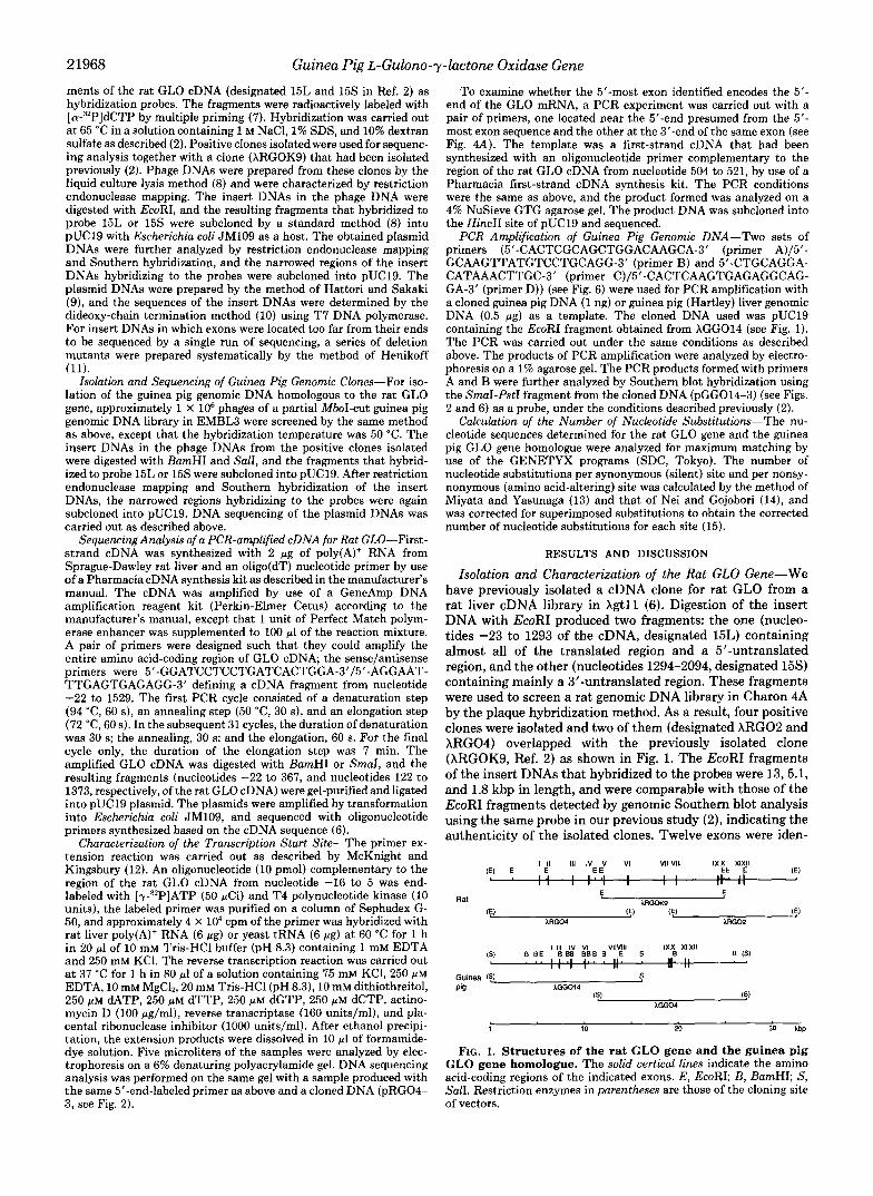
Isolation and Characterization of the Rat GLO Gene-We have previously isolated a cDNA clone for rat GLO from a rat liver cDNA library in Xgtll (6). Digestion of the insert DNA with EcoRI produced two fragments: the one (nucleo- tides -23 to 1293 of the cDNA, designated 15L) containing almost all of the translated region and a 5”untranslated region, and the other (nucleotides 1294-2094, designated 15s) containing mainly a 3”untranslated region. These fragments were used to screen a rat genomic DNA library in Charon 4A by the plaque hybridization method. As a result, four positive clones were isolated and two of them (designated ARGO2 and XRG04) overlapped with the previously isolated clone (XRGOK9, Ref. 2) as shown in Fig. 1. The EcoRI fragments of the insert DNAs that hybridized to the probes were 13,5.1, and 1.8 kbp in length, and were comparable with those of the EcoRI fragments detected by genomic Southern blot analysis using the same probe in our previous study (Z), indicating the authenticity of the isolated clones. Twelve exons were iden-
(S) B B E 888 BBB B E 5 B 11111 IV VI VllVlll IXX XIXII
I I . I I . . . 11. . 11. II I 1 I I I
B IS1
AGO04
1 10 20 30 kbp
FIG. 1. Structures of the rat GLO gene and the guinea pig GLO gene homologue. The solid vertical lines indicate the amino acid-coding regions of the indicated exons. E, EcoRI; B, BamHI; S, SalI. Restriction enzymes in parentheses are those of the cloning site of vectors.
Guinea Pig L-Gulono-y-lactone Oxidase Gene 21969
Guinea pig
- c - - I - " - -
B P s"1 P
X l l 1x11 ? pGGO4-IB
t c - - - 1 kb - - -
FIG. 2. Partial restriction maps and sequencing strategy for the rat GLO gene and the guinea pig GLO gene homologue. Arrows without a dot at the end indicate the direction and length of the sequencing runs with a universal or reverse primer for pUC19; arrows with a dot at the end indicate those of the sequencing runs with oligonucleotide primers synthesized on the basis of sequences that were determined on the opposite strands. The amino acid-coding regions are indicated by black boxes, and the regions corresponding to the untranslated regions of GLO mRNA, by open boxes. For the guinea pig gene homologue, only the amino-acid coding regions are shown. Names of subclones are shown aboue or beside the respective regions of the genes. E, EcoRI; P, PstI; H, HindIII, Sm, SmaI; X , XbaI; Sc, SacI; B, BamHI; Hc, HincII; Pu, PuuII; Sh, SphI. E in parentheses is the EcoRI site of the vector.
tified by sequence analysis (Figs. 2,3, and 4A) with reference to the sequence of the rat GLO cDNA (6).'
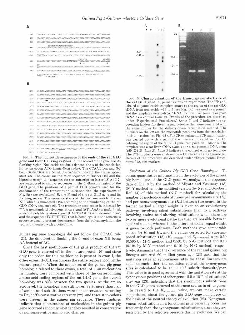
To determine the transcription start site, we carried out a primer extension experiment using, as a primer, an oligonu- cleotide complementary to 24 nucleotides at the 3' end of the 5'-most exon identified (see Fig. 4A) and rat liver poly(A)+ RNA as a template. Two major products and a minor one, which extended to positions -145, -123, and -142, respec- tively, from the translation initiation codon, were formed, indicating the presence of multiple transcription start sites (Fig. 5A). The fact that the 5'-most exon identified represents the 5'-end of the gene was evidenced by the following PCR experiment. If the entire 145-nucleotide 5"untranslated re- gion of the mRNA is coded by this exon, a forward primer (positions -136 to -119 of the sequence in Fig. 4A) located near the 5'-end presumed from the 5'-most exon sequence in
'The following nucleotides of the previously reported rat GLO cDNA (6) were found to have been read erroneously because of ambiguous sequence ladders. The nucleotide at position 567 should read C, and the C at position 1460 and the G at position 1504 should be deleted.
combination with a reverse primer (positions -19 to 5 ) located at the 3'-end of the same exon should amplify, from a rat liver cDNA, a sequence that is identical with what is to be amplified from the cloned genomic DNA. In fact, the PCR products of the expected size (141 bp) were generated with both templates as shown in Fig. 5B; the identity was further confirmed by sequencing the PCR product generated with the rat liver cDNA as a template. It should be noted that the transcription start sites determined are located in the two regions that are homologous to the consensus initiation se- quence (TCAGTCTT) identified recently by Bucher (16) (Fig. 4A). The GLO gene is expressed only in the liver; however, the promoter region of this gene has no TATA box, as do most tissue-specific genes. A CCAAT box and a GC box (GGGCGG) are present 6 and 90 bp upstream, respectively, from the first transcription start site. At positions -206 to -187 from the first transcription start site, there is a sequence that has a 11/13 nucleotide match to the bipartite recognition sequence (TGGACYYNXTGGCCC) that was found in the promoter regions of several liver-specific genes and was dem- onstrated to be the binding site of the transcription factor
The sequence of the 3'-end of the rat GLO genes is shown in Fig. 4B. The translation stop codon, TAA, is located in the 3'-most exon identified and precedes a nonstandard polyad- enylation signal (ATTAAA) (18) by 780 bp. The sequence CACTG, located 4 bp downstream from this sequence, matches a motif, CAYTG, which is usually present close to the polyadenylation site (19). Thirteen base pairs downstream from the polyadenylation signal is located a motif, TGTTTTTC, that has a 7/8 nucleotide match to the consen- sus sequence, YGTGTTYY, present downstream from the polyadenylation site in many eukaryotic genes (20). There- fore, this part is probably the end of the gene. Thus, the rat GLO gene contains 12 exons and 11 introns in the region spanning about 23 kbp. All the intron/exon boundaries fol- lowed the GT/AG rule (21). The nucleotide sequences of the exon regions were identical with those of the rat GLO cDNA reported previously (6) except for three differences. Two differences (G + A at position 252 and C + T at position 432 of the rat GLO cDNA) occur in the third position of the codons for glutamine and threonine, respectively, and are most probably due to allelic polymorphism. The other differ- ence (A + G at position 1211), which alters the codon for glutamine to that for arginine, may also be a polymorphism or may be an artifact arising during the cloning procedures. Sequencing analysis of a PCR-amplified cDNA from rat liver showed that the nucleotides at these three positions were identical with those of the previously isolated cDNA (6).
Isolation and Sequencing of the Guinea Pig GLO Gene Hom- ologue-A guinea pig genomic DNA library in EMBL3 was screened with the 15L fragment of the rat GLO cDNA as a probe. Twelve positive clones were isolated, and two overlap- ping ones were characterized. The BamHI fragments that contain rat GLO exon-related sequences were 7.5, 6.3, 2.5, 1.3, and 0.5 kbp in length (Fig. 1). The first three of them were detected by genomic Southern blot analysis using the 15L fragment as a probe in our previous study (Z), although the last two fragments were not detected, probably because of the shortness of the exon-related regions in them and the stringent conditions used in the hybridization. This finding indicates that the isolated clones represent the true GLO- related region in the guinea pig genome.
The regions corresponding to the rat GLO gene exons except for exons I and V were identified in the guinea pig gene, and their sequences were determined (Figs. 2 and 3).
LF-A1 (17).
21970 Guinea Pig L-Gulono-y-lactone Oxidase Gene
Rat Exon I M
.GACTTTCCGTGGGAACGCTTCAAGTCAAGTCGTCTGATCCTCCTGATCACTGG~RT~TAGGA. .TTTCTTCCTGGTACA TCCATGGGTACARRGGGGTCCAGTT
f G ! K G $ Q F V Q G H K G F D F
...... ........ ....... * L l t t ( l . . * ................ Guinea pig ......... .TGCTTCCCTGGCACA TCCAGGGGCACMGGCTTCTAGTT .
Q N W A K T Y G C S P E V Y Y Q P T S V E E V R E 0 . 0 0 0 Exon
V L A L A R CC~TTGGI:CAIV\GACCTATGGTTGCA~TCCAGAGGTGTACTACCAGCCCACCTCCGTGGAGGAGGTCAGAGA TAGGTG .......... .(It**. ........................................................ CC~CTGG(iTGRCTACTGCTGCTGCCTGGAGATGTACTACCAGTCGGCATCGGTGGAGGAGGTCCGAGA TGAGTG.'.....'.. Q N W V K T Y C C C L E M Y Y Q S A S V E E V R E V L A L A R
0 . 0 . 0 E Q K K K V K V V G G G H S P S D I A C T D G F M I H M G K M N R V L Q
0 .
GGGAGCAGAAI:liRGAAAGTGAAGGTGGTGGGTGGTGGCCACTCGCCTTCAGACATTGCCTGCACTGACGGTTTCATGATCCACATGGGCAAGATGAACCGGGTTCTCCA TACTGG ........ GGCAACAGAACAAGCGCGTGAAGGTTGTGGGCAGAGGCCACTCCCCCTCCGACATTGCCTGAACAGAGGGCATCATGATCCAC~GGGGCAAGATGAACCGGATCCTCCA TACCGC .. ................................................................................... . I . * . * . .** ........
Q Q N K R V K V V G R G H S P S D I A D T e G I n I H R G b n W R I L O
FIG. 3. Comparison of the nucleo- tide sequences of the exons of the rat GLO gene with their corre- sponding parts of the guinea pig GLO gene homologue. Amino acid- coding regions of the nucleotide se- quences are boxed. Asterisks indicate identical nucleotides between rat and guinea pig. The deduced amino acid se- quences for rat and guinea pig are shown above and below the respective se- quences. Open triangles represent dele- tions of nucleotides in the guinea pig sequence, and solid squares indicate stop codons. Open and closed circles above the amino acid sequence of the rat gene rep- resent conservative and nonconservative amino acid substitutions, respectively, according to Dayhoff's conservative cat- egory (22): C; S, T , P, A, G; N, D, E, &; H , R , K ; M , I , V , L ; a n d F , Y , W .
Exon IV 0 . 0 0 0 0 0 0 0 V D K E K K Q I T V E A G I L L A D L H P Q L D E H G L A M S N
..TGCT-GACTCTTCCA TGGACRRGGAG~RGAAGCAGATAACAGTGGAAGCCGGTATCCTCCTGGCTGACCTGCACCCACAGCTGGATGAGCAT~CCTGGCCATGTCC TAAGAGTCCC. .................................................................................................
..CCCTGGTCTCCTGCA(TGGACRRG~~RG~RTAAGCAGGTGACGGTGGAGACTGGCATCCTGCTGGCTCAGCTGCACTCGC~CTGGACAAGCACGGCCTGTCCCTGTCC TAAGAGACCC. V D K K N K Q V T V E T G I L L A E L H S Q L D K H G L S L S N
Exon V L G A V S D V T V A G V I G S G T H N T G I K H G I L A T Q
........ ... Exon VI . O . a o 0
V V A L T L M T A D G E V L E C S E S R N A D V F Q A A R V H L G C . """ACCTCTCCATGGCTA TGGTGGCCCTGACCCTGATGACAGCTGATGGAGAAGTTCTGGAATGTTCTGAGTCAAGAAATGCAGATGTGTTCCAGGCTGCACGGGTGCACCTGGGTTGC
....... AGCGTCCCTCTGCCA TGGTGGCACTGACACTGATGACAGCTGATGGGGTCATTCTGGAGTG=TCCGAGTCCAGCCACCCAGA--TGTTCCAGGC~~RCGCGGGTGCACCTGGGCTG~ .........................................................................................
V V A L T L M T A D G V I L E C S E S S H P A A F Q A T R V H L G D
L G I I L T V T L Q C V P Q F H L Q E T S F P S T L K E Exon VI1
CTGGGCATCATCCTCACTGTCACCCTGCAGTGTGTGCCTCAGTTTCACCTTCAGGAGACATCCTTCCCTTCGACCCTCARRGA~TAGTCA V L D .......... .. .* ............................................. . ,. ....
CTGGGCTCCTGCCTCGGGGTTG.AGTCC ......... 0.. . n n n
C~CCTAGACAGCCACCTGAAGAGGTCTGAGTACTTCCGCTTCCTCTGGTTTCCTCACACTGAGAACGTCAGCATCAT~TACCAAGACCACACCliRCAA T M C T G "GCATTAT ...... **..**,. **tl.t. ..................................................... *I* . .* . . . . . .* . . ..... N L D S H L K i s a Y F n F L W F P H i E N V S ~ I Y Q D ~ T N K ........ C~CCTTGACAGCCATCTGAAGAAGTTCAAGTACTTCTGCTTCCTGTGGTTCCCACACAGCGAGAACGTCAGTGTCATCTACCAGGACCGCACCAACAA TAGCCG ........ "ACATTTT N L D S H L K K F K Y F C F L W F P H S E N V S V I Y Q D R T N K
Exon Vlll 0 A P S S A S N W F W D Y A I G F Y L L E F L L W T S
0 0 . b . 0 . Exon IX CCCCCTCCTCTGCATCTAACTGGTTTTGGGACTATGCCATC~GGTT=T*CCTA~=G=AGT~TTGCT~TGGA~=A .TACGAA .......... **..*..,*** .... ..l..t**..*.1*1,.*..ttt.,,.*t...t.~.... .............. ........ CCCCCTCCTCTTCAGCCAGCTGGTTTTGGGACTATGCCATCGGGTTCTACCTACTGGAATTCCTGCCCCGGCCCC T.GTCT ..........
A P S S S A S W F W D Y A I G F Y L L E F L P R P H
0 0 0 0 . 0 . L P C L V G W I N R F F F W M L F N C K K E S S N L S H K I F T Y E C R F K Q H V
ACCTGCCATGCCTCGTGGGCTGGATCAACCGCTTCTTCTTCTGGATGCTGTTCAACTGCAAGAAGGAGAGCAGCAACCTCAGTCACAAGATCTTCACCTACGAGTGTCGCTTC~CAGCATGT ........................................................................................................... TCCTGCCGTGCCTCATGGGTTGGATCAATTGCTTCTTCTTCTGGCTGCTGTTCAACTGCAAGAAGGAGhRCTGTGACTTCAGCCAC~CATCTTCAGCTATGAGT~CGCTTCAAGCA~ACGT
L P C L M G W I N C F F F W L L F N C K K E N C D F S H K I F S Y E C R F K Q H V
Exon X n rn Q D W A I P R E K T K E A L L E L K A M L E A H P K V i A H .......... CTGGGCTGACGGGCA GAGAAGACChRGGAGGCCCTACTGGAGCTAAAGGCCATGCTGGAGGCCCACCCCARRGTGGTAGCCCAC ............................................................. t. ........... Q D W A I P R E K T K E A L L E L K S M L E A H P K V A A H
Y P V E V R F T R G D D I L L S P C F Q R D S C Y M N I I H Y R . O
TACCCCGTAGAGGTGCGCTTCACCCGAGGCGATGACATTCTGCTGAGCCCCTGCTTCCAGAGGGACAGCTGCTACATG~RCATCATTATGTACA.T.G .......... TTCCTCTCTACG ................................................................................. '4.. ... ....... TACCCTGTGGGGGTGCGCTTCACCCGGGGGGACGACATCCTGCTGAGCCCCTCCTTCCAGAGGGACAGCTGCTACATGAACATCTGCATATACA TGACAG""'""'CTCCCCTGTGCC I P V G V R F T R G D D I L L S P S F Q R D S C Y M N I C I Y R
Exon XI a 0 0 . e P Y G K D V P R L D Y W L A Y E T I M K K F G G R P H W A K
CTA CCCTATGGAAAGGACGTGCCTCGGCTAGACTACTGGCTGGCCTATGAGACCATCATGAAGAAGTTTGGAGGAAGACCCCACTGGGC ............................................................................ ........ CCA E CCCTACGGCAAGGACGTGCCTCAGCTTliRCTACTGGCTliRCCTGTGAGACCATCATGhRGliRGTTCCAGGGAAGACCCCATTGGGC
P Y G K D V P Q L N Y W L T C E T I M K K F Q G R P H W A K
Exon XI1 . . 0 0 .. 0 A H N C T R K D F E E M Y P T F H K T C D I R E K L D P T G M F L N S Y L E K V
0 .
.. ............................................................................................. CC~AACTGCACCTGGAAGGACTTTGAG~TGTACTCTGCCTTTCCCACGTTTTGCACCATCCGTG~GCTG~CCTCACTGG~~RTGTTCCTAAATTCCTACCTGGAGARRCTG
A H N C T W K D F E K M Y S A F P T F C T I R E K L N L T G M F L N S Y L E K V
F Y . .. ...... ACAA=GGGAACTGACC ....... .......................... (I f . f t . 1. . I . ........ .....
F Y D
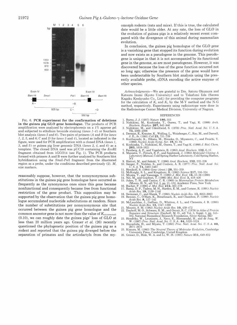
Sequencing of the entire region between exons IV and VI of the cloned gene showed that the sequence homologous to exon V of the rat GLO gene is absent in the guinea pig gene homologue. The possibility that the deletion of exon V is due to an artifact arising during the cloning process was ruled out by a PCR experiment (Fig. 6). Two overlapping PCR products were produced with two sets of oligonucleotide primers such that they covered the whole region between exons IV and VI. The PCR products had the same sizes whether they were formed with the cloned DNA or with the guinea pig genomic
DNA as a template. The sequence corresponding to a 3'-part (84 bp in size) of rat exon VI also appears to be deleted in the guinea pig gene homologue, since the alignment of the guinea pig sequence with the rat sequence necessitates many gaps and since the aligned sequence shows only low homology with the rat sequence (Fig. 3). This deletion was also confirmed by a PCR experiment as shown in Fig 6. In addition to these two major deletions, there is a 2-nucleotide deletion in the exon VI-related region of the guinea pig GLO gene homologue. One of the identified putative intron/exon boundaries of the
Guinea Pig L-Gulono-y-lactone Oxidase Gene 21971
A - 5 4 0
- 4 8 0
- 4 ? 0
-360
-330
- ? 4 0
-180
“73
-60
1
B
1 1 9 5
1 2 5 5
1315
1 3 7 5
1 4 3 5
1 4 9 5
1 5 5 5
1615
1 6 7 5
1 1 3 5
1 1 9 5
1 8 5 5
1 9 1 5
1 9 7 5
?035
2 0 9 5
2 1 5 5
CTGCAGCTCTGMTATTTTGCTCCTTTGGATCTTAACAAGTTTCTCAATAATTGTACRAA
ATGCTGTGTATCRAAGGACTGAGAAGAGTCAACCTTTTATGGTTTGAAGGGCCGATGAGG
CAGACTGTCCCRAACCCAGACACGTAGGCAGCTAGCCTAGTTCTGAGCCTGARAATAGGG
TGGACYYNNNNNNNTGCCCC CTTTAGCTCTGGACTCCCAAGGCTGGGCTCAGAACCCCAGCAATGGGTTTCAGRRGCACA
GCAGCTGCTGTTGAATCTCCATCTTTCAAGCTTCCTTGGTGCTCACAGACAGAGGGCAGG
~ACCCAGCTCCTGTTTACAGGGATTGCCTGGGTCTTGCAGGGGGAGGAGCCTGT
TCGACACTGTTTTCCTGCCTTTGTCZCL~IECCTTAGTATGGGT~ACXP.GXC TCAGTCTT TCAGTC
1 1 1 Ti C*GTTCTGGCTGACGTAACCCAGAGCTGAGGTTGCCTGACCACTGCTACCCAGGGCTTTG
TCCGACTTTCCGTGGGRRCGCTTClVIGTCAAGTCG?CTGATKT.C!XWCTG!iUX
~ACGAATGC?AGGGGTCCGTGAGACCGCCAGTGGATACAGTCAGCT~TGT - i n t r o n :
GCCCACAATTGCACCCGGRGACT~TGAGGRRATGTACCCCACCTTTCACAAGTTCTGT
GACATCCGTGAGAAGCTGGACCCCACTGGAATGTTCTTGAATTCGTACCTGGAGRAAGTC
TTCTACTRAAGCAGGAGTGGRAACAAACCACCCTGACCCCTCACACTTCTGCTGCCCCCG
GGCGTCTGGGGAGCAGAGAAGTGCCTCACAAGCACAATGGGAACTGACCTCTCCTCCTGA
CCACAAAGRRAGGCTGGGCTCTGGGCGGGTCCTCTCTGCCTTCGGCATCATTTCCCTTAC
ATCCAGGCAAGAAGTGGCCTCTCACTCRAATTCCTGTTAGCATTTCCATGGGTCACACAT
)IIWCTGCAATCCTC?CAGGAGAAGGGGGATCCCTGATACATCATATC?ATCCAGACTAAG
GATGTGGTTCTTCCTAGATTCTATGGCTCCACCAGGTATAGAGAGATTCCTGGGGCCTGC
AGTTCTCCATCCCTCTTCAGRRGGGAGGGATCCCTTGGCGAGAGTT?GGCTCAGAGGTGG
CATGAAGCATGCTCTGCTCTCTCTTACCCTTGAAGGTCCTTCGGATGCCCAGAGATGTCT
GCTGGTCCTGGGCAAGCCATCATTCRAACGGGTCCRRCCTGGCCTTCTGTCTGCCATGGC
CTGACCCTCGCAGTGTCTCTTCCAGAGGTGTTTAGAGTGGAACTCGCTTCAACCTCTTAA
CCAGTTGCTGATCCCTCTGTTTCTCTCCCTTCTCCTTGGAGACTACTCTTGGAGGGGGAT
CCCACCATGTCCTTGGCTTTCCCTGGGTATTGTTCTCCTCTTCCTCTTCACRAATATGAT
TTCAGTTTGATTTGTGGCCTTTCTGGAGTGTTCCTTGGAGAACCRRGATGTTCCAGCTAC
TTTGTAAGAaruraenGGCAAGA?G?T?~~TGCCCTTGAGRRATTCATATTTTlVI
TTCTTTATCTCATAGAGAGT
...
FIG. 4. The nucleotide sequences of the ends of the rat GLO gene and their flanking regions. A , the 5’-end of the gene and its flanking region. Nucleotide residue 1 denotes the A of the translation initiation codon ATG (underlined twice). The CCAAT box and GC box (GGGCGG) are boxed. Arrowheads indicate the transcription start site. The consensus initiation sequence of Bucher (16) and the bipartite recognition sequence for the transcription factor LF-A1 (17) are juxtaposed to similar sequences in the 5”flanking region of the GLO gene. The positions of a pair of PCR primers used for the confirmation of the transcription initiation site (the experiment of Fig. 5 R ) are underlined. R, the :3’-end of the rat GLO gene and its flanking region. The sequence starts at the first nucleotide of exon XII, which is numbered 1195 according to the numbering of the rat GLO cDNA sequence (6). The translation stop codon is indicated by ***. A nonstandard polyadenylation signal (ATTAAA) is underlined, a second polyadenylation signal (CACTGA)(19) is underlined twice, and the sequence (TGTTTTTC) that is homologous to the consensus sequence usually present downstream from the polyadenylation site (20) is underlined with a dotted line.
guinea pig gene homologue did not follow the GT/AG rule (21), the dinucleotide flanking the 5’-end of exon XI1 being AA instead of AG.
Since the first methionine of the gene product of the rat GLO gene is cleaved off in the mature protein (6) and since only the codon for this methionine is present in exon I, the other exons, 11-XII, encompass the entire region encoding the mature protein. When the sequences of the guinea pig gene homologue related to these exons, a total of 1140 nucleotides in number, were compared with those of the corresponding amino-acid coding regions of the rat GLO gene, the overall homology was 83% between the two species. At the amino acid level, the homology was still lower, 79%; more than half of amino acid substitutions were nonconservative according to Dayhoffs conservative category (22), and three stop codons were present in the guinea pig sequence. These findings indicate that substitutions of nucleotides in the guinea pig gene occurred randomly whether they resulted in conservative or nonconservative amino acid changes.
A B
bP ’ 1 2 T C M 1 2 3
-145 - . -142 - a 310 - i
I I i
d 194 - I I
-123 - 0 118 - 3
FIG. 5. Characterization of the transcription start site of the rat GLO gene. A , primer extension experiment. The ‘”P-end- labeled oligonucleotide complementary to the region of the rat GLO cDNA from nucleotide -16 to 5 (see Fig. 4 A ) was used as a primer, and the templates were poly(A)’ RNA from rat liver (lane I ) or yeast tRNA as a control (lane 2) . Details of the procedure are described under “Experimental Procedures.” Lanes 7’ and C indicate the se- quencing ladders for thymine and cytosine that were generated with the same primer by the dideoxy-chain termination method. The numbers on the left are the nucleotide positions from the translation initiation codon (see Fig. 4A) . H, PCR experiment. PCR amplification was carried out with a pair of the primers indicated in Fig. 4A, defining the region ofthe rat GLO gene from position -136 to 5. The template was a rat liver cDNA (lane 1 ) or a rat genomic DNA clone (pRG04-3) (lane 2). Lane 3 indicate the control with no template. The PCR products were analyzed on a 4r;i NuSieve GTG agarose gel. Details of the procedure are descrihed under “Experimental Proce- dures.” M, size markers.
Evolution of the Guinea Pig GLO Gene Homologue-To obtain quantitative information on the evolution of the guinea pig homologue of the GLO gene, we analyzed the sequence data of Fig. 3 by the method of Miyata and Yasunaga (13) (M-Y method) and the modified version (by Nei and Gojobori; Ref. 14) of this method (N-G method) for calculating the number of nucleotide substitutions per synonymous site (K,) and per nonsynonymous site (K,,) between two genes. In the former method a larger weight is given to an evolutionary pathway involving silent substitutions than to a pathway involving amino acid-altering substitutions when there are two or more evolutional pathways that are possible between a pair of codons, whereas in the latter method, an equal weight is given to both pathways. Both methods gave comparable values for K, and K,, and the values corrected for superim- posed substitution (15) (Ks~corl.cclc~~~ and Kn(rorrcrtrrl)) were 0.59 (0.595 by M-Y method and 0.591 by N-G method) and 0.10 (0.104 by M-Y method and 0.101 by N-G method), respec- tively. Assuming that the divergence of the rat and guinea pig lineages occurred 60 million years ago (23) and that the mutation rates a t synonymous sites for these lineages are equal to each other, the mutation rate at the synonymous sites is calculated to be 4.9 X lo-“ substitutions/site/year. This value is in good agreement with the mutation rate at the synonymous positions of other genes, 5.5 X substitutions/ site/year (23), indicating that the synonymous substitutions in the GLO genes occurred at the same rate as in other genes.
In regard to the KnIrorrcrted) value, we can make certain suppositions about the guinea pig GLO gene homologue on the basis of the neutral theory of evolution (25). Nonsynon- ymous substitutions in a functional gene generally occur less frequently than the synonymous substitutions, since they are restricted by the selective pressure during evolution. We can
21972
M I 2 3 4
1353 - 1078 - 872 - 603 -
Exon IV BamHI Smal
Guinea Pig L-Gulono-y-lactone Oxidase Gene
5 6
Exon VI
Psf I 8amHI Barn HI a I a- A - .B
925 bp c - - D I
592 bp
FIG. 6. PCR experiment for the confirmation of deletions in the guinea pig GLO gene homologue. The products of PCR amplification were analyzed by electrophoresis on a 1% agarose gel and subjected to ethidium bromide staining (lanes 1-4) or Southern blot analysis (lanes 5 and 6 ) . Two pairs of primers (A and B for lanes I, 2, 5, and 6; C and D for lanes 3 and 4 ) , located as indicated in the figure, were used for PCR amplification with a cloned DNA (lanes I, 3, and 5 ) or guinea pig liver genomic DNA (lanes 2, 4, and 6) as a template. The cloned DNA used was pUC19 containing the EcoRI fragment obtained from XGG014 (see Fig. 1). The PCR products formed with primers A and B were further analyzed by Southern blot hybridization using the SrnaI-PstI fragment from the illustrated region as a probe, under the conditions described previously (2). M, size markers.
reasonably suppose, however, that the nonsynonymous sub- stitutions in the guinea pig gene homologue have occurred as frequently as the synonymous ones since this gene became nonfunctional and consequently became free from functional restriction of the gene product. This supposition may be supported by the observation that the guinea pig gene homo- logue accumulated nucleotide substitutions at random. Since the number of substitutions per nonsynonymous site that occurred between the guinea pig gene homologue and the common ancestor gene is not more than the value of (0.10), we can roughly date the guinea pigs' loss of GLO a t less than 20 million years ago. Grauer et al. (26) recently questioned the phylogenetic position of the guinea pig as a rodent and reported that the guinea pig diverged before the separation of primates and the artiodactyls from the my-
omorph rodents (rats and mice). If this is true, the calculated date would be a little older. At any rate, the loss of GLO in the evolution of guinea pigs is a relatively recent event com- pared with the divergence of this animal during mammalian evolution.
In conclusion, the guinea pig homologue of the GLO gene is a vanishing gene that stopped its function during evolution and now exists as a pseudogene in the genome. This pseudo- gene is unique in that it is not accompanied by its functional gene in the genome, as are most pseudogenes. However, it was discovered because the loss of the gene function occurred not so long ago; otherwise the presence of the gene would have been undetectable by Southern blot analysis using the pres- ently available probe, cDNA encoding the active enzyme of other species.
Acknowledgments-We are grateful to Drs. Satoru Okumura and Katsura Izumi (Kyoto University) and to Takafumi Iida (Sanwa Kagaku Kenkyusho Co., Ltd.) for providing the computer programs for the calculation of K, and K. by the M-Y method and the N-G
the Radioisotope Center Medical Division, University of Nagoya. method, respectively. Experiments using radioisotope were done in
REFERENCES 1. Burns, d . J. (1957) Nature 180,553 2. Nishikimi, M., Koshizaka, T., Ozawa, T., and Yagi, K. (1988) Arch.
3. Nishikimi, M., and Udenfriend, S. (1976) Proc. Natl. Acad. Sci. U . S.A.
4. Derman, E., Krauter, K., Walling, L., Weinberger, C., Ray, M., and Darnell,
5. Kurihayashi, K., Hikata, M., Hiraoka, 0.. Miyamoto, C., and Fruichi. Y.
6. Koshizaka. T.. Nlshikimi. M.. Ozawa. T.. and Yagi K. (1988) J. Riol. Chern.
Hiochrm. Riophys. 2 6 7 , 842-846
73, 2066-2068
-1. E., Jr. (1981) Cell 23,731-799
(1988) NuclricAcids S.vmp. Ser. 1 9 , 61-64
263,16ig-i621 , ,
7. Feinherg, A. P., and Vogelstein, B. (1983) Anal. Riochem. 132,6-13 8. Maniatis, T., Fritsch, E. F., and Samhrook, .J. (1982) Molecular C h i n $ : A
!,n+tory Manual, Cold Spring Harbor Lahoratory, Cold Spring Harhor, N1
10. Sanger, F., Nicklen, S., and Coulson. A. R. (1977) Proc. Natl. Acad. Sci.
11. Henikoff, S. (1984) Gene (Amst.) 28,351-359 12. McKnight, S. L., and Kingshury, K. (1982) Science 217,316-324 13. Miyata, T., and Yasuna a, T. (1980) J. Mol. Euol. 1 6 , 2 3 4 6 (1980) 14. Nei, M., and Gojohori, 'f. (1986) Mol. Rml. Ecol. 3, 418-426 15. Jukes, T. H., and Cantor, C. R. (1969) in Mammalian Profrin Metabolism
(Munro, H. N., ed) Vol. 2, pp. 21-132, Academic Press, New York 16. Bucher, P. (1990) J. Mol. Riol. 212,563-578 17. Ramii. D. P.. Tadros. M. H.. Hardon. E. M.. and Cortese. R. (1991) Nuckic
9. Hattori, M., and Sakaki, Y. (1986) Anal. Riochem. 152,232-238
C. S. A. 74,5463-5467
18. 19.
20. McLauchlan, J., Gaffney, D., Whitton, J. L., and Clements, d . R. (1985) Acids Res. 8 , 127-142
21. Mounts, S. M. (1982) Nucleic Acids Res. 10, 459-472 Nucleic Acids Res. 13, 1347-1368
22. Da hoff, M. O., Schwarz, R. M., and Orcutt, R. C. (1978) in Atlasof Profrin & p e n c e and Structure (Dayhoff, M. 0.. ed) Vol. 5, Suppl. 3, pp. 345-
23. Hendriks, W., Leunissen, d., New, E., Hloemendal, H., and de .long, W . 352, National Biomedical Research Foundation, Silver Spring, MD
24. Hayashida, H., and Miyata, T. (1983) Proc. Nut / . Acad. Sci. U. S. A. 80, W . (1987) Proc. Nafl. Acad. Sci. U. S. A,. 84,5920-5324
25.
26.
%71-7675
A&sRes.'l9, 11g9-1146 Swimmer, C., and Shenk, T. (1985) Nucleic Acids Res. 13,8053-8069 Benoist, C.. O'Hare, K., Breathnach, R., and Chamhon, P. (1980) Nurkric
Kimura, M. (1983) The Neutral Theory of Molecular h d u f i o n , Camhridge
Grauer, D., Hide, W. A. and Li, W.-H. (1991) Nature 351,649-652
- - . - - - . - University Press, Camhridge, United Kingdom