Embed Size (px)
Citation preview

The Novel Kinase Peptidylglycine a-Amidating MonooxygenaseCytosolic Interactor Protein 2 Interacts with the Cytosolic RoutingDeterminants of the Peptide Processing Enzyme Peptidylglycine a-Amidating Monooxygenase*
(Received for publication, September 7, 1999, and in revised, September 29, 1999)
Benjamin D. Caldwell‡, Daniel N. Darlington§, Peter Penzes, Richard C. Johnson,Betty A. Eipper, and Richard E. Mains¶
From the Departments of Neuroscience and Physiology, The Johns Hopkins University School of Medicine, BaltimoreMaryland 21205 and the §Departments of Surgery and Physiology, University of Maryland School of Medicine,Baltimore, Maryland 21201
The cytosolic domain of the peptide-processing inte-gral membrane protein peptidylglycine a-amidating mo-nooxygenase (PAM; EC 1.14.17.3) contains multiple sig-nals determining its subcellular localization. ThreePAM cytosolic interactor proteins (P-CIPs) were identi-fied using the yeast two hybrid system (Alam, M. R.,Caldwel, B. D., Johnson, R. C., Darlington, D. N., Mains,R. E., and Eipper, B. A. (1996) J. Biol. Chem. 271, 28636–28640); the partial amino acid sequence of P-CIP2 sug-gested that it was a protein kinase. In situ hybridizationand immunocytochemistry show that P-CIP2 is ex-pressed widely throughout the brain; PAM and P-CIP2are expressed in the same neurons. Based on subcellularfractionation, the 47-kDa P-CIP2 protein is mostly cyto-solic. P-CIP2 is a highly selective kinase, phosphoryl-ating the cytosolic domain of PAM, but not the corre-sponding region of furin or carboxypeptidase D.Although P-CIP2 interacts with stathmin, it does notphosphorylate stathmin. Site-directed mutagenesis,phosphoamino acid analysis, and use of synthetic pep-tides demonstrate that PAM-Ser949 is the major sitephosphorylated by P-CIP2. Based on both in vitro bind-ing experiments and co-immunoprecipitation from cellextracts, P-CIP2 interacts with PAM proteins contain-ing the wild type cytosolic domain, but not with mutantforms of PAM whose trafficking is disrupted. P-CIP2,through its highly selective phosphorylation of a keysite in the cytosolic domain of PAM, appears to play acritical role in the trafficking of this protein.
The targeting of membrane proteins requires sorting signalsthat govern the interaction of the membrane protein with theproper cytosolic proteins. For integral membrane proteins tar-geted to trans-Golgi network (TGN),1 endosomes, lysosomes, or
plasma membrane, a number of distinct and often overlappingmotifs have been identified in the cytosolic domain (1–11).Tyr-containing motifs can recruit adaptor proteins and coatproteins to membranes (12–15) and phosphorylation can gov-ern individual trafficking steps (3, 4, 7, 9, 11, 16–18). Neuronsand endocrine cells store bioactive peptides in a specializedorganelle, the large dense core vesicle. Very little is knownabout the targeting signals governing membrane proteins thatfunction in large dense core vesicles. The peptide amidatingenzyme, peptidylglycine a-amidating monooxygenase (PAM),functions in the regulated secretory pathway and has beenused to study trafficking to large dense core vesicles.
For roughly half of all bioactive peptides, COOH-terminalamidation is the final step of activation following endoproteo-lytic digestion from inactive precursors (19, 20). PAM is a TypeI integral membrane protein with routing determinants withinits cytosolic COOH-terminal domain (CD). Membrane forms ofPAM are localized primarily in a post-Golgi tubuloreticularcompartment overlapping but not identical with the TGN38-containing compartment; PAM that reaches the plasma mem-brane recycles to the TGN region (21). Truncation of the CD ofPAM results in cell surface localization with little internaliza-tion (22). Fusion of the transmembrane domain/CD of PAM tothe lumenal domain of a plasma membrane protein or to asoluble secretory granule constituent results in TGN localiza-tion of both chimerae (21, 23). Thus the CD of PAM containsrouting signals which mediate the localization of membraneforms of PAM within the regulated secretory pathway.
We recently reported the use of the yeast two-hybrid systemto identify PAM COOH-terminal interactor proteins (P-CIPs)(24). Three cDNAs were identified; one encoded a putativeserine/threonine protein kinase followed by a RNA bindingmotif (P-CIP2) and another encoded Kalirin (formerlyP-CIP10), a novel member of the Dbl family of GDP/GTP ex-change factors that interacts with Rac1, a small GTP-bindingprotein of the Rho subfamily (25). Both P-CIP2 and Kalirinwere shown to interact directly with membrane PAM in vitrousing GST fusion proteins expressed in bacteria. P-CIP2 wasalso identified independently as an interactor of stathmin, acytosolic protein involved in microtubule destabilization (26).Stathmin is homologous to the neuronal protein SCG10, whichalso destabilizes microtubules (27).
In this study we set out to characterize P-CIP2 more fully
* This work was supported by National Institutes of Health GrantDK-32949. The costs of publication of this article were defrayed in partby the payment of page charges. This article must therefore be herebymarked “advertisement” in accordance with 18 U.S.C. Section 1734solely to indicate this fact.
‡ Current address: Dept. of Chemistry, Missouri Western State Col-lege, St. Joseph, MO 64507.
¶ To whom correspondence should be addressed: Dept. of Neuro-science, Rm. 907 WBSB, The Johns Hopkins University School of Med-icine, 725 North Wolfe St., Baltimore, MD 21205. Tel.: 410-955-6938;Fax: 410-955-0681; E-mail: [email protected].
1 The abbreviations used are: TGN, trans-Golgi network; PAM, peptidyl-glycine a-amidating monooxygenase; CD, COOH-terminal cytosolic domain;P-CIP, PAM COOH-terminal interactor protein; GST, glutathione S-trans-ferase; TES, N-tris(hydroxymethyl)methyl-2-aminoethanesulfonic acid;
PIPES, piperazine-N,N9-bis(2-ethanesulfonic acid); PMSF, phenylmethyl-sulfonyl fluoride; PAGE, polyacrylamide gel electrophoresis; kb, kilobase(s);PAL, peptidyl-a-hydroxyglycine-a-amidating lyase.
THE JOURNAL OF BIOLOGICAL CHEMISTRY Vol. 274, No. 49, Issue of December 3, pp. 34646–34656, 1999© 1999 by The American Society for Biochemistry and Molecular Biology, Inc. Printed in U.S.A.
This paper is available on line at http://www.jbc.org34646
by guest on March 9, 2019
http://ww
w.jbc.org/
Dow
nloaded from

and to determine whether P-CIP2 is a functional kinase capa-ble of phosphorylating PAM. For the interaction of P-CIP2 andPAM to be of functional significance, the two proteins must beexpressed in the same cell; we used in situ hybridization andimmunocytochemistry to demonstrate that the sites of P-CIP2and PAM expression overlapped extensively in the brain. Evi-dence of a functionally significant interaction of P-CIP2 withPAM was shown by the ability of endogenous P-CIP2 to bind toPAM expressed in AtT-20 corticotrope tumor cells and in testtube assays. In vitro binding assays and co-immunoprecipita-tion demonstrated that mutant PAM proteins that aremisrouted in AtT-20 cells do not interact well with P-CIP2. Thesite of PAM-CD phosphorylation by P-CIP2 was identified, andthe specificity of P-CIP2 for various other substrates was in-vestigated. The results indicate that P-CIP2 phosphorylates aspecific region of the cytosolic domain of PAM which containsrouting determinants essential for the correct localization ofmembrane PAM, but phosphorylates few other potentialsubstrates.
MATERIALS AND METHODS
Cloning of Full-length Rat P-CIP2—The 1.4-kb cDNA insert isolatedfrom the original yeast two-hybrid screen (24) was 32P-labeled by ran-dom priming (Prime It II, Stratagene) and used to screen a rat hip-pocampal random primed lZAPII cDNA library (Stratagene). Sevenpositive overlapping clones were isolated and purified from 6 3 105
plaque forming units. The seven library cDNAs were rescued, the 59ends were sequenced, and the start codon was identified by its Kozaksequence which matches 7 of 9 bases in an optimal translation initiationsite (59-GCCCGCACCATGG-39, mismatch is underlined) (28). The fullsequence has been deposited with GenBank (accession numberU70372).
Construction of Expression Vectors—The 1.4-kb P-CIP2 partial cDNAfragment (I-2) encoding P-CIP2 residues 28–419 (DNP-CIP2) (24) wasligated into pGEX5X2 to generate pGEX5X2/DNP-CIP2 for the expres-sion of GST/DNP-CIP2 fusion protein to be used as an antigen. Toconstruct a full-length P-CIP2 fusion protein expression vector,pGEX5X2/P-CIP2, the cDNA encoding P-CIP2 was modified to generatea SalI site in place of the start codon, and the 1.4-kb fragment wasinserted into pGEX5X2 digested with SalI/NotI, yielding pGEX5X2/P-CIP2. Expression of the GST fusion protein with full-length P-CIP2from pGEX5X2/P-CIP2 was carried out in BL21(DE3) cells (see below).
For mammalian expression, an optimal ribosomal binding site wasengineered to precede the initiation codon of P-CIP2 in pBS.KrP-CIP2(28, 29). Two mammalian expression vectors were used to expressP-CIP2 and P-CIP2(1–383) (P-CIP2cc) with the Myc epitope and aHis6-tag at their COOH terminus (pcDNA3.1/mycHis; Invitrogen) or atthe amino terminus (created by combining the epitope tag regions ofpEAK10-His and pEAK10-Myc from Edge Biosystems, Gaithersburg,MD). Plasmid pBS.KrP-CIP2 was digested with XhoI/EcoRV and in-serted into pcDNA3.1/mycHis digested with KpnI (blunted) and XhoI.For expression of P-CIP2cc, pBS.KrP-CIP2 was digested with XhoI/BsaBI and ligated. The full coding sequence of P-CIP2 was excised frompBluescript with NcoI-NotI and inserted into the pEAK10 vectors cutwith BspLU11I-NotI. The Stratagene Quikchange site-directed mu-tagenesis kit was used to create pcDNA.KrP-CIP2cc/K54A using thesense primer 59-GGCGCCCTCGCGCAGTTCCTGCCTCCGCC-39. AllPCR products and ligations were confirmed by DNA sequencing. The plas-mids pcDNA3.1/b-galactosidase-mycHis (Invitrogen) and pEGFP-N2(CLONTECH) were used as transfection controls.
Northern Blotting—Northern blots were performed as described pre-viously (30) except that total RNA was extracted from adult HarlanSprague-Dawley rat tissues with RNA Stat-60 (Tel Test). The SalI/NotI1.4-kb fragment isolated in the original two-hybrid screen was randomprimed using the Prime-it II kit (Stratagene) with [a-32P]dCTP.
Generation of Antisera—A glutathione S-transferase fusion proteinwith DNP-CIP2 was expressed in Escherichia coli and purified byglutathione-Sepharose affinity chromatography (24) for use as an anti-gen to generate anti-P-CIP2 rabbit antisera JH1998 and JH1999 (Co-vance, Denver, PA). Generation of the rabbit polyclonal antibodyJH2004 directed toward a 19-residue peptide near the COOH terminusof P-CIP2 was described previously (24).
In Situ Hybridization and Immunocytochemistry—In situ hybridiza-tion for P-CIP2 and double labeling by in situ hybridization for P-CIP2with immunocytochemical staining for PAM-1 or P-CIP2 were per-
formed as described previously (24, 25, 31). After completion of the insitu washes, sections were washed in phosphate-buffered saline,blocked with 10% normal goat serum (30 min) and treated with 3%H2O2 in phosphate-buffered saline for 30 min. PAM antibodies (AbJH1761 directed to the amino-terminal third of PAM or Ab 471 directedtoward the middle third of PAM) were used at 1:1000 dilutions onsections and coverslipped with Parafilm. For P-CIP2 immunocytochem-ical staining, Ab JH1998 was used at a dilution of 1:1000. Sections wereincubated at 4 °C overnight. Immunocytochemistry was performed us-ing an avidin-biotin kit (Vector, Burlingame, CA) and visualized withhorseradish peroxidase substrate. Sections were then dipped in photo-graphic emulsion as described previously (24, 31).
Tissue Extract Preparation and Subcellular Fractionation—Adultrat brain was separated into soluble and crude particulate fractions orsubjected to differential centrifugation. Equal volumes of soluble andparticulate fractions were subjected to Western blot analysis with AbJH2004. For preparation of soluble and crude particulate fractions,tissues were homogenized in a 10-fold volume excess of 0.25 M sucrosein 20 mM TES, pH 7.0, containing 0.3 mg/ml PMSF and proteaseinhibitor mixture (29). The homogenate was centrifuged at 10,000 3 gfor 10 min and the pellet discarded; the supernatant was centrifuged at372,000 3 g for 15 min. The pellet was re-suspended in a volume equalto the supernatant (soluble fraction).
Differential centrifugation of parietal cortex was carried out accord-ing to Huttner et al. (32) and Bennett et al. (33). Tissue was homoge-nized in 10 volumes of SHEEP buffer (0.32 M sucrose, 4 mM HEPES-KOH, pH 7.5, 0.1 mM EDTA, 0.1 mM EGTA, 0.3 mg/ml PMSF). Thehomogenate was centrifuged at 400 3 g for 10 min and the pellet wasdiscarded. The supernatant was centrifuged at 800 3 g for 10 min; thepellet was washed by re-suspension in the same volume of SHEEPbuffer yielding the crude nuclear pellet (P1) and pooled supernatants(S1) that were centrifuged at 9200 3 g for 10 min. The crude synapto-somal pellet was washed and resuspended in SHEEP buffer (P2). Thepooled supernatants (S2) were centrifuged at 165,000 3 g for 15 min;the microsomal pellet was washed as before and resuspended in SHEEPbuffer (P3). The final pooled supernatant (S3) is the cytosolic fraction.
Co-immunoprecipitation—Immunoprecipitations were carried outaccording to the method of Ratovitski et al. (34). Briefly, the culturemedium was removed from a confluent 100-mm dish and the cells wererinsed with phosphate-buffered saline. Extraction buffer (20 mM PIPESbuffer, 2 mM Na2EDTA, 50 mM sodium fluoride, 10 mM Na3P2O7, 1 mM
NaV3O4, 1% Triton X-100, with 0.3 mg/ml PMSF and protease inhibitormixture, adjusted to pH 6.8 with NaOH) was added to each dish (2.0 ml)on ice for 15 min. Cells were scraped from the dish, and debris wasremoved by high-speed centrifugation. Extracts (300 ml) were dilutedwith 300 ml of extraction buffer, 1 ml of PMSF (30 mg/ml), 5 ml ofinhibitor mixture, and 10 ml of P-CIP2 antibody (JH2004 or JH1998) or10 ml of P-CIP2 antibody pre-blocked for 1 h on ice by incubation with50 mg of P-CIP2 peptide (PKENPGRGQVFVEYANAGD, underlined inFig. 1) for JH2004 or 50 mg of GST/P-CIP2 for JH1998. Antibody-extractmixtures were incubated on ice for 1 h, followed by addition of ProteinA-agarose (Sigma) and agitation on ice for 1 h. Protein A-agarose beadswere washed and boiled into loading buffer for separation by SDS-PAGE (10% polyacrylamide gel). Western blots used antibodies to PAM(Ab 629, 1:2000 dilution, or monoclonal antibody 6E6, 1:20 dilution ofmedium (35)) and were visualized using the ECL kit (Amersham Phar-macia Biotech).
Tissue Culture and Expression of Proteins in Bacterial and Mamma-lian Cells—For mammalian expression of P-CIP2 or its mutants, AtT-20, COS, hEK-293, and pEAK-Rapid (Edge Biosystems) cells werecultured in Dulbecco’s modified Eagle’s medium/F-12 medium contain-ing 10% NuSerum (omitted for pEAK-Rapid cells) (Collaborative Re-search, Bedford, MA) and 10% fetal clone serum (Hyclone, Logan, UT)and plated in 75-cm2 flasks 1–2 days prior to transfection. Stable celllines were created as described previously (21), and selected by immun-ofluorescent staining and Western blotting with myc monoclonal anti-body 9E10 (36). Transient transfections were performed using Lipo-fectAMINE or LipofectAMINE Plus (Life Technologies, Inc.). Cells wereplated on glass slides and immunostained as described (21, 24) using aZeiss Axioskop equipped with a Princeton Instruments Micromax dig-ital camera. GST fusion proteins were purified from bacterial cell ly-sates by affinity chromatography on glutathione-Sepharose (AmershamPharmacia Biotech) and eluted with glutathione, which was removed bydialysis.
In Vitro Kinase Assays—Mammalian cells stably transfected withvectors encoding P-CIP2mH, P-CIP2ccmH, or b-galactosidase-mH werescraped from culture dishes and extracted with 20 mM Na-TES/10 mM
mannitol, pH 7.4, containing PMSF (30 mg/ml) and inhibitor mixture,
P-CIP2 Interacts with PAM Routing Determinants 34647
by guest on March 9, 2019
http://ww
w.jbc.org/
Dow
nloaded from

followed by three cycles of freezing and thawing. Extracts from mam-malian cell pellets were diluted with an equal volume of 23 metalchelate resin binding buffer (13 is 10 mM imidazole, 150 mM NaCl, 50mM Tris, pH 7.4). The diluted extracts were applied batchwise to His-bind metal affinity resin (Novagen) charged with Ni21, or to Ni21-NTA-agarose resin (Qiagen). Samples were incubated with resin for 30–60min at 4 °C with gentle mixing, washed three times with 40 mM imid-azole, 0.5 M NaCl, 50 mM Tris-HCl, pH 7.4, and twice with 5 mM MgCl2in Na-HEPES, pH 7.4. P-CIP2 was eluted with 1 M imidazole andrequired at least a 10-fold dilution before it was assayed.
pEAK-Rapid cells transiently transfected with the pEAK10 vectorwere extracted into isotonic sucrose buffer containing 20 mM Na-HEPES, pH 7.4, with protease inhibitors, centrifuged at 430,000 3 g for15 min and the pellet was re-extracted with 50 mM HEPES, 1 mM
MgCl2, pH 7.5, containing 1% Thesit and protease inhibitors. Most ofthe exogenous P-CIP2 was recovered in the pellet fraction followingtransient expression. P-CIP2 was purified from pEAK-Rapid extracts(#100 mg of protein/mg of resin) using the His-bind resin, Talon resin(CLONTECH), or the Ni-NTA resin according to the manufacturers’instructions, and eluates were desalted into 50 mM HEPES, 1 mM
MgCl2, pH 7.5, containing 0.1% Thesit using NICK columns (Amer-sham Pharmacia Biotech).
Enzyme was added to the reaction mixture (40 or 50 ml) containingrecombinant substrate (5 or 10 mg; PAM-CD, GST-PAM-CD, other GSTfusion proteins); kinase buffer (50 mM HEPES, 1 mM MgCl2, pH 7.5,0.1% Thesit) containing 1 or 2 mCi of [g-32P]ATP (3000 Ci/mmol, Am-ersham Pharmacia Biotech) was added to each sample. Reactions wereincubated at 37 °C for 30 or 60 min. Positive control reactions utilizedprotein kinase C purified from rat brain (kindly provided by CarolDoherty and Dr. Richard Huganir, Howard Hughes Medical Institutes/Johns Hopkins University School of Medicine), bovine heart proteinkinase A (Sigma), or recombinant human casein kinase II (Calbiochem).Reaction supernatants from stable transfections were precipitated with20 mg of bovine serum albumin and 15% trichloroacetic acid, washedwith acetone, boiled into Laemmli loading buffer, loaded onto 15%SDS-PAGE gels, and visualized by autoradiography. For transient ex-pression in pEAK-Rapid cells, the reaction mixtures were loaded di-rectly onto SDS-PAGE peptide gels (37), transferred to nylon mem-branes, and visualized by autoradiography, direct staining, or Westernanalyses. Gels were stopped before the dye ran off and a 3-cm regionbehind the dye was cut off before transfer, to remove the free [g-32P]ATP(38). Phosphoamino acid analysis of phosphorylated products was per-formed as described (39) using sequential pH 1.9 and pH 3.5 thin layerelectrophoresis.
P-CIP2 and commercial kinases were combined with 5 mg of syn-thetic peptide and 1 mCi of [g-32P]ATP in Hepes-Mg-Thesit for 30 min at37 oC, followed by addition of 5 mg of Trp-Met-Asp-Phe-NH2 and 100 mgof bovine serum albumin. PAM peptides were synthesized by Dr. HenryKeutmann, Massachusetts General Hospital (24, 39); standard kinasesubstrate peptides were from Calbiochem. Samples were left on ice for30 min, centrifuged, and aliquots of the supernatant spotted onto P81ion exchange paper (Whatman) prewetted with 1% phosphoric acid.One-eighth of each reaction was analyzed; samples were analyzed induplicate. Samples were washed repeatedly in 1% phosphoric acid,finally rinsed in acetone, air dried, and submitted to Cerenkov counting(38, 40). Data are reported as mean 6 S.E.
RESULTS
P-CIP2: A Putative Ser/Thr Protein Kinase with a RNABinding Motif—We previously used the yeast two-hybrid sys-tem to identify proteins that interact with the COOH-terminaldomain (CD) of PAM-1 (24). One of these PAM-COOH-terminalinteractor proteins (P-CIPs) contained an almost complete pu-tative protein kinase domain followed by a putative RNA bind-ing motif and a stop codon. Comparison of the predicted molec-ular mass (44 kDa) with the band observed in Western blots ofendogenous P-CIP2 in AtT-20 corticotrope tumor cell extracts(47 kDa) indicated that the complete cDNA would encode ad-ditional residues at the amino terminus.
Subsequent screening of a random primed rat hippocampalcDNA library identified an additional 27 amino acids precedingthe known P-CIP2 sequence. Thus, the full-length P-CIP2cDNA encodes a 419-residue protein with a predicted mass of46.5 kDa. Consistent with this, in vitro translation of full-length clones of P-CIP2 yielded a 46.5-kDa product (data not
shown) and the protein observed in AtT-20 cell extracts byWestern blot analysis has an apparent molecular mass (Mr) of47 kDa (24).
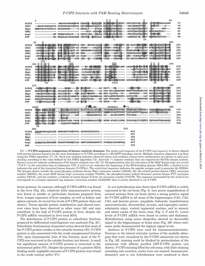
Fig. 1 shows an alignment of the amino acid sequence ofP-CIP2 with the most homologous protein kinases according toBLASTP analysis. The kinases shown, most of which are Ser/Thr protein kinases, share 22–30% identity with P-CIP2 withinthe catalytic core of 250 residues. The greatest sequence vari-ation among these kinases occurs in subdomains X and XI andfollowing subdomain XI. While the sequence Asp158-Leu-Lys-Pro-Arg-Asn in subdomain VI suggests that P-CIP2 belongs tothe serine/threonine protein kinase family, P-CIP2 also sharesmoderate homology with the dual specificity (Ser/Thr and Tyr)kinase PYT and has some residues normally considered diag-nostic for Tyr kinases (40). The non-conserved residues pre-sumably affect the substrate specificity of P-CIP2, which isremarkable (see below).
BLASTP comparison of P-CIP2 versus the Saccharomycescerevisiae Genome Data base showed that the most homologousyeast protein kinases are members of the MAP/ERK kinasefamily including SMK1, HOG1 (shown in Fig. 1), SLT2, KSS1,and FUS3; the most similar yeast kinase, HOG1, shows 14%identity and 30% similarity within the catalytic core. Theseyeast MAP kinases are involved in the mating pheromoneresponse pathway (41, 42), suggesting that P-CIP2 might beinvolved in signal transduction. MAP kinases have two closelyspaced phosphorylation sites (T-X-Y, circled in Fig. 1 for theHOG1 sequence) in subdomain VIII whose phosphorylation isrequired for activation of the kinase (43). All of the kinasesshown, including P-CIP2, share a Thr at the appropriate posi-tion in subdomain VIII, whereas only HOG1 contains bothsites. Rat CKII shows as much similarity to P-CIP2 as anykinase, which is interesting considering the substrate specific-ity of P-CIP2 (see below).
As noted previously, the COOH-terminal region of P-CIP2following subdomain XI shows significant homology with thethird ribonucleoprotein consensus sequence (RNCS) ofpre-mRNA splicing factor U2AF65 (24). A human expressedsequence tag matches 59 of 60 residues following subdomain XIinto the ribonucleoprotein consensus sequence region (Fig. 1,dashed box) with the only mismatched residue substituting anaspartate for glutamate. Only one other kinase is known tocontain an RNA-binding domain, PKR (for review, see Ref. 44).
P-CIP2 Is a Soluble Protein Found in Nervous and EndocrineTissues That Contain PAM—Northern blotting revealed thepresence of a single 9-kb transcript in every region of the brainexamined (Fig. 2A). PAM transcripts are known to be presentin all of the brain tissues tested (45). P-CIP2 mRNA was alsofound at high levels in anterior and neurointermediate pitui-tary, adrenal, kidney, and spleen and at lower levels in heartatrium, ventricle, duodenum, liver, and skeletal muscle. Onlyabout 1.5 kb of the 9-kb message encodes protein, and sequenceanalysis shows that the P-CIP2 transcript contains at least 2kb of 39-untranslated sequence. Transcripts with large 39-un-translated regions are often found in neuroendocrine tissuesand the 39-untranslated regions have been suggested to play arole in the regulation of translation (46, 47).
Expression of P-CIP2 protein in different tissues was com-pared using Western blotting to examine crude particulate andsoluble fractions. Western blot analysis of isotonic extractsfrom various brain tissues with similar P-CIP2 mRNA levelsshow that P-CIP2 protein is present at similar levels, andprimarily in the soluble fraction (Fig. 2B). Equal amounts ofthe pellet fractions contain less immunoreactive protein thanthe soluble fractions. This distribution of P-CIP2 protein isconsistent with a cytosolic protein that can interact with mem-
P-CIP2 Interacts with PAM Routing Determinants34648
by guest on March 9, 2019
http://ww
w.jbc.org/
Dow
nloaded from
brane proteins. In contrast, although P-CIP2 mRNA was foundin the liver (Fig. 2A), relatively little immunoreactive proteinwas found in soluble or particulate fractions prepared fromliver. Longer exposures of liver samples, as well as kidney andspleen extracts, do reveal low levels of P-CIP2 protein (data notshown). Tissue-specific protein stabilization and altered turn-over rates have been observed in other cases (48) and maycontribute to the lack of P-CIP2 protein in liver, despite theP-CIP2 mRNA visualized in liver total RNA.
The distribution of P-CIP2 protein in subcellular fractionsprepared by differential centrifugation was analyzed (Fig. 2C).Subcellular fractionation of parietal cortex showed that most ofthe P-CIP2 protein resides in the cytosolic fraction (S3). P-CIP2protein is also associated with the crude synaptosomal fraction(P2); upon hypoosmostic lysis of this fraction, most of the P-CIP2 was recovered in the soluble fraction (not shown). A smallbut significant amount of P-CIP2 protein is recovered in themicrosomal pellet (P3). Despite the presence of a putative RNAbinding motif, negligible amounts of P-CIP2 protein were foundin the crude nuclear pellet (P1).
In situ hybridization also shows that P-CIP2 mRNA is widelyexpressed in the rat brain (Fig. 3). Low power magnification ofcoronal sections from rat brain shows a strong positive signalfor P-CIP2 mRNA in all areas of the hippocampus (CA1, CA2,CA3, and dentate gyrus), amygdala, habenula, hypothalamus(paraventricular, dorsomedial, arcuate, and supraoptic nuclei),substantia nigra, ventral tegmental nucleus, and in sensoryand motor nuclei of the brain stem (Fig. 3, F and G). Lowerlevels of P-CIP2 mRNA were found in cortex and thalamus.Hybridization using sense riboprobes showed no discerniblesignal in the hippocampus or brain stem (Fig. 3B) where anti-sense probe demonstrated the highest signal levels.
Antisera to P-CIP2 were used for immunocytochemistry.Neurons in the lateral reticular nucleus of the medulla oblon-gata that were visualized by the P-CIP2 antiserum are shownin Fig. 3H; staining could be blocked by preincubation of theantiserum with affinity purified GST-P-CIP2 protein (notshown). P-CIP2 staining filled the cell soma, with little stainingextending into neurites; nuclei were not stained. Immunocyto-chemistry and in situ hybridization were combined to show
FIG. 1. P-CIP2 sequence: comparison of kinase catalytic domains. The amino acid sequence of rat P-CIP2 (top sequence) is shown alignedwith protein kinases found to be the most homologous to P-CIP2 according to a BLASTP homology search. Multiple sequence alignment was doneusing the PIMA algorithm (71, 72). Dark gray shading indicates identical amino acid residues; conservative substitutions are shown in light grayshading according to the rules defined by the PIMA algorithm (72). Asterisks (*) indicate residues that are required for Ser/Thr kinase activity.Roman numerals indicate subdomains of the kinase catalytic core (40, 73). Phosphorylation sites for activation of the MAP kinase hog1 are circled(-T-X-Y-) in the activation loop of subdomain VIII. A solid arrow indicates the beginning of the RNA-binding domain (RNA-BD); a dashed arrowindicates the end of the truncated kinase domain (P-CIP2cc). The underlined sequence indicates the peptide antigen used to generate Ab JH2004.The kinases shown include the yeast glycogen synthase kinase Skp1 (accession number L29449), the cdc-related protein kinase CRK1 (accessionnumber X60385), the yeast MAP kinase hog1 (accession number P32485), the phosphotyrosine picked threonine protein kinase PYT (accessionnumber I38144), and the catalytic a-subunit of casein kinase II from rat (accession number P19139). The sequence surrounded by the dashed boxcorresponds to a human expressed tag sequence (accession number AA452389) that is nearly identical to rat P-CIP2.
P-CIP2 Interacts with PAM Routing Determinants 34649
by guest on March 9, 2019
http://ww
w.jbc.org/
Dow
nloaded from
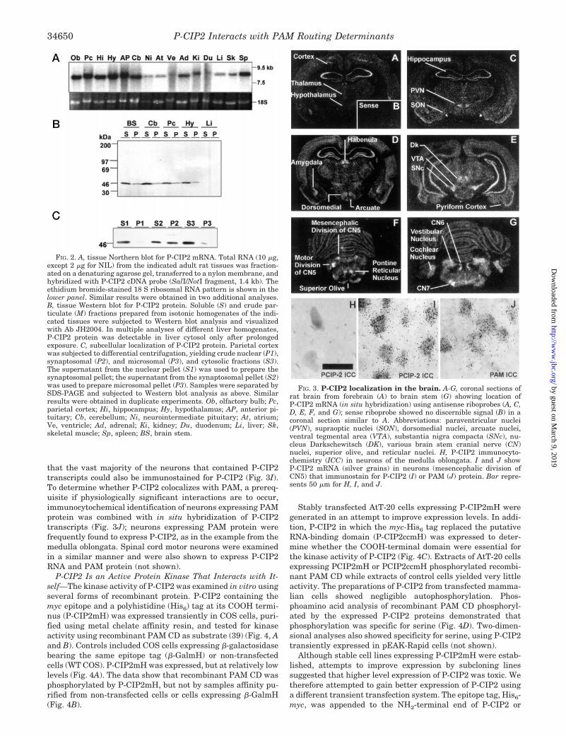
that the vast majority of the neurons that contained P-CIP2transcripts could also be immunostained for P-CIP2 (Fig. 3I).To determine whether P-CIP2 colocalizes with PAM, a prereq-uisite if physiologically significant interactions are to occur,immunocytochemical identification of neurons expressing PAMprotein was combined with in situ hybridization of P-CIP2transcripts (Fig. 3J); neurons expressing PAM protein werefrequently found to express P-CIP2, as in the example from themedulla oblongata. Spinal cord motor neurons were examinedin a similar manner and were also shown to express P-CIP2RNA and PAM protein (not shown).
P-CIP2 Is an Active Protein Kinase That Interacts with It-self—The kinase activity of P-CIP2 was examined in vitro usingseveral forms of recombinant protein. P-CIP2 containing themyc epitope and a polyhistidine (His6) tag at its COOH termi-nus (P-CIP2mH) was expressed transiently in COS cells, puri-fied using metal chelate affinity resin, and tested for kinaseactivity using recombinant PAM CD as substrate (39) (Fig. 4, Aand B). Controls included COS cells expressing b-galactosidasebearing the same epitope tag (b-GalmH) or non-transfectedcells (WT COS). P-CIP2mH was expressed, but at relatively lowlevels (Fig. 4A). The data show that recombinant PAM CD wasphosphorylated by P-CIP2mH, but not by samples affinity pu-rified from non-transfected cells or cells expressing b-GalmH(Fig. 4B).
Stably transfected AtT-20 cells expressing P-CIP2mH weregenerated in an attempt to improve expression levels. In addi-tion, P-CIP2 in which the myc-His6 tag replaced the putativeRNA-binding domain (P-CIP2ccmH) was expressed to deter-mine whether the COOH-terminal domain were essential forthe kinase activity of P-CIP2 (Fig. 4C). Extracts of AtT-20 cellsexpressing PCIP2mH or PCIP2ccmH phosphorylated recombi-nant PAM CD while extracts of control cells yielded very littleactivity. The preparations of P-CIP2 from transfected mamma-lian cells showed negligible autophosphorylation. Phos-phoamino acid analysis of recombinant PAM CD phosphoryl-ated by the expressed P-CIP2 proteins demonstrated thatphosphorylation was specific for serine (Fig. 4D). Two-dimen-sional analyses also showed specificity for serine, using P-CIP2transiently expressed in pEAK-Rapid cells (not shown).
Although stable cell lines expressing P-CIP2mH were estab-lished, attempts to improve expression by subcloning linessuggested that higher level expression of P-CIP2 was toxic. Wetherefore attempted to gain better expression of P-CIP2 usinga different transient transfection system. The epitope tag, His6-myc, was appended to the NH2-terminal end of P-CIP2 or
FIG. 2. A, tissue Northern blot for P-CIP2 mRNA. Total RNA (10 mg,except 2 mg for NIL) from the indicated adult rat tissues was fraction-ated on a denaturing agarose gel, transferred to a nylon membrane, andhybridized with P-CIP2 cDNA probe (SalI/NotI fragment, 1.4 kb). Theethidium bromide-stained 18 S ribosomal RNA pattern is shown in thelower panel. Similar results were obtained in two additional analyses.B, tissue Western blot for P-CIP2 protein. Soluble (S) and crude par-ticulate (M) fractions prepared from isotonic homogenates of the indi-cated tissues were subjected to Western blot analysis and visualizedwith Ab JH2004. In multiple analyses of different liver homogenates,P-CIP2 protein was detectable in liver cytosol only after prolongedexposure. C, subcellular localization of P-CIP2 protein. Parietal cortexwas subjected to differential centrifugation, yielding crude nuclear (P1),synaptosomal (P2), and microsomal (P3), and cytosolic fractions (S3).The supernatant from the nuclear pellet (S1) was used to prepare thesynaptosomal pellet; the supernatant from the synaptosomal pellet (S2)was used to prepare microsomal pellet (P3). Samples were separated bySDS-PAGE and subjected to Western blot analysis as above. Similarresults were obtained in duplicate experiments. Ob, olfactory bulb; Pc,parietal cortex; Hi, hippocampus; Hy, hypothalamus; AP, anterior pi-tuitary; Cb, cerebellum; Ni, neurointermediate pituitary; At, atrium;Ve, ventricle; Ad, adrenal; Ki, kidney; Du, duodenum; Li, liver; Sk,skeletal muscle; Sp, spleen; BS, brain stem.
FIG. 3. P-CIP2 localization in the brain. A-G, coronal sections ofrat brain from forebrain (A) to brain stem (G) showing location ofP-CIP2 mRNA (in situ hybridization) using antisense riboprobes (A, C,D, E, F, and G); sense riboprobe showed no discernible signal (B) in acoronal section similar to A. Abbreviations: paraventricular nuclei(PVN), supraoptic nuclei (SON), dorsomedial nuclei, arcuate nuclei,ventral tegmental area (VTA), substantia nigra compacta (SNc), nu-cleus Darkschewitsch (DK), various brain stem cranial nerve (CN)nuclei, superior olive, and reticular nuclei. H, P-CIP2 immunocyto-chemistry (ICC) in neurons of the medulla oblongata. I and J showP-CIP2 mRNA (silver grains) in neurons (mesencephalic division ofCN5) that immunostain for P-CIP2 (I) or PAM (J) protein. Bar repre-sents 50 mm for H, I, and J.
P-CIP2 Interacts with PAM Routing Determinants34650
by guest on March 9, 2019
http://ww
w.jbc.org/
Dow
nloaded from

P-CIP2cc bearing an ATP-binding site mutation (K54A) thatshould inactivate the kinase. Both proteins were expressed athigh levels in pEAK-Rapid hEK-293 cells. The transfectedHmP-CIP2 protein was present at a high level and much of itwas recovered in the isotonic sucrose pellet; in contrast, thesmall amount of endogenous P-CIP2 was recovered primarilyin the soluble fraction (Fig. 5A). Immunostainable HmP-CIP2expression was readily detected in over half of the transientlytransfected cells. Staining was observed in aggregates whichwere especially visible in cells which had rounded up andappeared to be detaching from the culture dish (not shown). Bycomparison, we could not reliably detect stable P-CIP2 expres-sion using the same reagents.
Both HmP-CIP2 and HmP-CIP2cc-K/A were purified fromsolubilized sucrose pellets in good yield using a metal-chelateresin (Fig. 5B). Our initial kinase assays unexpectedly indi-cated that HmP-CIP2cc-K/A had some activity (data notshown). Although the problem of spurious binding of kinase(s)to resins has been reported many times before (38), neithernontransfected cells nor GFP-transfected cells yielded back-ground kinase activity. An explanation for this observation issuggested by the fact that P-CIP2 interacted with itself in theyeast two-hybrid assay to produce yeast growth and b-galacto-sidase activity (Fig. 5C). Visualization of purified HmP-CIP2and HmP-CIP2cc-K/A using a P-CIP2 antibody instead of the
myc antibody identified a small amount of endogenous P-CIP2in the purified HmP-CIP2cc-K/A (Fig. 5B, asterisk). To elimi-nate the co-purification of endogenous P-CIP2, a differentbrand of metal chelate affinity resin was utilized (Fig. 5D). Abetter purification of HmP-CIP2 (Fig. 5D) was achieved, withno endogenous P-CIP2 detectable. Bacterially expressedP-CIP2 showed very low activity (,1% as much activity as foran equivalent Western blot signal from pEAK-Rapid cells) andwas not used in subsequent work.
P-CIP2 Phosphorylates the Region of the PAM Cytosolic Do-main Involved in Trafficking—The ability of purified mamma-lian recombinant HmP-CIP2 to phosphorylate recombinantPAM-CD and GST-PAM-CDt was compared (Fig. 6A). In GST-PAM-CDt, the PAM protein is truncated at residue 961, delet-ing the final 15 residues of the cytosolic domain; PAM proteinstruncated at this position are known to show normal traffickingin AtT-20 cells (21). P-CIP2 was found to phosphorylatePAM-CD and GST-PAM-CDt to a similar extent. The GSTcontrol was not phosphorylated by P-CIP2. The nontransfectedcells showed no activity in this assay (Fig. 6) and extracts ofpEAK-Rapid cells transfected with GFP vectors were inactive(not shown). Autophosphorylation of the recombinant HmP-CIP2 (at 47 kDa) was not detectable.
Truncation of PAM at residue 936 eliminates all cytosolictrafficking determinants (21), and P-CIP2 was unable to phos-phorylate the corresponding GST fusion protein (GST-CD-936s) (Fig. 6B). Mutation of Lys919 to Arg was shown to elim-
FIG. 4. P-CIP2 phosphorylates PAM-CD. A, confluent 100-mmplates of COS cells were transfected transiently with vector encodingPCIP2mH or similarly tagged b-galactosidase, b-GalmH, or not trans-fected (WT). Cells were extracted and the tagged proteins purified bymetal affinity chromatography. Bound proteins were eluted and sub-jected to Western blot analysis for the Myc epitope. Arrows indicateexpressed epitope tagged protein. B, samples from panel A were dilutedinto kinase buffer containing 10 mg of purified recombinant PAM CD(rCD), and incubated at 37 °C for 1 h. Kinase reaction products wereseparated on a 15% SDS-PAGE gel, and visualized by autoradiography.C, AtT-20 cells were stably transfected with vector encoding PCIP2mHor P-CIP2ccmH. Expressed P-CIP2 proteins were purified by metalaffinity chromatography. Purified imidazole eluted protein was dilutedinto kinase buffer with 10 mg of purified recombinant PAM CD, incu-bated at 37 °C for 1 h, separated by SDS-PAGE, and visualized byautoradiography. D, 32P-labeled recombinant CD bands from panel Cwere excised from the SDS-PAGE gel and subjected to acid hydrolysisfollowed by thin layer electrophoresis with the indicated phosphoaminoacid standards (39, 57). Standards were visualized with ninhydrinspray, and samples were visualized by autoradiography.
FIG. 5. Purification of P-CIP2. A, a control protein (green fluores-cent protein (GFP)) and HmP-CIP2 were transiently expressed inpEAK-Rapid hEK-293 cells, homogenized in isotonic sucrose, and thesupernatant (S) and pellet (P) were subjected to Western analysis usingthe GST-P-CIP2 fusion protein antibody (JH1998); the endogenousP-CIP2-reactive band is indicated (*). B, pEAK-Rapid cells were tran-siently transfected with pEAK10 vectors encoding HmP-CIP2 (full) orHmP-CIP2cc-K/A (cc-K/A) and the isotonic sucrose pellet was extractedand purified using the Talon metal-chelate protocol. Samples eluted inimidazole were desalted and subjected to Western analysis using themyc monoclonal antibody or the P-CIP2 antibody (JH1998). The smallamount of endogenous P-CIP2 that copurified with HmP-CIP2cc-K/A isindicated by an asterisk (*). C, a liquid phase b-galactosidase assay wasused to quantify the interaction of DN-P-CIP2 with itself in the yeasttwo-hybrid system (24); DN-P-CIP-2 was expressed as bait or as bothbait and prey. D, samples were prepared as in B, except that theNi-NTA resin was used and non-transfected cells were analyzed as acontrol; cells transfected with GFP or b-galactosidase vector showed thesame pattern as for the NT cells (not shown). Equal portions of all thesamples were analyzed; much of the input HmP-CIP2 was bound to andeluted from the Ni-NTA resin, while less was found in the resin flow-through (FT) and washes before elution, and very little remained on theresin after elution. Similar results were obtained in 4 additional exper-iments of this type.
P-CIP2 Interacts with PAM Routing Determinants 34651
by guest on March 9, 2019
http://ww
w.jbc.org/
Dow
nloaded from

inate the ability of DNP-CIP2 to interact with PAM-CD in theyeast two-hybrid system.2 Nevertheless, P-CIP2 phosphoryl-ated the K919R version of CDt. Since Kalirin, another PAM-CDinteractor, binds Rac1, the ability of P-CIP2 to phosphorylatemembers of the Rho subfamily of small GTP-binding proteinswas determined (Fig. 6). GST fusion proteins consisting ofRac1, RhoA, and Cdc42 were not detectably phosphorylated byP-CIP2; these GST fusion proteins all functioned as substratesfor protein kinase A, protein kinase C, or casein kinase II (notshown). Thus P-CIP2 exhibits specificity for the region of thePAM cytosolic domain involved in trafficking.
P-CIP2 Shows a Strong Preference for Ser949 in the PAM-CD—The substrate specificity of P-CIP2 was tested using GSTfusion proteins consisting of the cytosolic domains of two othermembrane proteins localized to the TGN region (Fig. 7). LikePAM, the endoprotease furin and the exoprotease carboxypep-tidase D are transmembrane proteins with their catalytic re-gions in the lumen of the TGN, and their carboxyl-terminalregions extending across the membrane and out into the cytosol(8, 49). P-CIP2 did not efficiently phosphorylate the cytosolicdomain of either furin or carboxypeptidase D (Fig. 7A). Incontrast, the furin and carboxypeptidase D cytosolic domainswere substrates for protein kinase A and/or casein kinase II(Fig. 7B) and protein kinase C (not shown).
P-CIP2 was also isolated independently as an interactorprotein for P19/stathmin, a small cytosolic phosphoprotein in-volved in microtubule destabilization (26). SCG10 is a neuronalhomolog of stathmin with similar microtubule destabilizingactivity (27). We used GST fusion proteins to evaluate theability of P-CIP2 to phosphorylate P19 and SCG10. Purifiedmammalian recombinant P-CIP2 did not phosphorylate SCG10or P19 detectably (Fig. 7A); in contrast, both GST fusion pro-teins were good substrates for protein kinase A (Fig. 7B). Bac-terially expressed GST-P-CIP2 did show some labeling of P19and SCG10, but only at levels of P-CIP2 protein at least 100-fold higher than the levels used in these assays (data notshown). Other substrates tested included the Exon A region of
PAM, casein and histone; none was significantly phosphoryl-ated by P-CIP2 (not shown). Thus, P-CIP2 is highly selectivefor the cytosolic domain of PAM.
The protein kinase A, protein kinase C, and casein kinase IIsites in recombinant PAM CD were identified previously (Fig.8) (39). GST fusion proteins in which each identified Ser resi-due was individually mutated to Asp were evaluated as sub-strates for P-CIP2 (Fig. 7C). Elimination of the protein kinaseC sites (Ser932 and Ser937) or the protein kinase A site (Ser921)did not affect the ability of P-CIP2 to phosphorylate CD. Incontrast, mutation of the sites phosphorylated by sea star ca-sein kinase II (Thr946 and Ser949) greatly reduced the ability ofP-CIP2 to phosphorylate CD. The S937A, S932A, and S921Amutants were also good substrates (data not shown). Since itwas established that P-CIP2 primarily phosphorylates Ser res-idues (Fig. 4D), these data implicate Ser949 as the primary targetof P-CIP2 phosphorylation. Importantly, the T946D,S949Dmutant of PAM (mimicking permanent phosphorylation of theThr946 and Ser949 residues) was not trafficked normally inAtT-20 cells (50).
To further evaluate the specificity of P-CIP2, synthetic pep-tides containing various potential phosphorylation sites weretested as substrates (Fig. 8). P-CIP2 phosphorylated PAM(942–953). In contrast, the same synthetic peptide containing phos-pho-Thr946 and phospho-Ser949 was not phosphorylated by P-CIP2. PAM(945–961), a peptide in which Ser949 is closer to theNH2 terminus, was not phosphorylated by P-CIP2. The otherPAM peptides tested did not serve as substrates for P-CIP2,nor did peptides known to serve as substrates for casein kinaseI, protein kinase A, protein kinase C, or calmodulin kinase II(Fig. 8). Although Thr946 and Ser949 are phosphorylation sitesfor sea star casein kinase II, they are not the preferred sites forhuman recombinant casein kinase II. PAM(942–953)/phospho-Thr946/phospho-Ser949, which contains only one potential ac-ceptor residue at Ser945, was efficiently phosphorylated by hu-man recombinant casein kinase II. Loss of phosphorylation bymutagenesis of the putative acceptor site provides strong evi-2 R. C. Johnson, R. E. Mains, and B. A. Eipper, unpublished results.
FIG. 6. P-CIP2 selectively phosphorylates GST-PAM-CD fusionproteins. Samples from pEAK-Rapid cells expressing HmP-CIP2 (up-per) or not transfected (NT; middle) were analyzed for kinase activity.The proteins on the NT gel were visualized with Coomassie BrilliantBlue (Stain; lower). A, phosphorylation of purified PAM-CD, GST con-trol, and GST-PAM-CD was tested, using recombinant proteins. B,phosphorylation of truncated PAM-CD-936s, mutant PAM-CD-K919R,and the three GST Rho-family fusion proteins was also tested. Similarresults were obtained in five additional experiments of this type.
FIG. 7. P-CIP2 is highly selective for PAM-CD. A, kinase assayswere carried out as described in the legend to Fig. 6 using purifiedHmP-CIP2 and the indicated GST fusion proteins. Expression of GST-fusion proteins was compared by staining the membrane with Coomas-sie. B, the ability of bovine heart protein kinase A (PKA) and humanrecombinant casein kinase II a-subunit (CKII) to phosphorylate thesame GST fusion proteins was compared. C, HmP-CIP2 was used tophosphorylate the indicated GST fusion proteins; the film was deliber-ately overexposed to determine the level of label in the TS/DD (Thr946-Asp/Ser949-Asp) mutant. Similar results were obtained in five addi-tional experiments of this type.
P-CIP2 Interacts with PAM Routing Determinants34652
by guest on March 9, 2019
http://ww
w.jbc.org/
Dow
nloaded from
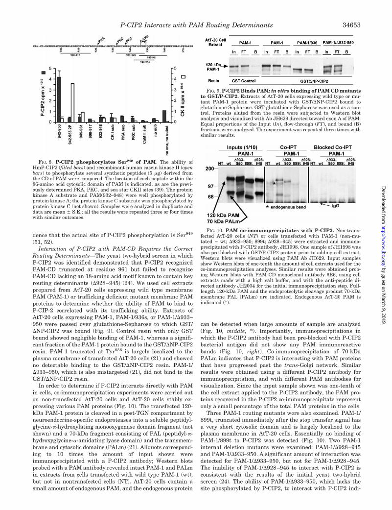
dence that the actual site of P-CIP2 phosphorylation is Ser949
(51, 52).Interaction of P-CIP2 with PAM-CD Requires the Correct
Routing Determinants—The yeast two-hybrid screen in whichP-CIP2 was identified demonstrated that P-CIP2 recognizedPAM-CD truncated at residue 961 but failed to recognizePAM-CD lacking an 18-amino acid motif known to contain keyrouting determinants (D928–945) (24). We used cell extractsprepared from AtT-20 cells expressing wild type membranePAM (PAM-1) or trafficking deficient mutant membrane PAMproteins to determine whether the ability of PAM to bind toP-CIP-2 correlated with its trafficking ability. Extracts ofAtT-20 cells expressing PAM-1, PAM-1/936s, or PAM-1/D933–950 were passed over glutathione-Sepharose to which GST/DNP-CIP2 was bound (Fig. 9). Control resin with only GSTbound showed negligible binding of PAM-1, whereas a signifi-cant fraction of the PAM-1 protein bound to the GST/DNP-CIP2resin. PAM-1 truncated at Tyr936 is largely localized to theplasma membrane of transfected AtT-20 cells (21) and showedno detectable binding to the GST/DNP-CIP2 resin. PAM-1/D933–950, which is also mistargeted (21), did not bind to theGST/DNP-CIP2 resin.
In order to determine if P-CIP2 interacts directly with PAMin cells, co-immunoprecipitation experiments were carried outon non-transfected AtT-20 cells and AtT-20 cells stably ex-pressing various PAM proteins (Fig. 10). The transfected 120-kDa PAM-1 protein is cleaved in a post-TGN compartment byneuroendocrine-specific endoproteases into a soluble peptidyl-glycine-a-hydroxylating monooxygenase domain fragment (notshown) and a 70-kDa fragment consisting of PAL (peptidyl-a-hydroxyglycine-a-amidating lyase domain) and the transmem-brane and cytosolic domains (PALm) (21). Aliquots correspond-ing to 10 times the amount of input shown wereimmunoprecipitated with a P-CIP2 antibody; Western blotsprobed with a PAM antibody revealed intact PAM-1 and PALmin extracts from cells transfected with wild type PAM-1 (wt),but not in nontransfected cells (NT). AtT-20 cells contain asmall amount of endogenous PAM, and the endogenous protein
can be detected when large amounts of sample are analyzed(Fig. 10, middle, *). Importantly, immunoprecipitations inwhich the P-CIP2 antibody had been pre-blocked with P-CIP2bacterial antigen did not show any PAM immunoreactivebands (Fig. 10, right). Co-immunoprecipitation of 70-kDaPALm indicates that P-CIP2 is interacting with PAM proteinsthat have progressed past the trans-Golgi network. Similarresults were obtained using a different P-CIP2 antibody forimmunoprecipitation, and with different PAM antibodies forvisualization. Since the input sample shown was one-tenth ofthe cell extract applied to the P-CIP2 antibody, the PAM pro-teins recovered in the P-CIP2 co-immunoprecipitate representonly a small percentage of the total PAM proteins in the cells.
Three PAM-1 routing mutants were also examined. PAM-1/899t, truncated immediately after the stop transfer signal hasa very short cytosolic domain and is largely localized to theplasma membrane in AtT-20 cells. Essentially no binding ofPAM-1/899t to P-CIP2 was detected (Fig. 10). Two PAM-1internal deletion mutants were examined: PAM-1/D928–945and PAM-1/D933–950. A significant amount of interaction wasdetected for PAM-1/D933–950, but not for PAM-1/D928–945.The inability of PAM-1/D928–945 to interact with P-CIP2 isconsistent with the results of the initial yeast two-hybridscreen (24). The ability of PAM-1/D933–950, which lacks thesite phosphorylated by P-CIP2, to interact with P-CIP2 indi-
FIG. 8. P-CIP2 phosphorylates Ser949 of PAM. The ability ofHmP-CIP2 (filled bars) and recombinant human casein kinase II (openbars) to phosphorylate several synthetic peptides (5 mg) derived fromthe CD of PAM were compared. The location of each peptide within the86-amino acid cytosolic domain of PAM is indicated, as are the previ-ously determined PKA, PKC, and sea star CKII sites (39). The proteinkinase A substrate and PAM(932–948) were well phosphorylated byprotein kinase A; the protein kinase C substrate was phosphorylated byprotein kinase C (not shown). Samples were analyzed in duplicate anddata are mean 6 S.E.; all the results were repeated three or four timeswith similar outcomes.
FIG. 9. P-CIP2 Binds PAM: in vitro binding of PAM CD mutantsto GST/P-CIP2. Extracts of AtT-20 cells expressing wild type or mu-tant PAM-1 protein were incubated with GST/DNP-CIP2 bound toglutathione-Sepharose. GST-glutathione-Sepharose was used as a con-trol. Proteins eluted from the resin were subjected to Western blotanalysis and visualized with Ab JH629 directed toward exon A of PAM.Equal proportions of the Input (In), flow-through (FT), and bound (B)fractions were analyzed. The experiment was repeated three times withsimilar results.
FIG. 10. PAM co-immunoprecipitates with P-CIP2. Non-trans-fected AtT-20 cells (NT) or cells transfected with PAM-1 (non-mu-tated 5 wt; D933–950; 899t; D928–945) were extracted and immuno-precipitated with P-CIP2 antibody, JH1998. One sample of JH1998 wasalso pre-blocked with GST/P-CIP2 protein prior to adding cell extract.Western blots were visualized using PAM Ab JH629. Input samplesshow Western blots of one-tenth the amount of cell extracts used for theco-immunoprecipitation analyses. Similar results were obtained prob-ing Western blots with PAM CD monoclonal antibody 6E6, using cellextracts made with a high salt buffer, and with the anti-peptide di-rected antibody JH2004 for the initial immunoprecipitation step. Full-length 120-kDa PAM and the endoproteolytic cleavage product 70-kDamembrane PAL (PALm) are indicated. Endogenous AtT-20 PAM isindicated (*).
P-CIP2 Interacts with PAM Routing Determinants 34653
by guest on March 9, 2019
http://ww
w.jbc.org/
Dow
nloaded from

cates that some determinants governing interaction are dis-tinct from those governing phosphorylation and proximal toresidue 933.
DISCUSSION
Proper targeting of proteins within the secretory pathway isessential for normal function. Complex arrays of overlappingand often redundant sorting signals have been identified in thecytosolic domains of many membrane proteins. While the im-portance of Tyr motifs, di-Leu motifs, and phosphorylationsites has been established for a number of membrane proteins(1–3, 8, 13, 21), we still know relatively little about the proteinsthat interact with these motifs. The COOH-terminal cytosolicdomain of membrane PAM contains targeting signals that in-clude a tyrosine-based internalization motif and phosphoryla-tion sites. In searching for cytosolic proteins that could interactwith membrane PAM, we anticipated finding coat proteins,cytoskeletal proteins, kinases, phosphatases, motor proteins,and/or regulatory factors. The first P-CIPs identified using ayeast two-hybrid screen included a protein kinase (P-CIP2), aGDP/GTP exchange factor for Rac1 (Kalirin) (24, 25), and anovel protein involved in endocytosis (P-CIP1) (53).
Phosphorylation is important in the trafficking of a widevariety of membrane proteins. For example, several steps inthe trafficking of furin (2, 8, 11, 54), intracellular cycling of themannose 6-phosphate receptor (2), transcytosis of the poly-meric immunoglobulin receptor (55), retention of TGN38 in thetrans-Golgi network (56), differences among NMDA subunits(57), and internalization of epidermal growth factor (58) recep-tors all involve phosphorylation. The cytosolic domain of PAMis phosphorylated at Ser937 in vivo, in addition to phosphoryl-ation at other Ser residues and Thr (39). The cycle of phospho-rylation and dephosphorylation at Ser937 is essential for nor-mal trafficking, and mutagenesis of Ser937 to Ala or Aspdemonstrates a role for phosphorylation in avoiding delivery ofPAM to lysosomes after internalization (39, 59). In addition,the trafficking of both newly synthesized PAM and PAMinternalized from the plasma membrane were altered whenThr946 and Ser949 were mutated to Ala or Asp (50). The role ofprotein kinases in regulating cytoskeletal function is also wellestablished (43).
We show here that P-CIP2, a 46.5-kDa cytosolic proteinkinase, phosphorylates recombinant PAM CD highly selec-tively in vitro. This specificity, along with the demonstratedco-expression of P-CIP2 and PAM throughout the nervous sys-tem, suggests that P-CIP2 contributes to the phosphorylationand targeting of membrane PAM in situ. Phosphorylation ofPAM CD by P-CIP2 is limited to Ser residues. Mutation ofpotential phosphorylation sites within the CD of PAM andphosphorylation of synthetic peptides identified Ser949 as themajor site phosphorylated by P-CIP2 (Figs. 6–8). The CD ofPAM is phosphorylated in vitro by protein kinase C (at Ser937
and Ser932) as well as by protein kinase A (at Ser921), and atThr946 and Ser949 by sea star casein kinase II (39). Unlike seastar casein kinase II, P-CIP2 does not phosphorylate Thr res-idues in PAM-CD. Interestingly, recombinant human caseinkinase II does not phosphorylate Ser949. Despite the presenceof a potential CKII site closer to the COOH terminus of PAM(Ser961), P-CIP2 is unable to phosphorylate a synthetic peptidethat includes this site. P-CIP2 also fails to phosphorylate apeptide that includes a trio of Ser residues near the transmem-brane domain (residues 907–909).
A kinase identical to P-CIP2 was identified by Maucuer et al.(26, 60) when using a yeast two-hybrid system to screen forproteins that could interact with stathmin. A number of extra-cellular signals associated with cell proliferation and differen-tiation regulate the phosphorylation of stathmin, a cytosolic
protein implicated in cell cycle control (61). The ability of stath-min to destabilize microtubules is regulated by its phosphoryl-ation state (62, 63). We confirmed the reported phosphorylationof stathmin by recombinant P-CIP2/KIS from bacterial sources(60) and tested the ability of recombinant P-CIP2 from mam-malian cells to phosphorylate stathmin and SCG10, a mem-brane-associated neuronal stathmin homologue (27). SCG10and stathmin were not significantly phosphorylated by recom-binant P-CIP2 purified from mammalian sources, althoughboth were good substrates for other kinases (Fig. 7). We foundthat nanogram amounts of purified P-CIP2 from mammaliansources were sufficient for readily detectable kinase activity(Figs. 4–7), while microgram amounts of P-CIP2 from bacterialsources were required, raising the worry that co-purifying mi-nor contaminants were responsible for some of the apparentactivity from the bacterial sources (38, 60).
The specificity of P-CIP2 for Ser949 of PAM is quite remark-able. P-CIP2 did not phosphorylate significantly the CDs ofcarboxypeptidase D or furin, integral membrane proteinswhose steady state localization and itineraries resemble thoseof PAM (5, 8, 11, 49, 54). The phosphorylation sites controllingfurin trafficking are typical of the acidic sites recognized bycasein kinase II (8, 54). P-CIP2 did not phosphorylate the smallGTP-binding proteins Rac1, RhoA, or Cdc42, at least in thenucleotide-free form; these were tested because Rac1 interactswith Kalirin, another P-CIP. P-CIP2 did not phosphorylate anyof the standard small peptide substrates used for protein ki-nase A, protein kinase C, or casein kinases (Fig. 8), histone,casein, or recombinant exon A. The striking specificity of P-CIP2 is undoubtedly key to its function (50). While some pro-tein kinases have a rather wide substrate specificity, others(e.g. myosin light chain kinase) have highly restricted sub-strate specificity (64). Factors regulating the activity of P-CIP2have not yet been identified. Homomeric interactions of P-CIP2, interactions of P-CIP2 with Kalirin, stathmin, or RNA aswell as phosphorylation/dephosphorylation might regulate thekinase activity of P-CIP2.
The protein kinases most closely related to P-CIP2 (Fig. 1)are involved in a number of intracellular signaling cascades,suggesting a signaling role for P-CIP2. Kalirin, one of the otherP-CIPs identified in the initial yeast two-hybrid screen, occursin a form that includes a putative protein kinase domain ho-mologous to the COOH-terminal region of human Trio2 (65).Interestingly, the kinase domain of rat P-CIP2 exhibits 20%identity and 28% similarity to the kinase domain of humanTrio. The kinase domain of Trio has not yet been shown tophosphorylate any substrates (66, 67). The homology of ratPCIP2 to rat casein kinase II (18% identity and 33% similarity)is interesting in light of the ability of P-CIP2 to phosphorylatea Ser residue in an acidic motif (Fig. 8).
The putative RNA-binding domain of P-CIP2 is not essentialto the expression of kinase activity toward the PAM-CD sub-strate. Only one other kinase, PKR, contains an RNA-bindingdomain (44). PKR is involved with the interferon antiviralresponse pathway and appears to be involved in cell growthand differentiation. PKR contains two RNA-binding domainsand specifically binds double-stranded RNA; binding of double-stranded RNA to PKR is required for kinase activity (44). Thefunction of the putative RNA-binding domain of P-CIP2 is stillunclear. Several possibilities exist. P-CIP2 may bind RNA andlocalize it within the cell. P-CIP2 may act to stabilize boundRNA. Alternatively, RNA may act to modulate the kinase ac-tivity of P-CIP2, as for PKR (44). Presently, it is not knownwhether P-CIP2 binds a specific RNA sequence or any RNA atall. However, it has recently been reported that a specific20-nucleotide sequence in the 39-untranslated region of PAM
P-CIP2 Interacts with PAM Routing Determinants34654
by guest on March 9, 2019
http://ww
w.jbc.org/
Dow
nloaded from

mRNA forms a complex with a 46-kDa cytosolic protein (68),and several other short nucleotide stretches in 39-untranslatedregions are known to have intracellular routing information(69, 70).
Identification of PAM as a substrate for P-CIP2 was possibleonly because PAM and P-CIP2 interacted in a yeast two-hybridscreen. Whether P-CIP2 has additional substrates is not yetclear. The determinants governing the interaction of P-CIP2with the CD of PAM are not limited to the region surroundingSer949, the site phosphorylated by P-CIP2, and involve regionsof the CD that are closer to the transmembrane domain. Thus,P-CIP2 interacts with PAM-CD bearing Ala or Asp at position949 and fails to interact with PAM-CD bearing mutations atLys919. PAM-1/D933–950, which lacks the site phosphorylatedby P-CIP2 can still interact with P-CIP2. As expected for aninteraction that may be involved in regulating a dynamic proc-ess such as the targeting of membrane proteins, the interactionof PAM with P-CIP2 may be transient; only a small fraction ofthe PAM protein in cells can be co-immunoprecipitated withP-CIP2 (Fig. 10). This is consistent with that fact that most ofthe P-CIP2 protein is cytosolic, with a small fraction associatedwith membranes (Fig. 2) (24). The fact that 70-kDa PAL ispresent in the co-immunoprecipitate along with full-length120-kDa PAM indicates that P-CIP2 can interact with PAMafter it has entered the immature secretory granules and un-dergone endoproteolytic cleavage. Recruitment of P-CIP2 tomembranes may involve PAM-CD as well as other soluble andmembrane proteins.
In conclusion, P-CIP2 is a highly selective cytosolic proteinkinase that interacts with membrane PAM in intact cells andphosphorylates the CD of PAM in vitro at Ser949. P-CIP2 andPAM clearly have an opportunity to interact, and the region ofPAM required for the interaction with P-CIP2 is critical to itsrouting. Future studies will examine the specific determinantswithin the PAM-CD which mediate P-CIP2 interaction andfactors regulating P-CIP2 localization and kinase activity.
Acknowledgments—We thank Gary Thomas (Vollum Institute, Uni-versity of Oregon) for providing GST/furin-CD plasmid, Lloyd Fricker(Albert Einstein College of Medicine) for providing GST-carboxypepti-dase D plasmid, David Anderson (California Technical Institute) forsupplying SCG10 plasmid, Ulrich Schubart (Albert Einstein) for theP19/stathmin plasmid, Richard Cerione (Cornell University) for theGST-Rac1/RhoA/Cdc42 plasmids, Henry Keutmann (MassachusettsGeneral Hospital) for synthetic peptides, and David Ginty (Johns Hop-kins University School of Medicine) for comments and suggestions onexperiments and the manuscript. We thank Marie Bell and CathyCaldwell for general laboratory assistance, Lixian Jin for tissue culture,and Greg Galano for assistance in working out the recombinant P-CIP2purification.
REFERENCES
1. Johnson, K. F., and Kornfeld, S. (1992) J. Biol. Chem. 267, 17110–171152. Johnson, K. F., and Kornfeld, S. (1992) J. Cell Sci. 119, 249–2573. Humphrey, J. S., Peters, P. J., Yuan, L. C., and Bonifacino, J. S. (1993) J. Cell
Biol. 120, 1123–11354. Wong, S. H., and Hong, W. (1993) J. Biol. Chem. 268, 22853–228625. Molloy, S. S., Thomas, L., Van Slyke, J. K., and Thomas, G. (1994) EMBO J.
13, 18–336. Sandoval, I. V., and Bakke, O. (1994) Trends Cell Biol. 4, 292–2977. Shum, L., Reeves, S. A., Kuo, A. C., Fromer, E. S., and Derynck, R. (1994)
J. Cell Biol. 125, 903–9168. Jones, B. G., Thomas, L., Molloy, S. S., Thulin, C. D., Fry, M. D., Walsh, K. A.,
and Thomas, G. (1995) EMBO J. 14, 5869–58839. Schafer, W., Stroh, A., Berghofer, J., Seiler, J., Vey, M., Kruse, M. L., Kern,
H. F., Klenk, H. D., and Garten, W. (1995) EMBO J. 14, 2424–243510. Rohrer, J., Schweizer, A., Johnson, K. F., and Kornfeld, S. (1995) J. Cell Biol.
130, 1297–130611. Takahashi, S., Nakagawa, T., Banno, T., Watanabe, T., Murakami, K., and
Nakayama, K. (1995) J. Biol. Chem. 270, 28397–2840112. Ohno, H., Stewart, J., Fouriier, M. C., Bosshart, H., Rhee, I., Miyatake, S.,
Saito, T., Galluser, A., Kirchenhausen, T., and Bonifacino, J. S. (1995)Science 169, 1872–1875
13. Honing, S., Griffith, J., Geuze, H. J., and Hunziker, W. (1996) EMBO J. 15,5230–5239
14. Rapoport, I., Miyazaki, M., Boll, W., Duckworth, B., Cantley, L. C., Shoelson,S., and Kirchenhausen, T. (1997) EMBO J. 16, 2240–2250
15. Stephens, D. G., Crump, C. M., Clarke, A. R., and Banting, G. (1997) J. Biol.Chem. 272, 14104–14109
16. LeBorgne, R., Schmidt, A., Mauxion, F., Griffiths, G., and Hoflack, B. (1993)J. Biol. Chem. 268, 22552–22556
17. Mauxion, F., Borgne, R. L., Munier-Lehmann, H., and Hoflack, B. (1996)J. Biol. Chem. 271, 2171–2178
18. Chen, H. J., Yuan, J., and Lobel, P. (1997) J. Biol. Chem. 272, 7003–701219. Mains, R. E., Dickerson, I. M., May, V., Stouffers, D. A., Perkins, S. N., Ouafik,
L., Husten, E. J., and Eipper, B. A. (1990) Front. Neuroendocrinol. 11,52–89
20. Zhou, A., Webb, G., Zhu, X., and Steiner, D. F. (1999) J. Biol. Chem. 274,20745–20748
21. Milgram, S. L., Mains, R. E., and Eipper, B. A. (1996) J. Biol. Chem. 271,17526–17535
22. Milgram, S. L., Mains, R. E., and Eipper, B. A. (1993) J. Cell Biol. 121, 23–3623. Milgram, S. L., Chang, E. Y., and Mains, R. E. (1996) Mol. Endocrinol. 10,
837–84624. Alam, M. R., Caldwell, B. D., Johnson, R. C., Darlington, D. N., Mains, R. E.,
and Eipper, B. A. (1996) J. Biol. Chem. 271, 28636–2834025. Alam, M. R., Johnson, R. C., Darlington, D. N., Hand, T. A., and Eipper, B. A.
(1997) J. Biol. Chem. 272, 12667–1267526. Maucuer, A., Camonis, J. H., and Sobel, A. (1995) Proc. Natl. Acad. Sci.
U. S. A. 92, 3100–310427. DiPaolo, G., Lutjens, R., Pellier, V., Stimpson, S. A., Beuchat, M.-H., Catsicas,
S., and Grenningloh, G. (1997) J. Biol. Chem. 272, 5175–518228. Kozak, M. (1991) J. Cell Biol. 115, 887–90329. Milgram, S. L., Johnson, R. C., and Mains, R. E. (1992) J. Cell Biol. 117,
717–72830. Bloomquist, B. T., Eipper, B. A., and Mains, R. E. (1991) Mol. Endocrinol. 5,
2014–202331. Darlington, D. N., Schiller, M. R., Mains, R. E., and Eipper, B. A. (1997)
J. Histochem. Cytochem. 45, 1265–127732. Huttner, W. B., Schiebler, W., Greengard, P., and De Camilli, P. (1983) J. Cell
Biol. 96, 1373–138833. Bennett, M. K., Calakos, N., Kreiner, T., and Scheller, R. H. (1992) J. Cell Biol.
116, 761–77534. Ratovitski, E. A., Alam, M. R., Quick, R. A., McMillan, A., Bao, C., Hand, T. A.,
Johnson, R. C., Mains, R. E., Eipper, B. A., and Lowenstein, C. J. (1999)J. Biol. Chem. 274, 993–999
35. Milgram, S. L., Kho, S. T., Martin, G. V., Mains, R. E., and Eipper, B. A. (1997)J. Cell Sci. 110, 695–706
36. Borjigin, J., and Nathans, J. (1994) J. Biol. Chem. 169, 14715–1472237. Zhou, A., Bloomquist, B. T., and Mains, R. E. (1993) J. Biol. Chem. 268,
1763–176938. Ferrari, S., and Thomas, G. (1998) in Protein Phosphorylation (Sefton, B. M.,
and Hunter, T., eds) pp. 371–381, Academic Press, Boston39. Yun, H. Y., Milgram, S. L., Keutmann, H., and Eipper, B. A. (1995) J. Biol.
Chem. 270, 30075–3008340. Hanks, S. K., and Hunter, T. (1998) in Protein Phosphorylation (Sefton, B. M.,
and Hunter, T., eds) pp. 36–63, Academic Press, Boston41. Liu, H., Styles, C. A., and Fink, G. R. (1993) Science 262, 1741–174442. Hunter, T., and Plowman, G. D. (1997) Trends Biochem. Sci. 22, 18–2243. Kyriakis, J. M., and Avruch, J. (1994) in Protein Kinases (Woodgett, J. R., ed)
pp. 85–148, IRL Press, New York44. Proud, C. G. (1995) Trends Biochem. Sci. 20, 241–24645. Eipper, B. A., Stoffers, D. A., and Mains, R. E. (1992) Annu. Rev. Neurosci. 15,
57–8546. Milner, R. J., and Sutcliffe, J. G. (1983) Nucleic Acids Res. 11, 5497–552047. Sutcliffe, J. G., Milner, R. J., Gottesfeld, J. M., and Reynolds, W. (1984) Science
225, 1308–131548. Daly, C., and Ziff, E. B. (1997) J. Neurosci. 17, 2365–237549. Xin, X., Varlamov, O., Day, R., Dong, W., Bridgett, M. M., Leiter, E. H., and
Fricker, L. D. (1997) DNA Cell Biol. 16, 897–90950. Steveson, T., Zhao, G. C., Mains, R. E., and Eipper, B. A. (1998) Mol. Biol. Cell
9, 327a51. Hunter, T. (1998) in Protein Phosphorylation (Sefton, B. M., and Hunter, T.,
eds) pp. 3–37, Academic Press, Boston52. Boyle, W. J., Geer, P. V. D., and Hunter, T. (1998) in Protein Phosphorylation
(Sefton, B. M., and Hunter, T., eds) pp. 201–240, Academic Press, Boston53. Chen, L., Johnson, R. C., and Milgram, S. L. (1998) J. Biol. Chem. 273,
33524–3353254. Wan, L., Molloy, S. S., Thomas, L., Liu, G., Xiang, S. L., Rybak, S. L., and
Thomas, G. (1998) Cell 94, 205–21655. Hirt, R. P., Hughes, G. J., Frutiger, S., Michetti, P., and Perregaux, C. (1998)
Cell 74, 245–25556. Roquemore, E. P., and Banting, G. (1998) Mol. Biol. Cell 9, 2125–214457. Lau, L. F., and Huganir, R. L. (1995) J. Biol. Chem. 270, 20036–2004158. Chen, W. S., Lazar, C. S., Poenie, M., Tsien, R. Y., Gill, G. N., and Rosenfield,
M. G. (1996) Nature 328, 820–82359. Steveson, T. C., Keutmann, H. T., Mains, R. E., and Eipper, B. A. (1999)
J. Biol. Chem. 274, 21128–2113860. Maucuer, A., Ozon, S., Monceau, V., Gavet, O., Lawler, S., Curmi, P., and
Sobel, A. (1997) J. Biol. Chem. 272, 23151–2315661. Marklund, U., Osterman, O., Melander, H., Bergh, A., and Gullberg, M. (1994)
J. Biol. Chem. 269, 30626–3063562. Belmont, L. D., and Mitchison, T. J. (1996) Cell 84, 623–63163. Horowitz, S. B., Shen, H.-J., He, L., Ditmar, P., Neef, R., Chen, J., and
Schubart, U. K. (1997) J. Biol. Chem. 272, 8129–813264. Kemp, B. E., Faux, M. C., Means, A. R., House, C., Tiganis, T., Hu, S. H., and
Mitchelhill, K. I. (1994) in Protein Kinases (Woodgett, J. R., ed) pp. 30–67,
P-CIP2 Interacts with PAM Routing Determinants 34655
by guest on March 9, 2019
http://ww
w.jbc.org/
Dow
nloaded from

IRL Press, New York65. Debant, A., Serra-Pages, C., Seipel, K., O’Brien, S., Tang, M., Park, S.-H., and
Streuli, M. (1996) Proc. Natl. Acad. Sci. U. S. A. 93, 5466–547166. Stevens, R., Kubieski, T. J., Zheng, H., Kulkarni, S., Mancillas, J., Morales,
A. R., Hogue, C. W. V., Pawson, T., and Culotti, T. (1998) Cell 92, 785–79567. Seipel, K., Medley, Q. G., Kerdersha, N. L., Zhang, X. A., O’Brien, S. P.,
Serra-Pages, C., Hemler, M. E., and Streuli, M. (1999) J. Cell Sci. 112,1825–1834
68. Fraboulet, S., Boudouresque, F., Delfino, C., and Ouafik, L. H. (1998) Endo-
crinology 139, 894–90469. Mohr, E., Morris, J. F., and Richter, D. (1995) Proc. Natl. Acad. Sci. U. S. A.
92, 4377–738170. Ainger, K., Avossa, D., Diana, S., Barry, C., Barbarese, E., and Carson, J. H.
(1997) J. Cell Biol. 138, 1077–108771. Smith, R. F., and Smith, T. F. (1990) Proc. Natl. Acad. Sci. U. S. A. 87,
118–12272. Smith, R. F., and Smith, T. F. (1992) Protein Eng. 5, 35–4173. Hanks, S. K., Quinn, A. M., and Hunter, T. (1988) Science 241, 42–52
P-CIP2 Interacts with PAM Routing Determinants34656
by guest on March 9, 2019
http://ww
w.jbc.org/
Dow
nloaded from

Eipper and Richard E. MainsBenjamin D. Caldwell, Daniel N. Darlington, Peter Penzes, Richard C. Johnson, Betty A.
-Amidating MonooxygenaseαProcessing Enzyme Peptidylglycine Protein 2 Interacts with the Cytosolic Routing Determinants of the Peptide
-Amidating Monooxygenase Cytosolic InteractorαThe Novel Kinase Peptidylglycine
doi: 10.1074/jbc.274.49.346461999, 274:34646-34656.J. Biol. Chem.
http://www.jbc.org/content/274/49/34646Access the most updated version of this article at
Alerts:
When a correction for this article is posted•
When this article is cited•
to choose from all of JBC's e-mail alertsClick here
http://www.jbc.org/content/274/49/34646.full.html#ref-list-1
This article cites 67 references, 41 of which can be accessed free at
by guest on March 9, 2019
http://ww
w.jbc.org/
Dow
nloaded from