Embed Size (px)
Citation preview
THE JOURNAL OF BIOLOGICAL CHEMISTRY Q 1991 by The American Society for Biochemistry and Molecular Biology, Inc.
Vol. 266, No. 4, Issue of February 5, pp. 2660-2668,1991 Printed in U. S. A.
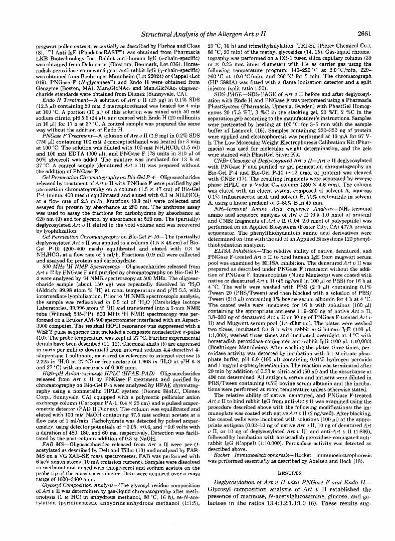
Structural Analysis of the Glycoprotein Allergen Art v I1 from the Pollen of Mugwort (Artemisia vulgaris L.)*
(Received for publication, August 13, 1990)
Bente M. Nilsen$& Knut Slettenn, and Berit Smestad PaulsenS From the Departments of $Pharmacy and TBiochemistry, University of Oslo, Oslo, Norway
Malcolm O’Neill and Herman van Halbeek From the Complex Carbohydrate Research Center and the Department of Biochemistry, University of Georgia, Athens, Georgta 30602
The glycoprotein allergen Art u 11, from the pollen of mugwort (Artemisia vulgaris L.) was treated with peptide:N-glycosidase F (PNGase F) to release aspar- agine-linked oligosaccharides. The oligosaccharides were isolated by gel permeation chromatography and their structures determined by 500-MHz ‘H NMR spec- troscopy, fast atom bombardment-mass spectrometry, and high-pH anion-exchange chromatography. The high-mannose oligosaccharides Man5GlcNAcz, Man6GlcNAcz, Man7GlcNAcz, MansGlcNAcz, and MansGlcNAcz were present in the ratios 2:49:19:24:6 and accounted for all the asparagine-linked oligosac- charides released from Art u I1 by PNGase F. The NH2- terminal amino acid sequences of Art u I1 and of four peptides generated by cyanogen bromide (CNBr) cleav- age of deglycosylated Art u I1 were determined. The first 30 amino acid residues of Art u I1 did not contain any potential N-glycosylation sites. One potential N- glycosylation site was identified in one of the CNBr fragments. The native protein conformation was shown by enzyme-linked immunosorbent assay inhi- bition assays to be essential for the binding of rabbit IgG to Art u I1 and for the binding of human IgE to the major IgE-binding epitope(s) in this allergen. At least one minor IgE-binding epitope still bound IgE after denaturation of the allergen. Removal of the high- mannose chains from denatured Art u I1 had no signif- icant effect on the binding of human IgE to the minor IgE-binding epitope(s).
Pollen of mugwort (Artemisia vulgaris L.) is one of the main causes of allergic reactions in late summer in Europe (1). It has been demonstrated that extracts of mugwort pollen con- tain 8-11 different allergens of variable clinical importance (2-4). No data about the molecular structure of any of the allergens from mugwort pollen have been reported to date.
* This investigation was supported by a grant from the Norwegian Research Council for Science and the Humanities (to B. M. N.), a Fulbright Grant (to B. M. N.), and National Institutes of Health Grant P41 RR-05351 from the Division of Research Resources (to H. v. H.). The costs of publication of this article were defrayed in part by the payment of page charges. This article must therefore be hereby marked “aduertisement” in accordance with 18 U.S.C. Section 1734 solely to indicate this fact.
5 Part of this work was performed at the Complex Carbohydrate Research Center while on leave from the Department of Pharmacy. To whom correspondence should be addressed.
Two mugwort pollen allergens,’ A r t u I and Art u 11, have recently been isolated (5, 6). These purified allergens were found to react with IgE from 70 and 45%, respectively, of sera from mugwort allergic patients (5, 6). A r t u I1 (previously denoted Ag7) has been identified as a concanavalin A-binding glycoprotein (7) that consists of at least 6 isoforms with PI values between 4.10 and 4.65 (6). The M, of A r t u I1 is approximately 35,000 and the allergen appears to be composed of two identical, or nearly identical, subunits linked together by disulfide bridge(s) (6). The isolated A r t u I1 preparation had a protein to carbohydrate ratio of 1O:l. Glycosyl compo- sition analysis of A r t u I1 revealed the presence of mannose, N-acetylglucosamine, glucose, and galactose in the ratios 13.4:3.2:1.3:1.0 (6). The glycosyl composition and the high affinity for concanavalin A suggest that at least part of the carbohydrate structure of Art u I1 consists of Asn-linked oligosaccharides of the high-mannose type.
In this study, the complete structural analysis of the Asn- linked oligosaccharide chains and partial amino acid sequence data of the glycoprotein allergen Art u I1 are reported. This is the first time that the complete structures of the Asn-linked oligosaccharides of a glycoprotein allergen from a plant have been determined. The IgE- and IgG-binding activity of Art u I1 after denaturation and denaturation followed by deglyco- sylation is also reported.
EXPERIMENTAL PROCEDURES
Materials-Art u I1 was purified from an aqueous phosphate-NaC1 buffer extract of mugwort (A. vulgaris L.) pollen by a combination of preparative isoelectric focusing in Ukrodex granulated gel (pH 3.5- 5), affinity chromatography on concanavalin A-Sepharose and HPLC2 size exclusion on Ultropac TSK G2000SW as previously described (6). A human serum pool containing specific IgE against mugwort pollen allergens including Art u I1 (Mugwort serum pool) was prepared from equal volumes of sera from 64 individuals with positive reactions against mugwort pollen extract (RAST-class 3) in the radioallergosorbent test (RAST). A rabbit antiserum against Art u I1 (anti-Art u 11) was prepared by immunization with immunopre- cipitates of Art u I1 obtained in crossed immunoelectrophoresis of
’ The nomenclature of the allergens referred to in this presentation follows the allergen nomenclature system recommended by the Inter- national Union of Immunological Societies (IUIS) ((1986) Bull. WHO 64, 767-770).
The abbreviations used are: HPLC, high performance liquid chro- matography; PNGase F, peptide:N-glycosidase F; Endo H, endo-@- N-acetylglucosaminidase H; WEFT, water-elimination Fourier- transform; HPAE, high-pH anion exchange; PAD, pulsed ampero- metric detector; FAB-MS, fast atom bombardment-mass spectrome- try; SDS, sodium dodecyl sulfate; PAGE, polyacrylamide gel electro- phoresis; ELISA, enzyme-linked immunosorbent assay; PBS, phos- phate-buffered saline.
2660
Structural Analysis of the Allergen Art u 11 2661
mugwort pollen extract, essentially as described by Harboe and Closs (8). "'1-Anti-IgE (PhadebasRAST'") was obtained from Pharmacia LKB Biotechnology Inc. Rabbit anti-human IgE (E-chain-specific) was obtained from Dakopatts (Glostrup, Denmark, Lot 036). Horse- radish peroxidase-conjugated goat anti-rabbit IgG (7-chain-specific) was obtained from Boehringer Mannheim (Lot 22024) or Cappel (Lot 019). PNGase F (N-glycanase") and Endo H were obtained from Genzyme (Boston, MA). Man5GlcNAcz and MangGlcNAcz oligosac- charide standards were obtained from Dionex (Sunnyvale, CAI.
Endo H Treatment-A solution of Art u I1 (25 pg) in 0.1% SDS (12.5 pl) containing 20 mM 2-mercaptoethanol was heated for 4 min at 100 'C. A portion (10 pl) of this solution was mixed with 50 mM sodium citrate, pH 5.5 (24 pl), and treated with Endo H (20 milliunits in 10 pl) for 17 h at 37 "C. A control sample was prepared the same way without the addition of Endo H.
PNGuse F Treatment-A solution ofArt u I1 (1.9 mg) in 0.2% SDS (750 pl) containing 100 mM 2-mercaptoethanol was heated for 3 min at 100 "C. The solution was diluted with 100 mM NH4HCO~ (1.5 ml) and 100 mM EDTA (300 pl), and PNGase F (78 units in 310 p1 of 50% glycerol) was added. The mixture was incubated for 13 h at 37 "C. A control sample (denatured Art u 11) was prepared without the addition of PNGase F.
Gel Permeation Chromatography on Bio-Gel P-4-Oligosaccharides released by treatment of Art u I1 with PNGase F were purified by gel permeation chromatography on a column (1.5 X 47 cm) of Bio-Gel P-4 (minus 400 mesh) equilibrated and eluted with 0.3 M NH4HC03 at a flow rate of 2.5 ml/h. Fractions (0.9 ml) were collected and assayed for protein by absorbance at 280 nm. The anthrone assay was used to assay the fractions for carbohydrate by absorbance at 620 nm (9) and for glycerol by absorbance at 520 nm. The (partially) deglycosylated Art u I1 eluted in the void volume and was recovered by lyophilization.
Gel Permeation Chromatography on Bw-Gel P-IO-The (partially) deglycosylated Art u I1 was applied to a column (1.5 X 46 cm) of Bio- Gel P-10 (200-400 mesh) equilibrated and eluted with 0.3 M NH4HC03 at a flow rate of 5 ml/h. Fractions (0.9 ml) were collected and assayed for protein and carbohydrate.
500 MHz 'H NMR Spectroscopy-Oligosaccharides released from Art v I1 by PNGase F and purified by chromatography on Bio-Gel P- 4 were analyzed by 'H NMR spectroscopy at 500 MHz. The oligosac- charide sample (about 150 pg) was repeatedly dissolved in 'H20 (Aldrich; 99.98 atom % 2H) at room temperature and pZH 5.5, with intermediate lyophilization. Prior to 'H NMR spectroscopic analysis, the sample was redissolved in 0.5 ml of *HZ0 (Cambridge Isotope Laboratories, 99.996 atom % 'H) and transferred into a 5-mm NMR tube (Wilmad; 535-PP). 500 MHz 'H NMR spectroscopy was per- formed on a Bruker AM-500 spectrometer interfaced with an Aspect- 3000 computer. The residual HOZH resonance was suppressed with a WEFT pulse sequence that included a composite nonselective *-pulse (10). The probe temperature was kept at 27 "C. Further experimental details have been described (11, 12). Chemical shifts (6) are expressed in parts per million downfield from internal sodium 4,4-dimethyl-4- silapentane 1-sulfonate, measured by reference to internal acetone ( 6 2.225 in 'Hz0 at 27 "C) or free acetate (6 1.908 in 'HZO at p2H 6-8 and 27 "C) with an accuracy of 0.002 ppm.
High-pH Anion-exchange HPLC (HPAE-PAD)-Oligosaccharides released from Art v I1 by PNGase F treatment and purified by chromatography on Bio-Gel P-4 were analyzed by HPAE chromatog- raphy using a nonmetallic HPLC system (Dionex BioLC, Dionex Corp., Sunnyvale, CA) equipped with a polymeric pellicular anion exchange column (Carbopac PA-l,O.4 X 25 cm) and a pulsed amper- ometric detector (PAD I1 Dionex). The column was equilibrated and eluted with 100 mM NaOH containing 37.5 mM sodium acetate at a flow rate of 1 ml/min. Carbohydrate was detected by pulsed amper- ometry, using detector potentials of -0.05, +0.6, and -0.6 volts with a duration of 480, 180, and 60 ms, respectively. Detection was facili- tated by the post-column addition of 0.3 M NaOH.
FAB-MS-Oligosaccharides released from Art u I1 were per-0- acetylated as described by Dell and Tiller (13) and analyzed by FAB- MS on a VG ZAB-SE mass spectrometer. FAB was performed with 6 keV xenon atoms (10 mA emission current). Samples were dissolved in methanol and mixed with thioglycerol and sodium acetate on the probe tip OE the mass spectrometer. Data were acquired over a mass range of 1000-3400 amu.
Glycosyl Composition Analysis-The glycosyl residue composition of Art u I1 was determined by gas-liquid chromatography after meth- anolysis (1 M HCl in anhydrous methanol, 80 "C, 16 h), re-N-ace- tylation (pyridine:acetic anhydride:anhydrous methanol (1:1:5),
20 "C, 16 h) and trimethylsilylation (TRI-Si1 (Pierce Chemical CO.), 80 "C, 20 min) of the methyl glycosides (14, 15). Gas-liquid chroma- tography was performed on a DB-1 fused silica capillary column (30 m X 0.25 mm inner diameter) with He as carrier gas using the following temperature program: 140-220 "C at 2.0 "C/min, 220- 260 "C at 10.0 "C/min, and 260 "C for 5 min. The chromatograph (HP 5880A) was fitted with a flame ionization detector and a split injector (split ratio 1:50).
SDS-PAGE-SDS-PAGE of Art u I1 before and after deglycosyl- ation with Endo H and PNGase F was performed using a Pharmacia PhastSystem (Pharmacia, Uppsala, Sweden) with PhastGel Homog- enous 20 (7.5 %T, 3 %C in the stacking gel, 20 %T, 2 %C in the separation gel) according to the manufacturer's instructions. Samples were pretreated by heating at 100 'C for 3-5 min with the sample buffer of Laemmli (16). Samples containing 230-350 ng of protein were applied and electrophoresis was performed at 10 mA for 97 V- h. The Low Molecular Weight Electrophoresis Calibration Kit (Phar- macia) was used for molecular weight determination, and the gels were stained with PhastGel Silver Kit.
CNBr Cleavage of Deglycosylated Art u 11-Art u I1 deglycosylated with PNGase F and purified by gel permeation chromatography on Bio-Gel P-4 and Bio-Gel P-10 (-12 nmol of protein) was cleaved with CNBr (17). The resulting fragments were separated by reverse phase HPLC on a Vydac CIR column (250 X 4.6 mm). The column was eluted with an eluent system composed of solvent A, aqueous 0.1% trifluoroacetic acid, and solvent B, 70% acetonitrile in solvent A, using a linear gradient of 0-80% B in 45 min.
NHz-terminal Amino Acid . Sequence Analysis-NH2-termina1 amino acid sequence analysis of Art u I1 (0.5-1.0 nmol of protein) and CNBr fragments of Art u I1 (0.04-2.0 nmol of polypeptide) was performed on an Applied Biosystems (Foster City, CA) 477A protein sequenator. The phenylthiohydantoin amino acid derivatives were determined on-line with the aid of an Applied Biosystems 120 phenyl- thiohydantoin analyzer.
ELISA Inhibition-The relative ability of native, denatured, and PNGase F-treated Art u I1 to bind human IgE from mugwort serum pool was examined by ELISA inhibition. The denatured Art u I1 was prepared as described under PNGase F treatment without the addi- tion of PNGase F. Immunoplates (Nunc Maxisorp) were coated with native or denatured Art u I1 (45 ng/well in 100 p1 of PBS) for 16 h at 4 "C. The wells were washed with PBS (210 pl) containing 0.1% Tween 20 (PBS/Tween) and then blocked with a solution of PBS/ Tween (210 pl) containing 1% bovine serum albumin for 4 h at 4 "C. The coated wells were incubated for 16 h with solutions (100 pl) containing the appropriate antigens (1.9-200 ng of native Art u 11, 3.8-200 ng of denatured Art u I1 or 30 ng of PNGase F-treated Art u 11) and Mugwort serum pool (1:4 dilution). The plates were washed two times, incubated for 8 h with rabbit anti-human IgE (100 pl, 1:1,000), washed three times and incubated overnight at 4 "C with horseradish peroxidase-conjugated anti-rabbit IgG (100 pl, 1:10,000) (Boehringer Mannheim). After washing the plates three times, per- oxidase activity was detected by incubation with 0.1 M citrate phos- phate buffer, pH 6.0 (100 PI) containing 0.01% hydrogen peroxide and 1 mg/ml o-phenylenediamine. The reaction was terminated after 20 min by addition of 0.33 M citric acid (50 pl) and the absorbance at 490 nm determined. All antigens, serum and antisera were diluted in PBS/Tween containing 0.5% bovine serum albumin and the incuba- tions were performed at room temperature unless otherwise stated.
The relative ability of native, denatured, and PNGase F-treated Art u I1 to bind rabbit IgG from anti-Art u I1 was examined using the procedure described above with the following modifications: the im- munoplate was coated with native Art u I1 (2 ng/well). After blocking, the coated wells were incubated with solutions (100 p1) of the appro- priate antigens (0.02-10 ng of native Art u 11, 10 ng of denatured Art u 11, or 10 ng of deglycosylated Art u 11) and anti-Art u I1 (1:800), followed by incubation with horseradish peroxidase-conjugated anti- rabbit IgG (Cappel) (1:10,000). Peroxidase activity was detected as described above.
Rocket Immunoelectrophoresis-Rocket immunoelectrophoresis was performed essentia!ly as described by Axelsen and Bock (18).
RESULTS
Deglycosylation of Art u 11 with PNGase F and Endo H- Glycosyl composition analysis of Art u I1 established the presence of mannose, N-acetylglucosamine, glucose, and ga- lactose in the ratios 13.4:3.2:1.3:1.0 (6) . These results sug-
2662 Structural Analysis of the Allergen Art u II
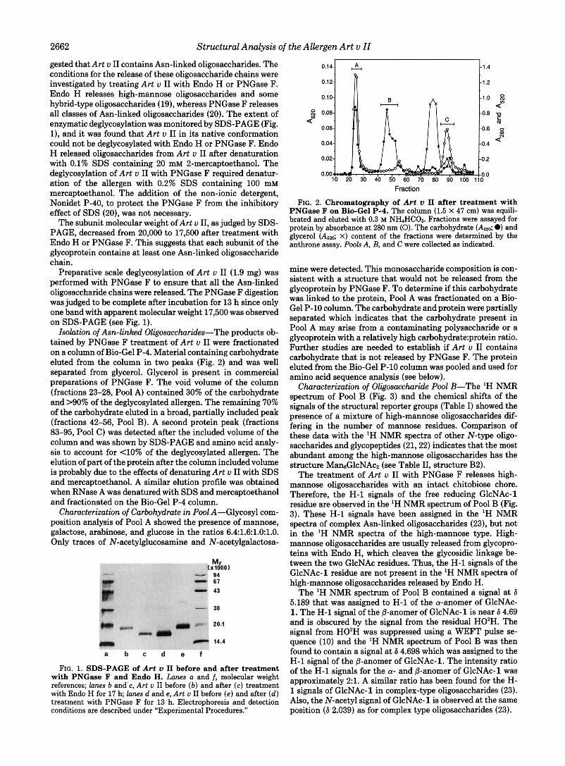
gested that Art u I1 contains Asn-linked oligosaccharides. The conditions for the release of these oligosaccharide chains were investigated by treating Art u I1 with Endo H or PNGase F. Endo H releases high-mannose oligosaccharides and some hybrid-type oligosaccharides (19), whereas PNGase F releases all classes of Asn-linked oligosaccharides (20). The extent of enzymatic deglycosylation was monitored by SDS-PAGE (Fig. l ) , and it was found that Art u I1 in its native conformation could not be deglycosylated with Endo H or PNGase F. Endo H released oligosaccharides from Art u I1 after denaturation with 0.1% SDS containing 20 mM 2-mercaptoethanol. The deglycosylation of Art u I1 with PNGase F required denatur- ation of the allergen with 0.2% SDS containing 100 mM mercaptoethanol. The addition of the non-ionic detergent, Nonidet P-40, to protect the PNGase F from the inhibitory effect of SDS (20), was not necessary.
The subunit molecular weight of Art u 11, as judged by SDS- PAGE, decreased from 20,000 to 17,500 after treatment with Endo H or PNGase F. This suggests that each subunit of the glycoprotein contains at least one Asn-linked oligosaccharide chain.
Preparative scale deglycosylation of Art u I1 (1.9 mg) was performed with PNGase F to ensure that all the Asn-linked oligosaccharide chains were released. The PNGase F digestion was judged to be complete after incubation for 13 h since only one band with apparent molecular weight 17,500 was observed on SDS-PAGE (see Fig. 1).
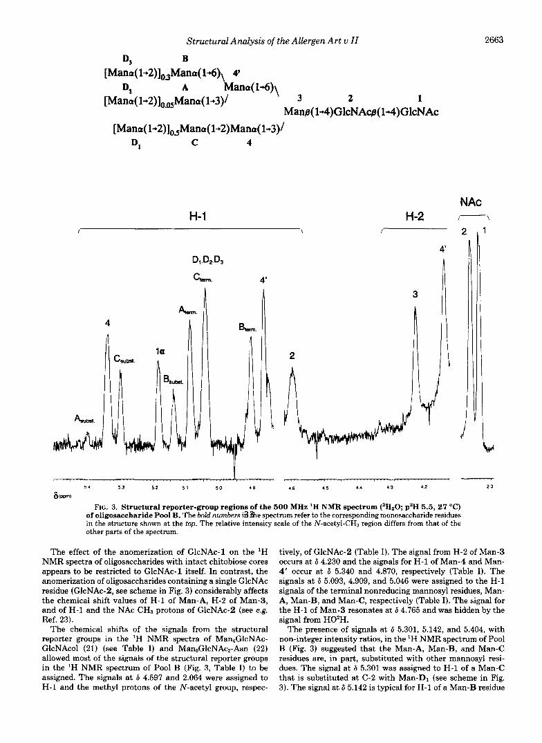
Isolation of Asn-linked Oligosaccharides-The products ob- tained by PNGase F treatment of Art u I1 were fractionated on a column of Bio-Gel P-4. Material containing carbohydrate eluted from the column in two peaks (Fig. 2) and was well separated from glycerol. Glycerol is present in commercial preparations of PNGase F. The void volume of the column (fractions 23-28, Pool A) contained 30% of the carbohydrate and >90% of the deglycosylated allergen. The remaining 70% of the carbohydrate eluted in a broad, partially included peak (fractions 42-56, Pool B). A second protein peak (fractions 83-95, Pool C) was detected after the included volume of the column and was shown by SDS-PAGE and amino acid analy- sis to account for (10% of the deglycosylated allergen. The elution of part of the protein after the column included volume is probably due to the effects of denaturing Art u I1 with SDS and mercaptoethanol. A similar elution profile was obtained when RNase A was denatured with SDS and mercaptoethanol and fractionated on the Bio-Gel P-4 column.
Characterization of Carbohydrate in Pool A-Glycosyl com- position analysis of Pool A showed the presence of mannose, galactose, arabinose, and glucose in the ratios 6.41.6:l.Ol.O. Only traces of N-acetylglucosamine and N-acetylgalactosa-
[XlOOO) Mr
94 67
43
30
20.1
I 14.4
a b c d e f
FIG. 1. SDS-PAGE of Art u I1 before and after treatment with PNGase F and Endo H. Lanes a and f, molecular weight references; lanes b and’c, Art u I1 before ( b ) and after (c) treatment with Endo H for 17 h; lanes d and e, Art u I1 before (e ) and after ( d ) treatment with PNGase F for 13 h. Electrophoresis and detection conditions are described under “Experimental Procedures.”
1.4
1.2
1.0 :: am
0.8 -0
0.6 g
0.4
C Q
am
0.2
3.0 I
Fraction
FIG. 2. Chromatography of Art u I1 after treatment with PNGase F on Bio-Gel P-4. The column (1.5 X 47 cm) was equili- brated and eluted with 0.3 M NH4HC03. Fractions were assayed for protein by absorbance at 280 nm (0). The carbohydrate (ASZO; 0) and glycerol (AsZ0; X) content of the fractions were determined by the anthrone assay. Pools A, B, and C were collected as indicated.
mine were detected. This monosaccharide composition is con- sistent with a structure that would not be released from the glycoprotein by PNGase F. To determine if this carbohydrate was linked to the protein, Pool A was fractionated on a Bio- Gel P-10 column. The carbohydrate and protein were partially separated which indicates that the carbohydrate present in Pool A may arise from a contaminating polysaccharide or a glycoprotein with a relatively high carb0hydrate:protein ratio. Further studies are needed to establish if Art u I1 contains carbohydrate that is not released by PNGase F. The protein eluted from the Bio-Gel P-10 column was pooled and used for amino acid sequence analysis (see below).
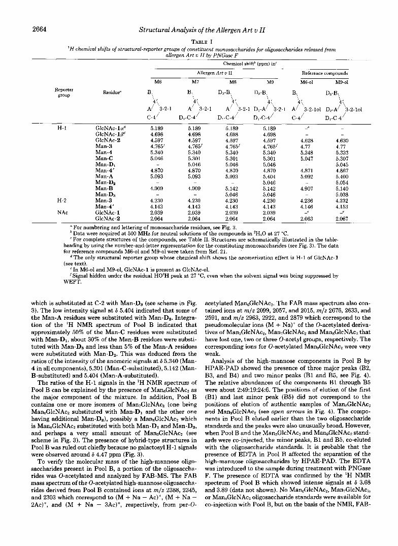
Characterization of Oligosaccharide Pool B-The ‘H NMR spectrum of Pool B (Fig. 3) and the chemical shifts of the signals of the structural reporter groups (Table I) showed the presence of a mixture of high-mannose oligosaccharides dif- fering in the number of mannose residues. Comparison of these data with the ’H NMR spectra of other N-type oligo- saccharides and glycopeptides (21,22) indicates that the most abundant among the high-mannose oligosaccharides has the structure Man6GlcNAc2 (see Table 11, structure B2).
The treatment of Art u I1 with PNGase F releases high- mannose oligosaccharides with an intact chitobiose chore. Therefore, the H-1 signals of the free reducing GlcNAc-1 residue are observed in the ’H NMR spectrum of Pool B (Fig. 3). These H-1 signals have been assigned in the ‘H NMR spectra of complex Asn-linked oligosaccharides (23), but not in the ‘H NMR spectra of the high-mannose type. High- mannose oligosaccharides are usually released from glycopro- teins with Endo H, which cleaves the glycosidic linkage be- tween the two GlcNAc residues. Thus, the H-l signals of the GlcNAc-1 residue are not present in the ‘H NMR spectra of high-mannose oligosaccharides released by Endo H.
The ‘H NMR spectrum of Pool B contained a signal at 6 5.189 that was assigned to H-1 of the a-anomer of GlcNAc- 1. The H-1 signal of the &anomer of GlcNAc-1 is near 6 4.69 and is obscured by the signal from the residual H02H. The signal from H02H was suppressed using a WEFT pulse se- quence (10) and the ’H NMR spectrum of Pool B was then found to contain a signal at 6 4.698 which was assigned to the H-1 signal of the /%anomer of GlcNAc-1. The intensity ratio of the H-1 signals for the a- and /%anomer of GlcNAc-1 was approximately 2:l. A similar ratio has been found for the H- 1 signals of GlcNAc-1 in complex-type oligosaccharides (23). Also, the N-acetyl signal of GlcNAc-1 is observed at the same position (6 2.039) as for complex type oligosaccharides (23).
Structural Analysis of the Allergen Art u 11
D3 B [Mano( l+2)lo,Mana( 14)\ 4'
[Mana( 1+2)], ,Maria( 1-.3)/ 3 2 1 D* A Mana( M) \
Man@( 1+4)GlcNAcg( 1+4)GlcNAc [Mano( 1+2)],,Mana( 1+2)Mana( 1+3)/
D, C 4
2663
H-1 H-2 I \ I
L. 4
4'
4'
I 3
1 ' _?_
I .-?. . . , - . . , . . , . . . . , . . . . . I . . . . , . . , \ . '
5 4 5 3 5 2 5 1 5.0 4 9 4 6 4.5 4.4 4.3 4.2
6(am, FIG. 3. Structural reporter-group regions of the 500 MHz 'H NMR spectrum ('HzO; pZH 5.5,27 "C)
of oligosaccharide Pool B. The bold numbers ia&e spectrum refer to the corresponding monosaccharide residues in the structure shown at the top. The relative intensity scale of the N-acetyl-CHs region differs from that of the
2.0
other parts of the spectrum.
The effect of the anomerization of GlcNAc-1 on the 'H NMR spectra of oligosaccharides with intact chitobiose cores appears to be restricted to GlcNAc-1 itself. In contrast, the anomerization of oligosaccharides containing a single GlcNAc residue (GlcNAc-2, see scheme in Fig. 3) considerably affects the chemical shift values of H-1 of Man-A, H-2 of Man-3, and of H-1 and the NAc CH3 protons of GlcNAc-2 (see e.g. Ref. 23).
The chemical shifts of the signals from the structural reporter groups in the 'H NMR spectra of Man6GlcNAc- GlcNAcol (21) (see Table I) and ManGGlcNAcz-Asn (22) allowed most of the signals of the structural reporter groups in the 'H NMR spectrum of Pool B (Fig. 3, Table I) to be assigned. The signals a t 6 4.597 and 2.064 were assigned to H-1 and the methyl protons of the N-acetyl group, respec-
tively, of GlcNAc-2 (Table I). The signal from H-2 of Man-3 occurs at 6 4.230 and the signals for H-1 of Man-4 and Man- 4' occur at 6 5.340 and 4.870, respectively (Table I). The signals at 6 5.093, 4.909, and 5.046 were assigned to the H-1 signals of the terminal nonreducing mannosyl residues, Man- A, Man-E, and Man-C, respectively (Table I). The signal for the H-1 of Man-3 resonates at 6 4.765 and was hidden by the signal from HO'H.
The presence of signals at 6 5.301, 5.142, and 5.404, with non-integer intensity ratios, in the 'H NMR spectrum of Pool B (Fig. 3) suggested that the Man-A, Man-B, and Man-C residues are, in part, substituted with other mannosyl resi- dues. The signal at 6 5.301 was assigned to H-1 of a Man-C that is substituted at C-2 with Man-D1 (see scheme in Fig. 3). The signal a t 6 5.142 is typical for H-1 of a Man-B residue
2664 Structural Analysis of the Allergen Art u 11
TABLE I 'H chemical shifts of structural-reporter groups of constituent monosaccharides for oligosaccharides released from
allergen Ar t u II by PNGase F Chemical shift' (ppm) in'
Allergen Art u I1 Reference compounds
M6 M7 M8 M9 M6-01 M9-01 Reporter Residue"
group B\ B,
A/ 3-2-1 A/ 3-2-1 A/ 3-2-1 D,-A/ 3-2-1 A/ 3-2-101 D,-A/ 3-2-101 hi \( D:l-B\ 4'\ D:l-B\ 4'\ B\ 4'\ D.l-B\ 4'\
c -4 ' D1-C-4 ' D,-C-4 ' D1-C-4' C-4 / Dl-C-4 / H-1 GlcNAc-lad
GlcNAc-1j3"
Man-3 Man-4 Man-C Man-D1 Man-4' Man-A Man-Dz Man-B Man-DB
Man-4'
GIcNAc-2
H-2 Man-3
NAc GlcNAc-1 GlcNAc-2
5.189 4.698 4.597 4.765' 5.340 5.046
4.870 5.093
4.909
4.230 4.143 2.039 2.064
-
-
-
5.189 4.698 4.597 4.765' 5.340 5.301 5.046 4.870 5.093
4.909
4.230 4.143 2.039 2.064
-
-
5.189 4.698 4.597 4.765' 5.340 5.301 5.046 4.870 5.093
5.142 5.046 4.230 4.143 2.039 2.064
-
5.189 4.698 4.597 4.765' 5.340 5.301 5.046 4.870 5.404 5.046 5.142 5.046 4.230 4.143 2.039 2.064
- -
- -
4.628 4.630 4.77 4.77 5.348 5.333 5.047 5.307 - 5.045
4.871 4.867 5.092 5.400 - 5.054
4.907 5.140 - 5.038
4.236 4.232 4.146 4.153
2.063 2.067 - -
a For numbering and lettering of monosaccharide residues, see Fig. 3. Data were acquired at 500 MHz for neutral solutions of the compounds in 'H,O at 27 "C. For complete structures of the compounds, see Table 11. Structures are schematically illustrated in the table-
heading by using the number-and-letter representation for the constituting monosaccharides (see Fig. 3). The data for reference compounds M6-01 and M9-01 were taken from Ref. 21.
The only structural reporter group whose chemical shift shows the anomerization effect is H-1 of GlcNAc- 1 (see text).
e In M6-01 and M9-01. GlcNAc-1 is Dresent as GlcNAc-01.
WEFT. ' Signal hidden under' the residual HO'H peak at 27 "C, even when the solvent signal was being suppressed by
which is substituted at C-2 with Man-D3 (see scheme in Fig. 3). The low intensity signal at 6 5.404 indicated that some of the Man-A residues were substituted with Man-D2. Integra- tion of the 'H NMR spectrum of Pool B indicated that approximately 50% of the Man-C residues were substituted with Man-Dl, about 30% of the Man-B residues were substi- tuted with Man-D3 and less than 5% of the Man-A residues were substituted with Man-Dz. This was deduced from the ratios of the intensity of the anomeric signals at 6 5.340 (Man- 4 in all components), 5.301 (Man-C-substituted), 5.142 (Man- B-substituted) and 5.404 (Man-A-substituted).
The ratios of the H-1 signals in the 'H NMR spectrum of Pool B can be explained by the presence of Man6GlcNAc2 as the major component of the mixture. In addition, Pool B contains one or more isomers of Man7GlcNAc2 (one being MansGlcNAcz substituted with Man-D1 and the other one having additional Man-D3), possibly a MansGlcNAcz which is MansGlcNAcz substituted with both Man-Dl and Man-Ds, and perhaps a very small amount of Man9GlcNAc2 (see scheme in Fig. 3). The presence of hybrid-type structures in Pool B was ruled out chiefly because no galactosyl H-1 signals were observed around 6 4.47 ppm (Fig. 3).
To verify the molecular mass of the high-mannose oligo- saccharides present in Pool B, a portion of the oligosaccha- rides was 0-acetylated and analyzed by FAB-MS. The FAB mass spectrum of the 0-acetylated high-mannose oligosaccha- rides derived from Pool B contained ions at m/z 2388, 2345, and 2303 which correspond to (M + Na - Ac)+, (M + Na - ~Ac)', and (M + Na - 3Ac)+, respectively, from per-0-
acetylated Man6GlcNAcz. The FAB mass spectrum also con- tained ions at m/z 2099, 2057, and 2015, mlz 2676, 2633, and 2591, and mlz 2963, 2922, and 2879 which correspond to the pseudomolecular ions (M + Na)+ of the 0-acetylated deriva- tives of Man5GlcNAc2, Man7GlcNAc2 and MansGlcNAcz that have lost one, two or three 0-acetyl groups, respectively. The corresponding ions for 0-acetylated Man9GlcNAcz were very weak.
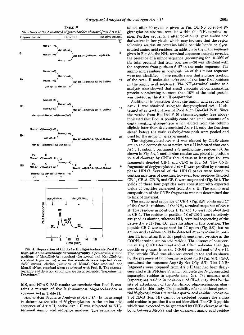
Analysis of the high-mannose components in Pool B by HPAE-PAD showed the presence of three major peaks (B2, B3, and B4) and two minor peaks (B1 and B5, see Fig. 4). The relative abundances of the components B1 through B5 were about 2:49:19:24:6. The positions of elution of the first (Bl) and last minor peak (B5) did not correspond to the positions of elution of authentic samples of Man5GlcNAc2 and Man9GlcNAcz (see open arrows in Fig. 4). The compo- nents in Pool B eluted earlier than the two oligosaccharide standards and the peaks were also unusually broad. However, when Pool B and the Man5GlcNAcz and Man9GlcNAcz stand- ards were co-injected, the minor peaks, B1 and B5, co-eluted with the oligosaccharide standards. It is probable that the presence of EDTA in Pool B affected the separation of the high-mannose oligosaccharides by HPAE-PAD. The EDTA was introduced to the sample during treatment with PNGase F. The presence of EDTA was confirmed by the 'H NMR spectrum of Pool B which showed intense signals at 6 3.68 and 3.89 (data not shown). No Man6GlcNAc2, Man7GlcNAc2, or MansGlcNAcP oligosaccharide standards were available for co-injection with Pool B, but on the basis of the NMR, FAB-
Structural Analysis of the Allergen Art u 11 2665
TABLE I1 Structures of the Asn-linked oliposaccharides obtained from Art v II
Olieosaccharide Structure Relative amount
B1
83
84
Man a ( l 4 ) Man a(1-S) \ Man a(1-A) ,
Man a ( l 4 ) Man a(14) Man a ( l 4 ) '
D3 Man a(1-Q) Man a ( l 4 ) \ 4,
B
D2 & \ S 2 1 Man a(1-A)
Man a ( l 4 ) Mana( l4 ) Man E ( 1 4 ) G l c N A c E(14)GkNAc
%
2
49
19
24
6
c 0
W :: a n d
82
I I I I I I 6 8 10 12 14 16
Time (min)
FIG. 4. Separation of the Art u I1 oligosaccharide Pool B by high-pH anion-exchange chromatography. Open arrows, elution positions of Man6GlcNAc2 standard (left arrow) and Man9GlcNAc2 standard (right arrow) when the standards were injected alone. Solid arrows, elution positions of Man6GlcNAcn-standard and Man,GlcNAcZ-standard when co-injected with Pool B. The chroma- tography and detection conditions are described under "Experimental Procedures."
MS, and HPAE-PAD results we conclude that Pool B con- tains a mixture of five high-mannose oligosaccharides as summarized in Table 11.
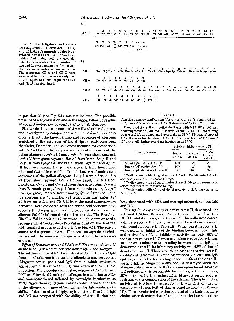
Amino Acid Sequence Analysis of Art u ZZ-In an attempt to determine the site of N-glycosylation in the amino acid sequence of Art u 11, native Art u I1 was subjected to NH2- terminal amino acid sequence analysis. The sequence ob-
tained after 30 cycles is given in Fig. 5A. No potential N- glycosylation site was revealed within this NHz-terminal re- gion. Further sequencing after position 30 gave amino acid derivatives in low yields, which may indicate that the region following residue 30 contains labile peptide bonds or glyco- sylated amino acid residues. In addition to the main sequence given in Fig. 5A, the NHn-terminal sequence analysis revealed the presence of a minor sequence (accounting for 10-30% of the total protein) that from position 5-26 was identical with the sequence from position 6-27 in the main sequence. The amino acid residues in positions 1-4 of this minor sequence were not identified. These results show that a minor fraction of the Art u 11-molecules lacks one of the four first residues in the amino acid sequence. The NHz-terminal amino acid analysis also showed that small amounts of contaminating protein constituting no more than 10% of the total protein was present in the Art u 11-preparation.
Additional information about the amino acid sequence of Art u I1 was obtained using the deglycosylated Art u I1 ob- tained after fractionation of Pool A on Bio-Gel P-10. Since the results from Bio-Gel P-10 chromatography (see above) indicated that Pool A possibly contained small amounts of a contaminating glycoprotein which eluted from the column slightly later than deglycosylated Art u 11, only the fractions eluted before the main carbohydrate peak were pooled and used for the sequencing experiments.
The deglycosylated Art u I1 was cleaved by CNBr since amino acid composition of native Art u I1 indicated that each Art u I1 subunit contained 2-3 methionine residues (6). As shown in Fig. 5A, 1 methionine residue was placed in position 27 and cleavage by CNBr should thus at least give the two fragments denoted CB-1 and CB-2 in Fig. 5A. The CNBr fragments of deglycosylated Art u I1 were purified by reversed phase HPLC. Several of the HPLC peaks were found to contain mixtures of peptides, however, four peptides denoted CB-1, CB-A, CB-B, and CB-C were sequenced (Fig. 5B) . The yields of these four peptides were consistent with expected yields of peptides generated from Art u 11. The amino acid composition of the CNBr fragments was not determined due to lack of material.
The amino acid sequence of CB-1 (Fig. 5B) confirmed 17 of the first 21 residues of the NHz-terminal sequence of Art u 11. The residues in positions 1, 12, and 16 were not identified in CB-1. The residue in position 18 of CB-1 was tentatively assigned as alanine, whereas NHz-terminal sequencing of the native Art u I1 (Fig. 5A) gave histidine in this position. The peptide CB-C was sequenced for 17 cycles (Fig. 5B) , but no amino acid residues could be detected after tyrosine in posi- tion 11, indicating that this peptide contained tyrosine as the COOH-terminal amino acid residue. The absence of homoser- ine in the COOH-terminal end of CB-C indicates that this peptide originates from the COOH-terminal end of Art u 11. The peptide CB-A was also sequenced to the end as shown by the presence of homoserine in position 9 (Fig. 5B) . CB-A contained the sequence Asp-Gly-Ser (Fig. 5 B ) . The CNBr fragments were prepared from Art u I1 that had been degly- cosylated with PNGase F, which converts the N-glycosylated asparagine residue to aspartic acid (24). The aspartic acid (asparagine) residue in position 6 of CB-A may thus be the site of attachment of the Asn-linked oligosaccharides char- acterized in this study. The possibility of an additional poten- tial N-glycosylation site at the aspartic acid residue in position 7 of CB-B (Fig. 5 B ) cannot be excluded because the amino acid residue in position 9 was not identified. The CB-2 peptide which was expected to be generated by CNBr cleavage of the bond between Met-27 and the unknown amino acid residue
2666 Structural Analysis of the Allergen Art v 11 A )
Arl V 11: Xxx - Glu - Thr - Tyr - Gly - Glu - Pro - Gly - Asn - Thr ~ Pro -Asp - Asp - Tyr- Val - His -Ala - His - Asn -(Am)- 118 ~
1 2 3 4 5 6 7 8 9 10 1 1 12 (3 14 15 16 17 18 19 x) 21
L CE-1
22 23 24 25 26 27 28 29 30 ~ r g -(m)- Val - Leu - GIY - Met - xxx - Pro - ~ y s ~
LY s ' CB-2 - FIG. 5. The NHz-terminal amino
acid sequence of native Art u I1 ( A ) and of CNBr fragments of deglyco- sylated Art u I1 ( B ) . X x x denotes an unidentified amino acid. Leu/Lys de- notes two cases where the separation of Leu and Lys was incomplete. Amino acid residues in parentheses are tentative. The fragments CB-A and CB-C were sequenced to the end, whereas only part of the sequences of the fragments CB-1 and CB-B was elucidated.
B)
CB-1: Xxx -(Glu)- Thr - Tyr - Gly - 1 2 3 4 5
Glu - Pro - 6 7
Gly - Asn . Thr - Pro - 8 9 10 1 1
x x x - 12
Asp - Tyr - 13 14
Val - Xxx - Ala -(Ala)- Asn -(Am)- Ile - 15 16 17 16 19 20 21
CB-A: Xxx - Gln - Gly - Ala - He - Asp - Gly - Ser - H S e r 1 2 3 4 5 6 7 8 9
CB-8: Xxx - Val - Gln - Leu - Xxx - Leu - Asp - Glu - Xxx - Leu - Asp - Tyr . Asp - Tyr - Asn - Xxx - Asn - Lys - Xxx . Ile - Lys - 1 2 3 4 5 6 7 6 9 10 1 1 12 13 14 15 16 17 18 19 20 21
1 2 3 4 5 6 7 8 9 1 0 1 1 CB-c: (Pro). Pro - Gly - Asn - Val - Val - Gly - Gln - Leu - Pro . Tyr
LY s
in position 28 (see Fig. 5 A ) was not isolated. The possible presence of a glycosylation site in the region following residue 30 could therefore not be verified after CNBr cleavage.
Similarities in the sequences of Art u I1 and other allergens, was investigated by comparing the amino acid sequence data of Art u I1 with the known amino acid sequences of allergens contained in the data base of Dr. H. Ipsen, ALK-Research, Hersholm, Denmark. The sequences included for comparison with Art u I1 were the complete amino acid sequences of the pollen allergens A m b a I11 and A m b a V from short ragweed, A m b t V from giant ragweed, Bet u I from birch, Lo1 p I1 and Lo1 p I11 from rye-grass, and the allergens Api m I and Api m I11 from bee venom, Der p I and Der p I1 from house dust mite, and Gad c I from codfish. In addition, partial amino acid sequences of the pollen allergens Aln g I from alder, A m b a VI from short ragweed, Cor a I from hazel, Car b I from hornbeam, Cry j I and Cry j I1 from Japanese cedar, Cyn d I from Bermuda grass, J u n p I from mountain cedar, Lo1 p I from rye-grass, Phl p V from timothy, Que a I from oak, and the allergens Der f I and Der m I from house dust mites, Fel d I from cat saliva, and Cla h I1 from the mold Cladosporium herbarum were compared with the amino acid sequence data of Art u 11. The partial amino acid sequence of the major cat allergen Fel d I (25) contained the hexapeptide Thr-Pro-Asp- Glu-Tyr-Val in position 17-22 which is highly similar to the sequence Thr-Pro-Asp-Asp-Tyr-Val in position 10-15 of the NH2-terminal sequence of Art u I1 (see Fig. 5A) . The partial amino acid sequence of Art u I1 showed no significant simi- larities with the amino acid sequences of the other allergens examined.
Effect of Denaturation and PNGase F Treatment of Art u 11 on the Binding of Human IgE and Rabbit IgG to the Allergen- The relative ability of PNGase F-treated Art u I1 to bind IgE from a pool of serum from patients allergic to mugwort pollen (Mugwort serum pool) and IgG from a rabbit antiserum against Art u I1 (anti-Art u 11) was examined by ELISA inhibition. The procedure for deglycosylation of Art u I1 with PNGase F involved heating the allergen in a solution of SDS and mercaptoethanol followed by overnight incubation at 37 "C. Since these conditions induce conformational changes in the allergen that may affect IgE and/or IgG binding, the ability of denatured and deglycosylated Art u I1 to bind IgE and IgG was compared with the ability of Art u 11, that had
TABLE I11 Relative antibody binding activities of native Art u II, denatured Art u II, and PNGase F-treated Art u 11 determined by ELISA inhibition
Denatured Art u I1 was boiled for 3 min with 0.2% SDS, 100 mM 2-mercaptoethanol, diluted 1:3.8 with 70 mM NH4HC03 containing 14 mM EDTA and incubated overnight at 37 "C. PNGase F-treated Art u I1 was as for denatured Art u I1 but with addition of PNGase F (27 units/ml) during overnight incubation at 37 "C.
Relative inhibitory activity (%)
Binding between Native Denatured F-treated PNGase
Art u I1 Art u I1 Art II
Rabbit IgG-native Art u II" 100 <1 <1 Human IgE-native Art u 11' 100 30 20 Human IgE-denatured Art u II' 93 100 94
Wells coated with 2 ng of native Art u 11. Rabbit anti-Art u I1
' Wells coated with 45 ng of native Art u 11. Mugwort serum pool
e Wells coated with 45 ng of denatured Art u 11. Otherwise as in
added together with inhibitor (10 ng).
added together with inhibitor (30 ng).
Footnote b.
been denatured with SDS and mercaptoethanol, to bind IgE and IgG.
The IgE-binding activity of native Art u 11, denatured Art u I1 and PNGase F-treated Art u I1 was compared in two ELISA inhibition assays, one in which the wells were coated with native Art u I1 and another where the wells were coated with denatured Art u I1 (Table 111). When denatured Art u I1 was used as an inhibitor of the binding between human IgE and native Ar t u 11, its inhibitory activity was only 30% of that of native Art u 11. Conversely, when native Art u I1 was used as an inhibitor of the binding between human IgE and denatured Art u 11, its inhibitory activity was 93% of that of denatured Art u 11. These results indicate that native Art u I1 contains at least two IgE-binding epitopes. At least one IgE epitope, responsible for binding of about 70% of the Art u II- specific IgE in Mugwort serum pool, is destroyed when the allergen is denatured with SDS and mercaptoethanol. Another IgE epitope, that is responsible for binding of the remaining 30% of the Ar t u 11-specific IgE in Mugwort serum pool, is resistant to the denaturation of the allergen. The IgE-binding activity of PNGase F-treated Art u I1 was 20% of that of native Art u I1 and 94% of that of denatured Art u I1 (Table 111). These results indicate that removal of the high-mannose chains after denaturation of the allergen had only a minor
Structural Analysis of the Allergen Art u 11 2667
effect on the remaining IgE-binding ability of the allergen. ELISA inhibition using a rabbit antiserum containing spe-
cific IgG against Art u I1 (anti-Art u 11), demonstrated that the IgG-binding activity of both denatured and PNGase F- treated Art u I1 was reduced to less than 1% of the IgG- binding activity of the native allergen (Table 111). This result was confirmed by rocket immunoelectrophoresis where de- natured and PNGase F-treated Art u I1 did not give any immunoprecipitates in the antibody containing gel (result not shown).
DISCUSSION
Asparagine-linked Oligosaccharides of Art u 11-A combi- nation of 'H NMR spectroscopy, FAB-MS, and HPAE-PAD established that the oligosaccharides released by treatment of the glycoprotein allergen Art u I1 with PNGase F were com- posed of 2 GlcNAc residues and between 5 and 9 mannosyl residues (Table 11). Analysis of the oligosaccharides by FAB- MS and HPAE-PAD (Fig. 4) showed Man,GlcNAcz (Bl), Man6GlcNAcP (B2), Man7GlcNAcz (B3), MansGlcNAcz (B4), and Man9GlcNAcz (B5) to be present in the ratios 2:49:19:24:6, respectively. The 'H NMR spectrum of Pool B (Fig. 3) showed that 50% or less of the Man-A, Man-B, and Man-C residues (see B3-B5, Table 11) were substituted by Man-D2, ManDs, and Man-Dl, respectively. Therefore Man6GlcNAcp devoid of Man-D residues:
B Mana( 1-6)
\ 4'
A /Man")
I Maria( 1-3) \ 3 2 1
Manp(l+4)GlcNAc/3(1+4)GlcNAc
Mana( 1+2)Mana( 1+3) C 4
accounts for 50% of the high-mannose oligosaccharides. The oligosaccharide Man5GlcNAcn was present in amounts too small (2% of the mixture) to be detected by 'H NMR. Inte- gration of the anomeric signals in the 'H NMR spectrum of Pool B showed that 5% of the Man-A residues were substi- tuted with Man-D2 and established that ManQGlcNAcz (see B5, Table 11) accounted for approximately 5% of the high- mannose oligosaccharides. Approximately 30% of the Man-B residues were substituted with Man-D3 (25% MansGlcNAcz and 5% ManQGlcNAcz). Therefore, Man6GlcNAcz that is sub- stituted with Man-D1 and Man-Ds (MansGlcNAcz) accounts for 25% of the oligosaccharides in Pool B (see B4, Table 11). Approximately 50% of the Man-C residues were substituted with Man-Dl (20% Man7GlcNAcz, 25% Ma&GlcNAcz, and 5% ManQGlcNAcz) which established that a single Man7GlcNAcz isomer in which Man-D1 is attached to MansGlcNAcz accounted for 20% of the high-mannose oligo- saccharides (see B3, Table 11). The Man7GlcNAcz isomer in which Man-Ds is attached to Man6GlcNAcz may be present in amounts too small ((5%) to be detected by 'H NMR. Our data strongly suggest that only a single isomer of Man7GlcNAcz and MansGlcNAcz are attached to asparagine residues of Art u 11. Further studies are necessary to establish if Art u I1 contains one or more 0-linked carbohydrate chains.
Comparison of the Asn-linked Oligosaccharides of Art u 11, Other Allergens and Plant Glycoproteins-The glycoprotein allergen, Art u 11, from the pollen of mugwort has been shown to contain Asn-linked oligosaccharides of the high-mannose
type (Table 11). Several allergens isolated from other plants contain carbohydrate (26), but a complete structural charac- terization of the oligosaccharides has not been reported. Ovo- mucoid (Gal d I), ovalbumin (Gal d 11), and ovotransferrin (Gal d 111) from hen egg white and phospholipase AZ (Api m I) from bee venom are glycoprotein allergens that contain Asn-linked oligosaccharides. Ovalbumin contains a mixture of high-mannose oligosaccharides ( M B ~ ~ - ~ G ~ C N A C ~ ) in addi- tion to hybrid-type oligosaccharides, whereas ovomucoid and ovotransferrin contain mixtures of multiantennary complex type oligosaccharides (Ref. 27 and references therein). Phos- pholipase AP has been reported to contain unusual "high- mannose" oligosaccharides in which the M B ~ ~ - ~ G ~ C N A C ~ core is extended with nonreducing terminal N-acetylglucosamine and fucose residues (28). These limited data indicate that there are a variety of oligosaccharide structures that are attached to the asparagine residues of glycoprotein allergens from plants and animals.
High-mannose oligosaccharides linked to asparagine resi- dues have been found in numerous glycoproteins from mam- malian cells (29). In contrast, the number of plant glycopro- teins that have been unambiguously shown to contain Asn- linked high-mannose oligosaccharides is small (30). The Man6_8GlcNAc2 isomers found in Art u I1 are identical to the predominant Man6-&lcNAcz isomers found in kidney bean glycoprotein I1 (phaseolin) and 7 s soybean storage protein (31, 32). The predominant Man7GlcNAcz isomer found in these plant glycoproteins is substituted with Man-Dl on Man- C (see scheme in Fig. 3 and Table 11). In contrast, the predominant Man7GlcNAcz isomer usually found in animal glycoproteins is the one in which Man-B is substituted with Man-Ds (33). The limited data available suggest that although the biosynthesis of Asn-linked high-mannose oligosaccharides of mammalian and plant glycoproteins is similar, the enzy- matic processing of the GlcsManQGlcNAcz precursor may differ (34). The finding that the predominant Man7GlcNAcz isomer present in plant glycoproteins are different from the predominant Man7GlcNAcz isomer found in animal glycopro- teins may perhaps reflect such differences in the processing.
Amino Acid Sequence of Art u II-The NHz-terminal se- quences of Art u I1 and of four peptides generated by CNBr- cleavage of deglycosylated Art u I1 were determined in an attempt to identify potential N-glycosylation sites. The se- quence Asp-Gly-Ser was located at positions 6-8 of the CNBr- cleavage product CB-A (Fig. 5B) and represents a potential N-glycosylation site. However, the location of this potential N-glycosylation site within the intact glycoprotein remains to be established. The peptides sequenced accounted for 71 of the approximately 150 amino acid residues each subunit of Art u I1 is composed of (6). Further studies are needed to determine the entire amino acid sequence of Art u I1 and the location of the high-mannose oligosaccharides.
The NHz-terminal sequence of Art u 11 and the sequence of the peptide CB-C which probably originates from the COOH- terminal end of Art u I1 have some characteristic features. Of the 9 proline residues present in each Art u I1 subunit (6), 3 were positioned within the first 29 residues from the NH,- terminal end and 2-3 were found within the first 11 residues from the COOH-terminal end. Thus, it seems that more than half of the proline residues are clustered within the NHz- terminal and COOH-terminal regions of this allergen. These terminal regions also contain pairs of identical amino acid residues, i.e. Asp-Asp in positions 12-13, Asn-Asn in position 19-20, and Arg-Arg in position 22-23 of the NHz-terminal sequence and Pro-Pro, Val-Val in positions 1-2 and 5-6, respectively, of peptide CB-C. It is not known if these struc-
2668 Structural Analysis of the Allergen Art v II tural features are related to the biological activity of this glycoprotein. Comparison of the amino acid sequence data of Art u I1 with amino acid sequence data of other allergens from a variety of sources showed significant similarity only between a hexapeptide sequence present in position 17-22 of the NH,- terminal sequence of Fel d I (25) and the hexapeptide sequence in positions 10-15 of the NHz-terminal sequence of Art u 11. Since the function of Art u I1 and Fel d I has not been determined, the functional significance of this similarity is not known.
Art u I1 has earlier been found to consist of at least 6 isoforms with PI values in the region 4.10-4.65 (6). Since the carbohydrate part of Art u I1 contained only neutral mono- saccharides, the PI heterogeneity was assumed to arise from (micr0)heterogeneities in the protein part of the molecule. NH2-terminal sequence analysis of native Art u I1 revealed that 10-30% of the molecules lacked one of the first four amino acid residues in the sequence. This may at least partly explain the PI heterogeneity of this allergen. No other major heterogeneities were observed in the amino acid sequence of the peptides sequenced, but may be present in parts of the sequence not determined here. Minor differences caused by partial deamidation of the amino acid residues asparagine and glutamine may also contribute to the PI heterogeneity of Art u 11.
Antibody-binding Activity of Art u II after Denaturation and Treatment with PNGase F-The contribution of the Asn- linked high-mannose oligosaccharides of Art u I1 to the im- munogenicity of the allergen was assessed by determining the relative IgE- and IgG-binding activities of the native allergen, denatured allergen, and denatured and deglycosylated allergen (Table 111). The IgG-binding activity of Art u I1 was com- pletely destroyed after the glycoprotein was denatured with SDS and mercaptoethanol. This shows that the native three- dimensional protein conformation of Art u I1 is essential for the binding of rabbit IgG indicating that at least part of the IgG epitope resides in the protein part of the molecule. The glycoprotein allergen could not be deglycosylated without prior denaturation with SDS and mercaptoethanol. Therefore, the effect of enzymatic deglycosylation of Art u I1 on the IgG- binding activity of this allergen could not be determined.
The IgE-binding activity of Art u I1 decreased by 70% when the allergen was denatured with SDS and mercaptoethanol (Table 111) showing that the native three-dimensional confor- mation of Art u I1 is required for binding of human IgE to the major IgE-binding epitope(s). The IgE-binding epitope(s) of Art u I1 that are resistant to denaturation with SDS and mercaptoethanol were not significantly affected by the enzy- matic deglycosylation of the allergen (Table 111).
Our results suggest that the Asn-linked high-mannose oli- gosaccharides of Art u I1 have no direct effect on the binding of IgE to the allergen. A possible role of these oligosaccharides either as part of the dominant IgE-binding epitope(s) or as a stabilizing element of the three-dimensional protein confor- mation necessary for binding of human IgE to the dominant IgE epitope(s) can, however, not be ruled out. The data obtained for the IgE-binding activity of Art u I1 were obtained using a pool of sera from 64 individuals who were allergic to mugwort pollen. Studies with the glycoprotein allergen Api m I (phospholipase Aq) from bee venom have shown that indi-
vidual patients may recognize different IgE-binding epitopes (35). I t is therefore possible that the IgE-binding epitope(s) recognized by some mugwort allergic individuals may include the Asn-linked high-mannose oligosaccharides of Art u 11. The biological role(s) of the carbohydrate portions of glyco- protein allergens will only be determined when the Asn-linked oligosaccharide chains can be enzymatically removed from the native glycoprotein.
Acknowledgments-We thank William S. York (CCRC, Athens, GA) for performing the FAB-MS experiments, Dr. Svein Haavik (Department of Pharmacy, University of Oslo) for valuable assistance in planning and performing the ELISA inhibition experiments, Jessie Juul (Department of Biochemistry, University of Oslo) for expert technical assistance in performing the amino acid sequencing, and Dr. H. Ipsen (ALK-Research, H~rsholm, Denmark) for checking possible similarities between the amino acid sequence data of Art u I1 and those of other allergens.
1.
2.
3.
4.
5.
6.
7.
8.
10. 9.
11.
12.
13.
14. 15.
16. 17. 18.
20. 19.
21.
22.
23.
24.
25.
26.
27.
28.
29. 30.
31.
32.
33. 34.
35.
REFERENCES Spieksma, F. T. M., Charpin, H., Nolard N., and Stix E. (1980) Clin. Allergy
Ipsen, H., Formgren, H., L~wenstein, H., and Ingemann, L. (1985) Allergy
Smestad Paulsen, B., Fb, L., Nesje, G., and Wold, J. K. (1985) Int. Arch.
Diener. C.. Frenzel. K.. Wiebicke. K.. Schlenvoiet, G.. Fuster, R.. and Jaaer,
10,319-329
4 0 , 289-294
Allergy Appl. Immunol. 78,206-212
L. (1988) Allerg.'Immunol. ( h ' i p z k ) 34,3-9- ' '
M., and Carreira, J. (1990) Mol. Immunol. 2 7 , 651-657
1056
Immunol. 80,113-121
359
. .
De La Hoz, F., Polo, F., Moscoso del Prado, J., Selles, J. G., Lombardero,
Nilsen, B. M., and Smestad Paulsen, B. (1990) Mol. Immunol. 2 7 , 1047-
Nilsen, B. M., and Smestad Paulsen, B. (1986) Int. Arch. Allergy Appl.
Harboe, M., and Closs, 0. (1983) S c a d . J. Immunol. 17 , Suppl. 10, 353-
Dische, Z. (1962) Methods Carbohydr. Chem. 1,478-512 Haasnoot, C. A. G. (1983) J. Magn. Reson. 52, 153-158 Green, E. D., Adelt, G., Baenziger, J. U., Wilson, S., and van Halbeek, H.
Argade, S. P., Hopfer, R. L., Strang, A,"., van Halbeek, H., and Alhadeff,
Dell, A., and Tiller, P. R. (1986) Biochem. Blophys. Res. Commun. 135 ,
(1988) J . Biol. Chem. 2 6 3 , 18253-18268
J. A. (1988) Arch. Biochem. Biophys. 266,227-247
Chambers, R. E., and Clamp, J. R. (1971) Biochem. J. 125 , 1009-1018 Sweeley, C. C., Bentley, R., Makita, M., and Wells, W. W. (1963) J. Am.
Laemmli, U. K. (1970) Nature 227,680-685 Sletten, K., and Husby, G. (1974) Eur. J. Biochem. 4 1 , 117-125 Axelsen, N. H., and Bock, E. (1983) S c a d . J. Immunol. 17 , Suppl. 10,
1126-1134
Chem. SOC. 85,2497-2507
Kobata, A. (1984) Biol. Carbohydr. 2,87-161 Tarentino, A. L., Gbmez, C. M., and Plummer, T. H., Jr. (1985) Biochem-
Mutsaers, J. H. G. M., van Halbeek, H., Kamerling, J. P., and Vliegenthart,
103-106
istry 24,4665-4671
J. F. G. (1985) Eur. J. Biochem. 147.569-574 Vliegenthart, J.' F. G., Dorland, L., and van Halbeek, H. (1983) Adu.
Carbohydr. Chem. Biochem. 41,209-374 Spellman, M. W., Basa, L. J., Leonard, C. K., Chakel, J. A,, O'Connor, J.
V., Wilson, S., and van Halbeek, H. (1989) J. Biol. Chem. 2 6 4 , 14100-
Tarentino, A. L., and Plummer, T. H. (1987) Methods Enzymol. 138,770- 14111
Chapman, M. D., Aalberse, R. C., Brown, M. J., and Platts-Mills, T. A. E. 778
Haavik, S., Smestad Paulsen, B., and Wold, J. K. (1985) Int. Arch. Allergy (1988) J. Immunol. 140,812-818
Van Halbeek, H., Vliegenthart, J. F. G., Iwase, H., Li, S. C., and Li, Y.-T. Appl. Immunol. 78, 197-205
Weber, A,, Marz, L., and Altmann, F. (1986) Comp. Biochem. Physiol. 83B, (1985) Glycoconjugate J. 2,235-253
Kornfeld, R., and Kornfeld, S. (1985) Annu. Reu. Biochem. 54,631-664 321-324
Faye, L., Johnson, K. D., Sturm, A., and Chrispeels, M. J. (1989) Physiol.
Neeser, J.-R., del Vedovo, S., Mutsaers, J. H. G. M., and Vliegenthart, J.
Sturm, A,, Van Kuik, J. A,, Vliegenthart, J. F. G., and Chrispeels, M. J.
Hubbard, S. C., and Ivatt, R. J. (1981) Annu. Reu. Biochem. 50,555-583 Elbein, A. D., and Kaushal, G. P. (1990) in Methods in Plant Biochemistry
Weber, A,, Sihroder, H., Thalberg, K., and Marz, L. (1987) Allergy 42,
Plant. 75,309-314
F. G. (1985) Glycoconjugate J . 2,355-364
(1987) J. Biol. Chem. 262,13392-13403
(Dey, P. M. ed) Vol. 2, pp. 79-110, Academic Press, London
464-470