Embed Size (px)
Citation preview

THE JOURNAL OF BIOLOGICAL CHEMISTRY 0 1986 by The American Society of Biological Chemists, Inc.
Vol. 261, No. 5, Issue of February 15, pp. 2376-2383,1986 Printed in U.S.A.
Structure of a Novel Sialylated Fucosyl Lacto-N-nor- ~ e ~ a o s y ~ c e r a ~ i d e Isolated from Chronic ~ y e ~ o g ~ n o u s Leukemia Cells*
(Received for publication, August 12,1985)
Michiko N. Fukudalfi, Anne DelllI, Philip R. TillerllII , Ajit Varki**$$, John C. Klock#fillll, and Minoru Fukuda$$ From the $Cancer Research Center, La Jolla Cancer Research Foundation, La Jolla, California 92037, the YDepartment of Biochemistry, Imperial College of Science and Technology, London, England, the **Cancer Center, University of California, San. Diego, California 92037, and the $$Medical Research Institute, San Francisco, California 941 15
A novel sialylated fucosyl glycolipid, which is pres- ent at an elevated level in chronic myelogenous leuke- mia cells, was isolated. The structure of this fucogan- glioside was elucidated by methylation analysis, fast atom bombardment-mass Spectrometry, and enzymatic degradation, followed by reaction with anti-le”, Gal@l-+ 4 (Fucal+ 3) GlcNAcBl-+, monoclonal anti- body. The structure of this ganglioside was found to be: NeuNAccw2+3Gal~l+4GlcNAc~1-+3
Gal~1+4GlcNAc,91+3Gal@l+4Glc@l~1Cer 3
1 Fuc
T 4
This structure is unique in that a fucose is attached to the internal N-acetylglucosamine but not to the sub- terminal N-acetylglucosamine. Since this gIycolipid is apparently absent in normal granulocytes or acute my- elogenous leukemia cells, it can be a specific marker for chronic myelogenous leukemia cells. Based on the structures of this fucoganglioside and normal granu- locyte glycolipids, a biosynthetic pathway of extension, sialylation, followed by fucosylation is proposed.
Chronic myelogenous leukemia (CML’} is a clonal myelo- proliferative disorder and characterized by a marked overpro- duction of granulocytes in peripheral blood. Although malig- nant cells arise from a stem cell common to granulocytes and erythrocytes, these malignant cells achieve significant mat- uration and granulocytes produced in the chronic phase of
* This work was supported by Grants ROl CA 33895 and CA 34014 from the National Cancer Institute and by grants awarded to Profes- sor H. R. Morris of the Imperial College by the British Medical and Science and Engineering Research Councils. The costs of publication of this article were defrayed in part by the payment of page charges. This article must therefore be hereby marked “aduertisement” in accordance with 18 U.S.C. Section 1734 solely to indicate this fact.
3 To whom correspondence should be addressed at La Jolla Cancer Research Foundation, 10901 North Torrey Pines Rd., La Jolla, CA 92037.
// Recipient of a studentship from the British Science and Engi- neering Research Council.
85: Recipient of National Institutes of Health Grant GM 32373. TIl Recipient of National Cancer Institute Grant CA 32632.
The abbreviations used are: CML, chronic myelogenous leukemia; FAB-MS, fast atom bombardment-mass spectrometry; HPTLC, high- performance thin layer chromatography; HPLC, high performance liquid chromatography; Cer, ceramide; CTH, ceramide trihexoside.
CML patients are very similar to normal mature granulocytes in morphology and ~ n c t i o n s (1). In fact, Dacremont and H ~ d e b r ~ d (Z), Klock et al. (3), . and Westrick et at. (4) reported that granulocytes produced in the chronic phase of CML contain glycolipids very similar to those present in mature granulocytes.
Recently, we have analyzed the structures of polylactosa- minoglycans isolated from CML granulocytes ( 5 ) and found that CML polylactosaminoglycans are distinctly different from normal granulocyte polylactosaminoglycans (6, 7), i.e. CML polylactosaminoglycans, are 1) highly sialylated, 2) shorter, and 3) characterized by a great abundance of the sialyl Lex, NeuNAc~+3Gal/3l+4(Fucal+3)GlcNAcpl+ ter- minal structure and the presence of the NeuNAccrZ- 3Gal~ l~4(Fuccr l -+3)GlcNAc~l+3~a l /3 l~4 (Fuca l~3) - GlcNAc structure. Our recent studies on normal g r a n u l o c ~ glycolipids indicate that polylactosaminoglycans and glyco- lipids share the same structures; in the majority of neutral glycolipids from granulocytes, fucose is attached to linear poly-N-acetyllactosaminyl backbones to form GalBl- 4(Fuccul-+3)GlcNAc~l~3 terminal structure, whereas sialic acid is joined to the same backbones by either a2+3 or a2+ 6 linkage in gangliosides (8). However, we have not detected the structures unique to CML polylactosaminoglycans in nor- mal granulocyte gangliosides (8).
These results led us to investigate CML glycolipids in detail to see whether cells from CML patients contain any unique glycolipids. We describe here the structural characterization of novel fucosyl gangliosi~es uniquely present in CML cells.
EXPERIM~NTAL PROCEDURES
Isolation of Glycosphingolipids from Granulocytic Cells of CML Patients-Patients were admitted to the hospital of the University of California at San Diego or the Medical Research Institute at San Francisco. All patients were in the chronic phase of CML and had blast counts of less than 10%. Leukocytes were enriched by therapeu- tic leukophoresis and were further purified by hypotonic lysis to remove contaminating erythrocytes as described (5,9). The prepara- tion obtained was found hy its morphology to be more than 90% granulocytic cells with various degrees of maturation (segmented and banded neutrophils, metamyelocytes, and myelocytes).
Isolation and purification of glycolipids were carried out as de- scribedpreviously @), except for the following. The granulocytic cells were extracted s~uent ia l ly with 5 times the volume of hot ethanol and ch1orofo~:methanol (2:1, v,k). The volume of c h l o r o f o ~ : methanol was 10 times that of the residue obtained after ethanol extraction. The crude glycolipid extract was then fractionated by Folch’s phase partition (10). The lower-phase glycolipids were freed from cholesterol and phospholipids by acetylation procedures (11). Deacetylated glycolipids were applied to QAE-Sephadex A-25 column
using the solvent systems described by Ando and Yu (12). Upper- chromatography and neutral and acidic glycolipids were separated by
phase glycolipids were also separated into neutral and acidic fractions
2376

A Novel Fucoganglioside from Chronic Myelogenous Leukemia Cells
in the same manner and acidic glycolipids of the upper and the lower layer were pooled.
Purification of Acidic Glycolipids-Acidic glycolipids were purified by high-performance liquid chromatography with a Varian HPLC apparatus (model 5000, Varian Associates, CA). The glycolipids dis- solved in a minimum amount of ch1oroform:methanol (2:1, v/v) were applied to a column (0.4 X 50 cm) of Iatrobeads (IRS 8010, 10 pm diameter, Iatron, Tokyo). The column was equilibrated with isopropyl alcoho1:hexane:water (55:40:5), eluted over 20 min by a gradient to isopropyl alcoho1:hexane:water (55:35:10), and followed by a shallower gradient to isopropyl alcoho1:hexane:water (55:29:16) over an addi- tional 180 min (8, 13). The flow rate was constant at 0.5 ml/min and the eluate was collected every 2 min. Glycolipids in each tube were analyzed by HPTLC (Si-HPF, J. T. Baker Chemical Co.) using a solvent system chloroform:methanol:3.5 M NH4OH (6035:8, v/v). The elution positions of glycolipids were as follows: GI, 70-72 min; GO, 72-74 min; GB, 77-86 min; G,, 82-88 min. Each glycolipid fraction recovered was then acetylated in pyridine:acetic anhydride (l : l , v/v) (11). Acetylated glycolipids were then separated on HPTLC (Si-HPF, J. T. Baker) using a solvent of dich1oroethane:acetone:water (50:50:1, v/v). Acetylated glycolipids detected by iodine vapor were eluted from thin layer plates and deacetylated with sodium in methanol (11). Glycolipids obtained were further purified by HPLC in the same manner as described above, with the solvent system as follows. The solvent gradient was programmed as isopropyl a1cohol:hexane:water (55:405 to 55:37:8) over 10 min, followed by the gradient to isopropyl alcoho1:hexane:water (55:29:16) over 70 min. The flow rate was con- stant at 0.5 ml/min and the eluate was collected every 1 min. Glyco- lipids in each tube were analyzed by HPTLC as described above. The elution positions of glycolipids were as follows: GI, 33 min; GO, 34-37 min; G3,37-39 min; G4, 40-42 min.
Methylation Analysis-The glycolipids were methylated by the method of Hakomori (14). The permethylated samples were purified by LH-20 column chromatography and further purified by partition with ch1oroform:water (1:l) and the chloroform layer was washed four times with water as described (8, 15).
The permethylated samples thus obtained were then subjected to FAB-MS for sequence analysis as described below. In order to obtain the information on linkages between monosaccharides, the permeth- ylated samples were subjected to acid hydrolysis with 0.5 N H o S O ~ in 90% acetic acid at 80 "C for 4 h. The hydrolysates were then neutral- ized and acetylated as described (15). The alditol acetates of partially methylated sugars were analyzed by gas liquid chromatography-mass spectrometry with a modification in column temperature as described (5, 15).
A B
so c - % so c
s,c . - s1 c
s2c ,-3D"--, sa c s3c =""p
s3 c s4 c s5 c +
- - CI --- - ." ~ -- - - ._
s4c '- - -- - :;i= 4mTZa- s6 -
s 1 2 3 4 5 S 1 6
FIG. 1. Thin layer chromatogram of gangliosides isolated from normal mature granulocytes, chronic myelogenous leu- kemia cells, and acute myelogenous leukemia cells. The glyco- lipids were developed on HPTLC three times using the solvent system of chloroform:methanol:3.5 M NH40H in water (60:35:8, v/v), and detected by orcinol-Hz-SO,. A, gangliosides from normal granulocytes ( S ) and from chronic myelogenous leukemia patients. Lanes 1-5 represent patients 1 through 5. The bands indicated by the arrows are CML specific glycolipids. SO, N e u N A c a 2 + 3 G a l ~ l 4 G l c ~ l + 1Cer; SI, NeuNAccu2+3Gal~l+4G1cNAc~l+3Gal~l+4Glcpl 1Cer; SO, NeuNAca2+6Gal~1+4G1cNAc~l+4Ga1/31+4Glc@l+ 1Cer; Sa, NeuNAc~2+3(Gal/3l4GlcNAcpl+3)~Gal~14Glc~1+ lCer S,, NeuNAca2+6(Gal~l+4G1cNAc~1+3)~Gal/31+4Glc@l~ lCer (from Ref. 8). B, gangliosides from CML patient 1 (lane 1) and from acute myelogenous leukemia patient (lane 6 ) . The band indi- cated by the arrow on the right side was due to contaminants.
A
C D H C
a C T H C
P G C .- N1 c N3C -- Ne+
B
F-
2377
* N1
* N3 * N6
N 1 3 5 N' 1 ' 3' 5 '
FIG. 2. Immunostaining of Folch's upper neutral glycolipids with monoclonal anti-Le", Gal@1+4(Fucal+3)GlcNAc@l+3, antibody. A , the plate was developed and visualized by orcinol- H,S04. Lanes N, 1, 3, and 5 correspond to glycolipids from normal granulocytes (N), CML patients 1,3, and 5, respectively. B, The same samples weFe subjected to immunostaining by anti-Le' (PM-81) an- tibody. Lanes N', 1 ', 3', and 5' correspond to lanes N, I, 3, and 5 in A. CDH, Gal@l"rlGlcpl+lCer; aCTH, GlcNAcpl+3Galpl+ 4Glcpl+lCer; PG, Gal~l4GlcNAc@l+3Gal~l4Glcj3l~lCer; N1, Gal@l+ 4(Fuca1+ 3)GlcNAcpl+ 4Glcp1+ 1Cer; N3, Galp l+ 4(Fuccul+3)GlcNAc~l+3Gal~l+4GlcNAc~l+3Gal~l+4Glc~l+ 1Cer; N6, Gal~l4(Fucal+3)GlcNAc~l+3Gal~l4GlcNAc~l+ 3Gal~l+4GlcNAc~1+3Gal~l4Glc~l+lCer (from Ref. 8). PM-81 antibody reacts with N1-glycolipid as well as Le"-glycolipids with longer carbohydrate chains (17).
s1 c
G 1 2 3 4 G FIG. 3. Thin-layer chromatogram of gangliosides purified
from CML cells. CML cells from patient 1 were used for starting materials. G, gangliosides from patient 1; lane I , G,; lane 2, GO; lane 3, Ga; lane 4 , G,. The mobility of So, S1, Sz, and SB (see legend to Fig. 1 for the structures) are shown on the left side.
Fast Atom Bombardment-Mass Spectrometry-FAB-MS was car- ried out on a VG analytical ZAB HF mass spectrometer (Imperial College, London) as previously described (8, 15). About 100-200 pg of glycolipids were permethylated and about 10-20 pg of each deriv- ative were loaded onto the glycerol/thioglycerol matrix for each FAB- MS run.

2378 A Novel Fucoganglioside from Chronic Myelogenous Leukemia Cells
Immunostaining of Glycolipid on TLC-Glycolipids were chromat- ographed on a HPTLC plate (alminium sheets Silica Gel 60, E. Merck) and were reacted with monoclonal anti-Lex antibodies ac- cording to the procedure described (16). Monoclonal antibody, PM- 81, which reacts ef~ciently with Le” structure (17), was kindly donated by Dr. E. D. Ball of Dartmouth Medical School.
RESULTS
Glycolipids from CML Cells-Fig. 1A shows the thin-layer chromatogram of the acidic glycolipids (gangliosides) pre- pared from CML cells. CML gangliosides provide a similar profile to that of normal granulocyte gangliosides except for the following. Doublets which migrate slightly faster than Sg were significantly increased in some cases of CMLs. This glycolipid, apparently unique to CML, was termed Gz. In addition, gangliosides migrating to the same position of S5 and those migrating more slowly than S5 were also increased in CML cells from most of the patients examined. In contrast, gangliosides from acute myelogenous leukemia patients were mainly composed of short-chain glycolipids (Fig. lB), which is consistent with the previous report (18).
Among the patients examined, patient 5 barely had any detectable amount of Gz (Fig. 1A). Apparently, higher gan- gliosides from patient 5 are not enriched. In order to verify these observations on gangliosides and determine whether there are differences in chain elongation, fucosylation, sialy- lation, neutral glycolipids with long chain carbohydrates pres- ent in Folcb’s upper layer were examined. As shown in Fig. 2A, the majority of higher neutral glycolipids are decreased in patients 1 and 3, whereas in patient 5 a comparable amount of higher neutral glycolipids to normal granulocytes was de- tected. Gly~olipids with the Le” structure, G a 1 / 3 1 ~ 4 ( ~ c a l ~ 3)GlcNAc, are also reduced in patients 1 and 3, whereas the granulocytes of patient 5 still contain a comparable amount of Le” active glycolipids to those in normal granulocytes (Fig. 2B). These results indicate that the glycolipids with long carbohydrate chains are more heavily sialylated in the gran- ulocytes of the majority of CML patients including patients 1 and 3 than those present in normal granulocytes.
Purification of CML Gangliosides-Since the level of unique gangliosides is apparently increased in some CML cells, gan-
HexNAc . Hex+ (m/z 1029), and NeuNAc Hex HexNAc+ (m/z 825) (Fig. 4). If Gz has a fucose residue at the subterminal ~-acetyIglucosamine residue, the fragment ion of m/z 999 should be prominent (Fig. 5Bf (see also Ref. 5). As shown in Fig. 4, the fragment ion of m/z 999 was barely detected. These results indicate that Gz has a sequence of NeuNAc+Hex+ HexNAc+Hex+(Fuc-)HexNAc+Hex-+Hex+Cer (Fig. 5A). In addition, the fragment ion of m/z 2183 corresponds to the M - Ciao acyl chain (2421-238) or M - C,S,, (2449- 266), confirming that the ceramide is composed of the sphin- gosine with dl,,. as the long-chain base and the C,,:, and C,,:, as the major fatty acids.
Methylation analysis of Gz produced 1 mol of 2,3,4-tri-0- methylfucose, 3 mol of 2,4,6-tri-O-methylgalactose, 1 mol of 6-O-methyl-N-acetylglucosamine, 1 mol of 3,6-di-O-methyl- N-acetylglucosamine, and 1 mol of 2,3,6-tri-O-methylglucose (Table I).
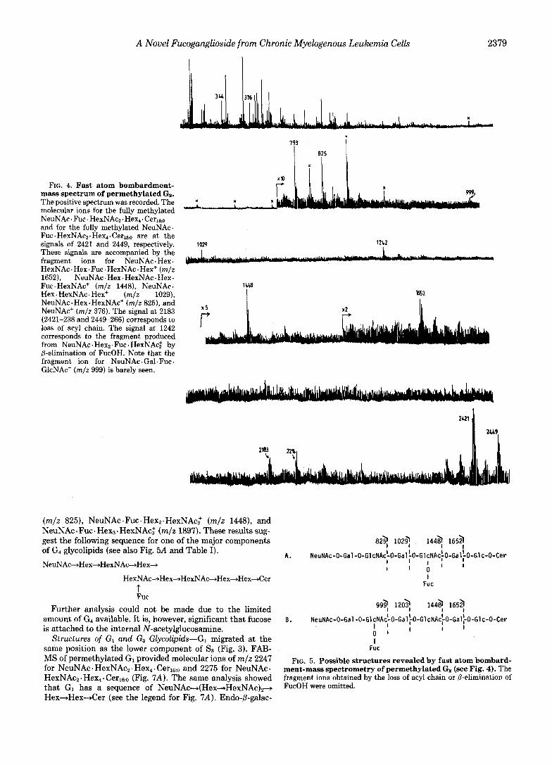
G, was sequentially digested with clostridial neuraminidase, @-galactosidase, and /3-N-acetylglucosaminidase. The product was reacted with anti-Lex monoclonal antibody, PM-81, and gave a positive spot at the same position of the N1-glycolipid, Gal@l-+4(Fuca14)GlcNAc/3l+3Gal@l”+4Glc@l~lCer (S) , whereas the starting material did not react with the same antibody (Fig. 6A). These results indicate the following struc- ture for G,. NeuNAc~~2-+Gal~l-+GlcNAcpl-tGalpl-s
4GlcNAc~l-3Gal~1-4Glc-Cer 3
1 Fuc
7..
Endo-&galactosidase from Escherichia freundii hydrolyzed Gz glycolipid to produce CMH and a large oligosaccharide (Fig. 6B).
It has previously been shown that endo-@-galactosidase cannot hydrolyze the @-galactosidic linkage adjacent to the fucosylated N-acetylglucosamine (6,19). Thus, the result can be interpreted as follows (where CMH represents Glc@1+ 1Cer):
*
c Heptasaccharide
gliosides from the CML cells of patient 1 were chosen for detailed structural analysis, After HPLC fractionation, the glycolipids were further purified through the procedures of acetylation, preparative HPTLC, and deacetylation followed by HPLC as described under “Experimental Procedures.” Fig. 3 presents the HPTLC of CniIL ganglioside preparations. In the following section, the structural analysis of G2 is described in detail, whereas the analysis of other glycolipids is described briefly.
~ t r u c ~ u r e of Gz-Gz migrated slightly faster than S4, N e u N A c ~ 2 + 6 G a l / 3 l ~ ~ G l c N A c @ l ~ 3 G a l @ l ~ ~ G l c N A c / 3 l ~ 3Gal@l4Glc+Cer (8) (Fig. 1A).
FAB-MS analysis of permethylated Gz provided the molec- ular ions for NeuNAcl. Fuc. . HexNAcz I Hex4. Cer16:o (m/z 2421) and NeuNAcl. Fuel. HexNAcz. Hex4. CerlS,o (m/z 2449), as shown in Fig. 4. The same analysis provided fragment ions corresponding to NeuNAc . Fuc . HexNAcz - Hex: (m/z 1652), NeuNAc Fuc . Hexn. HexNAct (m/z 1448), NeuNAc . Hex.
3 t
1 Fuc Endo-@galactosidase
4+ CMH +
These results obtained by FAB-MS, methylation analysis, immunostaining and endo-@-galactosidase digestion indicate that the structure of Gz glycolipid is VI~NeuNAcII13Fuc- LcnOsesCer as shown in Table 11.
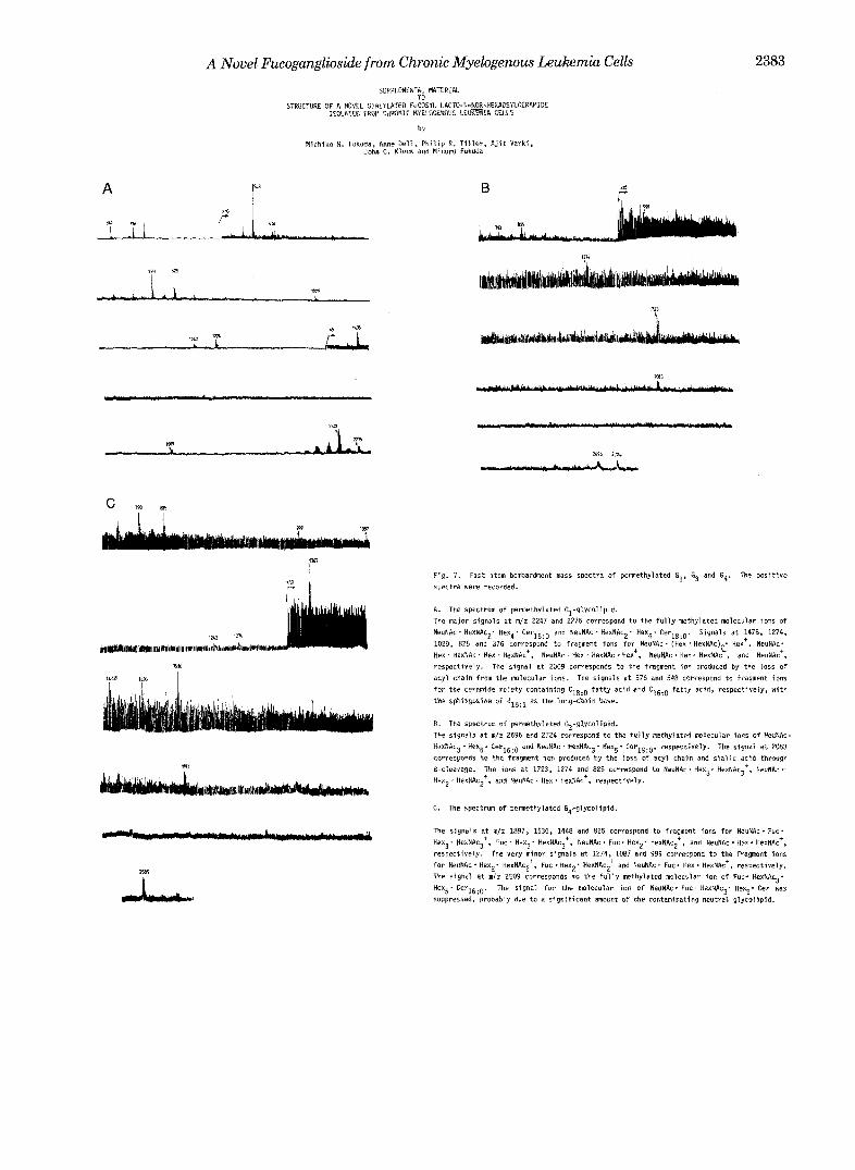
Structure of G4-G4 migrated slower than GB or Ss (Figs. 1 and 3);which has a composition of NeuNAc . Hexc,. HexNAcs- Cerleo (or (Fig. 7W.2
FAB-MS analysis {Fig. 7C) of permethylated G4 provided the fragment ions correspon~ng to NeuNAc a Rex. HexNAc+
-
Fig. 7 is presented in miniprint at the end of this paper. Miniprint is easily read with the aid of a standard magnifying glass. Full size photocopies are available from the Journal of Biological qhemistry, 9650 Rockville Pike, Bethesda, MD 20814. Request Document No. 85111-2676, cite the authors, and include a check or money order for $2.00 per set of photocopies. Full size photocopies are also included in the microfilm edition of the Journal that is available from Waverly Press.
A Novel Fucoganglioside from Chronic Myelogenous Leukemia Cells 2379
FIG. 4. Fast atom bombardment- mass spectrum of permethylated Gz. The positive spectrum was recorded. The molecular ions for the fully methylated NeuNAc . Fuc . HexNAcz. Hex4. Cerleo and for the fully methylated NeuNAc. Fuc . HexNAcz. Hex4. Cerls,o are at the signals of 2421 and 2449, respectively. These signals are accompanied by the fragment ions for NeuNAc . Hex. HexNAc . Hex. Fuc . HexNAc . Hex+ (m/z 1652), NeuNAc. Hex. HexNAc. Hex. Fuc. HexNAc+ (m/z 1448), NeuNAc. Hex. HexNAc . Hex+ (m/z 1029), NeuNAc. Hex. HexNAc+ (m/z 825), and NeuNAc+ (m/z 376). The signal at 2183 (2421-238 and 2449-266) corresponds to loss of acyl chain. The signal at 1242 corresponds to the fragment produced from NeuNAc. Hex2.Fuc. HexNAc: by 0-elimination of FucOH. Note that the fragment ion for NeuNAc . Gal. Fuc . GlcNAc+ (m/z 999) is barely seen.
x
1029 1212
x 5 I I
(m/z 825), NeuNAc-Fuc.Hexn.HexNAcz+ (m/z 1448), and NeuNAc . Fuc . Hex3. HexNAcg (m/z 1897). These results sug- gest the following sequence for one of the major components of G4 glycolipids (see also Fig. 5A and Table I). NeuNAc+Hex+HexNAc+Hex+
HexNAc+Hex+HexNAc+Hex+Hex+Cer T Fuc
Further analysis could not be made due to the limited amount of G4 available. It is, however, significant that fucose is attached to the internal N-acetylglucosamine.
Structures of GI and G3 Glycolipids-G1 migrated at the same position as the lower component of Ss (Fig. 3). FAB- MS of permethylated G1 provided molecular ions of m/z 2247 for NeuNAc . HexNAcz. Hex4. CerlG,o and 2275 for NeuNAc . HexNAcz-Hex4- Cerlko (Fig. 7A). The same analysis showed that G1 has a sequence of NeuNAc-(Hex+HexNAc)+ Hex-Hex-Cer (see the legend for Fig. 7A). Endo-P-galac-
82q 102q 144q 165a
A. NeuNAc-O-Gal-O-GlcNAc'0-Gal~O-GlcNAc~O-Gal~O-Glc-O-Cer 1 1
1 I A ' ' Fuc
I
999 1203 1449 1653
I ' I 1
I Fuc
B. NeuNAc-O-Gal-O-G1cNA~-O-GalfO-G1cNA~~O-Gal~-O-G1c-O-Cer
0 ' ' FIG. 5. Possible structures revealed by fast atom bombard-
ment-mass spectrometry of permethylated Gz (see Fig. 4). The fragment ions obtained by the loss of acyl chain or p-elimination of FucOH were omitted.
2380 A Novel Fucoganglioside from Chronic Myelogenous Leukemia Cells
TABLE I Relative proportions of methylated sugars obtained from gangliosides
of chronic mvelogenous leukemia cells Methvlated suwrs GI Go G, G.
Fucitol
Glucitol
Galactitol
~~
2,3,4-Tri-O-methyl 0 1.0 0 0.8
2,3,6-Tri-O-methyl 1.0 1.0 1.0 1.0
2,3,4,6-Tetra-O-methyl 0 0 0 0.2 2,4,6-Tri-O-methyl 3.0 3.0 3.8 3.7
deoxyglucitol 3,6-Di-O-methyl 2.0 1.0 3.0 2.1 6-Mono-0-methyl 0 1.0 0 1.0
2-N-Methylacetamido-2-
A 0
CMH U
CDHC
aCTHU
" P S NIC
N 1 2 3 L 1 2 3 4
FIG. 6. HPTLC of hydrolysis products of Gz by exo- and endo-@-galactosidase. A , immunostaining of glycolipids by anti- PM-81 monoclonal antibody. N and lane 2, Folch's upper phase neutral glycolipids ( N ) and Ng-glycolipid, Gal@l+4(Fuca1+ 3)GlcNAc~1+3Gal~l4GlcNAc~l+3Gal~l+4Glc~l+lCer (lane 2 ) . These are from normal granulocytes (8). Lane 1, untreated Gz; lane 3, G2 after clostridial neuraminidase, P-galactosidase, and P-N- acetylglucosaminidase. The monoclonal antibody reacts with GalP1-a 4(Fucal+3)GlcNAcfll+R terminal structure. The solvent system used was ch1oroform:methanol:water (6035:8, v/v). B, HPTLC of hydrolysis products of G1 (lanes 1 and 4 ) and Gz (lanes 2 and 3) gangliosides by endo-@-galactosidase. Glycolipids were digested under condition 1 as described (29, 30). Endo-0-galactosidase hydrolysis products were partitioned by ch1oroform:methanol:water (42:1, v/v) and each organic phase was applied to lanes 1 and 2 for analysis of shorter glycolipids. Water phases were applied to lanes 3 and 4 for analysis of oligosaccharides. The solvent systems used were chloro- form:methanol:water (6035:8, v/v) for glycolipids and l-bu- tano1:acetic acidwater (3:3:2, v/v) for oligosaccharides. L, Folch's lower phase glycolipid mixture from total blood cells as a Ref. 8. TC, sodium taurodeoxycholate; Di, GlcNAcpl+3Gal; Tetra, NeuNAca2- 3Galpl+4GlcNAcpl+3Gal. CMH, Glcfil+lCer; Glo, globoside, GalNAc~l+3Gala1+4Gal/3l+4Glc~Cer. Structures for other standard glycolipids are shown in Fig. 2.
tosidase treatment of G1 yielded lacto-N-triaosylceramide (amino CTH) and tetrasaccharide (Fig. 6B), as follows:
NeuNAc+Gal+GlcNAc+Gal-+GlcNAc+Gal+Glc+Cer
Endo-@-galactosidase
t Tetra - Amino CTH +
It was also apparent that a portion of amino CTH was further digested to produce Glc/3l+lCer and disaccharide (lanes I and 4 in Fig. 6B). Combining these results with methylation analysis (Table I), the structure of G1 was proposed to be NeuNAca2+3Gal/3l+4GlcNAc/31+3Gal/31+4GlcNAc/31~ 3Ga1/314Glc/3l+Cer with a Cleo or fatty acid. Thus, the GI glycolipid has the same carbohydrate sequence as the SS glycolipid which is present in normal granulocytes (8).
GS migrated approximately at the same position as S5 (Figs. 1A and 3). FAB-MS (Fig. 7B) of permethylated GS provided molecular ions for NeuNAc + Hex5. HexNAcs + Cerleo (m/z 2696) and NeuNAc Hex5. HexNAc3. Cerls:, (m/z 2724). The same analysis indicates that GS has a sequence of NeuNAc-, (Hex+HexNAc)3+Hex+Hex+Cer. The methylation analy- sis indicates that sialic acid is linked to galactose through a 2+3 linkage and that no branched galactose is present (Table I). Combining these results, the structure of GS was suggested to be NeuNAca2+3Gal/3l4GlcNAc/31+3Gal- /31+4G1cNAc/31+3Ga1/31~4G1cNAc/31+3Ga1/31+4G1c+ Cer with a Cleo or Ciao fatty acid.
T I
DISCUSSION
The present study revealed the presence of a unique fuco- syl ganglioside (G2) in chronic myelogenous leukemia cells and elucidated its structure as NeuNAca2+3Ga1/31+ 4G1cNAc/31+3Ga1/31+4(Fuca1+3)GlcNAc/31+3Ga1/31+ 4Glcpl+lCer. This is the first report of the isolation of this glycolipid although related fucogangliosides have been iso- lated from dolonic tumor tissues (20, 21) (see Table 11). Previously, we have established the structure of glycolipids present in normal granulocytes (8). Although a fucosyl gan- glioside, which has the same composition as G2, has been detected in the S6 fraction from normal granulocytes, the amount was significantly low (8). In addition, the S5 glycolipid migrated much slower than the G2 glycolipid on TLC, sug- gesting that Gz and S5 glycolipids are different in carbohydrate structure. Since the G2 glycolipid is not accumulated in acute myelogenous leukemia cells (Fig. lB), the accumulation of G2
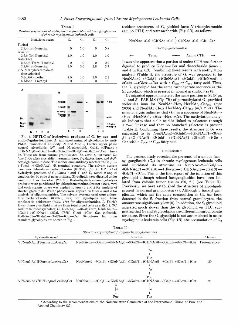
TABLE I1 Structures of sialylated fucosylnorhexaosylceramides
Svstematic name' Structure Reference
V13NeuNAcII13FucnorLcnOse&er NeuNAcol2+3Gal~1~G1cNAc~l+3Gal~ l~GlcNAc~l+3Gal~ l+4Glc~ l+ lCer Present study 3 ta 1
Fuc
3
1 Fuc
V16NeuNAcII13FucnorLcnOse&er NeuNAc~u2~Gal~l+4G1cNAcpl-*3Gal@l+4GlcNAc@l+3Gal@l~Glc~l+lCer 20
tff
V13NeuNAcV31113Fuc~norLcnOse&er NeuNAcol2-t3Gal~l+4GlcNAc~l+3Gal~l+4GlcNAc~l+3Gal~l+4Glc~l~lCer 21 3 3
1 1 Fuc Fuc
tff tff
a According to the recommendations of the Nomenclature Committee of the International Union of Pure and Applied Chemistry (27).
A Novel Fucoganglioside from Chronic Myelogenous Leukemia Cells 2381
glycolipid may be a characteristic marker for CML cells. It is significant that fucose is linked to the N-acetylglucos-
amine which is distal from the nonreducing end, but not to the subterminal N-acetylglucosamine. This was somewhat surprising to us since we have observed fucosylation at the subterminal N-acetylglucosamine which forms the sialyl Le’, NeuNAca2+3Gal~l+4(Fucal+3)GlcNAc~l+ structure in CML polylactosaminoglycans ( 5 ) . However, preferential fu- cosylation at internal N-acetylglucosamine residues in CML polylactosaminoglycans has also been noticed by FAB-MS; the signal a t m/z 1448 for NeuNAc+Gal+GlcNAc+Gak (Fuc+)GlcNAc+ or NeuNAc+Gal+(Fuc+)GlcNAc+Gal+ GlcNAc’ was stronger than that for NeuNAc+Gal+(Fuc+) GlcNAc+Gal+(Fuc+)GlcNAc+ (m/z 1622) or that for NeuNAc+Gal+(Fuc+)GlcNAc+ (m/z 999) (see Fig. 5A in Ref. 5 ) . The signal a t m/z 1448, therefore, mostly represents the NeuNAc+Gal+GlcNAc+Gal+(Fuc+)GlcNAc+ struc- ture. Thus, internal monofucosylation appears to take place more frequently than external monofucosylation or difucosy- lation at both internal and external sites.
The structure of Gz and the results of previous structural studies on granulocyte glycolipids provide significant infor- mation on the biosynthetic steps of poly-N-acetyllactosamine carbohydrates in terms of extension, fucosylation, and si- alylation. Previously, we have shown that no such structure as extended NI, Gal~l+4GlcNAc~l+3Gal~l+4(Fucal+3)- GlcNAc~l+3Gal~l-+4Glc~l+Ce~, is present in granulocyte glycolipids (8). This is consistent with the recent report that fucosylation takes place after lactosaminyl backbone is formed and that fucose is preferentially added to the subter- minal N-acetylglucosamine and then to the internal N-ace- tylglucosamine (22). These observations (8, 22) indicate that “extension precedes fucosylation” and thus the possibility that Gz is formed by the following pathway is less likely.
Galpl+4GlcNAc~l+R
Galp1+4GlcNAc~l+R .u
3
1 Fuc
tff
U Gal~l+4GlcNAc~l+3Gal~l”t4GlcNAc~l+R
3
1 Fuc
f f f
U NeuNAccuZ+3Gal~l+4GlcNAcpl+3Ga1~1+4GlcNAc~l+R
3
1 Fuc
tff
SCHEME I
Therefore, Gz must be formed by the following alternative pathway which is expressed as “extension, sialylation, then fucosylation.”
Galpl-+4GlcNAcpl+R
Gal~l+4GlcNAc~l”t3Gal~l+4GlcNAc~l~R
NeuNAca2+3Gal~l+4GlcNAc~l+3Gal~1+4G1cNAc~1+R
NeuNAca2+3Gal~l~4G1cNAc~l+3Gal~1+4G1cNAc~1+R
u. U U
3
1 Fuc
ta
SCHEME I1
Sialylation preceding fucosylation may explain why the fucose residue is present at the internal N-acetylglucosamine. The presence of sialic acid at the nonreducing terminal may hamper the addition of fucose to the subterminal N-acetylglu- cosamine. Thus, fucosylation takes place at the internal N - acetylghcosamine. If fucosylation takes place before sialyla- tion, fucose should be preferentially added at the subterminal N-acetylglucosamine as seen in neutral glycolipids (8, 22). Recently, 011-3 specific fucosyltransferases were detected in mutant Chinese hamster ovary cell lines or human lung cancer cell lines. These enzymes exclusively form a1-d fucosyl linkage on N-acetylglucosamine (22, 23), and al+3 specific fucosyltransferases can apparently add fucose to sialylated lactosaminyl chains (23). On the other hand, it has been shown that a1+3 and 011-4 fucosyltransferase activities cannot be separated in human milk (24, 25). In this enzyme system, fucosyltransferase could not add fucose to sialylated substrates (24). Therefore, at least two kinds of a1-3 fuco- syltransferases are known (for a detailed discussion, see Ref. 26). In CML cells, an enzyme similar to the former fucosyl- transferase may be responsible for the formation of Gz.
Table I1 lists the structures of the fucosyl gangliosides so far reported which have two N-acetylglucosamine residues available for fucosylation. In all of these structures, fucose residues are attached only at the internal N-acetylglucosa- mine or at both sites. This evidence is in agreement with the proposed sequence of sialylation followed by fucosylation (Scheme 11). It is plausible that fucose is preferentially added to the internal N-acetylglucosamine and then to the subter- minal N-acetylglucosamine, due to the steric hindrance caused by the terminal sialic acid residue. In order to test whether Scheme I1 is correct, it will be essential to character- ize an a1-3 fucosyltransferase by using a defined substrate, such as SI and SB glycolipids.
It is noted that CML gangliosides contain C18:o fatty acid in addition to Cleo and Cz4,1.3 We have previously shown that glycolipids from normal, mature granulocytes almost exclu- sively contain Cleo and CZk1 fatty acids (8). These results suggest that fatty acid metabolism is altered in CML cells and this is reflected in the glycolipids.
The present study also shows the heterogeneity of ganglio- side profiles among patients. Although cells from the majority of CML patients express a unique fucoganglioside, Gz, some of the patients do not show this glycolipid (Fig. 1A). There may be two possible explanations for this. One possibility is that this glycolipid is aberrently expressed in malignant cells. Another possibility is that the glycolipid is expressed in a subpopulation of immature CML cells which are not present in normal peripheral blood. For this, it will be important to correlate the presence or absence of Gz with maturational stages or cell types of dominant leukemic cells in a given stage of the disease. It will also be important to see whether any immature cells of normal granulocyte development also ex- press this carbohydrate structure. These studies will provide a basis for diagnosis and prognosis of chronic myelogenous leukemia and hopefully provide a basis for immunotherapy (28).
Acknowledgments-We thank Brian Bothner for assistance in mass spectrometric analysis, Priya Ramsamooj for technical assistance, and Anna Steve for secretarial assistance. We also thank Dr. David Ward for his kind arrangement for the leucophoresis of leukemic cells.
Our analysis showed that CML glycolipids corresponding to the upper band of S3 contain fatty acid.

2382 A Novel Fucoganglioside from Chronic Myelogenous Leukemia Cells
Note Added in Proof-After submitting our manuscript, the bio- synthetic pathway of sialylation followed by fucosylation has been shown in sialyl Le" oligosaccharide structure (Hansson, G. C., and Zopf, D. (1985) J. Bid. Chem. 260,9388-9392.
REFERENCES 1. Champlin, R. E., and Golde, D. W. (1985) Blood 65,1039-1047 2. Dacremont, G., and Hildebrand, J. (1976) Biochem. Biophys. Acta
3. Klock, J. C., DAngona, J. L., and Macher, B. A. (1981) J. Lipid Res. 22,1079-1083
4. Westrick, M. A., Lee, W. M. F., and Macher, B. A. (1983) Cancer Res. 43,5890-5894
5. Fukuda, M., Bothner, B., Ramsamooj, P., Dell, A,, Tiller, P. R., Varki, A., and Klock, J. C. (1985) J. Bid. Chem. 260, 12957- 12967
6. Spooncer, E., Fukuda, M., Klock, J. C., Oates, J. E., and Dell, A. (1984) J. Biol. Chem. 259,4792-4801
7. Fukuda, M., Spooncer, E., Oates, J. E., Dell, A., and Klock, J. C. (1984) J . Biol. Chem. 259,10925-10935
8. Fukuda, M. N., Dell, A., Oates, J. E., Wu, P., Klock, J. C., and Fukuda, M. (1985) J. Biol. Chem. 260,1067-1082
9. Klock, J. C., and Bainton, D. F. (1976) Blood 48,149-161
424,315-322
10. Folch, J., Arsove, S., and Heath, J. A. (1951) J. Biol. Chem. 191,
11. Hakomori, S., and Watanabe, K. (1976) in G ~ y c o ~ ~ i d ~ e € h o ~ o ~ g y (Witing, L. A., ed) pp. 13-47, American Oil Chemists Society, Champaign, IL
819-831
12. Ando, S., and Yu, R. K. (1977) J. Biol. Chem. 252,6247-6250 13. Watanabe, K., and Arao, Y. (1981) J. Lipid Res. 22,1020-1024 14. Hakomori, S. (1964) J. Biochem. (Tokyo) 55,205-208
15. Fukuda, M., Dell, A., and Fukuda, M. N. (1984) J. Bid. Chem.
16. Magnani, J. L., Nilsson, B., Brockhaus, M., Zopf, D., Steplewski, Z., Koprowski, H., and Ginsburg, V. (1982) J. Bioi. Chem. 257,
17. Magnani, J . L., Ball, E. D., Fanger, M. W., Hakomori, S., and Ginsburg, V. (1984) Arch. Biochem. Biophys. 233,501-506
18. Tsuboyama, A., Takaku, F., Sakamoto, S., Kano, Y., Ariga, T., and Miyatake, T. (1980) Br. J. Cancer 42,908-914
19. Kannagi, R., Nudelman, E., Levery, S. B., and Hakomori, S. (1982) J. Bioi. Chem. 257,14865-14874
20. Hakomori, S., Nudelman, E., Levery, S. B., and Patterson, C. M. (1983) Bwchem. Biophys. Res. Commun. 113,791-798
21. Fukushi, Y., Nudelman, E., Levery, S. B., Hakomori, S., and Rauvala, H. (1984) J. Biol. Chem. 259,10511-10517
22. Holmes, E. H., Ostrander, G. K., and Hakomori, S. (1985) J. Bwl. Chem. 260,7619-7627
23. Campbell, C., and Stanley, P. (1984) J. Biol. Chem. 259,11208- 11214
24. Prieeles, J.-P., Monnom, D., Dolmans, M., Beyer, T. A., and Hill, R. L. (1981) J. Bid. Chem. 256, 10456-10463
25. Johnson, P. M., Yates, A. D., and Watkins, W. M. (1981) Biochem. Biophys. Res. Commun. 100,1611-1618
26. Kornfeld, R., and Kornfeld, S. (1985) Annu. Reu. Biochem. 54,
27. IUPAC-IUB Commission on Biochemical Nomenclature (1977)
28. Fukuda, M. (1985) Biochim. Biophys. Acta 780,119-150 29. Fukuda, M. N., and Matsumura, G. (1976) J. Biol. Chem. 251,
30. Fukuda, M. I?., Watanabe, K., and Hakomori, S. (1978) J. BwL
259,4782-4791
14365-14369
631-664
Lipids 12,455-463
6218-6225
Chem. 253,6814-6819
A Novel I"ucogang1ioside from Chronic Myelogenous Leukemia Celb
SUPPLEMENTAL MATERIAL Tn
A liLa
STRUCTURE OF A NOVEL SlALYLATEU FUCkYL LA~rO-N-NOR-HE~OSYLCERAMIDE ISOLATE0 FRW C ~ ~ a ~ I ~ RYELOGENOUS LEUml& CELLS
by
Michiko N. Fukuda, Anne Dell, Philip R. Tiller, Ajit Varki, John C. Klock and Minoru Fukuda
2383
Fig. 7. Fast atom bombardment mass spectra of permethylated E.!, 4 and G4. The positive spectra were recorded.
A. The spectrum o f permethylated Gl-glycoIipid. The major signals at m/z 2247 and 2275 correspond tu the fully methylated molecular ions of NeuNRc' HexNAc?. Hexq' CerlsiO and NeuNAc. HexNAc2, Hex4. Cer18:0. Signals at 1478, 1274,
Hex ' HsxNAc ' Hex * HexNAc', NeuNAc - Hex . HexNAc Hex', NeuNAc . Hex. HexNAc , and NeuNAc+, 1029, 825 and 376 correspond to fragment ions for NeuNAc I (Hex . HexNAc)$ Her', NeuNAc.
acyl chain from the molecular ions. The signals at 576 and 54R correspond to fragment ions resprctively. The signal at 2009 Corresponds to the fragment ion produced by the loss of
for the ceramide moiety containing C18:o fatty acid and C,6:0 fatty acid, respectively, with the sphingosine o f d18:1 as the long-chain base.
The signals at n / z 2696 and 2724 correspond to the fully ~thylated Mlecuiar ions o f NeuRc. B. The spectrum o f pemethylated G3-glycolipid.
HexNAc3-Hex5. Cerlg:O and NeuN.4~. HexNAcg- Hex5* Cer18:0. respectively. The signal at 2083 carresponds tu the fraginent ion produced by the loss of acyl chain and sialic acid through
Hexp. nexNAc2*, and NeuNAc I Hex. HexNAc+, respectively. 8-cleavage. The ions at 1723, 1274 and 825 correspond to NeuNAc. Hexg. HexNRcg+, NeuNAc.
G. The spectrum of pennethylated G4-glycolipid.
The signals at m l z 1897, 1536, 1448 and 825 correspond tu fragment ions for NeuNAc. Fuc' Hexg. HexNAc3+, Fuc. Hex3. HexNAc3', NeuNAc . Fuc * Hex2' HexNAcZt, and NeuNAc . Hex. HexNAc+,
for NeuNAc. Hex2. HexNAc2+, Fuc.Hex2' HexNAc2+ and NeuNAc. FUC. Hex. H~xNAc', respectively. respectively. The very minor signals at 1274, 1087 and 999 correspond to the fragment ions
The signal at ndz 2509 corresponds to the fully methylated molecular ion of Fuc- HexNAc3* Hex5* Ccr16:0. The signal far the molecular ion o f NeuNAc. Fuc- HexNAcj. Hex5* Cer was suppressed, probably due tu a significant amount of the contaminating neutral glycolipid.