Embed Size (px)
Citation preview
Structural Analysis of Murine Zona Pellucida GlycansEVIDENCE FOR THE EXPRESSION OF CORE 2-TYPE O-GLYCANS AND THE Sda ANTIGEN*
(Received for publication, October 22, 1999, and in revised form, December 18, 1999)
Richard L. Easton‡, Manish S. Patankar§, Frank A. Lattanzio§, Trey H. Leaven§,Howard R. Morris‡¶, Gary F. Clark§¶, and Anne Dell‡¶
From the ‡Department of Biochemistry, Imperial College of Science, Technology and Medicine, London SW7 2AY, UnitedKingdom and §Department of Physiological Sciences, Eastern Virginia Medical School, Norfolk, Virginia 23501
Murine sperm initiate fertilization by binding to spe-cific oligosaccharides linked to the zona pellucida, thespecialized matrix coating the egg. Biophysical analyseshave revealed the presence of both high mannose andcomplex-type N-glycans in murine zona pellucida. Thepredominant high mannose-type glycan had the compo-sition Man5GlcNAc2, but larger oligosaccharides of thistype were also detected. Biantennary, triantennary, andtetraantennary complex-type N-glycans were found tobe terminated with the following antennae: Galb1–4GlcNAc, NeuAca2–3Galb1–4GlcNAc, NeuGca2–3Galb1–4GlcNAc, the Sda antigen (NeuAca2–3[GalNAcb1–4]Gal-b1–4GlcNAc, NeuGca2–3[GalNAcb1–4]Galb1–4GlcNAc),and terminal GlcNAc. Polylactosamine-type sequencewas also detected on a subset of the antennae. Analysisof the O-glycans indicated that the majority were core2-type (Galb1–4GlcNAcb1–6[Galb1–3]GalNAc). The b1–6-linked branches attached to these O-glycans were ter-minated with the same sequences as the N-glycans, ex-cept for terminal GlcNAc. Glycans bearing Galb1–4GlcNAcb1–6 branches have previously been suggestedto mediate initial murine gamete binding. Oligosaccha-rides terminated with GalNAcb1–4Gal have been impli-cated in the secondary binding interaction that occursfollowing the acrosome reaction. The significant impli-cations of these observations are discussed.
The initial event in the life of all sexually reproducing meta-zoans is the fertilization of an individual egg by a single sperm.Murine sperm begin this process by binding to the specializedextracellular matrix of the egg known as the mZP1 (1). Thismatrix has been shown to be composed of three major glyco-proteins (designated mZP1, mZP2, and mZP3) (2). There isstrong evidence to suggest that binding occurs via the interac-tion of mZP3-associated glycans with lectin-like proteins on the
sperm surface (3, 4) that in turn induce a signal transductionevent known as the acrosome reaction (5). During this reaction,the plasma membrane of the sperm fuses with the outer mem-brane of a lysosome-like organelle known as the acrosome lyingjust beneath the surface of the sperm. The resulting membranecomplex then blebs off to expose the inner acrosomal mem-brane. In the mouse model, this inner acrosomal membranethen undergoes secondary binding to mZP2. Sperm movethrough the mZP and fuse with the egg, thus completing theprocess of fertilization.
Initial studies performed by Wassarman and co-workers (3)indicate that either Pronase glycopeptides (3) or O-linked oli-gosaccharides (4) obtained from mZP3 block murine sperm-eggbinding. Several major models for the initial murine gametebinding interaction have subsequently been proposed that arebased upon specific carbohydrate recognition (6–9). A recentstudy involving recombinant mZP3 synthesized in murine F9embryonal carcinoma cells suggests that vicinal presentation ofO-linked oligosaccharides within a specific region of mZP3 isnecessary for initial sperm-egg binding (10). mZP2 has beenproposed to be the glycoprotein that mediates the secondarybinding interaction involving binding to the inner acrosomalmembrane (11). This binding event has also been postulated torely upon carbohydrate-mediated interactions (12).
Structural analysis of mZP2 and mZP3-associated glycanshas previously been performed using radioactive (13) and flu-orescent (14) detection methods. However, precise composi-tional and linkage data related to these oligosaccharides couldnot be obtained by employing such techniques. The presentstudy was undertaken to obtain this information using highsensitivity mass spectrometric techniques. We report evidencefor the presence of the Sda antigen in both the N- and O-linkedoligosaccharides derived from murine ZP. The O-glycans de-tected in this analysis are primarily of the core 2 glycan type.Potential structure-function relationships emerging from thisnew structural information will be discussed in depth.
EXPERIMENTAL PROCEDURES
Purification of Murine ZP—The method that was employed for zonapurification was a modification of an existing procedure kindly providedby Dr. Jeffrey Bleil.2 Flash-frozen mouse ovaries were obtained fromHarlan Bioproducts for Science (Indianapolis, IN) and stored at 280 °Cuntil processed. The total number of ovaries used in this study was4,000, processed in batches of 40–50 ovaries each. Each batch wasthawed and suspended in 6 ml of ice-cold triethylamine-NaCl buffer (25mM triethylamine, pH 8.5, containing 150 mM NaCl, 1 mM CaCl2, and0.02% NaN3). Turkey egg white trypsin inhibitor, DNase I, and hyalu-ronidase (Sigma) were added to this solution to give final concentrationsof 1, 0.1, and 0.1 mg/ml, respectively. The solution was homogenizedwith a Polytron homogenizer until the ovaries were particulate. Themixture was adjusted to 1% (v/v) Nonidet P-40 by the addition of a 20%
* This work was supported by a grant from the Biotechnology andBiological Sciences Research Council (to H. R. M. and A. D.) and a grantfrom the Wellcome Trust (to H. R. M. and A. D.). This study was alsosupported by the Jeffress Trust (to G. F. C.). The costs of publication ofthis article were defrayed in part by the payment of page charges. Thisarticle must therefore be hereby marked “advertisement” in accordancewith 18 U.S.C. Section 1734 solely to indicate this fact.
¶ To whom correspondence should be addressed: Tel.: 44 171 5945219; Fax: 44 171 225 0458: E-mail: [email protected] (A. Dell and H. R.Morris) or Tel.: 757 446 5650; Fax: 757 624 2270; E-mail: [email protected] (G. F. Clark).
1 The abbreviations used are: mZP, murine zona pellucida; FAB, fastatom bombardment; GC, gas chromatography; MS, mass spectrometry;ZP, zona pellucida; PNGase F, peptide N-glycosidase F; Hex, hexose;HexNAc, N-acetylhexosamine; Fuc, fucose, NeuAc, N-acetylneuraminicacid; NeuGc, N-glycolylneuraminic acid; Gal, galactose; GalNAcitol,reduced N-acetylgalactosamine; GlcNAc, N-acetylglucosamine; Man,mannose; GalNAc, N-acetylgalactosamine; Sda, NeuAca2–3[Gal-NAcb1–4]Galb1–4GlcNAc; BSA, bovine serum albumin. 2 J. Bleil, personal communication.
THE JOURNAL OF BIOLOGICAL CHEMISTRY Vol. 275, No. 11, Issue of March 17, pp. 7731–7742, 2000© 2000 by The American Society for Biochemistry and Molecular Biology, Inc. Printed in U.S.A.
This paper is available on line at http://www.jbc.org 7731
by guest on June 17, 2018http://w
ww
.jbc.org/D
ownloaded from
solution of this detergent in triethylamine-NaCl buffer. The solutionwas treated briefly with the Polytron homogenizer and then transferredto a Dounce homogenizer. The solution was further homogenized untilliquified and adjusted to a 1% concentration of deoxycholate by theaddition of a 20% solution of this detergent in triethylamine-NaClbuffer. The solution was further homogenized until the mixture becameopalescent. 3.8 mls of 90% Percoll in triethylamine-NaCl buffer wasadded to a 10-ml sealable Ultracentrifuge tube. The homogenate wascarefully layered on the Percoll solution without mixing. A solution of35% Percoll in triethylamine-NaCl buffer was carefully layered on topof the homogenate. The tube was centrifuged in Sorvall SS-34 rotor at19,000 rpm for 60 min at 18 °C. An identical sham tube containing thesame solutions was prepared and supplemented with Percoll gradientindicator beads. The length and/or the speed of the run was adjusted sothat the top blue bands of beads (specific gravity 5 1.018) migrated atleast 2/3 of the way to the top of the gradient. The position of the bluebeads matched the migration of the zonae in the Percoll gradient. Thezonae (4,000–5,000) were carefully removed with a syringe in a volumeof roughly 0.5 ml.
A small aliquot (5–10 ml) of each preparation was subjected to visualinspection under an Olympus phase contrast microscope to confirm thepresence of zonae. The purity of the preparation was also confirmedusing the same criteria established in the original isolation procedurepublished by Bleil and Wassarman (2).
The purified zona preparation was transferred to a 15-ml centrifugetube that was adjusted to a volume of 13 ml with phosphate-bufferedsaline (20 mM sodium phosphate, pH 7.4, containing 0.15 M NaCl and0.02% NaN3). After gentle mixing, the solution was centrifuged at500 3 g for 15 min to pellet the zonae. This step had to be repeated 12more times to remove all residual detergent that could interfere withthe biophysical analysis of the zona glycans. The zonae were resus-pended in 1 ml of phosphate-buffered saline and stored at 220 °C untilsubjected to further analysis.
Tryptic Digestion of Murine ZP—Suspensions of murine ZP werecentrifuged at 2600 3 g for 30 min to precipitate this extracellularmatrix. The ZP pellet was resuspended in 800 ml of 50 mM ammoniumhydrogen carbonate, pH 8.5. This ZP suspension was digested with 20
mg of trypsin (EC 3.4.21.4, Sigma) for 5 h at 37 °C. 20 mg of trypsin wasadded, and the digestion was allowed to proceed at 37 oC for 16 h. Thesample was placed in boiling water for 2 min to terminate the reactionand lyophilized.
PNGase F Digestion—The tryptic digest was dissolved in 200 ml of 50mM ammonium hydrogen carbonate, pH 8.5, and incubated with PN-Gase F (EC 3.5.1.52, Roche) at 37 °C overnight to release N-linkedoligosaccharides. The products were lyophilized and subjected to re-verse phase chromatography on a C18 Sep-Pak cartridge exactly asdescribed previously (15) to separate released oligosaccharides frompeptides and O-glycopeptides.
Reductive Elimination—The 20% and 40% 1-propanol Sep-Pak frac-tions obtained from the purification of the PNGase F digestion weredissolved in 200 ml of a solution of sodium borohydride (10 mg/ml) in0.05 M NaOH and incubated at 45 °C for 16 h. The reactions wereterminated by the addition of glacial acetic acid. Released O-glycanswere purified by Dowex as described previously (15).
Permethylation of Released Glycans and Preparation of PartiallyMethylated Alditol Acetates—Permethylation was performed using thesodium hydroxide/methyl iodide procedure exactly as established pre-viously (15). The permethylated N- and O-glycans were purified bySep-Pak chromatography using an established method (15). Prepara-tion of partially methylated alditol acetates was performed as described(16).
Glycosidase Digestions—The purified N-glycans were subjected todigestion with specific glycosidases to discern relevant structural fea-tures. Digestion with endo-b-galactosidase from Bacteroides fragilis(EC 3.2.1.103, Roche Molecular Biochemicals) was performed with 10milliunits of enzyme in 200 ml of 50 mM ammonium acetate, pH 5.5, for24 h at 37 °C. Terminal Gala1–3Gal sequences were hydrolyzed bytreatment with 10 milliunits of a-galactosidase from green coffee beans(EC 3.2.1.22, Roche Molecular Biochemicals) in 200 ml of 50 mM ammo-nium acetate, pH 6.0, for 24 h at 37 °C. Jack bean a-mannosidase (EC3.2.1.24, Roche Molecular Biochemicals) (0.5 units) digestion was per-formed under conditions identical to those used for endo-b-galactosid-ase digestion. The a-sialidase from Arthrobacter ureafaciens (EC3.2.1.18, Glyko, Inc.) (0.2 units) was incubated with the glycans in 200
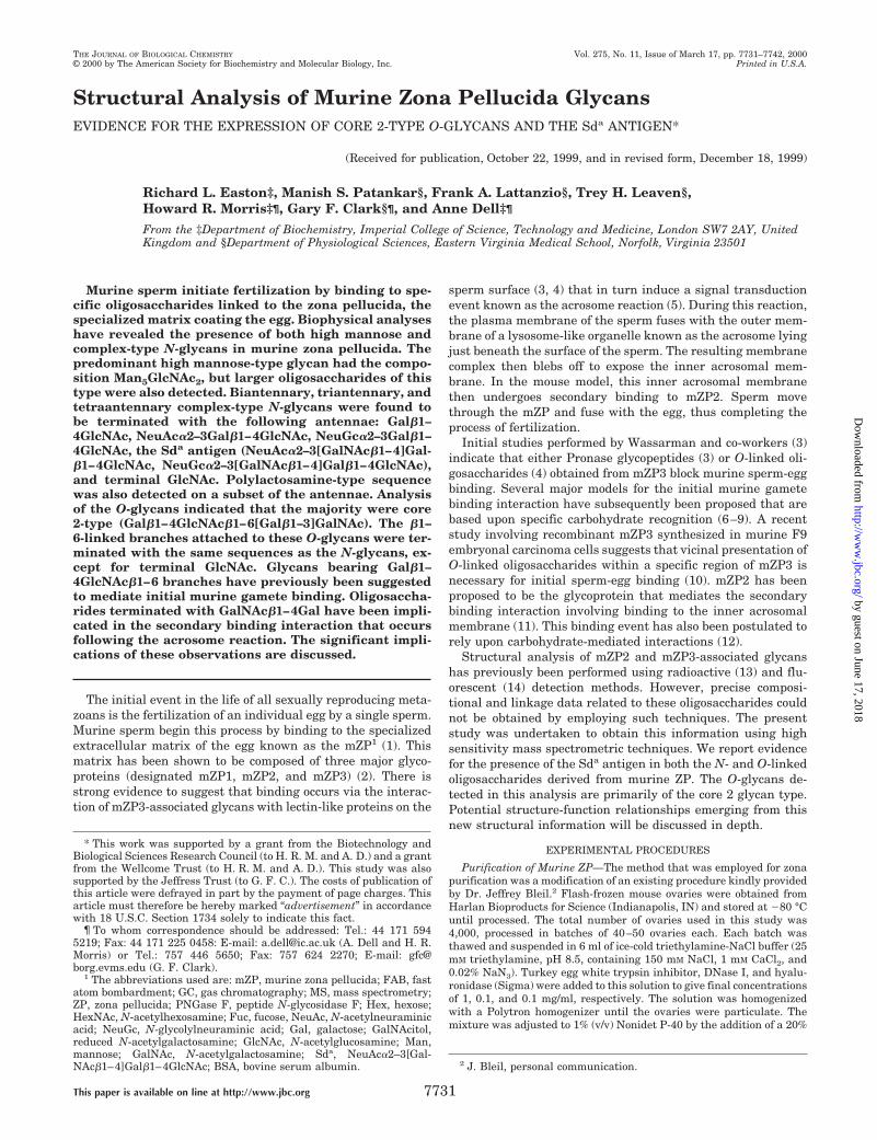
FIG. 1. FAB mass spectrum of permethylated N-glycans from murine zona pellucida. a, molecular ion region; b, fragment ion region.N-Glycans released by PNGase F from tryptic glycopeptides were purified by Sep-Pak, permethylated, and cleaned up by a second step of Sep-Pakpurification before FAB-MS analysis.
Murine Zona Pellucida-derived Glycans7732
by guest on June 17, 2018http://w
ww
.jbc.org/D
ownloaded from

ml of 100 mM sodium acetate, pH 5.0, at 37 °C for 48 h. Digestion withStreptomyces plicatus b-N-acetylhexosaminidase (recombinant fusionprotein, New England BioLabs) was performed with 50 units of enzymein 200 ml of 50 mM ammonium acetate, pH 4.5, at 37 °C for 24 h.
FAB-MS Analysis—FAB mass spectra of permethylated oligosaccha-rides were acquired using a ZAB-2S.E.-2FPD double-focusing massspectrometer fitted with a cesium ion gun operating at 30 kV. Dataanalysis was performed using VG Analytical Opus®software. Solventand matrices were as described previously (15).
GC-MS Analysis—GC-MS analysis was performed on a Fisons In-struments MD800 machine. Separation was achieved using an RTX-5fused silica capillary column (30 m 3 0.25 mm, Restek Corp). Partiallymethylated alditol acetates were dissolved in hexanes and loaded di-rectly onto the column at 65 °C. The column was held at 65 °C for 1 minand then increased to 290 °C at a rate of 8 °C/min.
RESULTS
Isolation and Characterization of ZP—Murine ZP were iso-lated by a modification of an original procedure (2) provided tous by Dr. Jeffrey Bleil (Scripts Research Institute, La Jolla,CA). Visual analysis of the preparation indicated the presenceof individual zonae exactly as was observed in the originalisolation. SDS-gel electrophoresis of the isolated ZP using thesame conditions employed in this earlier study indicated thepresence of three major diffuse bands consistent with molecu-lar weights of mZP1, mZP2, and mZP3 proposed earlier (2)(data not shown). Based on these criteria, the isolated zonae
were subjected to carbohydrate structural analysis.Mapping of the N-Glycan Population—Purified ZP were di-
gested with trypsin and incubated with PNGase F to releaseN-glycans. The oligosaccharides were separated from peptidesand glycopeptides via reverse phase chromatography on a C18Sep-Pak cartridge. The N-glycans were permethylated and sep-arated from other reactants and products by a second step ofSep-Pak reverse phase purification (15).
The permethylated glycans were subjected to FAB-MS anal-ysis using an established method (15). The data indicated thepresence of a range of N-glycans of both the high mannose andcomplex-types (Fig. 1, Table I). The following structural fea-tures were evident. (i) The most abundant component was thehigh mannose structure Hex5HexNAc2 indicated by the molec-ular ion at m/z 1580. Lower levels of larger high mannose-typeglycans (Hex6–9HexNAc2) were also indicated by the presenceof molecular ions at m/z 1784, 1988, 2192, and 2396 (ii) Com-plex-type biantennary N-glycans decorated with fucose andN-acetylneuraminic acid were also found (Hex5HexNAc4Fuc atm/z 2244; NeuAcHex5HexNAc4 at m/z 2432, NeuAc2Hex5-HexNAc4Fuc at m/z 2966). (iii) Some of these biantennaryglycans contain N-glycolylneuraminic acid, as indicated by sig-nals consistent with NeuGcHex5HexNAc4 (m/z 2462),NeuGcHex5HexNAc4Fuc(m/z2636),andNeuGc2Hex5HexNAc4-Fuc (m/z 3027). The presence of N-glycolylneuraminic acid wasalso confirmed by the detection of A-type fragment ions at m/z855 (NeuGcHexHexNAc1) and m/z 406 (NeuGc1). (iv) BothN-acetylneuraminic acid and N-glycolylneuraminic acid can belocated on the same N-glycan, as indicated by the molecular ionat m/z 2996, consistent with NeuAcNeuGcHex5HexNAc4. (v)Minor amounts of lactosamine repeats are present, as indicatedby the ion at m/z 913 (Hex2HexNAc2
1). (vi) A fragment ion atm/z 668 (Hex2HexNAc1) is consistent with the presence ofterminal Gala1–3Gal. (vii) The presence of A-type ions at m/z1070 and m/z 1100 suggest unusual terminal epitopes with thecompositions NeuAcHexHexNAc2
1 and NeuGcHexHexNAc21,
respectively.Linkage Analysis of Released Murine ZP N-Glycans—Perm-
ethylated N-glycans were acid-hydrolyzed, deuteroreduced,and peracetylated. The resulting partially methylated alditolacetates were analyzed by GC-MS (Table II). Notable featuresof the methylation analysis were the presence of 3,4-disubsti-tuted galactose and terminal N-acetylgalactosamine. The de-tection of 2 (major)-, 2,4 (minor)-, and 2,6 (minor)-linked man-
TABLE IAssignments of FAB-MS peaks observed for the molecular and
fragment ions of the permethylated N-glycans released from murinezona pellucida
Signal Assignment
m/z
High mass region1580 Hex5HexNAc2 1 Na1
1784 Hex6HexNAc2 1 Na1
1836 Hex3HexNAc4Fuc 1 Na1
1988 Hex7HexNAc2 1 Na1
2040 Hex4HexNAc4Fuc 1 Na1
2081 Hex3HexNAc5Fuc 1 Na1
2192 Hex8HexNAc2 1 Na1
2244 Hex5HexNAc4Fuc 1 Na1
2285 Hex4HexNAc5Fuc 1 Na1
2396 Hex9HexNAc2 1 Na1
2432 NeuAcHex5HexNAc4 1 Na1
2462 NeuGcHex5HexNAc4 1 Na1
2490 Hex5HexNAc5Fuc 1 Na1
2606 NeuAcHex5HexNAc4Fuc 1 Na1
2636 NeuGcHex5HexNAc4Fuc 1 Na1
2810 NeuAcHex6HexNAc4Fuc 1 Na1
2840 NeuGcHex6HexNAc4Fuc 1 Na1
2852 NeuGc2Hex5HexNAc4 1 Na1
2966 NeuAc2Hex5HexNAc4Fuc 1 Na1
2996 NeuAcNeuGcHex5HexNAc4 1 Na1
3027 NeuGc2Hex5HexNAc4Fuc 1 Na1
3243 NeuAc2Hex6HexNAc5 1 Na1
3417 NeuAc2Hex6HexNAc5Fuc 1 Na1
3808 NeuAc2NeuGcHex6HexNAc5Fuc 1 Na1
Low mass region260 HexNAc1
344 Loss of methanol from m/z 376374 Loss of methanol from m/z 406376 NeuAc1
406 NeuGc1
432 Loss of methanol from m/z 464464 HexHexNAc1
505 HexNAc21
636 Loss of methanol from m/z 668668 Hex2HexNAc1
793 Loss of methanol from m/z 825823 Loss of methanol from m/z 855825 NeuAcHexHexNAc1
855 NeuGcHexHexNAc1
913 Hex2HexNAc21
1070 NeuAcHexHexNAc21
1100 NeuGcHexHexNAc21
TABLE IIGC-MS analysis of the partially methylated alditol acetates obtained
from the N-glycans of murine zona pellucida
Elutiontime Characteristic fragment ions Assignment
min
17.23 115, 118, 131, 175 Terminal fucose18.83 102, 118, 129, 145, 161, 162, 205 Terminal mannose19.13a 102, 118, 129, 145, 161, 162, 205 Terminal galactose20.05 129, 139, 161, 190 2-Linked mannose20.36b 118, 129, 161, 234 3-Linked galactose20.93b 99, 102, 118, 129, 161, 162, 189, 233 6-Linked galactose21.07b 118, 129, 143, 185, 203, 232, 305 3,4-Linked galactose21.32 130, 190, 233 2,4-Linked mannose21.75 129, 130, 189, 190 2,6-Linked mannose21.93 118, 129, 189, 234 3,6-Linked mannose22.13 118, 129, 189, 234 3,6-Linked galactose22.40 118, 333 3,4,6-Linked mannose22.97 117, 159, 203, 205 Terminal GlcNAc23.45 117, 159, 203, 205 Terminal GalNAc23.89 117, 159, 233 4-Linked GlcNAc25.29 117, 159, 261 4,6-Linked GlcNAc
a Signals increased after treatment of N-glycans with A. ureafacienssialidase.
b Signals reduced or absent after treatment of N-glycans.
Murine Zona Pellucida-derived Glycans 7733
by guest on June 17, 2018http://w
ww
.jbc.org/D
ownloaded from

nose indicated that tri- and tetraantennary structures werepresent in addition to the major biantennary structures. Bi-sected-type glycan structures also exist, as shown by the pres-ence of 3,4,6-linked mannose, albeit as very minor components.
Endo-b-galactosidase Digestion of Murine ZP N-Gly-cans—To analyze the terminal structures of the minorpolylactosamine-containing N-glycans, PNGase F-releasedN-linked oligosaccharides were digested with endo-b-galacto-sidase. After digestion, a fraction of the reaction products waspermethylated, separated from contaminants by reverse phaseseparation on a Sep-Pak cartridge, and analyzed by FAB-MS.The data (Fig. 2, Table III) indicate the presence of severalcomponents not observed before digestion. These componentsrepresent the antennae of complex N-glycans bearing a rangeof different terminal structures. The high mannose-type struc-tures and shorter complex structures were unaffected by thisenzyme treatment. Because of the specificity of the enzyme, theoligosaccharides released from the nonreducing termini ofpolylactosamine-containing antennae all have a reducing Galresidue. Thus, significant signals were observed at m/z 926(Hex2HexNAc1-Gal), which likely represents structures termi-nating in a-linked galactose, m/z 1328 (NeuAcHex1HexNAc2-Gal), and its N-glycolylneuraminic acid counterpart at m/z1358 (NeuGcHex1HexNAc2-Gal). The large signal at m/z 518represents the disaccharide GlcNAc-Gal, the major productexpected after endo-b-galactosidase of polylactosamine-typechains.
Confirmation That a-Linked Galactose Is Associated withMurine ZP Glycans—The products of endo-b-galactosidase di-gestion were further digested with a-galactosidase, permethy-lated, and analyzed by FAB-MS. The signal at m/z 926 (Fig. 2)
FIG. 2. FAB mass spectra of the products of digestion of N-glycans from murine zona pellucida with endo-b-galactosidase. a, 35%acetonitrile Sep-Pak fraction; b, 50% acetonitrile Sep-Pak fraction. After digestion, the products were permethylated and purified by Sep-Pakbefore mass spectrometric analysis.
TABLE IIIAssignments of the molecular ions observed in the FAB mass spectraof murine zona pellucida N-glycans following digestion with endo-b-
galactosidase and permethylation
Signal Assignment
m/z
35% acetonitrile fraction518 HexNAcHex 1 Na1
700 Hex2HexNAc 1 H1
722 Hex2HexNAc 1 Na1
904 Hex3HexNAc 1 H1
926 Hex3HexNAc 1 Na1
1083 NeuAcHex2HexNAc 1 Na1
1113 NeuGcHex2HexNAc 1 Na1
1306 NeuAcHex2HexNAc2 1 H1
1328 NeuAcHex2HexNAc2 1 Na1
1336 NeuGcHex2HexNAc2 1 H1
1358 NeuGcHex2HexNAc2 1 Na1
50% acetonitrile fraction1580 Hex5HexNAc2 1 Na1
1784 Hex6HexNAc2 1 Na1
1988 Hex7HexNAc2 1 Na1
2040 Hex4HexNAc4Fuc 1 Na1
2081 Hex3HexNAc5Fuc 1 Na1
2192 Hex8HexNAc2 1 Na1
2244 Hex5HexNAc4Fuc 1 Na1
2285 Hex4HexNAc5Fuc 1 Na1
2396 Hex9HexNAc2 1 Na1
2490 Hex5HexNAc5Fuc 1 Na1
2531 Hex4HexNAc6Fuc 1 Na1
2606 NeuAcHex5HexNAc4Fuc 1 Na1
2636 NeuGcHex5HexNAc4Fuc 1 Na1
2735 Hex5HexNAc6Fuc 1 Na1
2852 NeuGc2Hex5HexNAc4 1 Na1
2922 NeuGcHex4HexNAc6Fuc 1 Na1
3126 NeuGcHex5HexNAc6Fuc 1 Na1
3517 NeuGc2Hex5HexNAc6Fuc 1 Na1
Murine Zona Pellucida-derived Glycans7734
by guest on June 17, 2018http://w
ww
.jbc.org/D
ownloaded from

was absent after a-galactosidase digestion (data not shown),demonstrating that it contained a-linked galactose.
Confirmation of the Presence of High Mannose-type N-Gly-cans on Murine ZP—Another aliquot of the endo-b-galactosid-ase-treated N-glycans was digested with a-mannosidase. Theproducts of the digestion were analyzed by FAB-MS after per-methylation. All signals corresponding to the high mannose-type sequences were absent, but those of the complex struc-tures were still detected (Fig. 3). This result is consistent withthe presence of high mannose-type sequences in this mixture.
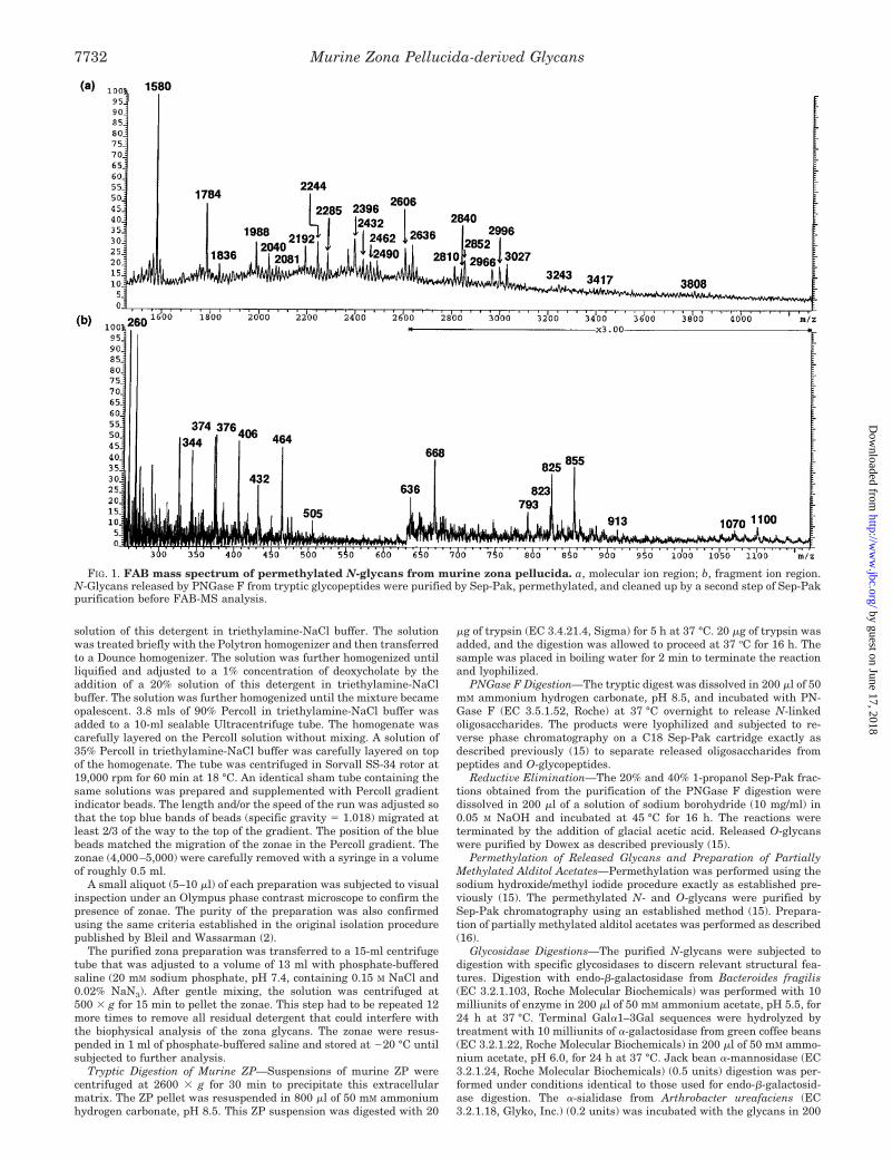
Identification of the Sda Determinant on Murine ZPN-Linked Oligosaccharides—The A-type fragment ions at m/z1070 and m/z 1100 observed in the N-glycan mapping experi-ment have compositions consistent with the Sda determinant(GalNAcb1–4(NeuAca2–3)Galb14GlcNAc) and its N-glycolyl-neuraminic acid counterpart. In addition, the results of thelinkage analysis and endo-b-galactosidase experiments sup-port the presence of this epitope (see above). Further informa-tion on the structures of these epitopes was obtained from thefollowing experiments. FAB-MS of the permethylated reactionproducts of a-sialidase digestion of the PNGase F-releasedmurine ZP N-glycans showed the release of both N-acetyl-neuraminic acid and N-glycolylneuraminic acid (Fig. 4, TableIV). Thus the fragment ions attributable to NeuAc-containingstructures originally detected in the undigested N-glycans atm/z 376, m/z 825, and m/z 1070 were absent after digestion. TheNeuGc-containing structures showed differing susceptibilitiesto the sialidase. The linear structure NeuGcHexHexNAc1 wasfully digested as shown by the absence of m/z 855, but thepotentially branched sequence, NeuGc[HexNAc]HexHexNAc,which is detected as an A-type ion at m/z 1100, was still presentafter digestion. A new fragment ion was present at m/z 709,corresponding to HexHexNAc2
1, the expected ion producedafter the removal of N-acetylneuraminic or N-glycolylneura-
minic acid from the Sda determinant. The small signal presentat m/z 1362 demonstrated the presence of low levels of polylac-tosamine repeats. This signal was not observed before sialidasedigestion, demonstrating that the polylactosamine repeats arecapped with N-acetylneuraminic acid or N-glycolylneuraminicacid. Linkage analysis of the products of digestion confirmedthe reduction in abundance of 3,4-linked galactose and anincrease in 4-linked galactose.
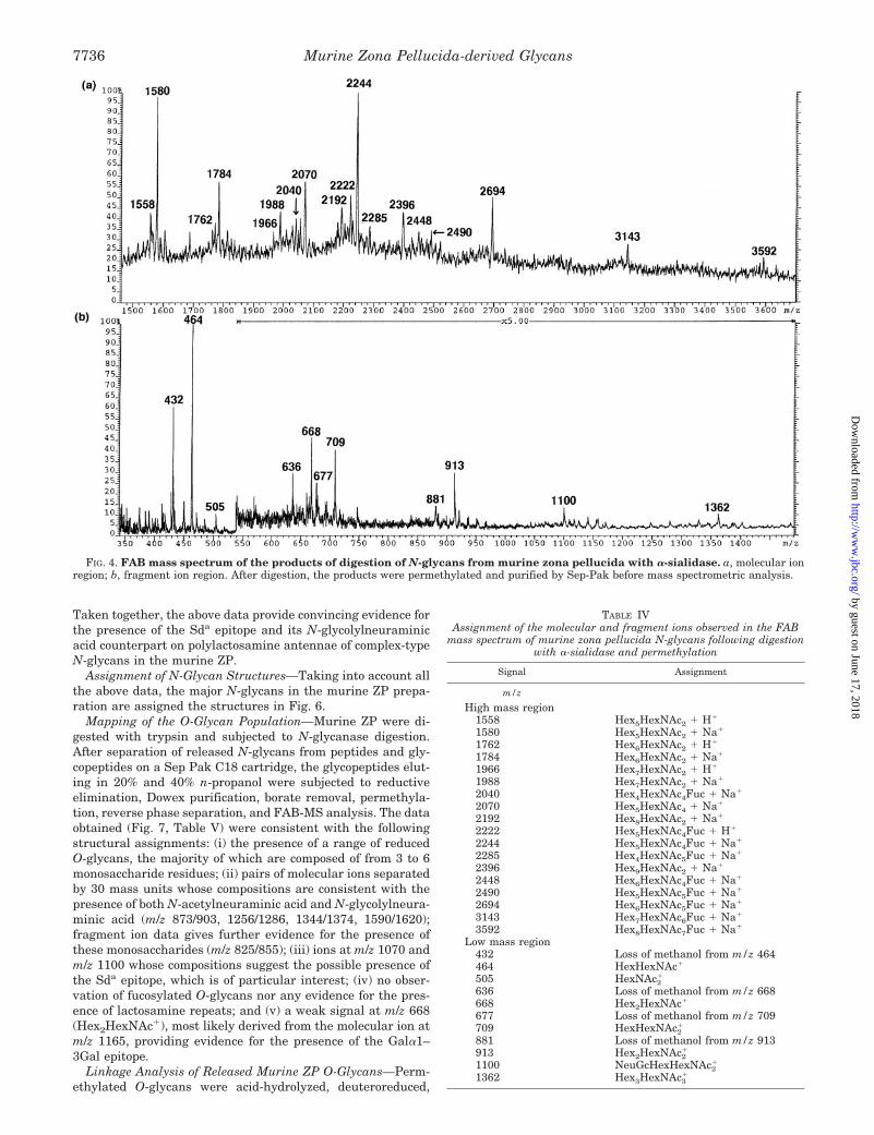
To provide further evidence for the putative Sda structure,an aliquot of the endo-b-galactosidase-treated murine ZP N-glycans was digested with a-sialidase. A portion of this reactionmixture was permethylated and analyzed by FAB-MS (Fig. 5a).The data indicated that N-acetylneuraminic acid had beenremoved (loss of m/z 1328), the signal at m/z 1358(NeuGcHex1HexNAc2-Gal) had been reduced in intensity, anda new signal had appeared at m/z 967, corresponding toHex1HexNAc2-Gal1Na1.
The remainder of the a-sialidase-treated fraction was furtherdigested with b-N-acetylhexosaminidase. An aliquot was re-moved for permethylation and FAB-MS analysis (Fig. 5b). Thedata showed significant reduction of the signal at m/z 967(Hex1HexNAc2-Gal1Na1) and the loss of the signal at 518(GlcNAc-Gal1Na1). The new minor signal at m/z 1171(Hex3HexNAc21Na1) is due to the loss of N-acetylglucosaminefrom truncated complex structures. To confirm that N-acetyl-galactosamine was removed, linkage analysis was performedon the a-sialidase-treated N-glycans and on the a-sialidaseplus b-N-acetylhexosaminidase-treated glycans. Comparison ofthe data indicated that terminal N-acetylgalactosamine waspresent before b-N-acetylhexosaminidase digestion but was ab-sent afterward. N-Acetylglucosamine continued to be detectedafter b-N-acetylhexosaminidase digestion, data that are con-sistent with the assignments of structures present in theFAB-MS spectrum after this full range of enzyme digests.
FIG. 3. FAB mass spectrum of the products of sequential digestion of N-glycans from murine zona pellucida with endo-b-galactosidase and a-mannosidase. After digestion, the products were permethylated and purified by Sep-Pak before mass spectrometricanalysis. For assignments of the signals, see Table I. Note the absence of signals corresponding to high mannose structures.
Murine Zona Pellucida-derived Glycans 7735
by guest on June 17, 2018http://w
ww
.jbc.org/D
ownloaded from
Taken together, the above data provide convincing evidence forthe presence of the Sda epitope and its N-glycolylneuraminicacid counterpart on polylactosamine antennae of complex-typeN-glycans in the murine ZP.
Assignment of N-Glycan Structures—Taking into account allthe above data, the major N-glycans in the murine ZP prepa-ration are assigned the structures in Fig. 6.
Mapping of the O-Glycan Population—Murine ZP were di-gested with trypsin and subjected to N-glycanase digestion.After separation of released N-glycans from peptides and gly-copeptides on a Sep Pak C18 cartridge, the glycopeptides elut-ing in 20% and 40% n-propanol were subjected to reductiveelimination, Dowex purification, borate removal, permethyla-tion, reverse phase separation, and FAB-MS analysis. The dataobtained (Fig. 7, Table V) were consistent with the followingstructural assignments: (i) the presence of a range of reducedO-glycans, the majority of which are composed of from 3 to 6monosaccharide residues; (ii) pairs of molecular ions separatedby 30 mass units whose compositions are consistent with thepresence of both N-acetylneuraminic acid and N-glycolylneura-minic acid (m/z 873/903, 1256/1286, 1344/1374, 1590/1620);fragment ion data gives further evidence for the presence ofthese monosaccharides (m/z 825/855); (iii) ions at m/z 1070 andm/z 1100 whose compositions suggest the possible presence ofthe Sda epitope, which is of particular interest; (iv) no obser-vation of fucosylated O-glycans nor any evidence for the pres-ence of lactosamine repeats; and (v) a weak signal at m/z 668(Hex2HexNAc1), most likely derived from the molecular ion atm/z 1165, providing evidence for the presence of the Gala1–3Gal epitope.
Linkage Analysis of Released Murine ZP O-Glycans—Perm-ethylated O-glycans were acid-hydrolyzed, deuteroreduced,
FIG. 4. FAB mass spectrum of the products of digestion of N-glycans from murine zona pellucida with a-sialidase. a, molecular ionregion; b, fragment ion region. After digestion, the products were permethylated and purified by Sep-Pak before mass spectrometric analysis.
TABLE IVAssignment of the molecular and fragment ions observed in the FAB
mass spectrum of murine zona pellucida N-glycans following digestionwith a-sialidase and permethylation
Signal Assignment
m/z
High mass region1558 Hex5HexNAc2 1 H1
1580 Hex5HexNAc2 1 Na1
1762 Hex6HexNAc2 1 H1
1784 Hex6HexNAc2 1 Na1
1966 Hex7HexNAc2 1 H1
1988 Hex7HexNAc2 1 Na1
2040 Hex4HexNAc4Fuc 1 Na1
2070 Hex5HexNAc4 1 Na1
2192 Hex8HexNAc2 1 Na1
2222 Hex5HexNAc4Fuc 1 H1
2244 Hex5HexNAc4Fuc 1 Na1
2285 Hex4HexNAc5Fuc 1 Na1
2396 Hex9HexNAc2 1 Na1
2448 Hex6HexNAc4Fuc 1 Na1
2490 Hex5HexNAc5Fuc 1 Na1
2694 Hex6HexNAc5Fuc 1 Na1
3143 Hex7HexNAc6Fuc 1 Na1
3592 Hex8HexNAc7Fuc 1 Na1
Low mass region432 Loss of methanol from m/z 464464 HexHexNAc1
505 HexNAc21
636 Loss of methanol from m/z 668668 Hex2HexNAc1
677 Loss of methanol from m/z 709709 HexHexNAc2
1
881 Loss of methanol from m/z 913913 Hex2HexNAc2
1
1100 NeuGcHexHexNAc21
1362 Hex3HexNAc31
Murine Zona Pellucida-derived Glycans7736
by guest on June 17, 2018http://w
ww
.jbc.org/D
ownloaded from
acetylated, and analyzed by GC-MS (Table VI). The presence of3-linked galactose and the absence of 6-linked galactose indi-cates that both N-acetylneuraminic acid and N-glycolylneura-minic acid are 3-linked to this monosaccharide. A weak spec-trum was obtained for 3,4-linked galactose, providing furtherevidence that the Sda structure is expressed in the O-glycans.Although the O-glycan preparation is contaminated with lowlevels of N-glycans as indicated by the signals for high mannosestructures in Fig. 6 and the variously linked mannoses in thelinkage data (Table VI), it is unlikely that the 3,4-linked ga-lactose is derived from N-glycan contaminants. This conclusionis arrived at by taking into consideration the low levels of Sda
containing N-glycans in the total N-glycan population and thefact that complex-type N-glycans were not detectable in theFAB spectra of the O-glycan preparation.
Assignment of O-Glycan Structures—Rigorous characteriza-tion of each of the O-glycans is particularly challenging becauseof the low levels of material available and the complexity of theO-glycan population. Nevertheless some firm conclusions canbe drawn from the data. In particular the observation of 3,6-linked GalNAcitol and the absence of detectable levels of3-linked GalNAcitol in the linkage analysis indicate that themajority of O-glycans are likely to have core-type 2 structures.Fig. 8 shows the sequences that have been assigned from theFAB and linkage data.
DISCUSSION
This report outlines the first characterization of murine ZPglycans using very sensitive biophysical methods of analysis todetermine their precise structural features. Structural analysisof glycans derived from mZP2 and mZP3 have been performedin previous studies (13, 14). However, because of the analytical
methods used, these investigations did not provide the explicitkind of data that was generated in the present study. Thefindings reported in this study are crucial because the mouse isthe most flexible mammalian model for experimental manipu-lation. Therefore this study provides complementary data thatmay now finally enable the murine gamete binding interactionto be understood at the molecular level. As will be discussed,this information may also provide more insight into potentialimmunological relationships present in the eutherian repro-ductive system.
The proposed structures for the major N-linked oligosaccha-rides in mZP are shown in Fig. 6. Based on the current study,the majority of the glycans associated with this extracellularmatrix are primarily high mannose and biantennary complexglycans, with lesser amounts of tri- and tetraantennary com-plex-type oligosaccharides. The complex N-glycans exhibit arange of terminal structures, the great majority of which havebeen previously identified during an investigation of mZP2-and mZP3-derived N-glycans (14). Our data confirm the con-clusion of Noguchi and Nakano (14) that polylactosamine se-quences and terminal a-linked galactose are associated withmZP glycoproteins. An additional structural feature revealedin our work is the presence of the Sda antigen on a subset of theglycans, a sequence not reported in the earlier structural anal-ysis (14). However, Noguchi and Nakano were required toremove sialic acid from the acidic glycans associated withmZP2 and mZP3 before obtaining structural analysis of thecore sequences (14). These investigators reported the presenceof terminal GalNAcb1-Galb1–4GlcNAc (terminal GalNAc link-age not defined) on 4% of the terminal sequences associatedwith mZP3 (14). This nonreducing trisaccharide sequence
FIG. 5. FAB mass spectra of the products of sequential digestion of N-glycans from murine zona pellucida with endo-b-galacto-sidase and a-sialidase (a) and a-sialidase (b) followed by b-N-acetylhexosaminidase. After digestion, the products were permethylatedand purified by Sep-Pak before mass spectrometric analysis.
Murine Zona Pellucida-derived Glycans 7737
by guest on June 17, 2018http://w
ww
.jbc.org/D
ownloaded from
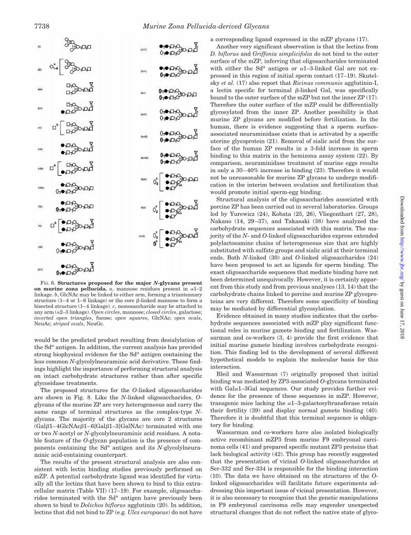
would be the predicted product resulting from desialylation ofthe Sda antigen. In addition, the current analysis has providedstrong biophysical evidence for the Sda antigen containing theless common N-glycolylneuraminic acid derivative. These find-ings highlight the importance of performing structural analysison intact carbohydrate structures rather than after specificglycosidase treatments.
The proposed structures for the O-linked oligosaccharidesare shown in Fig. 8. Like the N-linked oligosaccharides, O-glycans of the murine ZP are very heterogeneous and carry thesame range of terminal structures as the complex-type N-glycans. The majority of the glycans are core 2 structures(Galb1–4GlcNAcb1–6[Galb1–3]GalNAc) terminated with oneor two N-acetyl or N-glycolylneuraminic acid residues. A nota-ble feature of the O-glycan population is the presence of com-ponents containing the Sda antigen and its N-glycolylneura-minic acid-containing counterpart.
The results of the present structural analysis are also con-sistent with lectin binding studies previously performed onmZP. A potential carbohydrate ligand was identified for virtu-ally all the lectins that have been shown to bind to this extra-cellular matrix (Table VII) (17–19). For example, oligosaccha-rides terminated with the Sda antigen have previously beenshown to bind to Dolichos biflorus agglutinin (20). In addition,lectins that did not bind to ZP (e.g. Ulex europaeus) do not have
a corresponding ligand expressed in the mZP glycans (17).Another very significant observation is that the lectins from
D. biflorus and Griffonia simplicifolia do not bind to the outersurface of the mZP, inferring that oligosaccharides terminatedwith either the Sda antigen or a1–3-linked Gal are not ex-pressed in this region of initial sperm contact (17–19). Skutel-sky et al. (17) also report that Ricinus communis agglutinin-I,a lectin specific for terminal b-linked Gal, was specificallybound to the outer surface of the mZP but not the inner ZP (17).Therefore the outer surface of the mZP could be differentiallyglycosylated from the inner ZP. Another possibility is thatmurine ZP glycans are modified before fertilization. In thehuman, there is evidence suggesting that a sperm surface-associated neuraminidase exists that is activated by a specificuterine glycoprotein (21). Removal of sialic acid from the sur-face of the human ZP results in a 3-fold increase in spermbinding to this matrix in the hemizona assay system (22). Bycomparison, neuraminidase treatment of murine eggs resultsin only a 30–40% increase in binding (23). Therefore it wouldnot be unreasonable for murine ZP glycans to undergo modifi-cation in the interim between ovulation and fertilization thatwould promote initial sperm-egg binding.
Structural analysis of the oligosaccharides associated withporcine ZP has been carried out in several laboratories. Groupsled by Yurewicz (24), Kobata (25, 26), Vliegenthart (27, 28),Nakano (14, 29–37), and Takasaki (38) have analyzed thecarbohydrate sequences associated with this matrix. The ma-jority of the N- and O-linked oligosaccharides express extendedpolylactosamine chains of heterogeneous size that are highlysubstituted with sulfate groups and sialic acid at their terminalends. Both N-linked (30) and O-linked oligosaccharides (24)have been proposed to act as ligands for sperm binding. Theexact oligosaccharide sequences that mediate binding have notbeen determined unequivocally. However, it is certainly appar-ent from this study and from previous analyses (13, 14) that thecarbohydrate chains linked to porcine and murine ZP glycopro-teins are very different. Therefore some specificity of bindingmay be mediated by differential glycosylation.
Evidence obtained in many studies indicates that the carbo-hydrate sequences associated with mZP play significant func-tional roles in murine gamete binding and fertilization. Was-sarman and co-workers (3, 4) provide the first evidence thatinitial murine gamete binding involves carbohydrate recogni-tion. This finding led to the development of several differenthypothetical models to explain the molecular basis for thisinteraction.
Bleil and Wassarman (7) originally proposed that initialbinding was mediated by ZP3-associated O-glycans terminatedwith Gala1–3Gal sequences. Our study provides further evi-dence for the presence of these sequences in mZP. However,transgenic mice lacking the a1–3-galactosyltransferase retaintheir fertility (39) and display normal gamete binding (40).Therefore it is doubtful that this terminal sequence is obliga-tory for binding.
Wassarman and co-workers have also isolated biologicallyactive recombinant mZP3 from murine F9 embryonal carci-noma cells (41) and prepared specific mutant ZP3 proteins thatlack biological activity (42). This group has recently suggestedthat the presentation of vicinal O-linked oligosaccharides atSer-332 and Ser-334 is responsible for the binding interaction(10). The data we have obtained on the structures of the O-linked oligosaccharides will facilitate future experiments ad-dressing this important issue of vicinal presentation. However,it is also necessary to recognize that the genetic manipulationsin F9 embryonal carcinoma cells may engender unexpectedstructural changes that do not reflect the native state of glyco-
FIG. 6. Structures proposed for the major N-glycans presenton murine zona pellucida. a, mannose residues present in a1–2linkage. b, GlcNAc may be linked to either arm, forming a triantennarystructure (1–4 or 1–6 linkage) or the core b-linked mannose to form abisected structure (1–4 linkage). c, monosaccharide may be attached toany arm (a2–3 linkage). Open circles, mannose; closed circles, galactose;inverted open triangles, fucose; open squares, GlcNAc; open ovals,NeuAc; striped ovals, NeuGc.
Murine Zona Pellucida-derived Glycans7738
by guest on June 17, 2018http://w
ww
.jbc.org/D
ownloaded from

sylation of mZP glycoproteins.Shur and co-workers (6, 43) propose that a sperm-specific
b1–4-galactosyltransferase mediates binding by recognizingmZP3-associated O-glycans terminated with GlcNAc. Our cur-rent results indicate the presence of minor amounts of GlcNAc-terminated N-glycans, whereas terminal GlcNAc was not de-tectable in the O-glycans. Terminal GalNAc was also found, butit is presented in the context of Sda antigen. In addition, thelack of D. biflorus agglutinin binding to the mZP surface (TableVII) suggests that this b-galactosyltransferase does not haveimmediate access to Sda-terminated oligosaccharides. Finally,studies performed by Lu and Shur (41) indicate that transgenicmice lacking this specific b1–4-galactosyltransferase display3–4-fold higher binding to eggs compared with control mice(44). Therefore, based on all available data, it is very unlikelythat this galactosyltransferase plays any significant role in theinitial binding process.
Johnston et al. (9) recently proposed that glycans terminatedwith either Gala1–3Galb1–4[Fuca1–3]GlcNAc or Lewisx-ac-tive sequences (Galb1–4[Fuca1–3]GlcNAcb1–4GlcNAc) couldact as high affinity ligands that mediate initial sperm-eggbinding. In addition, these investigators also report that thetrisaccharide Galb1–4GlcNAcb1–4GlcNAc maximally inhib-ited sperm-egg binding by 47% at the highest tested concentra-tion (72 mM). A synergistic effect was observed when Gala1–3Galb1–4[Fuca1–3]GlcNAc and Galb1–4GlcNAcb1–4GlcNAcwere included together. Based upon this evidence, these inves-tigators proposed that Gala1–3Galb1–4[Fuca1–3]GlcNAccould be binding to sp56, a sperm protein previously implicatedin binding to ZP3 (45). They also suggested that another sperm-
associated calcium-dependent lectin that binds terminalGalb1–4GlcNAc sequences (46, 47) interacted with low affinityligands like Galb1–4GlcNAcb1–4GlcNAc (9).
Our work indicates that glycans carrying terminal Gala1–3Galb1–4[Fuca1–3]GlcNAc or Galb1–4[Fuca1–3]GlcNAc arenot expressed in murine ZP, consistent with the results of aprevious study (14). We have found that fucose is attached tothe N-glycans but apparently only via a1–6 linkage to thechitobiose core. No evidence for the fucosylation of O-glycanswas found. In addition, antibodies directed against the Lewisx
sequence did not bind to eggs obtained from transgenic micelacking a-galactosyltransferase.3 Therefore, although the inhi-bition mediated by the small fucosylated oligosaccharides isgenuinely interesting, it is very unlikely that such ligands arephysiologically relevant in the murine gamete binding system.
Tulsiani and co-workers (8) suggest that initial sperm-eggbinding is mediated via recognition of terminal a-mannosylresidues by a specific sperm surface a-mannosidase. High man-nose-type glycans are located in mZP, based on the results ofthe present study. The physiological relationship between thisa-mannosidase and sperm adhesion needs to be more fullyinvestigated.
It is also significant that murine sperm will bind to surfacesother than their homologous eggs. For example, murine spermundergo rapid and very tight binding to rabbit erythrocytes (48,49). Electron microscopy of sperm-erythrocyte interaction indi-cates that this binding is between the plasma membranes of
3 A. Thall, personal communication.
FIG. 7. FAB mass spectrum of the permethylated O-glycans from murine zona pellucida. Tryptic glycopeptides derived from murinezona pellucida were treated with PNGase F followed by reductive elimination of the Sep-Pak-purified O-glycopeptides, Dowex purification, borateremoval, permethylation, Sep-Pak clean up and FAB-MS analysis. The inset shows the region from m/z 950 to m/z 1300 following the addition ofa small amount of acid to enhance A-type ion formation.
Murine Zona Pellucida-derived Glycans 7739
by guest on June 17, 2018http://w
ww
.jbc.org/D
ownloaded from

the two cell types (50). Periodate oxidation of rabbit erythro-cytes (10 mM NaIO4, 0.15 M NaCl, 1 h, 23 °C) results in a .98%reduction in binding.4 Thus sperm binding to rabbit erythro-cytes (49) is both carbohydrate-dependent and requires acro-some-intact sperm, precisely the same requirements associatedwith murine sperm-ZP binding. This result suggests that bothsperm-erythrocyte binding and initial sperm-egg binding in-volve specific lectins associated with the plasma membrane ofmurine sperm.
Previous studies indicate that the O-glycans associated withmZP3 are responsible for mediating adhesion (4, 43). Based onthe current data, the carbohydrate sequence common to mZPO-glycans and rabbit erythrocytes is the b1–6-linkedN-acetyllactosamine (Galb1–4GlcNAcb1–6) sequence. Unlikered blood cells from other species, rabbit erythrocytes profuselyexpress branches of this type on polylactosamine sequencesassociated with their glycolipids (51) and possibly their N-glycans but not on their O-linked oligosaccharides (52). Virtu-ally all of the rabbit erythrocyte glycolipids are also terminatedwith Gala1–3Gal sequences, but murine sperm bind to rabbiterythrocytes even after exhaustive digestion with a-galactosid-ase (49). As stated beforehand, murine sperm-egg binding isalso not dependent upon the presence of terminal a1–3-linkedgalactose (39, 40). If the mutation studies involving recombi-nant mZP3 are indeed correct (10), then vicinal presentation ofcore 2 O-glycans (each presenting terminal b1–6-linked N-acetyllactosamine units) at Ser-332 and Ser-334 could be re-sponsible for mediating the initial binding interaction. Thishypothesis must be thoroughly investigated.
Cahova and Draber (12) report that an IgM monoclonalantibody (Tec-02) directed against terminal GalNAcb1–4Galbinds to murine ZP and inhibits fertilization in a concentration-dependent manner. However, Tec-02 did not interfere with theinitial sperm-ZP binding but did inhibit secondary binding thatoccurs after the induction of the acrosome reaction. Cahova andDraber (12) therefore proposed that glycans terminated withGalNAcb1–4Gal sequences mediate secondary gamete bindingin the mouse. In our study, we have clearly shown that theglycans bearing terminal GalNAcb1–4Gal sequences are pres-ent but are specifically associated with the Sda antigen. Thusthe structural data in combination with the antibody inhibitiondata indicates that the secondary binding interaction may bedependent upon the presence of the sequences terminated withthe Sda antigen. Again, this hypothesis needs to be thoroughlytested (12).
4 M. Patankar and G. Clark, unpublished observation.
FIG. 8. Structures proposed for themajor O-glycans present on murinezona pellucida. a, monosaccharide maybe attached to either the 3 or 6 branch ofthe O-glycan. Closed squares, GalNAc;closed circles, galactose; open squares, Gl-cNAc; open ovals, NeuAc; striped ovals,NeuGc
TABLE VIGC-MS analysis of the partially methylated alditol acetates obtained
from the O-glycan preparation of murine zona pellucida
Elutiontime Characteristic fragment ions Assignment
min
18.83 102, 118, 129, 145, 161, 162, 205 Terminal mannose19.13 102, 118, 129, 145, 161, 162, 205 Terminal galactose20.05 129, 139, 161, 190 2-Linked mannose20.36 118, 129, 161, 234 3-Linked galactose21.07 118, 129, 143, 185, 203, 232, 305 3,4-Linked galactose21.93 118, 129, 189, 234 3,6-Linked mannose23.40 130, 246, 318 3,6-Linked GalNAcitol23.89 117, 159, 233 4-Linked GlcNAc
TABLE VAssignment of FAB-MS peaks observed for the molecular and
fragment ions of the permethylated O-glycans released from murinezona pellucida
Signal Assignment
m/z
793 Loss of methanol from m/z 825825 NeuAcHexHexNAc1
855 NeuGcHexHexNAc1
873 NeuAcHexHexNAcitol 1 H1
895 NeuAcHexHexNAcitol 1 Na1
903 NeuAcHexHexNAcitol 1 H1
925 NeuGcHexHexNAcitol 1 Na1
983 Hex2HexNAcHexNAcitol 1 Na1
1070 NeuAcHexHexNAc21
1100 NeuGcHexHexNAc21
1140 NeuAcHexHexNAcHexNAcitol 1 Na1
1165 Hex3HexNAcHexNAcitol 1 H1
1187 Hex3HexNAcHexNAcitol 1 Na1
1234 NeuAc2HexHexNAcitol 1 H1
1256 NeuAc2HexHexNAcitol 1 Na1
1264 NeuAcNeuGcHexHexNAcitol 1 H1
1286 NeuAcNeuGcHexHexNAcitol 1 Na1
1294 NeuGc2HexHexNAcitol 1 H1
1316 NeuGc2HexHexNAcitol 1 Na1
1344 NeuAcHex2HexNAcHexNAcitol 1 Na1
1374 NeuGcHex2HexNAcHexNAcitol 1 Na1
1432 Hex3HexNAc2HexNAcitol 1 Na1
1531 NeuAcNeuGcHexHexNAcHexNAcitol 1 Na1
1549 NeuAcHex3HexNAcHexNAcitol 1 Na1
1590 NeuAcHex2HexNAc2HexNAcitol 1 Na1
1595 Hex5HexNAcHexNAcitol 1 Na1
1620 NeuGcHex2HexNAc2HexNAcitol 1 Na1
1800 Hex6HexNAcHexNAcitol 1 Na1
1824 NeuGcHex3HexNAc2HexNAcitol 1 Na1
1865 NeuGcHex2HexNAc3HexNAcitol 1 Na1
Murine Zona Pellucida-derived Glycans7740
by guest on June 17, 2018http://w
ww
.jbc.org/D
ownloaded from

Another very significant recent observation involves the in-duction of the acrosome reaction. Bovine serum albumin (BSA)-based neoglycoproteins terminated at multiple positions with asingle monosaccharide (GalNAc-BSA, GlcNAc-BSA, or Man-BSA) induce the acrosome reaction in mice (53). The currentstudy indicates that oligosaccharides with GalNAc, GlcNAc, orMan at the nonreducing ends are also present in the mZP.More study will be required to determine if mZP glycans par-ticipate in mediating signal transduction events during murinefertilization.
Initial human sperm binding to homologous zona pellucida isinhibited at low concentrations by glycodelin-A (54), a uterineglycoprotein with potent immunosuppressive activities (55).Structural analysis of the oligosaccharides associated with gly-codelin-A indicate that it expresses unusual fucosylated lacdi-NAc-type sequences (GalNAcb1–4[Fuca1–3]GlcNAc) on themajority of its glycans (56). Oligosaccharides of this type havepreviously been shown to be potent inhibitors of selectin-medi-ated adhesions (57). Based upon these both functional andstructural studies, we suggested the possibility that similarcarbohydrate sequences are utilized during immune and ga-mete recognition events in the human (56).
It is therefore significant to note in this context that theexpression of core 2 O-glycan sequences and the Sda antigenare significantly up-regulated on interleukin-2-stimulated Tlymphocytes (58, 59) and cytotoxic T lymphocytes (60–62) inthe mouse, respectively. Another major goal in the future is todetermine if the shared expression of these carbohydrate se-quences on murine ZP and activated lymphocytes has anyrelevant physiological implications in the mouse.
REFERENCES
1. Wassarman, P. M. (1990) Development 108, 1–172. Bleil, J. D., and Wassarman, P. M. (1980) Dev. Biol. 76, 185–2023. Florman, H. M., Bechtol, K. B., and Wassarman, P. M. (1984) Dev. Biol. 106,
243–2554. Florman, H. M., and Wassarman, P. M. (1985) Cell 41, 313–3245. Bleil, J. D., and Wassarman, P. M. (1983) Dev. Biol. 95, 317–3246. Shur, B. D., and Hall, N. G. (1982) J. Cell Biol. 95, 574–5797. Bleil, J. D., and Wassarman, P. M. (1988) Proc. Natl. Acad. Sci. U. S. A. 85,
6778–67828. Cornwall, G. A., Tulsiani, D. R., and Orgebin-Crist, M. C. (1991) Biol. Reprod.
44, 913–9219. Johnston, D. S., Wright, W. W., Shaper, J. H., Hokke, C. H., Van den Eijnden,
D. H., and Joziasse, D. H. (1998) J. Biol. Chem. 273, 1888–189510. Chen, J., Litscher, E. S., and Wassarman, P. M. (1998) Proc. Natl. Acad. Sci.
U. S. A. 95, 6193–619711. Bleil, J. D., Greve, J. M., and Wassarman, P. M. (1988) Dev. Biol. 128, 376–38512. Cahova, M., and Draber, P. (1992) J. Reprod. Immunol. 21, 241–25613. Nagdas, S. K., Araki, Y., Chayko, C. A., Orgebin-Crist, M. C., and Tulsiani,
D. R. (1994) Biol. Reprod. 51, 262–27214. Noguchi, S., and Nakano, M. (1993) Biochim. Biophys. Acta 1158, 217–22615. Dell, A., Khoo, K.-H., Panico, M., McDowell, R. A., Etienne, A. T., Reason, A. J.,
and Morris, H. R. (1993) in Glycobiology: A Practical Approach (Fukuda, M.,and Kobata, A., eds) pp. 187–222, Oxford University Press, Oxford
16. Albersheim, P., Nevins, D. J., English, P. D., and Karr, A. (1967) Carbohydr.
Res. 5, 340–34517. Skutelsky, E., Ranen, E., and Shalgi, R. (1994) J. Reprod. Fertil. 100, 35–4118. Aviles, M., Jaber, L., Castells, M. T., Ballesta, J., and Kan, F. W. (1997) Biol.
Reprod. 57, 1155–116319. Aviles, M., Castells, M. T., Abascal, I., Martinez-Menarguez, J. A., Draber, P.,
Kan, F. W., and Ballesta, J. (1999) Cell Tissue Res. 295, 269–27720. Blanchard, D., Cartron, J. P., Fournet, B., Montreuil, J., van Halbeek, H., and
Vliegenthart, J. F. (1983) J. Biol. Chem. 258, 7691–769521. Banerjee, M., and Chowdhury, M. (1997) Mol. Hum. Reprod. 3, 109–11422. Ozgur, K., Patankar, M. S., Oehninger, S., and Clark, G. F. (1998) Mol. Hum.
Reprod. 4, 318–32423. Mori, E., Mori, T., and Takasaki, S. (1997) Biochem. Biophys. Res. Commun.
238, 95–9924. Yurewicz, E. C., Pack, B. A., and Sacco, A. G. (1991) Mol. Reprod. Dev. 30,
126–13425. Mori, E., Takasaki, S., Hedrick, J. L., Wardrip, N. J., Mori, T., and Kobata, A.
(1991) Biochemistry 30, 2078–208726. Hirano, T., Takasaki, S., Hedrick, J. L., Wardrip, N. J., Amano, J., and Kobata,
A. (1993) Eur. J. Biochem. 214, 763–76927. Hokke, C. H., Damm, J. B., Penninkhof, B., Aitken, R. J., Kamerling, J. P., and
Vliegenthart, J. F. (1994) Eur. J. Biochem. 221, 491–51228. Hokke, C. H., Damm, J. B., Kamerling, J. P., and Vliegenthart, J. F. (1993)
FEBS Lett. 329, 29–3429. Noguchi, S., and Nakano, M. (1992) Eur. J. Biochem. 209, 883–89430. Noguchi, S., Hatanaka, Y., Tobita, T., and Nakano, M. (1992) Eur. J. Biochem.
204, 1089–110031. Noguchi, S., Hatanaka, Y., Tobita, T., and Nakano, M. (1992) Eur. J. Biochem.
207, 113032. Yonezawa, N., Aoki, H., Hatanaka, Y., and Nakano, M. (1995) Eur. J. Biochem.
233, 35–4133. Katsumata, T., Noguchi, S., Yonezawa, N., Tanokura, M., and Nakano, M.
(1996) Eur. J. Biochem. 240, 448–45334. Nakano, M., Yonezawa, N., Hatanaka, Y., and Noguchi, S. (1996) J. Reprod.
Fertil. 50, (suppl.) 25–3435. Yonezawa, N., Mitsui, S., Kudo, K., and Nakano, M. (1997) Eur. J. Biochem.
248, 86–9236. Kudo, K., Yonezawa, N., Katsumata, T., Aoki, H., and Nakano, M. (1998) Eur.
J. Biochem. 252, 492–49937. Yonezawa, N., Fukui, N., Kudo, K., and Nakano, M. (1999) Eur. J. Biochem.
260, 57–6338. Mori, E., Hedrick, J. L., Wardrip, N. J., Mori, T., and Takasaki, S. (1998)
Glycoconj. J. 15, 447–45639. Thall, A. D., Maly, P., and Lowe, J. B. (1995) J. Biol. Chem. 270, 21437–2144040. Liu, D. Y., Baker, H. W., Pearse, M. J., and d’Apice, A. J. (1997) Mol. Hum.
Reprod. 3, 1015–101641. Kinloch, R. A., Mortillo, S., Stewart, C. L., and Wassarman, P. M. (1991) J. Cell
Biol. 115, 655–66442. Liu, C., Litscher, E. S., and Wassarman, P. M. (1995) Mol. Biol. Cell 6, 577–58543. Miller, D. J., Macek, M. B., and Shur, B. D. (1992) Nature 357, 589–59344. Lu, Q., and Shur, B. D. (1997) Development 124, 4121–413145. Bookbinder, L. H., Cheng, A., and Bleil, J. D. (1995) Science 269, 86–8946. Goluboff, E. T., Mertz, J. R., Tres, L. L., and Kierszenbaum, A. L. (1995) Mol.
Reprod. Dev. 40, 460–46647. Abdullah, M., and Kierszenbaum, A. L. (1989) J. Cell Biol. 108, 367–37548. Yamagata, T., Ito, M., and Takahashi, K. (1983) in The Glycoconjugates
(Chester, M. A., Heinegard, A., and Svensson, S., eds) pp. 623–624, Rhams,New York
49. Sandow, B. A., and Clark, G. F. (1993) Biol. Reprod. 48, 166 (abstr.)50. Clark, G. F., Oehninger, S., and Seppala, M. (1996) Mol. Hum. Reprod. 2,
513–51751. Hanfland, P., Kordowicz, M., Peter Katalinic, J., Egge, H., Dabrowski, J., and
Dabrowski, U. (1988) Carbohydr. Res. 178, 1–2152. Fukuda, K., Honma, K., Manabe, H., Utsumi, H., and Hamada, A. (1987)
Biochim. Biophys. Acta 926, 132–13853. Loeser, C. R., and Tulsiani, D. R. (1999) Biol. Reprod. 60, 94–10154. Oehninger, S., Coddington, C. C., Hodgen, G. D., and Seppala, M. (1995) Fertil.
Steril. 63, 377–38355. Clark, G. F., Oehninger, S., Patankar, M. S., Koistinen, R., Dell, A., Morris,
TABLE VIILectin binding to murine zona pellucida reported in previous studies
Species of origin Location ofbindinga Potential ligands detected in the current study
Triticum vulgaris E Sialylated glycans, polylactosamine sequences (63–65)Triticum vulgaris (succinylated) E Polylactosamine sequences (66)Arachis hypogaea E Galb1–3GalNAc sequences (67)D. biflorus I Sda determinant (20)Maackia amurensis E Terminal Neu5Aca2–3Gal (68)G. simplicifolia I Terminal Gala1–3Gal sequences (69)Concanavalia ensiformis E High mannose, hybrid, and biantennary complex type N-linked glycans (70)Helix pomatia I Sda determinant (71)Aleuria aurantia E N-Linked glycans with fucose linked a1–6 to the core GlcNAc covalently bound to Asn (72)Datura stramonium E Tri- and tetraantennary complex N-linked glycans with b1–6- or b1–4-linked
N-acetyllactosamine units; polylactosamine sequences (73)R. communis E, Ob Terminal Galb1–4GlcNAc (74, 75)Limax flavus E Neu5Aca2–3Galb1–3GalNAc (76)
a E, binding to entire ZP; I, binding to inner ZP but not to outer surface; O, binding to outer surface only.b Entire ZP (18); outer surface (17).
Murine Zona Pellucida-derived Glycans 7741
by guest on June 17, 2018http://w
ww
.jbc.org/D
ownloaded from

H. R., Koistinen, H., and Seppala, M. (1996) Hum. Reprod. 11, 467–47356. Dell, A., Morris, H. R., Easton, R. L., Panico, M., Patankar, M., Oehninger, S.,
Koistinen, R., Koistinen, H., Seppala, M., and Clark, G. F. (1995) J. Biol.Chem. 270, 24116–24126
57. Grinnell, B. W., Hermann, R. B., and Yan, S. B. (1994) Glycobiology 4, 221–22558. Tsuboi, S., and Fukuda, M. (1997) EMBO J. 16, 6364–637359. Tsuboi, S., and Fukuda, M. (1998) J. Biol. Chem. 273, 30680–3068760. Lefrancois, L., and Bevan, M. J. (1985) Nature 314, 449–45261. Lefrancois, L., and Bevan, M. J. (1985) J. Immunol. 135, 374–38362. Lefrancois, L., Puddington, L., Machamer, C. E., and Bevan, M. J. (1985) J.
Exp. Med. 162, 1275–129363. Peters, B. P., Ebisu, S., Goldstein, I. J., and Flashner, M. (1979) Biochemistry
18, 5505–551164. Clark, G. F., and Ivatt, R. J. (1984) Fed. Proc. 43, 1696 (abstr.)65. Gallagher, J. T., Morris, A., and Dexter, T. M. (1985) Biochem. J. 231, 115–12266. Monsigny, M., Roche, A. C., Sene, C., Maget-Dana, R., and Delmotte, F. (1980)
Eur. J. Biochem. 104, 147–153
67. Uhlenbruck, G., Pardoe, G. I., and Bird, G. W. G. (1969) Z. Immunitaetsforsch.Allerg. Klin. Immunol. 138, 423–433
68. Wang, W. C., and Cummings, R. D. (1988) J. Biol. Chem. 263, 4576–458569. Hayes, C. E., and Goldstein, I. J. (1974) J. Biol. Chem. 249, 1904–191470. Cummings, R. D., and Kornfeld, S. (1982) J. Biol. Chem. 257, 11235–1124071. Blanchard, D., Piller, F., Gillard, B., Marcus, D., and Cartron, J. P. (1985)
J. Biol. Chem. 260, 7813–781672. Yamashita, K., Kochibe, N., Ohkura, T., Ueda, I., and Kobata, A. (1985) J. Biol.
Chem. 260, 4688–469373. Yamashita, K., Totani, K., Ohkura, T., Takasaki, S., Goldstein, I. J., and
Kobata, A. (1987) J. Biol. Chem. 262, 1602–160774. Olsnes, S., Saltvedt, E., and Pihl, A. (1974) J. Biol. Chem. 249, 803–81075. Smith, D. F., Prieto, P. A., McCrumb, D. K., and Wang, W. C. (1987) J. Biol.
Chem. 262, 12040–1204776. Ravindranath, M. H., Higa, H. H., Cooper, E. L., and Paulson, J. C. (1985)
J. Biol. Chem. 260, 8850–8856
Murine Zona Pellucida-derived Glycans7742
by guest on June 17, 2018http://w
ww
.jbc.org/D
ownloaded from

Morris, Gary F. Clark and Anne DellRichard L. Easton, Manish S. Patankar, Frank A. Lattanzio, Trey H. Leaven, Howard R.
EXPRESSION OF CORE 2-TYPE O-GLYCANS AND THE Sda ANTIGEN Structural Analysis of Murine Zona Pellucida Glycans: EVIDENCE FOR THE
doi: 10.1074/jbc.275.11.77312000, 275:7731-7742.J. Biol. Chem.
http://www.jbc.org/content/275/11/7731Access the most updated version of this article at
Alerts:
When a correction for this article is posted•
When this article is cited•
to choose from all of JBC's e-mail alertsClick here
http://www.jbc.org/content/275/11/7731.full.html#ref-list-1
This article cites 74 references, 32 of which can be accessed free at
by guest on June 17, 2018http://w
ww
.jbc.org/D
ownloaded from

![arXiv:2006.00067v2 [cs.CV] 20 Jul 2020of zona pellucida segmentation, all these features are used for embryo selec tion [1,27,2,24]; we segment the zona pellucida both to improve the](https://img.pdfslide.us/doc/110x75/60af951b4e64854d4508408b/arxiv200600067v2-cscv-20-jul-2020-of-zona-pellucida-segmentation-all-these.jpg)