Embed Size (px)
Citation preview
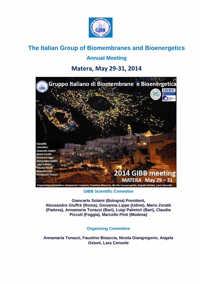
GIBB Scientific Commitee
Giancarlo Solaini (Bologna) President, Alessandro Giuffrè (Roma), Giovanna Lippe (Udine), Mario Zoratti (Padova), Annamaria Tonazzi (Bari), Luigi Palmieri (Bari), Claudia
Piccoli (Foggia), Marcello Pinti (Modena)
Organizing Committee
Annamaria Tonazzi, Faustino Bisaccia, Nicola Giangregorio, Angela
Ostuni, Lara Console
The Italian Group of Biomembranes and Bioenergetics
Annual Meeting
Matera, May 29-31, 2014
Thursday- May 29
14:00 - 15:30 Registration
15:30.-.16.00 Welcome address (GIBB President & The Organizing Committee, Authorities)
MECHANISMS OF CELLULAR BIOENERGETICS
Chairperson: Alessandro Giuffrè, Annamaria Tonazzi
16.00-17:00 Opening Plenary lecture. Prof. Ian Max Møller (Aarhus University ) OXIDATIVE STRESS IN (PLANT) MITOCHONDRIA
17:00-17:20 HUMAN CYSTATHIONINE ΒETA-SYNTHASE AND THE CROSSTALK
BETWEEN THE GASOTRANSMITTERS NO, CO AND H2S João B. Vicente, Henrique G. Colaço, Daniela Mastroni cola, Marisa I.S. Mendes, Paolo
Sarti, Paula Leandro, Alessandro Giuffrè (CNR, Roma)
17:20-17:40 FAD SYNTHESIS AND DEGRADATION IN THE NUCLEUS: A POSSIBLE LINK BETWEEN METABOLISM AND GENE ESPRESSION REGULATION
Teresa Anna Giancaspero, Matilde Colella, Angelica Miccolis, Giuseppina Maria Fiorino, Emilia Dipalo, Vania Cosma Liuzzi, Maria Barile (Università degli Studi di Bari)
17:40-18:00 Coffee break
18:00-18:20 SIRTUIN 3 INTERACTS WITH LON PROTEASE AND REGULATES ITS ACETYLATION STATUS
Lara Gibellini , Francesca Beretti, Ciro Leonardo Pierri, Gianluca Carnevale, Sara De Biasi, Milena Nasi, Andrea Cossarizza, Marcello Pinti (Università degli studi di Modena e Reggio Emilia)
18:20-18:40 ANALYSIS OF REGULATIVE ROLE OF DMPORIN1 ALTERNATIVE 5’UTR SEQUENCES IN PORIN1 TRANSLATION
Loredana Leggio , Rosita Accardi, Massimo Tommasino, Vito De Pinto, Angela Messina (Università degli Studi di Catania)
18:40:19:00 EXTRAMITOCHONDRIAL OXIDATIVE PHOSPHORYLATION: A NEW MODEL FOR COUPLING BETWEEN ATP SYNTHASE AND RESPIRATORY COMPLEXES
Alessandro Morelli, Silvia, Ravera, Martina Bartolucci, Daniela Calzia, Isabella Panfoli (Università degli studi di Genova)
19:00 End of session and Matera’s Sassi tour (optional)
Friday- May 30
MEMBRANE TRANSPORTERS
Chairperson: Mario Zoratti, Angela Ostuni
09:00-10:00 Plenary lecture. Prof. Edmund R.S. Kunji (Mitochondrial Biology Unit, MRC, Cambridge) MOLECULAR BASIS OF TRANSPORT AND REGULATION OF
MITOCHONDRIAL CARRIERS
10:00-10:20 UCP2 EXPORTS C4 METABOLITES OUT OF MITOCHONDRIA IN EXCHANGE FOR PHOSPHATE
Daniela Amorese, Angelo Vozza, Giovanni Parisi, Francesco De Leonardis, Francesco M. Lasorsa, Alessandra Castegna, Luigi Palmieri, Daniel Ricquier, Eleonora Paradies, Pasquale Scarcia, Ferdinando Palmieri, Frédéric Bouillaud and Giuseppe Fiermonte (Università degli Studi Bari)
10:20-10:40 KINETICS AND REGULATION OF THE AMINO ACID TRANSPORTER hASCT2
Mariafrancesca Scalise, Lorena Pochini, Simona Panni, Piero Pingitore, Kristina Hedfalk, Cesare Indiveri (Università della Calabria)
10:40-11:10 Coffee break.
11:10-11:30 ELECTROPHYSIOLOGICAL CHARACTERIZATION AND YEAST PHENOTYPIC ASSAYS OF hVDAC3 CYS MUTANTS
Simona Reina, Vanessa Checchetto, Flora Tomasello, Andrea Magrì, Loredana Leggio, Ildikò Szabò and Vito De Pinto (Università degli Studi di Catania)
11:30-11:50 BIOCHEMICAL CHARACTERIZATION OF MULTIDRUG RESISTANCE PROTEIN 6 (MRP6)
F. Cuviello, A. Ostuni, M.A. Castiglione Morelli, M.F. Armentano, R. Miglionico, M. Monnè, M. Carmosino, A. Salvia, and F. Bisaccia (Università della Basilicata)
11.50-12:10 INCREASED MITOCHONDRIAL PYRUVATE DISSIMILATION IN YEAST
Maria C. Mena , Gennaro Agrimi, Kazuki Izumi, Isabella Pisano, Lucrezia Germinario, Hisashi Fukuzaki, Luigi Palmieri, Lars M. Blank and Hiroshi Kitagaki (Università degli Studi di Bari)
12:10-12:30 THE GLUTAMINE B0AT1 TRANSPORTER, A MOLECULAR TARGET OF NIMESULIDE
Lorena Pochini, Angela Seidita, Cristina Sensi, Mariafrancesca Scalise, Ivano Eberini and Cesare Indiveri (Università della Calabria)
12:30-12:50 A HIGH-THROUGHPUT SCREENING IDENTIFIES NOVEL INHIBITORS OF THE MITOCHONDRIAL PERMEABILITY TRANSITION PORE
Justina Šileikytė, Paolo Bernardi and Michael Forte (Università degli Studi di Padova)
13:00-15:00 Lunch
DISEASES AND MITOCHONDRIAL DYSFUNCTION
Chairperson: Luigi Palmieri, Claudia Piccoli
15:00-15:20 MOLECULAR AND CYTOTOXIC PROPERTIES OF HIAPP17-29 AND RIAPP17-29 FRAGMENTS: A COMPARATIVE STUDY WITH THE RESPECTIVE FULL-LENGTH PARENT POLYPEPTIDES.
Marianna Flora Tomasello, Alessandro Sinopoli, Francesco Attanasio, Maria Laura Giuffrida, Tiziana Campagna, Danilo Milardi, Giuseppe Pappalardo (Università degli Studi di Catania)
15:20-15:40 STUDY OF INTERACTION BETWEEN VDAC1 AND ALS-LINKED SOD1 MUTANTS USING YEAST PHENOTYPIC ASSAY
Andrea Magrì, Simona Reina, Maria Carmela Di Rosa, Flora Tomasello, Danya Ben-Hail, Varda Shoshan-Barmatz and Angela Messina (Università degli Studi di Catania)
15:40-16:00 A NOVEL MUTATION IN THE SLC25A1 GENE CAUSES NEUROMUSCULAR JUNCTION DYSFUNCTION
Vito Porcelli, Amina Chaouch, Pasquale Scarcia, Anna De Grassi, Daniel Cox, Steven H. Laval, Helen Griffin, Juliane S. Müller, Teresinha Evangelista, Volker Straub, Rita Horvath, Simon Edvardson, Ann Sadaa, Orly Elpeleg, Hanns Lochmuller, Luigi Palmieri (Università degli Studi di Bari)
16:00-16:20 3,5-DIIODO-L-THYRONINE ADMINISTRATION TO HYPOTHYROID RATS RAPIDLY ENHANCES FATTY ACID OXIDATION RATE AND BIOENERGETIC PARAMETERS IN LIVER CELLS
Antonio Cavallo, Paola Priore, Gabriele Vincenzo Gnoni, Sergio Papa, Franco Zanotti, Antonio Gnoni (Università degli Studi di Bari)
16:20-16:50 Coffee break
16:50-17:10 THE HEPATITIS B X ANTIGEN EFFECTOR, URG7: NEW INSIGHTS AND CHARACTERIZATIONS
Maria Francesca Armentano, Angela Ostuni, Monica Carmosino, Magnus Monnè, Marianna Caterino, Rocchina Miglionico, Piero Pucci, Faustino Bisaccia (Università degli Studi della Basilicata)
17:10-17:30 NIM811, A CYCLOPHILIN INHIBITOR WITHOUT IMMUNOSUPPRESSIVE ACTIVITY, IS BENEFICIAL IN COLLAGEN VI CONGENITAL MUSCULAR DYSTROPHY MODELS
Marco Schiavone, Alessandra Zulian, Erika Rizzo, Elena Palma, Francesca,Tagliavini, Bert Blaauw, Luciano Merlini, Nadir Mario Maraldi, Patrizia Sabatelli,Paola Braghetta, Paolo Bonaldo, Francesco Argenton and Paolo Bernardi (Università degli Studi di Padova)
17:30-17:50 ALTERED ORGANIZATION OF THE RESPIRATORY CHAIN SUPERCOMPLEXES IN CELLS BEARING A NOVEL PATHOGENIC MT-CYB MICRO-DELETION
Concetta Valentina Tropeano, Maria Antonietta Calvaruso, Leonardo Caporali, Valerio Carelli, Michela Rugolo, Fevzi Daldal and Anna Maria Ghelli (Università degli Studi di Bologna)
17:50-18:10 THE MITOCHONDRIAL CHAPERONE TRAP1 PROTECTS TUMOR CELLS FROM OXIDATIVE STRESS
Giulia Guzzo, Marco Sciacovelli, Paolo Bernardi, and Andrea Rasola (Università degli Studi di Padova)
18:15 GIBB members meeting
20:15 Social dinner
Saturday- May 31
COMPLEXES OF OXIDATIVE PHOSPHORYLATION AND PHOTOSYNTHESIS
Chairperson: Giancarlo Solaini, Francesco Francia
09:00-09:20 THE CONFORMATIONAL DYNAMICS OF PHOTOSYNTHETIC REACTION CENTERS IN TREHALOSE AND SUCROSE GLASSES: THE EFFECT OF PROTEIN CONCENTRATION
Marco Malferrari, Anna Nalepa, Anton Savitsky, Francesco Francia, Wolfgang Lubitz, Klaus Möbius and Giovanni Venturoli (Università degli Studi di Bologna)
09:20-09:40 UNREVEALING THE REDOX REGULATION AND STRUCTURE OF CALVIN-BENSON-BASSHAM CYCLE ENZYMES FROM CHLAMYDOMONAS REINHARDTII: PRELIMINARY DATA ON TRANSKETOLASE
Miriam Pasquini, Laure Michelet, Stephane D. Lemaire, Francesco Francia, Mirko Zaffagnini (Università degli Studi di Bologna)
09:40-10:00 CHARACTERIZATION OF FOF1 ATP SYNTHASE FROM PEA STEM MITOCONDRIA: DOES IT FORM THE MITOCHONDRIAL PERMEABILITY TRANSITION PORE IN PLANTS?
Sonia Patui, Carlo Peresson, Valentina De Col, Enrico Braidot, Elisa Petrussa, Valentino Casolo, Alberto Bertolini, Giovanna Lippe, Angelo Vianello, Marco Zancani (Università degli Studi di Udine)
10:00-10:20 OLIGOMERIC ORGANIZATION OF F1F0-ATPase IN IF1-SILENCED HUMAN OSTEOSARCOMA CELL LINE
Giulia Gorini, Gianluca Sgarbi, Simona Barbato, Alessandra Baracca, Giancarlo Solaini (Università degli Studi di Bologna)
10:20-10:40 MODULATION OF F-ATP SYNTHASE BY pH: ROLE OF HIS112 PROTONATION OF OSCP
Manuela Antoniel, Barbara Spolaore,Valentina Giorgio, Federico Fogolari, Valeria Petronilli, Paolo Bernardi and Giovanna Lippe (Università degli Studi di Padova)
10:40-11:10 Coffee break
11:10-11:50 Awards and closing remarks
OXIDATIVE STRESS IN (PLANT) MITOCHONDRIA
Ian Max Møller,
Department of Molecular Biology and Genetics, Aarhus University, Forsøgsvej 1, DK-4200 Slagelse, Denmark
The mitochondria are the main site (animal cells) or one of the main sites (plant cells) of Reactive
Oxygen Species (ROS) production. Superoxide, and subsequently hydrogen peroxide, is produced
by the electron transport chain (ETC) as well as by other reactions inside the mitochondria. This
production is higher under conditions where the ETC is highly reduced. The biochemical
consequences for the cell appear to be similar in animal and plant cells. A number of enzyme
systems are found in the mitochondrial matrix for removing hydrogen peroxide, which can cause
damage especially if it interacts with free metal ions in the Fenton reaction to produce the highly
reactive hydroxyl radical. ROS can oxidize proteins, lipids and nucleic acids. A wide range of
proteases and peptidases are found in mitochondria to degrade oxidized proteins and the resulting
peptides may participate in retrograde signaling to activate nuclear genes to alleviate the oxidative
stress. However, hydrogen peroxide has also been considered to be a stress messenger and the
feasibility of this will be considered.
Relevant literature
Brand MD (2010) The sites and topology of mitochondrial superoxide production. Exp Gerontol
45:466-472
Møller, I.M. (2001) Plant mitochondria and oxidative stress. Electron transport, NADPH turnover
and metabolism of reactive oxygen species. Annu. Rev. Plant Physiol. Plant Mol. Biol. 52: 561-591.
Møller, I.M., Jensen, P.E & Hansson, A. (2007) Oxidative modifications to cellular components in
plants. Annu. Rev. Plant Biol, 58: 459-481
Møller, I.M. & Sweetlove, L.J. (2010) ROS signalling – Specificity is required. Trends Plant Sci. 15:
370-374
Møller, I.M., Rogowska-Wrzesinska, A. & Rao, R.S.P. (2011) Protein carbonylation and metal-
catalyzed protein oxidation in a cellular perspective. J. Proteomics 74: 2228-2242.
Salvato, F., Havelund, J.F., Chen, M., Rao, R.S.P., Wrzesinska-Rogowska, A., Jensen, O.N.,
Gang, D.R., Thelen, J.J. & Møller, I.M. (2014) The potato tuber mitochondrial proteome. Plant
Physiol. 164: 637–653
Vestergaard, C.L., Flyvbjerg, H. & Møller, I.M. (2012) Intracellular signalling by diffusion – Can
waves of hydrogen peroxide transmit intracellular information in plant cells? Front. Plant Sci. 3:295.
doi: 10.3389/ fpls.2012.00295
HUMAN CYSTATHIONINE ΒETA-SYNTHASE AND THE CROSSTALK BETWEEN THE GASOTRANSMITTERS NO, CO AND H2S
João B. Vicente1,2, Henrique G. Colaço1, Daniela Mastronicola3, Marisa I.S. Mendes1, Paolo Sarti3,4, Paula Leandro1,2, Alessandro Giuffrè3
1Metabolism and Genetics Group, Research Institute for Medicines and Pharmaceutical Sciences,
Faculty of Pharmacy, University of Lisbon, Portugal 2Department of Biochemistry and Human Biology, Faculty of Pharmacy, University of Lisbon,
Portugal 3CNR Institute of Biology, Molecular Medicine and Nanobiotechnology, Rome, Italy 4Department of Biochemical Sciences and Istituto Pasteur-Fondazione Cenci Bolognetti, Sapienza
University of Rome, Italy
Besides controlling vasodilation, neurotransmission, cell death and the response to oxidative
stress, the ‘gasotransmitters’ nitric oxide (NO), carbon monoxide (CO) and hydrogen sulphide
(H2S) can also regulate mitochondrial energy metabolism. While all these gaseous molecules are
inhibitors of cytochrome c oxidase [1,2], H2S at low concentration can also act as a reducing
substrate for the respiratory chain [3]. Cystathionine beta-synthase (CBS), a key enzyme
catalyzing the condensation of homocysteine and serine to cystathionine in the transsulfuration
pathway, is one of the major sources of H2S in humans, and CBS-mediated H2S production has
been associated with colorectal cancer progression. The enzyme harbours a heme moiety acting
as a redox sensor [4]. In the reduced state, the heme is able to bind CO or NO, impairing catalytic
activity at the pyridoxal-5’-phosphate active site. The binding of CO and NO to the ferrous heme of
recombinant human CBS was herein investigated by static and stopped-flow spectroscopy [5]. As
a novel finding, we report that CBS binds NO much more quickly (kon ≈ 8 x 10³ M-1 s-1) and tightly
(Kd ≤ 0.23 μM) than currently thought, in line with the in vivo role of NO in modulation of CBS
activity. These findings support the idea that CBS contributes to regulation of cell energy
metabolism by integrating the interplay between the gaseous signalling molecules CO, NO and
H2S.
[1] C.E. Cooper and G.C. Brown, The inhibition of mitochondrial cytochrome oxidase by the gases
carbon monoxide, nitric oxide, hydrogen cyanide and hydrogen sulfide: chemical mechanism and
physiological significance, J. Bioenerg. Biomembr. 40 (2008) 533–9.
[2] P. Sarti, E. Forte, D. Mastronicola, A. Giuffrè, M. Arese, Cytochrome c oxidase and nitric oxide
in action: molecular mechanisms and pathophysiological implications, Biochim. Biophys. Acta 1817
(2012) 610–9.
[3] C. Szabo, C. Ransy, K. Módis, M. Andriamihaja, B. Murghes, C. Coletta, G. Olah, K. Yanagi, F.
Bouillaud, Regulation of mitochondrial bioenergetic function by hydrogen sulfide. Part I.
Biochemical and physiological mechanisms, Br. J. Pharmacol. 171 (2014) 2099–122.
[4] S. Singh, P. Madzelan, R. Banerjee, Properties of an unusual heme cofactor in PLP-dependent
cystathionine β-synthase, Nat. Prod. Rep. 24 (2007) 631–9.
[5] J.B. Vicente, H.G. Colaço, M.I. Mendes, P. Sarti, P. Leandro, A. Giuffrè, NO binds human
cystathionine β-synthase quickly and tightly, J. Biol. Chem. 289 (2014) 8579–87.
FAD SYNTHESIS AND DEGRADATION IN THE NUCLEUS: A POSSIBLE LINK BETWEEN METABOLISM AND GENE ESPRESSION
Teresa Anna Giancaspero, Matilde Colella, Angelica Miccolis, Giuseppina Maria Fiorino, Emilia
Dipalo, Vania Cosma Liuzzi, Maria Barile
Dipartimento di Bioscienze, Biotecnologie e Biofarmaceutica, Università degli Studi di Bari “A. Moro”, via Orabona 4, I-70126, Bari, Italia; Istituto di Biomembrane e Bioenergetica, CNR, via Amendola 165/A, I-70126, Bari, Italia FAD is a redox cofactor ensuring the activity of many flavoenzymes, mainly located in mitochondria, but also relevant for a number of nuclear redox enzymes, among which lysine-specific demethylase (LSD1, EC 1.14.11.B2) that epigenetically regulates energy-expenditure genes [1]. We got here some insight into nuclear localization of the last enzyme in the metabolic pathway producing FAD, namely FAD synthase (FADS, EC 2.7.7.2), an enzyme that we have characterized in some detail and that was already known to be localized both in cytosol and in mitochondria [2]. By confocal microscopy and immunoblotting experiments, we demonstrate the existence of FADS in the nucleus of different experimental models and we measured the rate of FAD synthesis of rat liver nuclei (about 18 pmol·min-1·mg-1 protein) by both HPLC and continuous coupled enzymatic spectrophotometric assays [3]. Assuming that FADS acts as a FAD donor inside nucleus during flavinylation of apo-LSD1 we evaluated the possibility of a FADS-LSD1 physical association by dot-blot and co-immunoprecipitation assays. Rat liver nuclei were also shown to be endowed with a FAD pyrophosphatase that hydrolyzes FAD with an optimum at alkaline pH and is significantly inhibited by adenylate-containing nucleotides. The nature of this activity will be discussed. The coordinate activity of these FAD forming and degrading enzymes provides a potential mechanism by which a dynamic pool of flavin cofactor is created in the nucleus. These data, which significantly add to the biochemical comprehension of flavin metabolism and its subcellular compartmentation, may also provide the basis for a more detailed comprehension of the role of flavin homeostasis in biologically and clinically relevant epigenetic events. Acknowledgments
This work was supported by grants from PON-ricerca e competitività 2007-2013 (PON project 01_00937: “Modelli sperimentali biotecnologici integrati per la produzione ed il monitoraggio di biomolecole di interesse per la salute dell’uomo”) to M.B.
References
1. Hino, S., A. Sakamoto, K. Nagaoka, K. Anan, Y. Wang, S. Mimasu, T. Umehara, S. Yokoyama, K. Kosai, and M. Nakao, FAD-dependent lysine-specific demethylase-1 regulates cellular energy expenditure. Nat. Commun., 2012. 3: p. 758.
2. Barile, M., T.A. Giancaspero, C. Brizio, C. Panebianco, C. Indiveri, M. Galluccio, L. Vergani, I. Eberini, and E. Gianazza, Biosynthesis of flavin cofactors in man: implications in health and disease. Curr. Pharm. Des., 2013. 19: p. 2649-75.
3. Giancaspero, T.A., G. Busco, C. Panebianco, C. Carmone, A. Miccolis, G.M. Liuzzi, M. Colella, and M. Barile, FAD synthesis and degradation in the nucleus create a local flavin cofactor pool. J Biol Chem, 2013. 288: p. 29069-80.
SIRTUIN 3 INTERACTS WITH LON PROTEASE AND REGULATES ITS ACETYLATION STATUS
Lara Gibellini1 , Francesca Beretti1 , Ciro Leonardo Pierri2, Gianluca Carnevale1, Sara De Biasi1, Milena Nasi1, Andrea Cossarizza1, Marcello Pinti3
1. Department of Surgery, Medicine, Dentistry and Morphological Sciences, University of Modena and Reggio Emilia, Via Campi 287, 41125 Modena. 2. Department of Biosciences, Biotechnology and Biopharmaceutics, University of Bari Via Orabona 4, 70125 Bari. 3. Department of Life Sciences, University of Modena and Reggio Emilia, Via Campi 287, 41125
Modena Lon is a nuclear-encoded, mitochondrial ATP-dependent protease that degrades oxidized damaged proteins, assists protein folding and participates in maintaining mitochondrial DNA levels. Lon is upregulated at the mRNA level by several stressors, including (but not limited to) ROS, heat shock and hypoxia. However, we have observed that in many cases changes in mRNA levels, protein levels and activity are not directly correlated, suggesting the possibility that Lon activity could be regulated at post transcriptional and translational level. Lysine acetylation has emerged as an important post translational modification used to regulate mitochondrial proteins. While acetylation within mitochondria is basically a non enzymatic process, lysine deacetylation is carried out by sirtuins, a class of NAD+-dependent class III histone deacetylases. Thus, we hypothesized that Lon activity could be regulated by Sirt3, the most important mitochondrial Sirtuin. We first analysed the colocalization of Lon and Sirt3 in mitochondria by confocal microscopy, in three different human cell lines (MCF7, RKO, HepG2). The two proteins colocalize, with an estimated 70% of colocalization signal. In hypoxic conditions, or under oxidative stress, the colocalization signal increased to 80%, at least in RKO cells. Coimmunoprecipitation experiments confirmed a direct interaction between Lon and Sirt3, both in basal conditions, and in the conditions of hypoxia or oxidative stress. Silencing of Lon did not alter Sirt3 levels, suggesting that Sirt3 is not a substrate of Lon proteolytic activity. Conversely, silencing of Sirt3 caused a marked increase of Lon protein levels and of its acetylation, suggesting that Lon is a target of Sirt3 deacetylation activity. An in silico analysis of Lon primary sequence indicated K357, K361 and K917 as possible acetylation targets. We focused our attention on K917, a Lys on the surface of the catalytic domain of Lon. Protein-protein docking analysis suggested that acetylated K917 can enter the cavity of Sirt3 known to be involved in the deacetylation of the Ac-ACS peptide – a substrate proved to be deacetylated by Sirt3- and that same residues involved in the binding and deacetylation of this substrate are involved in the interaction with Lon. In conclusion, Lon appeared to be a substrate of Sirt3, and its proteolytic activity could be regulated by the acetylation status of K917.
ANALYSIS OF REGULATIVE ROLE OF DMPORIN1 ALTERNATIVE 5’UTR SEQUENCES IN PORIN1 TRANSLATION
Loredana Leggio 1, Rosita Accardi2, Massimo Tommasino2, Vito De Pinto1, Angela Messina1
1Dept of Biological, Geological and Environmental Sciences, Section of Molecular Biology, University of Catania, Catania, Italy, 2 International Agency for Research on Cancer, Section of Infection and Cancer Biology , Lyon, France
The Voltage-Dependent Anion Channel (VDAC) is a 30kDa protein inserted in the outer mitochondrial membrane of all eukaryotic organisms. VDAC1 is permeable to molecules up to 5000 Da and plays a key role in cell metabolism: it is involved in ATP/ADP and ions transport, calcium homeostasis and apoptosis pathway regulation [1]. In mammals there are three VDAC homologous genes encoding for three different isoforms, called VDAC1, 2 and 3. In D. melanogaster, the corresponding DmPorin1 gene shows 60% of sequence identity with human
VDAC1 gene [2,3] and, in contrast to the other organisms, its mRNA shows peculiar features. Indeed, the mRNA undergoes alternative splicing, producing two different transcripts characterized by an identical coding sequence but different 5’-UTRs, namely 1A and 1B. The two alternative splice variants are present in the same tissues at the same time, but only 1A-mRNA is translated into a functional protein; on the contrary, no translation of 1B-mRNA was observed in any tissue or in any development stage of the fly. Aim of this work is to investigate the molecular basis of the translation process following the alternative splicing of DmPorin1 mRNAs. For this purpose, we analyzed the DmPorin1 expression process in embryonic D. melanogaster SL2 cells and in a yeast S. cerevisiae strain devoid of endogenous porin (Δpor1). Our preliminary data show that both in SL2 and yeast cells transfection with 1A-sequence results in the expression of DmPorin1 protein; furthermore the expression of 1A-mRNA DmPorin1 in yeast cells led to a recovery of the typical growth defect on glycerol at 37°C, associated with the deficiency of endogenous porin (Δpor1). On the contrary, yeast cells transformed with 1B-mRNA are not able to grow in these restrictive conditions, presumably because porin is not expressed. To investigate this phenomenon, 8 different mutants carrying progressive deletions of 15 bps in the 1B 5’UTR sequence were created, and used to transform Δpor1 yeast cells. The yeast complementation assay showed that only one of these mutants, the 1B-DmPorin1 Δ16-31, was able to recover the growth defect on glycerol at 37°C. In addition, bioinformatic analysis of the sequence 16-31 revealed the presence of 3 binding sites for RNA binding proteins (RBPs), suggesting that the linking of RBPs to 5’UTR-1B could be responsible of the translation inhibition of corresponding mRNA and of any protein-coding sequence fused downward. Our results suggest that the 5’-UTR sequences may act as a molecular switch and be responsible of a specific regulatory mechanism. 1) Shoshan-Barmatz et al, 2010, Mol Aspects Med. 31(3):227-85 2) Oliva et al, 2002, Mol Genet Genomics. 267(6):746-56 3) Oliva et al, 1998, FEBS Lett. 430(3):327-32
MOLECULAR BASIS OF TRANSPORT AND REGULATION OF MITOCHONDRIAL CARRIERS
Edmund R.S. Kunji
The Medical Research Council, Mitochondrial Biology Unit, Cambridge, CB2 0XY, United Kingdom [email protected]
Mitochondrial carriers transport nucleotides, amino acids, inorganic ions, fatty acids, keto acids, and cofactors across the mitochondrial inner membrane. They link the biochemical pathways in the cytoplasm with those in the mitochondrial matrix, playing key roles in cellular metabolism and regulation (1). There are many metabolic and neuromuscular diseases associated with defective mitochondrial carriers (2). Here we report on the progress made in defining the structural mechanism of transport and regulation of two mitochondrial carriers. The mitochondrial ADP/ATP carrier, also called adenine nucleotide translocase, imports ADP from the cytosol and exports ATP from the mitochondrial matrix. The carriers cycle between the cytoplasmic state, in which the binding site for ADP is open to the cytoplasm, and the matrix state, in which the binding site for ATP is open to the mitochondrial matrix. We have determined the atomic structures of the yeast ADP/ATP carriers Aac2p and Aac3p in the cytoplasmic state to understand their transport mechanism (3). The carriers consist of three homologous domains, which move as rigid bodies in the transport cycle. At the matrix side the carriers are closed by three inter-domain salt-bridge interactions and a glutamine brace, preventing the conversion to the matrix state unless substrate binding occurs. At the cytoplasmic side a second set of charged residues is present, which are not interacting, but they form an inter-domain salt bridge network when the carrier is in the matrix state. The interconversion between states involves movement of the even-numbered α-helices across the surfaces of the odd-numbered α-helices by rotation of the domains, providing an alternating access mechanism of transport. The mitochondrial aspartate/glutamate carrier imports glutamate plus proton and exports aspartate, and is involved in the asparate-malate shuttle, urea cycle, myelin synthesis and gluconeogenesis (4,5). The carrier is a chimera, consisting of an N-terminal domain with 8 EF-hands involved in calcium binding, a carrier domain involved in transport, and a C-terminal domain with unknown function (5). We have determined the structure of the regulatory domain in three different states, elucidating the molecular mechanism of calcium regulation of the mitochondrial aspartate/glutamate carrier.
1. Palmieri, F. (2013) The mitochondrial transporter family SLC25: identification, properties
and physiopathology. Mol Aspects Med 34, 465-484
2. Palmieri, F. (2008) Diseases caused by defects of mitochondrial carriers: A review. Biochim. Biophys. Acta 1777, 564-578
3. Ruprecht, J. J., Hellawell, A. M., Harding, M., Crichton, P. G., Mccoy, A. J., and Kunji, E. R. S. (2014) Structures of yeast mitochondrial ADP/ATP carriers support a domain-based alternating-access transport mechanism. Proc. Natl. Acad. Sci. U.S.A 111, E426-E434
4. Wibom, R., Lasorsa, F. M., Tohonen, V., Barbaro, M., Sterky, F. H., Kucinski, T., Naess, K., Jonsson, M., Pierri, C. L., Palmieri, F., and Wedell, A. (2009) AGC1 deficiency associated with global cerebral hypomyelination. N Engl J Med 361, 489-495
5. Palmieri, L., Pardo, B., Lasorsa, F. M., del Arco, A., Kobayashi, K., Iijima, M., Runswick, M. J., Walker, J. E., Saheki, T., Satrustegui, J., and Palmieri, F. (2001) Citrin and aralar1 are Ca(2+)-stimulated aspartate/glutamate transporters in mitochondria. EMBO J. 20, 5060-5069
UCP2 EXPORTS C4 METABOLITES OUT OF MITOCHONDRIA IN EXCHANGE FOR PHOSPHATE
Daniela Amorese1, Angelo Vozza1, Giovanni Parisi1, Francesco De Leonardis1, Francesco M. Lasorsa1, Alessandra Castegna1, Luigi Palmieri1, Daniel Ricquier2, Eleonora Paradies1, Pasquale Scarcia1, Ferdinando Palmieri1, Frédéric Bouillaud2 and Giuseppe Fiermonte1
1Department of Biosciences, Biotechnologies and Biopharmaceutics, University of Bari, 70125 Bari, Italy 2Institut Cochin, INSERM-U1016 CNRS UMR9078, Université Paris Descartes, 75014 Paris France
Uncoupling protein-2 (UCP2) is involved in various cellular physiopathological processes such as the glucose and glutamine oxidation, diabetes, stem cell differentiation, ROS production and cancer. Attempts to explain the numerous metabolic alterations associated with gain or loss of function of UCP2 have focused on potential roles in uncoupling mitochondrial respiration from ATP generation through a less efficient UCP1-like protonophoric activity. Although this «uncoupling theory» has been questioned [1] no alternative mechanisms have been demonstrated up to date. Being UCP2 a member of the mitochondrial carrier family, we tested the hypothesis that transport of substrates, other than protons, could better explain its metabolic effects. The bacterially-expressed human UCP2 reconstituted into liposomes was shown to exchange aspartate, malate and oxaloacetate with phosphate. The UCP2 substrate specificity partially overlaps that of the mitochondrial dicarboxylate carrier and other putative plant UCPs and is consistent with the presence in its sequence of structural characteristics typical of the carboxylic acid and keto acid class of mitochondrial carriers [2]. Yeast phosphate carrier-knockout mitochondria expressing UCP2 catalyzed an uptake of Pi and H+. The higher levels of citric acid cycle intermediates in the mitochondria of siUCP2-HepG2 compared to those of wild-type cells and the transport data indicate that UCP2 catalyzes an exchange of intramitochondrial four carbon (C4) intermediates for cytosolic Pi by a H+-assisted mechanism, which would be stimulated in vivo by the electrochemical
gradient across the mitochondrial membrane of respiring cells. By exporting C4 out of mitochondria UCP2 regulates the entry of acetyl-CoA in the Krebs cycle and the mitochondrial energetic potential. Our work reveals a novel regulatory mechanism in cell bioenergetics and provokes a substantial reconsideration of the physiological and pathological functions ascribed to UCP2 based on its purported uncoupling properties.
References [1] F. Bouillaud, UCP2, not a physiologically relevant uncoupler but a glucose sparing switch
impacting ROS production and glucose sensing, Biochim. Biophys. Acta. 1787 (2009) 377–383.
[2] A.J. Robinson, C. Overy, E.R.S. Kunji, The mechanism of transport by mitochondrial carriers based on analysis of symmetry, Proc. Natl. Acad. Sci. U.S.a. 105 (2008) 17766–17771.
KINETICS AND REGULATION OF THE AMINO ACID TRANSPORTER hASCT2
Mariafrancesca Scalise1, Lorena Pochini1, Simona Panni 1, Piero Pingitore1, Kristina Hedfalk2, Cesare Indiveri1
1Department BEST (Biologia, Ecologia, Scienze della Terra) Unit of Biochemistry and Molecular Biotechnology, University of Calabria, Via P. Bucci 4c 87036 Arcavacata di Rende, Italy. 2Department of Chemistry and Molecular Biology, University of Gothenburg, PO Box 462, SE-405 30 Göteborg, Sweden.
The human plasma membrane transporter SLC1A5, named ASCT2, catalyses the transport of neutral amino acids. In spite of the acronym AlaSerCysTransporter2, the amino acid which underlies the special roles of this transporter in human physiology and pathology is glutamine. This rely on the wide expression of ASCT2 in several tissue regulating, thus, the glutamine disposition. Over the years, ASCT2 emerged as key player in cancer development and progression. This phenomenon is linked to the typical metabolic features of cancer cells, collectively known as Warburg effect (1,2). According to these characteristics cancer cells require high amounts of glutamine for energy and growth purposes through a complex pathway involving glutaminolysis and a truncated form of the citric acid cycle. This network serves both as source of ATP synthesis at the substrate level and of NADPH for reductive biosyntheses. Moreover, ASCT2 has been linked to mTOR signaling important for cell cycle and autophagy (3). All these features make the ASCT2 transporter objects of hot research topics. Clarifying and completing the knowledge of the human (h)ASCT2 is of great importance for understanding the biological functions of this transporter and, hence, its involvement in human pathology. The human isoform of ASCT2 has been over-expressed in P.pastoris, purified by Ni-chelating chromatography and reconstituted in
proteoliposomes for functional characterization (4). Kinetic mechanism has been determined by pseudo-bi-substrate kinetic analysis of the Na+-glutamineex/glutaminein transport reaction. A random simultaneous mechanism resulted from the experimental analysis suggesting an oligomeric structure. Purified functional hASCT2 is, indeed, chemically cross-linked to a stable dimeric form. Another functional and regulatory aspect here investigated is the electric nature of the transport reaction; this was determined imposing a negative inside membrane potential generated by K+ gradients in the presence of valinomycin. The transport activity of reconstituted ASCT2increased in the presence of imposed membrane potential, suggesting an electrogenic mechanism of transport. This is due to the inwardly directed Na+ transport . Moreover, native and deglycosylated hASCT2 extracted from HeLa showed the same transport features demonstrating that the glycosyl moiety has no role in transport function, as suggested for other transporters. The hASCT2 C-ter, harbors a consensus sequence for PDZ binding proteins. Then, both in vitro and in vivo interactions of hASCT2 with the scaffold protein PDZK1 has been revealed suggesting a new
regulator of hASCT2 transporter.
ELECTROPHYSIOLOGICAL CHARACTERIZATION AND YEAST PHENOTYPIC ASSAYS OF hVDAC3 CYS MUTANTS
Simona Reina1, Vanessa Checchetto2, Flora Tomasello3, Andrea Magrì1, Loredana Leggio1, Ildikò Szabò2 and Vito De Pinto1
1 Department of Biological, Geological and Environmental Science, University of Catania 2 Department of Biology, Institute "A. Vallisneri" University of Padua 3 CNR-Institute of Biostructure and Bioimaging, Section of Catania
Mitochondria are either the largest producers of free radicals and the main target of their harmful effects. Several lines of evidence suggest VDAC as a redox-sensitive target involved in the response to oxidative stress. VDAC is a highly conserved protein of the mitochondrial outer membrane that mediates cross-talk between mitochondria and cytosol. It consists of 19 β-strands arranged to form a β-barrel with a N-terminal α-helix. Typical features of VDAC are ion selectivity and voltage dependence. The opening/closing of VDAC acts on mitochondrial membrane potential, regulating mitochondria activity. In mammals there are three VDAC isoforms, called VDAC1, VDAC2 and VDAC3, that differ in function and distribution. VDAC3 is the most ancient member of the family of mitochondrial porins [1]. It shares 60-70% sequence identity with VDAC1 and VDAC2, but the mitochondrial function of VDAC3 is not clearly defined. VDAC3 is expressed ubiquitously at a low level with higher proportions in testis. Unlike VDAC1 and VDAC2, recombinant VDAC3 does not form channels in artificial membranes and its expression in yeast cells lacking the endogenous porin (Δpor1) does not complement the growth defect on glycerol at 37°C. In [2] we demonstrated that the substitution of hVDAC3 N-terminus with the corresponding region of hVDAC1, allows isoform 3 to fully restore the yeast wild type phenotype and to increase Δpor1 cells resistance to pro-apoptotic agents such as acetic acid and hydrogen peroxide, similar to hVDAC1 and hVDAC2. In [3], planar lipid bilayer experiments highlighted the ability of the chimeric protein hN1-VDAC3 to form channels similar to hVDAC1. These results have driven the design of further experiments to identify residues essential in the formation of the pore and in the response to oxidative stress. hVDAC3 has six cysteines while hVDAC1 has only two. Bioinformatic predictions suggest that the oxidative modification of these residues in hVDAC3 can somehow hamper the formation of the channel. Since, as noted above, VDAC3 is expressed only in cells with a high oxygen metabolism and therefore a high production of ROS, we hypothesize that the cysteine residues have a critical role in the activity of the protein. Here we report that, both in vitro and in cellulo, the mutagenesis of
hVDAC3 cysteines provokes a drastic change in the pore-forming activity and in the ability to restore a normal oxidative function of mitochondria. This result highlights the role of cysteines in VDAC3 and explains the presence of different isoforms in the cell.
1. Messina A. et al BBA 2012
2. Reina S. et al. FEBS Lett. 2010
3. Reina S. et al. BBA 2013
BIOCHEMICAL CHARACTERIZATION OF MULTIDRUG RESISTANCE PROTEIN 6 (MRP6)
F. Cuviello, A. Ostuni, M.A. Castiglione Morelli, M.F. Armentano, R. Miglionico, M. Monnè, M. Carmosino, A. Salvia, and F. Bisaccia. Department of Science, University of Basilicata, Viale dell’Ateneo Lucano 10- 85100 Potenza, Italy In humans, ATP-binding cassette (ABC) transporter superfamily includes 48 proteins divided into 7 subfamilies (A-G) that hydrolyze ATP and transport a wide variety of substrates across membranes. Multidrug Resistance Protein 6 (MRP6) is codified from ABCC6 gene and it is expressed primarly in liver and kidneys at the basolateral membrane [1]. MRP6 transports glutathione S-conjugates and BQ123, but its natural substrates remain undefined; it confers low levels of resistance to several anticancer drugs leading to multidrug resistance, the major complication in the treatment of cancer [2]. Mutations of ABCC6 gene cause Pseudoxanthoma Elasticum (PXE), a recessive genetic disorder characterized by ectopic calcification of connective fibers of the skin, ocular bleeding and cardiovascular diseases [3]. MRP6 topology presents three transmembrane domains and two nucleotide-binding domains (NBD1 and NBD2), that function cooperatively to bind and hydrolyze ATP for the transport of substrates across biological membranes [4-6]. NBD1 has a higher tendency to form an active homodimer while NBD2 binds ATP and presents ATPase activity although significantly lower compared with isolated NBD1. The mixture of NBD2 and NBD1 exhibited an activity similar to NBD2 alone, indicating that NBD1 and NBD2 form a heterodimer with the latter limiting ATP hydrolysis. Moreover, since MRP6 contains the third NH2-terminal transmembrane domain and since the functional role of this MRP6 domain is not characterized, we decided to investigate about its function. As the pathophysiological mechanism whereby MRP6 deficiency results in the onset of PXE is unknown [7], we evaluate in ABCC6-silenced HepG2 the expression of genes involved in the pathogenesis of ectopic mineralization. References [1] Dean M. et. al. (2001) The human ATP-binding cassette (ABC) transporter superfamily. J. Lipid Res. 11(7):1156–66.
[2] Belinsky M. et al. (2002) Characterization of the drug resistance and transport properties of multidrug resistance protein 6 (MRP6, ABCC6). Cancer Res. 62:6172-7. [3] Finger RP. et al. (2009) Pseudoxanthoma elasticum: genetics, clinical manifestations and therapeutic approaches. Surv Ophthalmol. 54(2):272-85.
[4] Ostuni A. et al. (2010) Biochemical characterization and NMR study of the region E748-A785 of the human protein MRP6/ABCC6. Protein Pept Lett.17(7):861-6.
[5] Ostuni A. et al. (2010) Study of the nucleotide-binding domain 1 of the human transporter protein MRP6. Protein Pept Lett. 17(12):1553-8.
[6] Ostuni A. et al. (2011) The nucleotide-binding domain 2 of the human transporter protein MRP6. J Bioenerg Biomembr. 43(5):465-71.
[7] Klement J. et al. (2005). Targeted ablation of the abcc6 gene results in ectopic mineralization of connective tissues. Mol. Cel. Biol. 25:8299–310
INCREASED MITOCHONDRIAL PYRUVATE DISSIMILATION IN YEAST
Maria C. Mena 1, Gennaro Agrimi 1*, Kazuki Izumi 2 , Isabella Pisano 1, Lucrezia Germinario
1, Hisashi Fukuzaki 2 , Luigi Palmieri 1, Lars M. Blank 3 and Hiroshi Kitagaki 2,4
1 Department of Biosciences, Biotechnologies and Biopharmaceutics, University of Bari, Via Orabona 4,
70125 Bari, Italy
2 Department of Environmental Sciences, Faculty of Agriculture, Saga University, 1 Honjo-cho, Saga 840-
8502, Japan
3 Institute of Applied Microbiology - iAMB, ABBt – Aachen Biology and Biotechnology Department, RWTH
Aachen University, 52074 Aachen, Germany
4 Department of Biochemistry and Applied Biosciences, United Graduate School of Agricultural Sciences,
In yeast, pyruvate is placed at the crossroad of fermentation, oxidative metabolism and biosynthetic pathways. In this study we have characterized a previously developed pyruvate undersecreting sake yeast obtained by isolating a strain (TCR7) tolerant to ethyl α-transcyanocinnamate, an inhibitor of pyruvate transport into mitochondria. To obtain insights into pyruvate metabolism, we investigated the mitochondrial activity of TCR7 by oxigraphy and 13C-metabolic flux analysis. The mutant strain (TCR7), displayed an higher mitochondrial pyruvate influx and oxidation, and a decreased glycerol production compared to the reference strain. These results indicate that mitochondrial activity is elevated in the TCR7 strain with the consequence of decreased pyruvate extracellular secretion. Surprisingly mitochondrial activity was much higher in the sake yeast compared to CEN.PK 113-7D, the reference strain in metabolic engineering. When shifted from aerobic to anaerobic conditions, sake yeast retained a branched mitochondrial structure for a longer time than laboratory strains. Further studies are needed to unveil the molecular mechanisms underlying these phenotypes.
THE GLUTAMINE B0AT1 TRANSPORTER, A MOLECULAR TARGET OF NIMESULIDE
Lorena Pochini1, Angela Seidita1, Cristina Sensi2, Mariafrancesca Scalise1, Ivano Eberini2 and
Cesare Indiveri1
1Dipartimento DiBEST (Biologia, Ecologia, Scienze della Terra) Unità di Biochimca e Biotecnologie Molecolari, Università della Calabria, 87036 Arcavacata di Rende, Italia 2Laboratorio di Biochimica e Biofisica Computazionale, Dipartimento di Scienze Farmacologiche e Biomolecolari Sezione di Biochimica, Biofisica, Fisiologia ed Immunopatologia Universita` degli Studi di Milano Via Trentacoste, 22134 Milano, Italy
B0AT1 is a transporter for glutamine and other neutral amino acids expressed in many tissues such as skin, pancreas, prostate, stomach and liver where it regulates the trafficking of neutral amino acids. Indeed, it is mainly involved in the absorption of glutamine in intestine and its reabsorption in kidney. Its localization makes B0AT1 a potential first-level off-site interactor of xenobiotics (1). B0AT1 structure harbours LeuT fold such as other sodium-coupled transporters (GABA transporter and SERT) which interact with several therapeutic compounds. Thus, the effect of pharmaceutical compounds on the rat kidney B0AT1 reconstituted in proteoliposomes has been screened. B0AT1 was reconstituted by the cyclic detergent removal procedure. Transport activity and inhibition were measured following Na+-[3H]glutamine co-transport in the presence of membrane potential positive outside. Among the tested compounds, only the anti-inflammatory drug nimesulide exerted potent inhibition on B0AT1. From dose response analysis an IC50 of 23 µM was found. Inhibition kinetic analysis was performed: noncompetitive inhibition of the glutamine transport was observed while competitive behaviour was found when the inhibition was analyzed with respect to the Na+ concentration. Several molecules harbouring functional groups of nimesulide were tested as inhibitors. None among the tested molecules had the capacity to inhibit the transport with the exception of the compound NS-398, whose chemical structure is very close to that of nimesulide. The IC50 for this compound was 131 µM. Inhibition kinetics showed behaviour of NS-398 identical to that of nimesulide. Molecular docking of nimesulide suggested that this drug is able to bind B0AT1 in an external dedicated binding site and that its binding produces a steric hindrance effect of the protein translocation path abolishing the transporter activity. These data gives an important indication on the role of B0AT1 as off-site target of drugs, in particular of nimesulide (2). The combined proteoliposome-bioinformatics experimental strategy used for B0AT1, can be very helpful in predicting interaction of drugs with transporters also when their structures are still not available but could be obtained by homology modelling. The results obtained from this experimental strategy have important outcome in guiding drug design and development.
1) Oppedisano F et al. Biochim Biophys Acta. 2011 Oct;1808(10):2551-8. 2) Lounkine E et al. Nature 2012; 486, 361-367 This work was supported by grants from: PON-ricerca e competitività 2007-2013 (PON project 01_00937: “Modelli sperimentali biotecnologici integrati per la produzione ed il monitoraggio di biomolecole di interesse per la salute dell’uomo”)
A HIGH-THROUGHPUT SCREENING IDENTIFIES NOVEL INHIBITORS OF THE MITOCHONDRIAL PERMEABILITY TRANSITION PORE
Justina Šileikytė1, Paolo Bernardi1 and Michael Forte2
1C.N.R. Institute of Neuroscience and Department of Biomedical Sciences, University of Padova, I-35121 Padova, Italy; 2Vollum Institute , Oregon Health and Science University, Portland, OR 97239
The mitochondrial permeability transition pore (PTP), a voltage- and Ca2+- dependent channel of the inner mitochondrial membrane (IMM), plays a key role in a wide variety of human diseases whose common pathology is based in mitochondrial dysfunction. Persistent opening of the PTP results in collapse of the IMM potential (which is required for Ca2+ accumulation and ATP synthesis), depletion of pyridine nucleotides and respiratory substrates (which causes respiratory inhibition), release of pro-apoptotic proteins and, finally, cell death. Currently there is a very limited number of PTP inhibitors. The gold standards cyclosporin A and its derivatives are not blockers of the channel and their biological potency is limited to that of their target, cyclophilin D. Despite the facts that robust assays for the activity of the PTP have been established and small molecules are of obvious therapeutic importance, the identification of small molecules specifically targeting the PTP has not advanced. Using resources within the NIH Molecular Libraries Probe Production Centers Network (MLPCN), a ~360,000-compound chemical library was screened and a number of novel small molecules that serve as inhibitors of PTP opening were identified and optimized. One of these compounds inhibits mitochondrial swelling with an EC50 < 0.39 μM, while it perturbs mitochondrial coupling only at > 100 μM. This compound increases the calcium retention capacity (CRC) of mitochondria 9-fold at 12.5 μM, a concentration at which the compound appears to be not toxic to mitochondria. Compared to the prior art, the new compound is the best-in-class inhibitor of the PTP and is a promising basis for the development of the therapeutic agents for some of the most wide-spread human diseases. The high-throughput screening was carried out at the Conrad Prebys Center for Chemical Genomics at the Sanford-Burnham Medical Research Institute, La Jolla, CA, supervised by M. Hedrick. The Medicinal Chemistry optimization was performed by Sudeshna Roy and Frank Schoenen at Specialized Chemistry Center at the University of Kansas, Lawrence, KS. Supported by NIH grant 1 R03 MH096534-01
MOLECULAR AND CYTOTOXIC PROPERTIES OF HIAPP17-29 AND RIAPP17-29 FRAGMENTS: A COMPARATIVE STUDY WITH THE
RESPECTIVE FULL-LENGTH PARENT POLYPEPTIDES.
Marianna Flora Tomasello,a Alessandro Sinopoli,b Francesco Attanasio,a Maria Laura Giuffrida,a
Tiziana Campagna,a Danilo Milardi,a Giuseppe Pappalardo.*a
aCNR-Institute of Biostructures and Bioimaging, Via P. Gaifami 18, 95126 Catania (Italy) bInternational PhD Program in Translational Biomedicine, University of Catania,V.le A. Doria 6, 95125 Catania (Italy)
The human islet polypeptide (hIAPP) or amylin is a 37-residue peptide hormone secreted by β-cells found in islet of Langherans in the pancreas. Unlike the rat variant of IAPP (rIAPP), human amylin is highly amyloidogenic and is found as amyloid deposits in nearly 95% of patients afflicted with type 2 diabetes mellitus (T2DM). Human and rat IAPP have nearly identical primary sequences differing at only six positions which are encompassed within the 17-29 aminoacid region. Using Circular Dichroism (CD), Dynamic light Scattering (DLS) and ThT-fluorescence (Th-T), we examined the aggregation properties of both full-length hIAPP 1-37 and the related peptide fragment hIAPP17-29. For the sake of comparison, similar experiments were carried out on the respective rat variants rIAPP1-37 and rIAPP17-29. These studies were conducted at physiological pH in buffered solution not containing fluorinated co-solvents as well as in the presence of model membranes (LUV). In addition, the cytotoxic activity of the investigated peptides was determined towards different pancreatic β-cell lines. All the peptides studied in this work resulted cytotoxic despite the β-sheet structure being observed, in vitro, for the hIAPP1-37 only. This suggests that β-sheet conformational transition, that generally precedes the fibril formation, is not a prerequisite for toxicity towards β-cells. Interestingly, confocal microscopy indicated that the IAPP peptides can enter the cell and might exert their toxic action at an intracellular level. Our studies revealed an initial accumulation of the fluorescents hIAPP 1-37 and hIAPP17-29 peptides at the cell surface that may be interpreted in terms of IAPP peptide chains starting oligomerization at the membrane surface. Then IAPP peptides were internalized and gained access to the mitochondrial or lysosomal compartment where they caused dysfonctions. Oxidative stress has been involved in various neurodegenerative disorders associated with increased amiloydogenesis. In agreement with this notion, in this work we have demonstrated that either the human IAPP peptides or the rat homologues caused ROS to increase. Moreover, cell death induced by IAPP is blocked by treatment with antioxidant compounds. Thus both the human and rat peptides appear as mitotoxic and cytotoxic molecules.
Keywords: Spectroscopy, Type 2 Diabetes, Confocal Microscopy, mitochondria, Amyloid, Model
Membranes.
STUDY OF INTERACTION BETWEEN VDAC1 AND ALS-LINKED SOD1 MUTANTS USING YEAST PHENOTYPIC ASSAY
Andrea Magrì1, Simona Reina1, Maria Carmela Di Rosa1, Flora Tomasello2, Danya Ben-Hail3, Varda Shoshan-Barmatz3 and Angela Messina1
1 Department of Biological, Geological and Enviromental Sciences, Section of Biochemistry and Molecular Biology, University of Catania, Italy 2 CNR – Institute of Biostructure and Bioimaging, Section of Catania, Italy 3 Department of Life Science and the National Institute for Biotechnology in the Negev, Ben-Gurion University, Beer-Sheva, Israel Superoxide dismutase isoform 1 (SOD1), ubiquitously expressed in any eukaryotic cells, represents one of the most important enzymatic defenses against superoxide anion. Mutations in the human SOD1 gene have been related to the onset of familial form of Amyotrophic Lateral Sclerosis (fALS), a neurodegenerative disease characterized by loss of spinal cord motor neurons. Although the ALS-linked SOD1 mutants show enzymatic activity, the typical hallmarks of neurodegeneration, such as aggregation of SOD1 proteins and impairment of mitochondrial function, were observed in affected tissues. Furthermore SOD1 aggregates may play a key role in mitochondrial degeneration since the most diffused SOD1 mutants, G93A and G85R, were found associated with the outer mitochondrial membranes (OMM) [1]. This phenomenon is made possible by the presence of the Voltage-Dependent Anion Channel isoform 1 (VDAC1), the main pore-forming protein of OMM, that acts as docking site for these proteins [2], as recently reported. VDAC1 is directly involved in metabolic cross-talk between mitochondria and cytoplasm, in regulation of apoptosis and cellular homeostasis [3]. In addition, VDAC1 is highly conserved in all eukaryotes: e.g., high level of sequence conservation was found in por1, the VDAC1 homologous in yeast S. cerevisiae. Several experiments have demonstrated that the expression of the
corresponding yeast forms of ALS-linked SOD1 mutants is able to promote accumulation of these proteins on OMM [4], underlining the suitability of yeast model to study the distribution of mutants SOD1 between cytosol and mitochondria. In our work, a yeast strain devoid of endogenous porin 1 (Δpor1) was used to study the role of VDAC in mitochondrial impairment SOD1-mediated. As reported, Δpor1 is not able to grow in a not fermentable carbon source, such as glycerol at 37°C, while VDAC1 and 2 complement the growth defect. Exploiting this feature, we analyzed the effect on Δpor1 yeast phenotype promoted by over-expression of wild-type and ALS-linked SOD1 mutants concurrently to the expression of VDAC isoforms and mutants. Our preliminary data show that the co-expression of wild-type or misfolded G85R hSOD1 with hVDAC1 strongly inhibits yeast growth on glycerol, while the G93A and G37R hSOD1 mutants are able to restore and ameliorate yeast growth. In addition, the only presence of both wt and mutants hSOD1 in Δpor1 strain is able to totally recover the growth defect in these restrictive condition, highlighting how the activity of SOD1 proteins is linked to the mitochondrial metabolism mediated by VDAC. Furthermore, the use of VDAC1 mutants or chimeras as in [5] has allowed us to propose the VDAC regions involved in the process. Authors acknowledge ARISLA and PRIN 2010 funding. [1] Vande Velde C et al (2008) PNAS 105, 4022-4027 [2] Israelson A et al (2010) Neuron 67, 575-587 [3] Messina A et al (2012) BBA 1818, 1466-1476 [4] Klöppel C et al (2010) Biochem Bioph Res Comm 403, 114-119 [5] Reina S et al (2010) FEBS Lett 584, 2837-2844
A NOVEL MUTATION IN THE SLC25A1 GENE CAUSES NEUROMUSCULAR JUNCTION DYSFUNCTION
Vito Porcelli1, Amina Chaouch2, Pasquale Scarcia1, Anna De Grassi1, Daniel Cox2, Steven H. Laval2, Helen Griffin2, Juliane S. Müller2, Teresinha Evangelista2, Volker Straub3, Rita Horvath3, Simon Edvardson4, Ann Sadaa4, Orly Elpeleg4, Hanns Lochmuller2, Luigi Palmieri1,5
1Department of Biosciences, Biotechnology and Biopharmaceutics, Laboratory of Biochemistry and Molecular Biology, University of Bari Aldo Moro, Bari, Italy 2Institute of Genetic Medicine, Newcastle University, Newcastle upon Tyne, UK 3Institute of Human Genetic, Newcastle University, Newcastle upon Tyne, UK 4Monique and Jacques Roboh Department of Genetic Research, Hadassah, Hebrew University Medical Center, Jerusalem, Israel 5CNR Institute of Biomembranes and Bioenergetics, Bari, Italy The human mitochondrial citrate carrier (CIC), encoded by the SLC25A1 gene, catalyzes the export of citrate or isocitrate from mitochondria in exchange with cytosolic malate (1). Citrate is a precursor of acetyl-coenzyme A which is used for fatty acid and sterol biosynthesis. Alternatively, isocitrate is used by isocitrate dehydrogenase for NADPH production in the cytosol. Recently, we have identified two pathogenic variants in the SLC25A1 gene of an Ashkenazi-Jewish patient (2). These variants, p.G130D and p.R282H, caused impairment of CIC activity and the patient showed agenesis of corpus callosum, optic nerve hypoplasia and high urinary levels of hydroxyglutaric acid. Now we report a novel homozygous mutation in the SLC25A1 gene in an affected English sib pair. Both patients suffer of myasthenia, impaired neuromuscular junction transmission whilst no neurodevelopment disorder has been observed. The identified variant, c.740G>A; p.R247Q, leads to the substitution of the electrically charged arginine to the neutral glutamine. Arginine247 is a highly conserved residue in fungi and metazoan citrate carriers. It is located at the level of the m-gate, in correspondence of the second basic amino acid of the MCF sequence motif Px[D/E]xx[K/R]x[K/R]. This residue should play a crucial role in intra-repeat interactions, important for closing and opening the carrier during the transport cycle (3). Human CIC has in yeast its orthologous protein, namely Ctp1p. The kinetics analysis of yeast recombinant p.R241Q (corresponding to p.R247Q in humans) compared to the WT Ctp1p has been performed upon reconstitution into liposomes. The mutation causes a marked loss of activity. At the same time, we have knocked down the two SLC25A1 orthologues in zebrafish embryos to explore the effect of SLC25A1 reduced activity on neuromuscular junction. We observe delayed neuromuscular junction
development, altered tail morphology and erratic outgrowth of neuronal axons toward muscle fiber. The milder phenotype associated to the English patients may be masked by the more severe neurodevelopment disorder present in the Jewish child due to different effect of the pathogenic mutations on CIC activity. Indeed, the residual activity of p.R247Q is higher than that measured with the variants previously found in the Ashkenazi-Jewish patient. This higher activity and the milder phenotype are also consistent with the prediction of an evolutionary-based in silico tool
developed in our lab that predicts the degree of functional relevance of each protein residue of the human mitochondrial carriers (4). [1] Palmieri 2004 Pflugers Arch. 447:689-709 [2] Edvardson et al. 2013 J Med Genet. 50:240-5 [3] Palmieri and Pierri 2010, FEBS Lett. 584:1931-9 [4] Pierri et al. 2014 Cell Mol Life Sci.71:349-64
3,5-DIIODO-L-THYRONINE ADMINISTRATION TO HYPOTHYROID RATS RAPIDLY ENHANCES FATTY ACID OXIDATION RATE AND BIOENERGETIC PARAMETERS IN LIVER CELLS. Antonio Cavalloa, Paola Priorea, Gabriele Vincenzo Gnonia, Sergio Papab, Franco Zanottic, Antonio Gnonic a Laboratory of Biochemistry, Department of Biological and Environmental Sciences and Technologies, University of Salento, Lecce, Italy b Institute of Biomembranes and Bioenergetics, National Research Council (CNR), Bari, Italy c Department of Basic Medical Science, University of Bari Aldo Moro, Bari, Italy. Growing evidence shows that, among triiodothyronine derivatives, 3,5 diiodo-L-thyronine (T2) plays an important role in energy metabolism and fat storage. In the present study, short-term effects of T2 administration to hypothyroid rats on fatty acid oxidation rate and bioenergetic parameters were investigated. Within 1 h following T2 injection, state 3 and state 4 respiration rates, which were reduced in hypothyroid mitochondria, were noticeably increased particularly in succinate- with respect to glutamate/malate-energized mitochondria. Maximal respiratory activity, observed when glutamate/malate/succinate were simultaneously present in the respiratory medium, was significantly stimulated by T2 treatment. A T2-induced increase in respiratory rates was also observed when palmitoyl-CoA or L-palmitoylcarnitine were used as substrates. No significant change in respiratory control index and ADP/O ratio was observed. The activities of the mitochondrial respiratory chain complexes, especially Complex II, were increased in T2-treated rats. In the latter, Complex V activities, assayed in both ATP synthesis and hydrolysis direction, were enhanced. The rate of fatty acid oxidation, followed by conversion of [14C] palmitate to CO2 and ketone bodies, was higher in hepatocytes isolated from T2-treated rats. This increase occurs in parallel with the raise in the activity of carnitine palmitoyltransferase-I, the rate limiting enzyme of fatty acid β-oxidation, assayed in situ in digitonin-permeabilized hepatocytes. Overall, these results indicate that T2 rapidly increases the ability of mitochondria to import and oxidize fatty acids. An emerging idea in the literature is the ability of T2 to reduce adiposity and dyslipidemia and to prevent the development in liver steatosis. The results of the present study, showing a rapid T2-induced increase in the ability of mitochondria to import and oxidize fatty acids, may contribute to understand the biochemical mechanisms of T2-metabolic effects.
THE HEPATITIS B X ANTIGEN EFFECTOR, URG7: NEW INSIGHTS AND CHARACTERIZATIONS.
Maria Francesca Armentano1, Angela Ostuni1, Monica Carmosino1, Magnus Monnè1, Marianna Caterino2§, Rocchina Miglionico1, Piero Pucci3, Faustino Bisaccia1. 1Department of Sciences, University of Basilicata, Potenza, Italy 2 Fondazione IRCCS SDN, Naples, Italy § CEINGE Biotecnologie Avanzate scarl, Naples, Italy 3Department of Chemical Science, “Federico II” University of Naples, Naples, Italy
Hepatitis B x antigen up-regulates the liver expression of URG7 that contributes to sustain chronic virus infection and to increase the risk for hepatocellular carcinoma by its antiapoptotic activity [1]. It has been reported that URG7 inhibits TNFα-mediated apoptosis through blocking one or more caspases, probably by activating anti-apoptotic signaling through phosphoinositol-3-kinase and β-catenin [2]. We have investigated the subcellular localization of URG7 overexpressed in HepG2 cells and determined its molecular partners by co-immunoprecipitation assay, crucial experiments to understand its biological role. The results demonstrate that URG7 is located to the endoplasmic reticulum membrane [3]. Moreover, some putative interactors might be related to its antiapoptotic activity.
[1] Lian, Z., Liu, J., Pan, J., Satiroglu Tufan, N.L., Zhu, M., Arbuthnot, P., Kew, M., Clayton, M.M. and Feitelson, M.A. (2001) A cellular gene up-regulated by hepatitis B virus-encoded X antigen promotes hepatocellular growth and survival. Hepatology 34, 146–157. [2] Pan, J., Lian, Z., Wallett, S. and Feitelson, M.A. (2007) The hepatitis B x antigen effector, URG7, blocks tumour necrosis factor alpha-mediated apoptosis by activation of phosphoinositol 3-kinase and beta-catenin. J. Gen. Virol. 88,3275–3285. [3] Ostuni, A., Lara,P., Armentano, M.F., Miglionico, R., Salvia, A.M., Mönnich, M., Carmosino, M., Lasorsa, F.M., Monné, M., Nilsson, I., Bisaccia, F. (2013) The hepatitis B x antigen anti-apoptotic effector URG7 is localized to the endoplasmic reticulum membrane. FEBS Letters 587, 3058–3062
NIM811, A CYCLOPHILIN INHIBITOR WITHOUT IMMUNOSUPPRESSIVE ACTIVITY, IS BENEFICIAL IN COLLAGEN VI CONGENITAL MUSCULAR
DYSTROPHY MODELS
Marco Schiavone1,3, Alessandra Zulian1,2, Erika Rizzo1,2, Elena Palma1, Francesca Tagliavini4, Bert Blaauw1, Luciano Merlini4, Nadir Mario Maraldi4, Patrizia Sabatelli4,5, Paola Braghetta6, Paolo Bonaldo6, Francesco Argenton3 and Paolo Bernardi1,2
1.Department of Biomedical Sciences, University of Padova, I-35131 Padova, Italy; 2.Consiglio Nazionale delle Ricerche Neuroscience Institute, I-35131 Padova, Italy; 3.Department of Biology,University of Padova, I-35131 Padova, Italy; 4.Laboratory of Musculoskeletal Cell Biology, Istituto Ortopedico Rizzoli, I-40136 Bologna, Italy; 5.Consiglio Nazionale delle Ricerche, Institute of Molecular Genetics, I-40136, Bologna Italy; 6.Department of Molecular Medicine, University of Padova, I-35131 Padova, Italy Ullrich Congenital Muscular Dystrophy (UCMD) and Bethlem Myopathy (BM) are inherited muscle diseases due to mutations in the genes encoding the extracellular matrix protein collagen (Col) VI. Opening of the cyclosporin A-sensitive mitochondrial permeability transition pore is a causative event in disease pathogenesis, and a potential target for therapy. Here we have tested the effect of N-methyl-4-isoleucine-cyclosporin (NIM811), a non-immunosuppressive cyclophilin inhibitor, in a
zebrafish model of ColVI myopathy obtained by deletion of the N-terminal region of the ColVI α1 triple helical domain, a common mutation of UCMD. Treatment with antisense morpholino sequences targeting col6a1 exon 9 at the 1-4 cell stage (within 1 hour post fertilization, hpf) caused
severe ultrastructural and motor abnormalities as assessed by electron and fluorescence microscopy, birefringence, spontaneous coiling events and touch-evoked responses measured at 24-48 hpf. Structural and functional abnormalities were largely prevented when NIM811 -which proved significantly more effective than cyclosporin A- was administered at 21 hpf, while FK506 was ineffective. Beneficial effects of NIM811 were also detected (i) in primary muscle-derived cell cultures from UCMD and BM patients, where the typical mitochondrial alterations and depolarizing response to rotenone and oligomycin were significantly reduced; and (ii) in the Col6a1-/- myopathic
mouse model, where apoptosis was prevented and muscle strength was increased. Since the permeability transition pore of zebrafish shares its key regulatory features with the mammalian pore, our results suggest that early treatment with NIM811 should be tested as a potential therapy for UCMD and BM.
ALTERED ORGANIZATION OF THE RESPIRATORY CHAIN SUPERCOMPLEXES IN CELLS BEARING A NOVEL PATHOGENIC MT-
CYB MICRO-DELETION
Concetta Valentina Tropeano1, Maria Antonietta Calvaruso1, Leonardo Caporali2,3, Valerio Carelli2,3, Michela Rugolo1, Fevzi Daldal4 and Anna Maria Ghelli1. 1Dept of Pharmacy and Biotechnology (FABIT) and 2Department of Biomedical and Neuromotor Sciences (DIBINEM), University of Bologna, Bologna, Italy; and 3IRCCS Istituto delle Scienze Neurologiche di Bologna, Bellaria Hospital, Bologna, Italy; 4Department of Biology, University of Pennsylvania, Philadelphia, PA 19104, USA. Email: [email protected]
Complex III (ubiquinol:cytochrome c oxidoreductase, CIII) is a multifunctional, homodimeric
complex localized to the inner mitochondrial membrane. CIII is the central component of the cellular respiratory chain. Together with the complex I (CI) and the complex IV (CIV) it contributes to generation of the proton gradient driving ATP synthesis through the complex V. In mammals, CIII contains 11 subunits, among them cytochrome b, the only CIII mtDNA-encoded subunit, forms its catalytic core together with cytochrome c1 and Fe/S cluster. CI and CIV can interact with CIII to
form supramolecular structures called supercomplexes or “respirasomes”. Alterations of CIII are commonly associated with mutations in the cytochrome b gene (MTCYB). These mutations result
in CIII deficiency alone or in combination of CI, indicating the critical role of CIII in the stability of CI in respirasomes. Here, we report a novel heteroplasmic mtDNA micro-deletion affecting MTCYB gene, identified in a patient suffering a multisystem disease. The deletion is 18 base pair long, encompassing the nucleotide positions 15649-15666 and causing the loss of six amino acids in the sixth transmembrane helix of cytochrome b, leaving the remaining of the MTCYB sequence in frame. We generated cybrid cell lines from patient’s fibroblasts and selected syngenic clones with different mutation load (from wild-type to 100% mutated) to investigate the consequences of this microdeletion. The cell viability in galactose medium and the activity and assembly of CIII were affected depending on the amount of mutation load, thus unequivocally establishing its pathogenic role. In addition, we showed that the amounts of supercomplexes III2IV1 and those I1III2IVn were reduced, suggesting a progressive loss of CIII and CIV interactions. Moreover, a very large supercomplex that contains CI, CIII and CIV was found in the heteroplasmic cybrids, suggesting that the mutant and wild type CIII interact differently with each other and/or with CI and CIV. Finally, CI+CIII integrated activity as well as the ATP synthesis driven by CI showed that they are highly conserved despite the progressive CIII deficiency
THE MITOCHONDRIAL CHAPERONE TRAP1 PROTECTS TUMOR CELLS FROM OXIDATIVE STRESS
Giulia Guzzo1, Marco Sciacovelli1, Paolo Bernardi1, and Andrea Rasola1
1 CNR Institute of Neuroscience, Department of Biomedical Sciences, and Venetian Institute of Molecular Medicine, University of Padova, 35121, Padova, Italy The mitochondrial chaperone TRAP1 belongs to the Heat Shock Protein 90 family. TRAP1 is reported to be overexpressed in diverse tumor types (Rasola et al, 2014), where it contributes to the adaptation of neoplastic cells to oxidant exposure, but the molecular mechanisms underlying this protection have never been investigated. We found that intracellular hydrogen peroxide and mitochondrial superoxide anion were constitutively increased when TRAP1 expression was knocked-down. Down-regulation of TRAP1 made tumor cells more sensitive to death induced by stimuli that promote oxidative stress, such as starvation and treatment with the BH3 mimetic EM20-25. The pro-oxidant effect of these treatments was confirmed by the protection afforded by the reactive oxygen species (ROS) scavenger N-acetyl-cysteine (NAC). We had previously demonstrated that TRAP1 binds and inhibits complex II, leading to HIF1 stabilization through increased levels of succinate (Sciacovelli, 2013). Therefore we decided to investigate if TRAP1 could protect from ROS produced by complex II, as increasing experimental evidence indicates that SDH is an important site of ROS production (Lemarie, 2011). We found that TRAP1-dependent inhibition of complex II could shield cells from oxidative stress, thus increasing the threshold for opening of the mitochondrial permeability transition pore (mPTP) in cells exposed to conditions that elicit oxidative stress, such as starvation or EM20-25, inhibiting the ensuing cell death. Interestingly, we found that during in vitro transformation mitochondrial superoxide levels increased only in shTRAP1 cells, and this increase became evident only 1-2 days before these cells underwent massive death. Moreover, antioxidants increased colony formation only in shTRAP1 cells. We can conclude that the respiratory down-regulation elicited by TRAP1 interaction with SDH promotes tumorigenesis by minimizing oxidative damage, as it abrogates ROS generation by SDH and the lethal opening of the mitochondrial permeability transition pore.
THE CONFORMATIONAL DYNAMICS OF PHOTOSYNTHETIC REACTION CENTERS IN TREHALOSE AND SUCROSE GLASSES: THE EFFECT OF
PROTEIN CONCENTRATION
Marco Malferrari1, Anna Nalepa2, Anton Savitsky2 , Francesco Francia1 , Wolfgang Lubitz2, Klaus Möbius2 and Giovanni Venturoli1
1 Dipartimento di Farmacia e Biotecnologie, FaBiT, Università di Bologna, 40126 Bologna, Italy. 2 Max-Planck-Institut für Chemische Energiekonversion, Stiftstr. 34-36, D-45470 Mülheim (Ruhr), Germany. A variety of organisms, which includes representatives from bacteria to plants and animals, is able to survive extreme drought by entering a condition of suspended metabolism known as anhydrobiosis (1). While entering this state, they synthesize large amounts of sugars, mainly trehalose and sucrose (1). To elucidate the molecular bases of the bioprotective effect of these disaccharides, in vitro studies have been performed in ternary systems of sugar, water and one
among the two main macromolecular constituents of the cell structure, i.e. phospholipid membranes and proteins. The photosynthetic reaction center (RC) of Rb. sphaeroides has been
taken as a model system to investigate the effects of the surrounding sugar matrix on protein dynamics: remarkably, it has been demonstrated that in disaccharide-protein glassy matrices the recombination kinetics of the primary photo-induced, charge-separated state (P+QA
-) and thermal denaturation assays can be used as probes to measure the degree of inhibition of protein dynamics caused by dehydration of the surrounding sugar matrix. By comparing sucrose and trehalose glasses characterized by a disaccharide/RC molar ratio equal to 104, a dramatic inefficiency in hindering RC dynamics has been reported for sucrose as compared to trehalose (2). To understand this different behaviour, glassy disaccharide-water binary systems, incorporating a nitroxide radical, have been studied by FTIR and W-band EPR spectroscopies as a function of the water content (3). The EPR spectra showed that the structure and the dynamics of the dehydrated matrices differ substantially in trehalose and sucrose. The residual water and the nitroxide probe are homogeneously distributed in the trehalose glass, but not in the sucrose matrix. Additionally, disaccharide-water-RC ternary systems of trehalose or sucrose were compared by measuring P+QA
- recombination kinetics by optical and EPR spectroscopies (3): the results showed that at low water content the protein–matrix coupling is modulated by the sugar/protein molar ratio in sucrose matrices only. These differences have been confirmed by thermal denaturation assays of sucrose/RC glasses at different molar ratios. For sucrose, at variance with trehalose, a tight protein-matrix coupling occurs only at high protein concentrations. The comparison of binary and ternary systems emphasized the relevance of the protein in determining the structural and dynamical properties of the glassy matrix. These results can be related to the preference of sucrose, rather than trehalose, as a bioprotective disaccharide in some anhydrobiotic organisms. References 1. Clegg, J.S. (2001) CBP 128: 613. 2. Francia, F., et al. (2008) JACS 130: 10240. 3. Malferrari, M., et al.., PCCP in press, DOI: 10.1039/c3cp54043j.
UNREVEALING THE REDOX REGULATION AND STRUCTURE OF
CALVIN-BENSON-BASSHAM CYCLE ENZYMES FROM
CHLAMYDOMONAS REINHARDTII: PRELIMINARY DATA ON
TRANSKETOLASE.
Miriam Pasquini1, Laure Michelet2, Stephane D. Lemaire2, Francesco Francia1, Mirko Zaffagnini1.
1 University of Bologna, Department of Pharmacy and Biotechnology (FABIT), 40126 Bologna, Italy. 2 Laboratoire de Biologie Moléculaire et Cellulaire des Eucaryotes, FRE3354 Centre National de la
Recherche Scientifique, Institut de de Biologie Physico-Chimique, Université Pierre et Marie Curie,
Paris, France.
e-mail address: [email protected]
Different studies identified the Calvin–Benson-Bassham (CBB) cycle, the photosynthetic pathway
responsible for carbon fixation, as a redox regulated process. New biochemical and proteomic
approaches suggest that all the CBB cycle enzymes and several associated regulatory proteins,
such as thioredoxins (TRXs), may withstand redox regulation through multiple redox post-
translational modifications (PTMs), like disulfide bond formation, glutathionylation and nitrosylation
(1). This redox control is likely directed to the adaptation to environmental conditions, including
biotic stress, abiotic stress and oxidative burst (due to ROS action). This work is part of a
collaborative project aimed to get insight into the redox regulation and the structural modifications
of the different CBB cycle enzymes from the green microalga Chlamydomonas reinhardtii. We
focused on chloroplastic transketolase (TK), which was proposed as a putative TRX target (1) and
more recently, it was found to contain a glutathionylation site (Cys84) (2). Therefore, we induced
Chlamydomonas TK expression in E. coli and purified it to homogeneity through metal affinity
chromatography. In vitro activity assays showed that the purified enzyme displays a redox-
sensitive activity. In addition, homology modelling using Z. mays TK as a template (3) suggests that
some cysteine residues are located either at suitable distance for disulfide bridge formation or in
close proximity to catalytic residues. Enzyme structure characterization will be carried out through
crystallographic trials, circular dichroism, dynamic light scattering and Fourier transform infrared
spectroscopy. In the future, disulfide bridge TRX-dependent regulation, glutathionylation,
nitrosylation and other PTMs will be examined by means of in vitro enzymatic assays, mass
spectrometry techniques, thiol and redox titration and site-directed mutagenesis.
Bibliography:
(1) Michelet L, Zaffagnini M, Morisse S, Sparla F, Pérez-Pérez ME, Francia F, Danon A, Marchand
CH, Fermani S, Trost P, Lemaire SD. (2013) Front Plant Sci. 4: 470, 1-21.
(2) Zaffagnini M, Bedhomme M, Groni H, Marchand CH, Puppo C, Gontero B, Cassier-Chauvat
C, Decottignies P, Lemaire SD. (2012) Mol Cell Proteomics. 11, 1-15.
(3) Gerhardt S, Echt S, Busch M, Freigang J, Auerbach G, Bader G, Martin WF, Bacher A, Huber
R, Fischer M. (2003) Plant Physiol. 132, 1941–1949.
CHARACTERIZATION OF FOF1 ATP SYNTHASE FROM PEA STEM
MITOCONDRIA: DOES IT FORM THE MITOCHONDRIAL PERMEABILITY
TRANSITION PORE IN PLANTS?
Sonia Patui1, Carlo Peresson1, Valentina De Col1, Enrico Braidot1, Elisa Petrussa1, Valentino Casolo1, Alberto Bertolini1, Giovanna Lippe2, Angelo Vianello1, Marco Zancani1. 1 Department of Agricultural and Environmental Science, Plant Biology Unit, University of Udine, Italy 2 Department of Food Sciences, University of Udine, Italy
Recent results suggest that the mitochondrial permeability transition pore (PTP) in mammals is actually formed by FOF1 ATP synthase dimers, showing electrophysiological properties identical to those of the mitochondrial megachannel (Giorgio et al., 2013; Bernardi et al., 2013). Nevertheless, in plant mitochondria the permeability transition (PT) is still elusive being evidenced only in few peculiar cases (Vianello et al., 2012). Therefore, the analysis of ATP synthase components and its activity in plant mitochondria is crucial to compare such an enzyme in relation to its mammalian counterpart. Purified mitochondria from pea stem were subjected to BN-PAGE and after the electrophoretic separation the gel was stained for ATPase activity. The active bands with an apparent molecular weight of ATP synthase dimers were then run under SDS-PAGE and blotted onto nitrocellulose. The immunodecoration with primary antibodies raised against mammalian cyclophilin D (CyPD) and oligomycin sensitivity-conferring protein (OSCP), respectively, strongly suggested the presence of such proteins associated to ATP synthase even if CyPD interaction with ATP synthase depended on the presence of inorganic phosphate. ATP synthase was also characterized in sub-mitochondrial particles (SMP), evaluating its activity both as ATP hydrolysis (scalar activity, detected as phosphate release) and proton pumping activity (vectorial activity, evaluated as acridine orange quenching). The scalar ATPase activity was dependent on divalent ions such as Mg2+, Mn2+ and even Ca2+. On the other hand, ATP induced the formation of a proton gradient in SMP in the presence of Mg2+ and Mn2+, while Ca2+ was ineffective. Oligomycin was always inhibitory on ATPase activity, regardless the divalent cation utilized, and on both scalar and vectorial activities. Our results show that ATP synthase in plant mitochondria possesses a similar structural and functional properties respect with the animal counterpart (Dabbeni-Sala et al., 2012). Nevertheless, even if CyPD seems to associate to the ATP synthase complex, there is still no evidence about the functional consequences on enzyme catalysis, in particular, on its capacity to modulate the ATP synthase switch to form the PTP in plant mitochondria. Bernardi et al., 2013. Front. Physiol. 4: 95. Dabbeni-Sala et al., 2012. Proteomics - Human Diseases and Protein Functions. 161-188. Giorgio et al., 2013. PNAS 110(15):5887-92. Vianello et al., 2012. BBA 1817(11):2072-86.
OLIGOMERIC ORGANIZATION OF F1F0-ATPase IN IF1-SILENCED HUMAN OSTEOSARCOMA CELL LINE
Giulia Gorini, Gianluca Sgarbi, Simona Barbato, Alessandra Baracca, Giancarlo Solaini Dipartimento di Scienze Biomediche e Neuromotorie, Università di Bologna, Via Irnerio 48, 40126 Bologna, Italy
The ATP synthase (F1F0-ATPase) is the key enzyme for ATP production in mammalian cells. Normally, the enzyme phosphorylates ADP to ATP using the proton electrochemical gradient created by the mitochondrial electron transport chain. When pH and mitochondrial membrane potential (Δψm) are reduced, possibly due to decreased oxygen tension, the ATP synthase works in reverse hydrolyzing cytosolic ATP to pump protons out of the mitochondrial matrix. This process is controlled by the inhibitor protein IF1 that binds the F1 subunit, to avoid the wasting of glycolytic ATP [1]. Interestingly, a number of papers report overexpression of IF1 in various human cancer cells [2-3]. Some studies have shown that IF1 is involved in regulation of the oligomeric state of ATP synthase, favoring its dimerization [4-5]. Supramolecular assemblies of the F1F0-ATPase complexes has been proposed to be beneficial to the cell because dimerization and rows of dimers lead to a bending of the inner mitochondrial membrane that helps to optimize ATP synthesis. In addition, the entrapment of protons within the cristae can preserve ATP synthesis during ischemia[6]. The initial purpose of our work was to evaluate the oligomeric organization of ATP synthase in mitochondria extracted from human osteosarcoma 143B cells and from stable IF1-silenced 143B cells. In order to examine the role played by IF1 in monomeric and oligomeric distribution of ATP synthase, we adopted Blue Native gel electrophoresis followed by Western Blotting technique. We first defined the correct protein/digitonin ratio to extract the F1F0-ATPase assemblies and chose the 1:4 ratio as the most appropriate to visualize the separation of the oligomers on a reasonable yield. Our preliminary data seem not to suggest important difference in oligomeric F1F0-ATPase organization between parental and IF1-silenced 143B cells mitochondria: indeed, the major part of the complex was in the monomeric form in mitochondria of both cell lines. Results will be discussed in relation to the enzyme activities.
1. Solaini G and Harris DA. Biochem J. 2005, 390:377-94 2. Sánchez-Cenizo L, Formentini L, Aldea M, Ortega AD, García-Huerta P, Sánchez-Aragó M,
Cuezva JM. J Biol Chem 2010, 285:25308-13 3. Solaini G, Sgarbi G. and Baracca A. Biochim Biophys Acta 2011, 1807:534-42 4. García JJ, Morales-Ríos E, Cortés-Hernandez P, Rodríguez-Zavala JS. Biochemistry. 2006
Oct 24; 45(42):12695-703 5. Faccenda D, Campanella M. Int J Cell Biol. 2012;2012:367934. doi: 10.1155/2012/367934 6. Strauss M, Hofhaus G, Schröder RR, Kühlbrandt W. EMBO J. 2008 Apr 9;27(7):1154-60
MODULATION OF F-ATP SYNTHASE BY pH: ROLE OF HIS112 PROTONATION OF OSCP
Manuela Antoniel1,2, Barbara Spolaore3,Valentina Giorgio1, Federico Fogolari4, Valeria Petronilli1, Paolo Bernardi1 and Giovanna Lippe2
1 Department of Biomedical Sciences, University of Padua, Italy 2 Department of Food Science, University of Udine, Italy 3 CRIBI Biotechnology Centre, University of Padua, Italy 4 Department of Biomedical Sciences, University of Udine, Udine, Italy
The mitochondrial FOF1ATP synthase is the terminal enzyme of the oxidative phosphorylation pathway. It is located in the inner membrane cristae, where it forms dimers organized in long rows of oligomers. Conserved from yeast to man, this rotary enzyme is composed of the catalytic F1 sector and the membranous FO sector linked by central and peripheral stalks. Recently we demonstrated (i) that in mammalian mitochondria cyclophilin D (CyPD, the only known mitochondrial immunophilin) binds to the FO OSCP subunit, resulting in partial enzyme inhibition; (ii) that CyPD binding requires high Pi, while the CyPD inhibitor CsA displaces CyPD from OSCP resulting in enzyme reactivation; and (iii) that ATP synthase dimers of mammalian and yeast mitochondria generate Ca2+-dependent currents with features indistinguishable from those of the permeability transition pore (PTP). These results show that the PTP forms from a Ca2+-dependent conformational change of FOF1 dimers, as well as that this feature is conserved. These findings imply that many modulators of the PTP may act on the ATP synthase. The most potent inhibitors of the PTP are H+ ions; indeed, the pore is blocked at acidic matrix pH values that also promote release of CyPD from the inner membrane in mammalian mitochondria. Diethylpyrocarbonate (DPC) prevents PTP inhibition by H+ through carbethoxylation of histidyl residues, and also prevents the release of CyPD from the inner membrane induced by acidic pH. We found that DPC also prevents the release of CyPD from ATP synthase induced by acidic pH, as revealed by immuno-detection of CyPD bound to OSCP after immunoprecipitation of the ATP synthase from bovine heart mitochondria. As is the case for the PTP, the effect of DPC was reversed by hydroxylamine, indicating that it can be traced to carbethoxylation of histidyl residue(s). This in turn suggests that reversible protonation of the unique histidyl residue of OSCP (His112 in bovine mitochondria) may play a critical role in modulation of the CyPD-ATP synthase interaction. The possible involvement of OSCP His112 in modulation of ATP synthase through CyPD (un)binding is also supported by data we obtained on the catalytic activity. Moreover, when OSCP subunit separated from mitochondria treated with DPC was digested with trypsin and analysed by ESI-MS, a mass shift of +72 Da of the OSCP 95-113 peptide was determined, which is consistent with carbethoxylation of the unique His112. In conclusion, DPC is proving very useful to address the role of OSCP His112 in modulation of CyPD binding to ATP synthase/PTP by matrix pH, which will be further addressed by mutagenesis.
![THE STRUCTURAL DYNAMICS OF BIOMEMBRANES · THE STRUCTURAL DYNAMICS OF BIOMEMBRANES ... topología, reología y termodinámica estadística combinados, ... [20,21] leading to cell](https://img.pdfslide.us/doc/110x75/5bac5c6f09d3f279368d8a92/the-structural-dynamics-of-the-structural-dynamics-of-biomembranes-topologia.jpg)