Embed Size (px)
Citation preview
The International Journal of Periodontics & Restorative Dentistry
Volume 37, Number 1, 2017
3
©2017 by Quintessence Publishing Co Inc.
1 Adjunct Assistant Professor, Graduate & Postgraduate Prosthodontics, Department of Prosthodontics and Operative Dentistry, TUFTS University, Boston, Massachusetts, USA; Visiting Professor, Department of Prosthodontics, University of Padova School of Dentistry, Padova, Italy.
2 Chairman, Department of Periodontology, Universitat Internacional de Catalunya, Barcelona, Spain.
3 Visiting Professor, Department of Periodontology, University of Padova School of Dentistry. Padova, Italy.
4 Dean, University of Padova School of Dentistry, Professor and Chairman, Department of Prosthodontics, University of Padova School of Dentistry, Padova, Italy.
5 Private Practice, Padova, Italy.6 Professor and Chairman, Department of Periodontology, University of Padova School of Dentistry, Padova, Italy. Correspondence to: Dr Gianluca Paniz, Department of Prosthodontics, University of Padova School of Dental Medicine, c/o Studio Dentistico Associato Paniz, via Cesarotti 31, 35123 Padova, Italy. Email: [email protected]
Clinical Periodontal Response to Anterior All-Ceramic Crowns with Either Chamfer or Featheredge Subgingival Tooth Preparations: Six-Month Results and Patient Perception
Subgingival margins are often required for biologic, mechanical, or esthetic reasons. Several investigations have demonstrated that their use is associated with adverse periodontal reactions, such as inflammation or recession. The purpose of this prospective randomized clinical study was to determine if two different subgingival margin designs influence the periodontal parameters and patient perception. Deep chamfer and featheredge preparations were compared on 58 patients with 6 months follow-up. Statistically significant differences were present for bleeding on probing, gingival recession, and patient satisfaction. Featheredge was associated with increased bleeding on probing; deep chamfer with increased recession; improved patient comfort was registered with chamfer margin design. Subgingival margins are technique sensitive, especially when featheredge is selected. This margin design may facilitate soft tissue stability but can expose the patient to an increased risk of gingival inflammation. Int J Periodontics Restorative Dent 2017;37:xxx–xxx. doi: 10.11607/prd.2765
Full-coverage restorations should restore the prepared tooth to its initial form following prosthodon-tic principles for tooth preparation, impression making, crown fabrica-tion, and cementation.1,2 Subgingi-val margins may promote adverse inflammatory periodontal reactions, even if bacterial plaque is well con-trolled.3,4 Restorations with subgin-gival margins have been associated with inaccurate margins,5,6 overcon-toured profiles,7,8 impaired oral hy-giene,9,10 increased pathogenicity of the subgingival dental plaque,11 and violation of the biologic width.12,13 Localized gingival inflammation, increased Plaque Index and Gingi-val Index, and increased probing depths have been recorded around subgingival margins.3,4,14 Addition-ally, restorations with subgingival margins have demonstrated in-creased attachment loss.15,16 Even if plaque is well controlled, subgin-gival margins present increased bleeding on probing compared with supragingival ones.17,18,19
Subgingival margins are indi-cated in multiple clinical situations, including presence of existing sub-gingival restorations, dental caries, tooth fracture, abfraction, abrasion, chemical erosion, tooth discol-oration, or to enhance retention and resistance and develop a fer-rule effect.20–22 The esthetic ben-efits of subgingival margins are well
Gianluca Paniz DDS, MS1
Jose Nart DDS, MS, PhD2
Luca Gobbato DDS, MS3
Fabio Mazzocco DDS, MS3/Edoardo Stellini DDS4
Giacomo De Simone DDS5/Eriberto Bressan, DDS6
The International Journal of Periodontics & Restorative Dentistry
4
established and contribute to a bet-ter crown contour and more natural gingival scalloping.23–25 In healthy sites with minimal probing depth, it is important to be cautious not to infringe on the biologic attachment apparatus minimizing to a depth of 0.5 to 0.7 mm into the gingival crev-ice.5,12,26,27 Violations of these di-mensions might invade the biologic width and result in gingival inflam-mation or recession.5,13,28–30
Restorations with subgingival margins exhibited increased gin-gival recession, especially with thin gingival biotypes, even if managed properly.15,19,29–34 It is unlikely that these margins will remain subgin-gival over time, and exposure of tooth structure might be expected. Recently, improved soft tissue sta-bility, better gingival scalloping, and better esthetics have been reported with the application of featheredge tooth preparation and a biologically oriented preparation technique.25 These recent reports use tooth preparation apical to the base of the sulcus, potentially into the attachment to allow space for tissue thickening. Margins are then identified more coronally, verified after a healing period, and limited to 0.5 to 0.7 mm subgingival. How-ever, distinctions must be made be-tween tooth preparations extending 0.5 mm intrasulcular and those ex-tending further subgingivally.
The purpose of this prospec-tive randomized clinical study was to evaluate the influence of a deep chamfer intrasulcular margin de-sign on the periodontal soft tissue parameters when compared with a featheredge subgingival tooth
preparation and intrasulcular mar-gin. The secondary purpose was to determine whether the patient per-ceives any difference between the two types of finishing lines in terms of esthetics and functional comfort.
Material and methods
Patient selection
The patient inclusion criteria were as follows: (1) patient requiring a single-unit maxillary anterior or first premo-lar crown restoration; (2) periodontal probing depth prior to tooth prepa-ration ≤ 4 mm, with no bleeding on probing; (3) > 21 years of age; (4) full-mouth plaque score (FMPS) and full mouth bleeding scores (FMBS) < 20% at study baseline; and (5) > 2 mm of keratinized tissue. The patient exclusion criteria were as fol-lows: (1) medical history in which any dental intervention would be con-traindicated; (2) any local or system-ic disease, condition, or medication that might compromise healing and affect the periodontium; (3) dental caries or periodontal disease in the remaining teeth; (4) smoker; and (5) inability or unwillingness to return for follow-up visits.
Prosthetic protocol
After a screening appointment to verify patient eligibility, 58 patients were included in the study. All pa-tients signed the informed consent form. The teeth planned for full-coverage restoration were randomly assigned to the treatment groups:
group 1 (deep chamfer) or group 2 (featheredge). Rounded shoulder and featheredge burs were used for patients of group 1 and 2, respec-tively (Figs 1 to 3). After recruitment, oral hygiene instructions were given to the patients and a prophylaxis was performed by a periodontist to establish optimal plaque control and gingival health. After 1 week, the fol-lowing periodontal measurements were registered by two experienced periodontists: periodontal probing depth (PPD) at three different facial sites (mesial, midpoint, distal) with a periodontal probe (UNC periodon-tal probe, Hu-Friedy), rounding the measurements to the nearest mil-limeter; plaque index (PI), according to Löe and Silness35; gingival index (GI), according to Löe and Silness35; gingival bleeding on probing (BoP), according to Ainamo and Bay.36 In-traexaminer calibration took place before initiation of the study by ex-amination of 10 patients twice, 24 hours apart. The sequence of exam-iners was random. Measurements were accepted as calibrated if 90 % of the recordings could be repro-duced within a difference of 1 mm. The interexaminer agreement for the assessment of the variables was determined with the intraclass cor-relation coefficient (ICC). For the two examiners, t test (α = .05) revealed no statistically significant differences.All restorative procedures were performed under local anesthesia (articaine with 1:100.000 epineph-rine) by a single experienced prosth-odontist. A classic preparation for an all-ceramic full-coverage restora-tion was employed for all teeth, with an initial depth of 0.8 mm and a final
Volume 37, Number 1, 2017
5
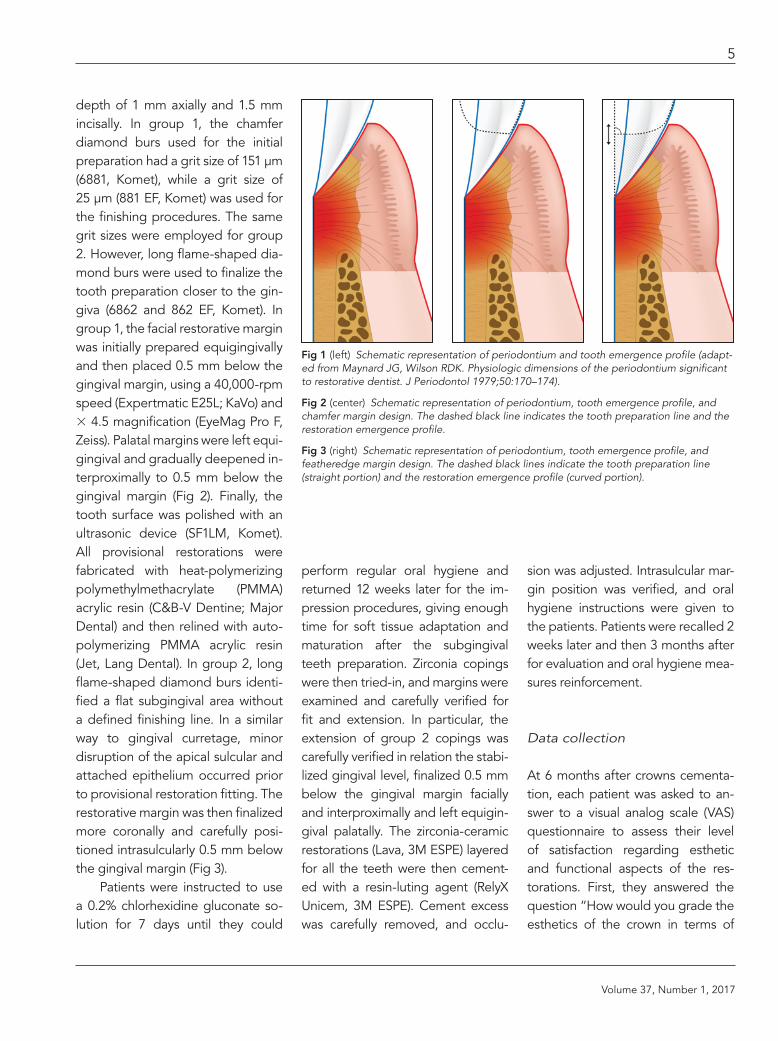
depth of 1 mm axially and 1.5 mm incisally. In group 1, the chamfer diamond burs used for the initial preparation had a grit size of 151 μm (6881, Komet), while a grit size of 25 μm (881 EF, Komet) was used for the finishing procedures. The same grit sizes were employed for group 2. However, long flame-shaped dia-mond burs were used to finalize the tooth preparation closer to the gin-giva (6862 and 862 EF, Komet). In group 1, the facial restorative margin was initially prepared equigingivally and then placed 0.5 mm below the gingival margin, using a 40,000-rpm speed (Expertmatic E25L; KaVo) and × 4.5 magnification (EyeMag Pro F, Zeiss). Palatal margins were left equi-gingival and gradually deepened in-terproximally to 0.5 mm below the gingival margin (Fig 2). Finally, the tooth surface was polished with an ultrasonic device (SF1LM, Komet). All provisional restorations were fabricated with heat-polymerizing polymethylmethacrylate (PMMA) acrylic resin (C&B-V Dentine; Major Dental) and then relined with auto-polymerizing PMMA acrylic resin (Jet, Lang Dental). In group 2, long flame-shaped diamond burs identi-fied a flat subgingival area without a defined finishing line. In a similar way to gingival curretage, minor disruption of the apical sulcular and attached epithelium occurred prior to provisional restoration fitting. The restorative margin was then finalized more coronally and carefully posi-tioned intrasulcularly 0.5 mm below the gingival margin (Fig 3).
Patients were instructed to use a 0.2% chlorhexidine gluconate so-lution for 7 days until they could
perform regular oral hygiene and returned 12 weeks later for the im-pression procedures, giving enough time for soft tissue adaptation and maturation after the subgingival teeth preparation. Zirconia copings were then tried-in, and margins were examined and carefully verified for fit and extension. In particular, the extension of group 2 copings was carefully verified in relation the stabi-lized gingival level, finalized 0.5 mm below the gingival margin facially and interproximally and left equigin-gival palatally. The zirconia-ceramic restorations (Lava, 3M ESPE) layered for all the teeth were then cement-ed with a resin-luting agent (RelyX Unicem, 3M ESPE). Cement excess was carefully removed, and occlu-
sion was adjusted. Intrasulcular mar-gin position was verified, and oral hygiene instructions were given to the patients. Patients were recalled 2 weeks later and then 3 months after for evaluation and oral hygiene mea-sures reinforcement.
Data collection
At 6 months after crowns cementa-tion, each patient was asked to an-swer to a visual analog scale (VAS) questionnaire to assess their level of satisfaction regarding esthetic and functional aspects of the res-torations. First, they answered the question “How would you grade the esthetics of the crown in terms of
Fig 1 (left) Schematic representation of periodontium and tooth emergence profile (adapt-ed from Maynard JG, Wilson RDK. Physiologic dimensions of the periodontium significant to restorative dentist. J Periodontol 1979;50:170–174).
Fig 2 (center) Schematic representation of periodontium, tooth emergence profile, and chamfer margin design. The dashed black line indicates the tooth preparation line and the restoration emergence profile.
Fig 3 (right) Schematic representation of periodontium, tooth emergence profile, and featheredge margin design. The dashed black lines indicate the tooth preparation line (straight portion) and the restoration emergence profile (curved portion).

The International Journal of Periodontics & Restorative Dentistry
6
relationship between the white ce-ramic component and the pink soft tissue?” marking a cross on a straight 100-mm line where the left end read “not satisfied at all” and the right end read “completely satisfied.” For the next question, “How would you grade the integration of the crown in the mouth?” patients marked a cross on another line where the left end read “not satisfied at all; I would like to change my crown” and the right end read “completely satisfied; I cannot recognize that I have a crown with my tongue.” All the answers were measured from left to right to obtain a numeric value for each pa-tient answer.
After the satisfaction question-naire, the same clinical measure-ments registered at baseline were taken again by the two blinded
experienced periodontists. The restorative margin position in rela-tion to the gingival margin was also registered. This was classified as subgingival (not visible), equigingi-val (slightly visible), or supragingival (visible).
Data analysis
Descriptive statistics were ex-pressed as mean (SD) and valid percentage for continuous and categorical data, respectively. The baseline comparisons between study groups were performed us-ing chi-square test (Fisher exact test with observed frequencies < 5) for categorical variables whereas con-tinuous variables were tested using t test (U-Mann Whitney test if the
variables were not normally distrib-uted). Outcomes were analyzed using analysis of covariance (AN-COVA), once assumptions for the convenience of this analysis were confirmed, with baseline values and age as covariates and study group as independent variable.37 Least square (LS) mean ± standard error (SE) was calculated for variables in-volving each outcome. Paired t test or McNemar test (if applicable) was used to compare outcomes at 6 months and baseline. Level of sig-nificance was set at .05. SPSS ver-sion 21 software (IBM) was used for all analyses.
Results
A total of 58 patients (27 men and 31 women, aged 30 to 64 years, mean age 50.3 years) received 200 full-coverage restorations, of which 106 were included in group 1 and 94 in group 2. All participants com-pleted the 6-month follow-up. Of these restorations, 2 were lost prior to the 6-month follow-up, 1 due to abutment root fracture and 1 due to porcelain fracture. These 2 sites were not included in the statistical analysis (Table 1).
At 6 months follow-up, changes from baseline were observed in GI, PI, and BoP. At 6 months, 12.6% of the sites presented dental plaque, while at baseline dental plaque was not present. Patients at baseline did not show any degree of gingival in-flamation or BoP, while at 6 months 43.4% of patients scored from 1 to 3 in the GI and about 39% present-ed bleeding. Statistically significant
Table 1 Sample Characteristics at Baseline and 6 Months
Variable Baseline (n = 198)
6 mo follow-up (n = 198) P
Age (y)a 52.4 (11.0)
Sex (men) 27 (48.2)
GI (n[%]) 0 1 2 3
198 (100)–––
112 (56.6)66 (33.3)18 (9.1)2 (1)
NA
PI (n[%]) 0 1
198 (100)–
173 (87.4)25 (12.6)
NA
BoP (n[%]) 0 1
198 (100)–
121 (61.1)77 (38.9)
NA
PPD mesial, mma 2.43 (0.60) 2.71 (0.78) 0.001b
PPD facial, mma 1.96 (0.57) 1.64 (0.59) 0.001b
PPD distal, mma 2.37 (0.60) 2.54 (0.72) 0.001b aMean (SD). b Paired t test (quantitative variables). NA = not applicable; GI = Gingival Index; PI = Plaque Index; BoP = bleeding on probing; PPD = periodontal probing depth.
Volume 37, Number 1, 2017
7
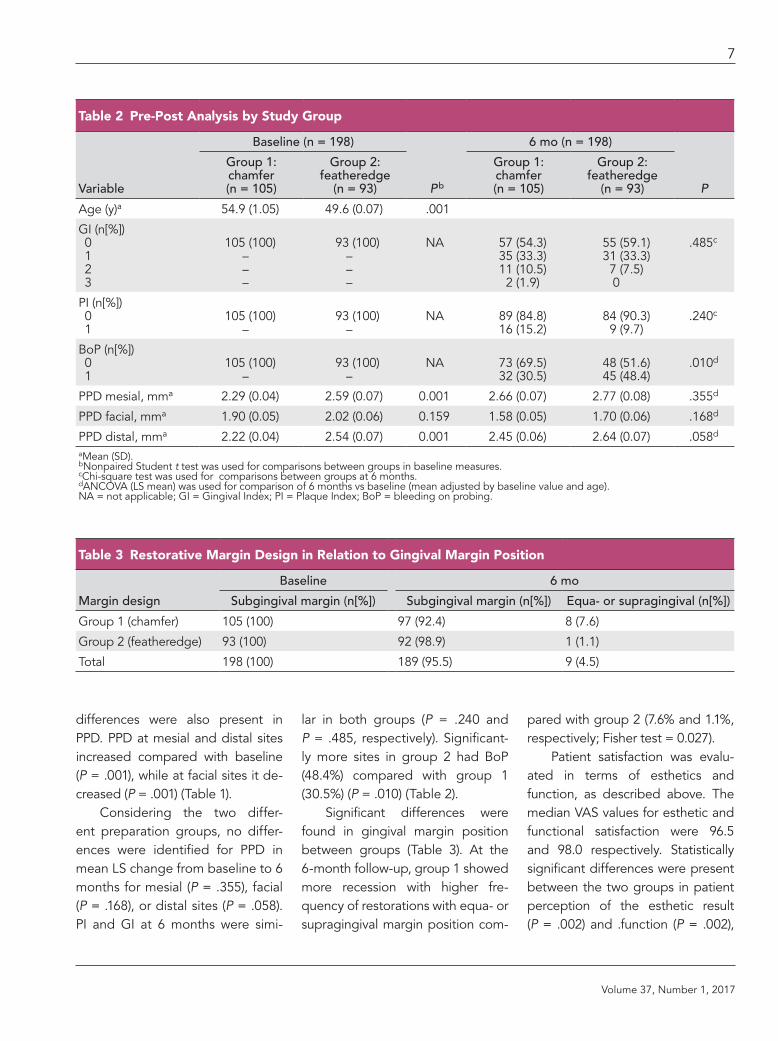
differences were also present in PPD. PPD at mesial and distal sites increased compared with baseline (P = .001), while at facial sites it de-creased (P = .001) (Table 1).
Considering the two differ-ent preparation groups, no differ-ences were identified for PPD in mean LS change from baseline to 6 months for mesial (P = .355), facial (P = .168), or distal sites (P = .058). PI and GI at 6 months were simi-
lar in both groups (P = .240 and P = .485, respectively). Significant-ly more sites in group 2 had BoP (48.4%) compared with group 1 (30.5%) (P = .010) (Table 2).
Significant differences were found in gingival margin position between groups (Table 3). At the 6-month follow-up, group 1 showed more recession with higher fre-quency of restorations with equa- or supragingival margin position com-
pared with group 2 (7.6% and 1.1%, respectively; Fisher test = 0.027).
Patient satisfaction was evalu-ated in terms of esthetics and function, as described above. The median VAS values for esthetic and functional satisfaction were 96.5 and 98.0 respectively. Statistically significant differences were present between the two groups in patient perception of the esthetic result (P = .002) and .function (P = .002),
Table 2 Pre-Post Analysis by Study Group
Variable
Baseline (n = 198)
Pb
6 mo (n = 198)
P
Group 1: chamfer (n = 105)
Group 2: featheredge
(n = 93)
Group 1: chamfer (n = 105)
Group 2: featheredge
(n = 93)
Age (y)a 54.9 (1.05) 49.6 (0.07) .001
GI (n[%]) 0 1 2 3
105 (100)–––
93 (100)–––
NA 57 (54.3) 35 (33.3) 11 (10.5) 2 (1.9)
55 (59.1) 31 (33.3) 7 (7.5) 0
.485c
PI (n[%]) 0 1
105 (100)–
93 (100)–
NA 89 (84.8)16 (15.2)
84 (90.3)9 (9.7)
.240c
BoP (n[%]) 0 1
105 (100)–
93 (100)–
NA 73 (69.5)32 (30.5)
48 (51.6) 45 (48.4)
.010d
PPD mesial, mma 2.29 (0.04) 2.59 (0.07) 0.001 2.66 (0.07) 2.77 (0.08) .355d
PPD facial, mma 1.90 (0.05) 2.02 (0.06) 0.159 1.58 (0.05) 1.70 (0.06) .168d
PPD distal, mma 2.22 (0.04) 2.54 (0.07) 0.001 2.45 (0.06) 2.64 (0.07) .058d aMean (SD). bNonpaired Student t test was used for comparisons between groups in baseline measures. cChi-square test was used for comparisons between groups at 6 months. dANCOVA (LS mean) was used for comparison of 6 months vs baseline (mean adjusted by baseline value and age). NA = not applicable; GI = Gingival Index; PI = Plaque Index; BoP = bleeding on probing.
Table 3 Restorative Margin Design in Relation to Gingival Margin Position
Margin design
Baseline 6 mo
Subgingival margin (n[%]) Subgingival margin (n[%]) Equa- or supragingival (n[%])
Group 1 (chamfer) 105 (100) 97 (92.4) 8 (7.6)
Group 2 (featheredge) 93 (100) 92 (98.9) 1 (1.1)
Total 198 (100) 189 (95.5) 9 (4.5)
The International Journal of Periodontics & Restorative Dentistry
8
with higher VAS median values for patients in group 1 (Table 4).
Discussion
The present research focused on subgingival margins in the esthetic zone since margins are often po-sitioned subgingivally to improve the treatment outcome1,2,4,21,22 and especially to enhance the natural es-thetic result and the gingival archi-tecture.20,23,24 Different indications have been described in the litera-ture in regard to finish line form,
but at the end the selection criteria should be based on personal prefer-ence, esthetics, formation ease, and type of restoration.21 In the present research, two different restorative margins, deep chamfer and feather-edge, were compared in regard to periodontal tissue response.
Deep chamfer was prepared intrasulcular. Featheredge was also positioned intrasulcular, but after a deeper subgingival tooth prepara-tion. A comparison with intrasulcu-lar featheredge tooth preparation was not performed. At the 6-month evaluation, PI and GI were increased
as in the previous literature,11 with no statistically significant differenc-es between the two types of finish-ing lines (Table 2). In the comparison of the two groups, statistically signif-icant differences were found in BoP. In accordance with published ar-ticles, a general increased BoP was noticed around subgingival mar-gins.28,32,39 More specifically, even when the gingival tissues appeared similar at the 6-month follow-up (similar GI), increased BoP was pres-ent with featheredge compared with chamfer. While no differences were present between the groups at baseline, at the 6-month follow-up 48.4% of sites in group 2 had BoP versus 30.5% in group 1 (P = .010) (Table 2).
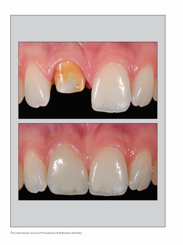
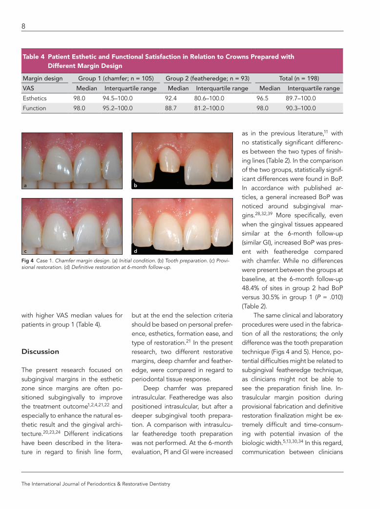
The same clinical and laboratory procedures were used in the fabrica-tion of all the restorations; the only difference was the tooth preparation technique (Figs 4 and 5). Hence, po-tential difficulties might be related to subgingival featheredge technique, as clinicians might not be able to see the preparation finish line. In-trasulcular margin position during provisional fabrication and definitive restoration finalization might be ex-tremely difficult and time-consum-ing with potential invasion of the biologic width.5,13,30,34 In this regard, communication between clinicians
Fig 4 Case 1. Chamfer margin design. (a) Initial condition. (b) Tooth preparation. (c) Provi-sional restoration. (d) Definitive restoration at 6-month follow-up.
Table 4 Patient Esthetic and Functional Satisfaction in Relation to Crowns Prepared with Different Margin Design
Margin design Group 1 (chamfer; n = 105) Group 2 (featheredge; n = 93) Total (n = 198)
VAS Median Interquartile range Median Interquartile range Median Interquartile range
Esthetics 98.0 94.5–100.0 92.4 80.6–100.0 96.5 89.7–100.0
Function 98.0 95.2–100.0 88.7 81.2–100.0 98.0 90.3–100.0
a
c
b
d
Volume 37, Number 1, 2017
9
and technicians must be clear to overcome the technician’s inability to visualize the exact position of the intrasulcular margin in the finishing area. In a similar way, the emergence profile could be challenging to de-termine, for both the provisional and the definitive restoration.40,41 To im-prove the gingival scalloping, espe-cially on teeth with triangular-shape roots, or to increase the strength of the ceramic at the cervical area, po-tential overcontouring might be ex-pected.4,42–44
Restorations with subgingival margins have been associated with increased gingival recession, espe-cially with thin gingival biotypes.29–34 Subgingival margins, examined for a mean period of 4 to 12 years, pre-sented gingival recession in 34% of the restorations. This was much more than around supragingival margins, where recession occurred only on 6% of the crowns.31 Simi-larly, in a longitudinal study by the same authors with a 15-year follow-up, crowns with subgingival margins had a 2.65 times greater chance of gingival recession when compared with the contralateral teeth.32
In the present study at 6 months after delivery of the restorations, 4.5% had gingival recession limited to 0.5 mm, with restorative margins exposure. Featheredge preparation performed statistically better than chamfer: only 1 crown in group 1 had gingival recession compared with 8 in the chamfer group. (Table 3) This potential benefit might be related to the described increased thickness of the periodontal bio-type, a consequence of the rotary curettage during subgingival feath-
eredge tooth preparation. However, as increased BoP was observed, lon-ger-term data will be needed to rule out the potential negative effect of gingival inflammation in terms of tis-sue stability. For this reason, the re-sults of the present study might be considered preliminary, as longer observational periods are needed to establish better correlations be-tween the examined parameters.
As a secondary level of analy-sis, patient functional and esthetic satisfaction has been investigated. The focus has been centered on one of the most crucial aspects in the esthetic zone: the cervical portion and its interaction with the gingival tissues.45,46 The results of this study suggest that the interaction be-tween all-ceramic restorations and the gingival tissue is well graded by patients (Table 4). Considering both esthetic and functional per-ceptions, patients preferred cham-fer preparation (P < .001). This result
could be explained by the possibil-ity that chamfer follows more closely the tooth emergence profile with-out interfering too much with the periodontium, while subgingival featheredge preparations are more related to a newly developed emer-gence profile. Thus, patients do not seem to experience the described benefits determined by the modifi-cation of the emergence profile with improved esthetic results and better soft tissue stability.
Conclusions
Within the limitations of this study, more BoP is present around feather-edge margins and significantly more gingival recession is present around deep chamfer margins. Intrasulcu-lar margins are technique sensitive, especially when subgingival tooth preparation with a featheredge mar-gin is selected.
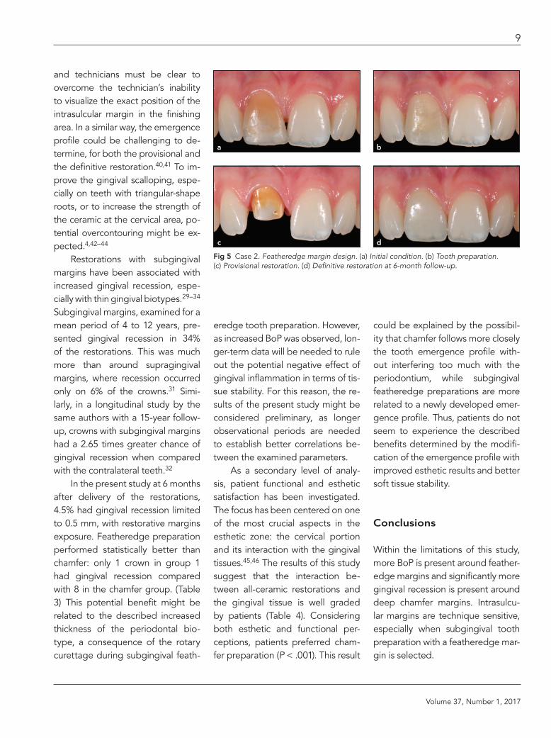
Fig 5 Case 2. Featheredge margin design. (a) Initial condition. (b) Tooth preparation. (c) Provisional restoration. (d) Definitive restoration at 6-month follow-up.
a
c
b
d
The International Journal of Periodontics & Restorative Dentistry
10
Acknowledgments
The authors reported no conflicts of interest related to this study.
References
1. Rosenstiel S, Land M, Fujimoto J. Con-temporary Fixed Prosthodontics, ed 4. St Louis: Mosby Elsevier, 2006.
2. Shillinburg HT. Fundamentals of Fixed Prosthodontics, ed 3. Chicago: Quintes-sence, 1987.
3. Bader J, Rozier RG, McFall WT Jr. The ef-fect of crown receipt on measures of gingi-val status. J Dent Res 1991;70:1386–1389.
4. Padbury A Jr, Eber R, Wang HL. Interac-tions between the gingiva and the mar-gin of restorations. J Clin Periodontol 2003;30:379–385.
5. Waerhaug J, Philos D. Periodontology and partial prosthesis. Int Dent J 1968;18: 101–107.
6. Silness J, Hegdahl T. Area of the exposed zinc phosphate cement surfaces in fixed restorations. Scand J Dent Res 1970;78: 163–177.
7. Perel ML. Periodontal considerations of crown contours. J Prosthet Dent 1971;26: 627–630.
8. Weisgold A. Contours of the full crown res-toration. Alpha Omegan 1977;70:77–89.
9. Marcum J. The effect of crown marginal depth upon gingival tissue. J Prosthet Dent 1967;17:479–487.
10. Newcomb GM. The relationship between the location of subgingival crown margins and gingival inflammation. J Periodontol 1974;45:151–154.
11. Flores-de-Jacoby L, Zafiropoulos GG, Ciancio S. The effect of crown margin lo-cation on plaque and periodontal health. Int J Periodont Rest Dent 1989;9:197–205.
12. Maynard JG Jr, Wilson RD. Physiologic di-mensions of the periodontium significant to the restorative dentist. J Periodontol 1979; 50:107–174.
13. Nevins M, Skurow HM. The intracrevicular restorative margin, the biologic width, and the maintenance of the gingival margin. Int J Periodont Rest Dent 1984;4:30–49.
14. Lindhe J. Textbook of clinical periodon-tology, ed 2. Copenhagen; Munksgaard, 1989.
15. Koke U, Sander C, Heinecke A, Müller HP. A possible influence of gingival dimen-sions on attachment loss and gingival re-cession following placement of artificial crowns. Int J Periodontics Restorative Dent 2003;23:439–445.
16. Giollo MD, Valle PM, Gomes SC, Rösing CK. A retrospective clinical, radiographic and microbiological study of periodon-tal conditions of teeth with and without crowns. Braz Oral Res 2007;21:348–354.
17. Orkin DA, Reddy J, Bradshaw D. The rela-tionship of the position of crown margins to gingival health. J Prosthet Dent 1987; 57:421–442.
18. Gemalmaz D, Ergin S. Clinical evaluation of all-ceramic crowns. J Prosthet Dent 2002; 87:189–196.
19. Valderhaug J, Birkeland JM. Periodontal conditions in patients 5 years following in-sertion of fixed prostheses. J Oral Rehabil 1976;3:237–243.
20. Chiche GJ, Pinault A. Esthetics of anterior fixed prosthodontics. Chicago: Quintes-sence, 1994.
21. Goodacre CJ, Campagni WV, Aquilino SA. Tooth preparations for complete crowns: An art form based on scientific principles. J Prosthet Dent 2001;85:363–376.
22. Tan PL, Aquilino SA, Gratton DG, et al. In vitro fracture resistance of endodontically treated central incisors with varying fer-rule heights and configurations. J Prosthet Dent 2005;93:331–336.
23. Rufenacht CR. Fundamentals of Esthetics. Chicago: Quintessence, 1990.
24. Kois JC, Spear FM. Periodontal prosthe-sis: Creating successful restorations. J Am Dent Assoc 1992;123:108–115.
25. Loi I, Di Felice A. Biologically oriented preparation technique (BOPT): A new approach for prosthetic restoration of periodontically healthy teeth. Eur J Esthet Dent 2013;8:10–23.
26. Gargiulo AW, Wentz FM, Orban BJ. Di-mensions and relations of the dentogingi-val junction in humans. J Periodontol 1961; 32:261–267.
27. Vacek JS, Gher ME, Assad DA, Richard-son AC, Giambarresi LI. The dimension of the human dentogingival junction. Int J Periodontics Restorative Dent 1994;14: 115–165.
28. Müller HP. The effect of artificial crown margins at the gingival margin on the peri-odontal conditions in a group of periodon-tally supervised patients treated with fixed bridges. J Clin Periodontol 1986;13:97–102.
29. Miller PD Jr. A classification of marginal tissue recession. Int J Periodontics Restor-ative Dent 1985;5:8–13.
30. Kao RT, Pasquinelli K. Thick vs. thin gin-gival tissue: A key determinant in tissue response to disease and restorative treat-ment. J Calif Dent Assoc 2002;30:521–526.
31. Valderhaug J. Periodontal conditions and carious lesions following the insertion of fixed prostheses: A 10-year follow-up study. Int Dent J 1980;30:296–304.
32. Valderhaug J, Ellingsen JE, Jokstad A. Oral hygiene, periodontal conditions and carious lesions in patients treated with dental bridges. A 15-year clinical and ra-diographic follow-up study. J Clin Peri-odontol 1993;20:482–489.
33. Tao J, Wu Y, Chen J, Su J. A follow-up study of up to 5 years of metal-ceramic crowns in the maxillary central incisors for different gingival biotypes. Int J Periodont Rest Dent 2014;34:e85–e92.
34. Müller HP, Heinecke A, Schaller N, Eger T. Masticatory mucosa in subjects with different periodontal phenotypes. J Clin Periodontol 2000;27:621–626.
35. Löe H, Silness J. Periodontal disease in pregnancy. I Prevalence and severity, Acta Odont Scand 1963;21:533–551.
36. Ainamo J, Bay I. Problems and proposals for recording gingivitis and plaque. Int Dent J 1975;25:229–235.
37. Miller GA, Chapman JP. Misunderstand-ing analysis of covariance. J Abnorm Psy-chol 2001;110:40–48.
38. Lang NP, Kiel RA, Anderhalden K. Clinical and microbiological effects of subgingi-val restorations with overhanging or clini-cally perfect margins. J Clin Periodontol 1983;10:563–578.
39. Schätzle M, Land NP, Anerud A, Boysen H, Bürgin W, Löe H. The influence of mar-gins of restorations of the periodontal tissues over 26 years. J Clin Periodontol 2001;28:57–64.
40. Dragoo MR, Williams GB. Periodontal tis-sue reactions to restorative procedures. Part I. Int J Periodont Rest Dent 1981; 2:8–29.
41. Dragoo MR, Williams GB. Periodontal tis-sue reactions to restorative procedures. Part II. Int J Periodontics Restorative Dent 1982;2:34–42.
42. Yuodelis RA, Weaver JD, Sapkos S. Facial and lingual contours of artificial complete crown restorations and their effects on the periodontium. J Prosthet Dent 1973; 29:61–66.
43. Silva NR, Bonfante EA, Martins LM, et al. Reliability of reduced-thickness and thinly veneered lithium disilicate crowns. J Dent Res 2012;91:305–310.
44. Reeves WG. Restorative margin place-ment and periodontal health. J Prosthet Dent 1991;66:733–736.
45. Paniz G, Kang KH, Kim Y, Kumagai N, Hirayama H. Influence of coping design on the cervical color of ceramic crowns. J Prosthet Dent 2013;110:495–500.
46. Heffernan MJ, Aquilino SA, Diaz-Arnold AM, Haselton DR, Stanford CM, Vargas MA. Relative translucency of six all-ceram-ic systems. Part II: Core and veneer materi-als. JProsthet Dent 2002;88:10–15.