Embed Size (px)
Citation preview

Accepted Manuscript
PII: S0006-2952(13)00153-6DOI: http://dx.doi.org/doi:10.1016/j.bcp.2013.02.030Reference: BCP 11571
To appear in: BCP
Received date: 31-1-2013Revised date: 24-2-2013Accepted date: 27-2-2013
Please cite this article as: Bende NS, Kang E, Herzig V, Bosmans F, Nicholson GM,Mobli M, King GF, The insecticidal neurotoxin Aps III is an atypical knottin peptidethat potently blocks insect voltage-gated sodium channels, Biochemical Pharmacology(2013), http://dx.doi.org/10.1016/j.bcp.2013.02.030
This is a PDF file of an unedited manuscript that has been accepted for publication.As a service to our customers we are providing this early version of the manuscript.The manuscript will undergo copyediting, typesetting, and review of the resulting proofbefore it is published in its final form. Please note that during the production processerrors may be discovered which could affect the content, and all legal disclaimers thatapply to the journal pertain.

Page 1 of 49
Accep
ted
Man
uscr
ipt
1
The insecticidal neurotoxin Aps III is an atypical knottin peptide
that potently blocks insect voltage-gated sodium channels
Niraj S. Bendea, Eunji Kangb, Volker Herziga, Frank Bosmansc,d,
Graham M. Nicholsonb, Mehdi Moblia, and Glenn F. Kinga,*
aInstitute for Molecular Bioscience, The University of Queensland,
St Lucia, QLD 4072, Australia
bSchool of Medical & Molecular Biosciences, University of Technology, Sydney,
Broadway NSW 2007, Australia
cDepartment of Physiology and dSolomon H. Snyder Department of Neuroscience, Johns
Hopkins University – School of Medicine, Baltimore MD 21205, USA
*Address for correspondence: Prof. Glenn F. King, Institute for Molecular Bioscience, The
University of Queensland, 306 Carmody Road, St Lucia, Queensland 4072, Australia; Phone:
+61 7 3346-2025; Fax: +61 7 3346-2021; Email: [email protected]

Page 2 of 49
Accep
ted
Man
uscr
ipt
2
Abstract
One of the most potent insecticidal venom peptides described to date is Aps III from the
venom of the trapdoor spider Apomastus schlingeri. Aps III is highly neurotoxic to
lepidopteran crop pests, making it a promising candidate for bioinsecticide development.
However, its disulfide-connectivity, three-dimensional structure, and mode of action have not
been determined. Here we show that recombinant Aps III (rAps III) is an atypical knottin
peptide; three of the disulfide bridges form a classical inhibitor cystine knot motif while the
fourth disulfide acts as a molecular staple that restricts the flexibility of an unusually large
hairpin loop that often houses the pharmacophore in this class of toxins. We demonstrate that
the irreversible paralysis induced in insects by rAps III results from a potent block of insect
voltage-gated sodium channels. Channel block by rAps III is voltage-independent insofar as it
occurs without significant alteration in the voltage-dependence of channel activation or
steady-state inactivation. Thus, rAps III appears to be a pore blocker that plugs the outer
vestibule of insect voltage-gated sodium channels. This mechanism of action contrasts
strikingly with virtually all other sodium channel modulators isolated from spider venoms that
act as gating modifiers by interacting with one or more of the four voltage-sensing domains of
the channel.
Keywords: voltage-gated sodium channel; neurotoxin; spider-venom peptide; pore blocker;
gating modifier; inhibitor cystine knot.

Page 3 of 49
Accep
ted
Man
uscr
ipt
3
1. Introduction
Insects serve as vectors for a wide range of debilitating and potentially lethal human diseases
such as malaria, dengue, Chagas disease, and yellow fever [1]. About 3.3 billion people,
almost half of the world’s population, are at risk of contracting vector-borne disease [2].
Moreover, despite intensive control measures, insect pests reduce world crop yields by
10–14% annually [3, 4].
Despite the widespread introduction of insect-resistant transgenic crops, chemical insecticides
remain the dominant method for controlling insect pests in both the agricultural and public
health arenas. These chemicals target a very small number of molecular targets in the insect
nervous system [5]. As a result, their widespread use over several decades has promoted the
evolution of resistant insect populations, with >600 insects and mites now resistant to one or
more classes of chemical insecticides [6]. In addition, key classes of insecticides have been
withdrawn from sale or their use has been restricted by regulatory authorities due to growing
environmental and human health concerns [7]. Thus, there is an urgent need to develop novel
classes of insecticides or alternative methods of insect pest control.
A promising approach in the agricultural sector is to engineer crops to produce insecticidal
toxins. By 2010, 148 million hectares of genetically modified (GM) crops had been planted in
29 countries, representing 10% of all cropland [8]. While the introduction of GM crops that
express Bacillus thuringiensis (Bt) toxins has provided an alternative and potentially safer
method of insect control than chemical insecticides, alternative insect-toxin transgenes are
urgently needed as constitutive expression of Bt toxin in transgenic plants is likely to expedite
resistance development [9].

Page 4 of 49
Accep
ted
Man
uscr
ipt
4
There are very few well characterised toxins that could be considered as alternatives or
adjuncts to Bt. However, some of the most promising candidates are novel insecticidal
peptides that have been isolated from the venom of spiders [7, 10-12], the most successful
insect predators on the planet. Most of these peptides are highly stable because they contain
an inhibitor cystine knot (ICK) motif [13, 14] that provides them with resistance to extremes
of pH, high temperatures, and proteolytic enzymes [7, 15]. One of the first insecticidal spider-
venom peptides to be reported was Aps III from the venom of the trapdoor spider Apomastus
schlingeri [16]. With a reported LD50 of 133 pmol/g against the tobacco hornworm Manduca
sexta, this peptide is one of the most potent insect toxins described to date according to
ArachnoServer [17, 18]. Aps III comprises 37-residues with four disulfide-bonds, but its
three-dimensional structure and mode of action are unknown.
Here we describe the development of an efficient E. coli expression system that was used to
produce recombinant Aps III (rAps III) for functional and structural studies. The 3D solution
structure of rAps III determined using NMR spectroscopy revealed an inhibitor cystine knot
motif that is commonly found in spider-venom peptides. However, rAps III contains an
additional disulfide bridge that is employed as a molecular staple to tie together the ends of a
very large -hairpin loop. We demonstrate that the insecticidal activity of rAps III results
from a potent block of insect voltage-gated sodium (Nav) channels in combination with a
weaker block of insect voltage-gated calcium (Cav) channels. However, in striking contrast to
previously characterised Nav channel blockers from spiders, all of which are gating modifiers
[19], rAps III appears to be a pore blocker that plugs the outer vestibule of insect Nav
channels.

Page 5 of 49
Accep
ted
Man
uscr
ipt
5
2. Material and Methods
2.1 Chemicals
All chemicals were purchased from Sigma-Aldrich Australia (Castle Hill, NSW, Australia),
Sigma-Aldrich USA (St Louis, MO, USA), or Merck Chemicals (Kilsyth, Victoria, Australia)
with the exception of isopropyl-β-D-thiogalactopyranoside (IPTG) and streptomycin (Life
Technologies, Victoria, Australia), tetrodotoxin (Alomone Labs, Israel), and HPLC-grade
acetonitrile (RCI Labscan, Bangkok, Thailand). 13C6-glucose and 15NH4Cl were from Sigma-
Aldrich Australia. Recombinant His6-TEV protease (EC 3.4.22.44) was produced in-house
used a published protocol [20].
2.2 Production of recombinant Aps III
A synthetic gene encoding Aps III, with codons optimised for expression in Escherichia coli,
was produced and cloned into a variant of the pLIC-MBP expression vector [21] by GeneArt
(Invitrogen, Regensburg, Germany). This vector (pLIC-NSB1) encodes a MalE signal
sequence for periplasmic export [22], a His6 tag for affinity purification, a maltose binding
protein (MBP) fusion tag to aid solubility [23], and a tobacco etch virus (TEV) protease
recognition site directly preceding the codon-optimised Aps III gene (Fig. 1A).
The plasmid encoding Aps III was transformed into E. coli strain BL21(DE3) for
recombinant toxin production. Protein expression and purification were performed as
described previously [24] with minor modifications. Briefly, cultures were grown in Terrific
Broth at 37C with shaking at 120 rpm. Toxin gene expression was induced with 1 mM IPTG
at an OD600 of 1.1–1.2, then cells were grown at 18C for a further 12 h before harvesting by
centrifugation for 15 min at 8000 rpm. For production of uniformly 13C/15N-labelled rAps III,
cultures were grown in minimal medium supplemented with 13C6-glucose and 15NH4Cl as the

Page 6 of 49
Accep
ted
Man
uscr
ipt
6
sole carbon and nitrogen sources, respectively.
The His6-MBP-toxin fusion protein was extracted from the bacterial periplasm by cell
disruption at 26 kPa (TS Series Cell Disrupter, Constant Systems Ltd, Northants, UK), then
captured by passing the extract (buffered in 40 mM Tris, 500 mM NaCl, pH 8.0) over
Ni-NTA Superflow resin (Qiagen). Proteins bound non-specifically were removed by
washing with 10 mM imidazole then the fusion protein was eluted with 500 mM imidazole.
The eluted fusion protein was concentrated to 10 ml and the buffer was exchanged to remove
imidazole. Reduced and oxidised glutathione were then added to 0.6 mM and 0.4 mM,
respectively, to maintain TEV protease activity and promote folding of the protein.
Approximately 100 μg of His6-tagged TEV protease was added per mg of rAps III, then the
cleavage reaction was allowed to proceed at room temperature for 12 h. The cleaved His6-
MBP and His6-TEV were removed by passing the solution over Ni-NTA Superflow resin,
while the eluate containing rAps III was collected for further purification using reverse-phase
HPLC (RP-HPLC). RP-HPLC was performed on a Vydac C18 column (250 4.6 mm,
particle size 5 μm) using a flow rate of 1 ml/min and a gradient of 20–40% Solvent B
(0.043% trifluoroacetic acid (TFA) in 90% acetonitrile) in Solvent A (0.05% TFA in water)
over 20 min. rAps III contains a non-native N-terminal serine residue (a vestige of the TEV
protease cleavage site), making it one-residue longer than native Aps III (Fig. 1B).
2.3 Mass spectrometry
Toxin masses were confirmed by matrix assisted laser desorption ionization–time of flight
mass spectrometry (MALDI-TOF MS) using a Model 4700 Proteomics Bioanalyser (Applied
Biosystems, CA, USA). RP-HPLC fractions were mixed (1:1 v:v) with α-cyano-4 hydroxy-
cinnamic acid matrix (5 mg/ml in 50/50 acetonitrile/H2O) and MALDI-TOF spectra were

Page 7 of 49
Accep
ted
Man
uscr
ipt
7
acquired in positive reflector mode. All reported masses are for monoisotopic [M+H]+ ions.
2.4 Insecticidal assays
rAps III dissolved in insect-saline [25] was injected into the ventro-lateral thoracic region of
sheep blowflies (Lucilia cuprina; mass 19.7–23.9 mg) using a 1.0 ml Terumo Insulin syringe
(B-D Ultra-Fine, Terumo Medical Corporation, MD, USA) with a fixed 29 G needle fitted to
an Arnold hand micro-applicator (Burkard Manufacturing Co. Ltd., England). A maximum
volume of 2 l was injected per fly. Thereafter, flies were individually housed in 2 ml tubes
and the paralytic activity was determined after 24 h. A total of three tests were carried out and
for each test seven doses of rAps III (n = 10 flies per dose) and the appropriate control (insect
saline; n = 30 flies each) were used. PD50 values were calculated as described previously [26].
2.5 Electrophysiological measurements
2.5.1 Primary cell culture
Dorsal unpaired median (DUM) neurons were isolated from unsexed adult American
cockroaches (Periplaneta americana) as described previously [27, 28]. Briefly, terminal
abdominal ganglia were removed and placed in normal insect saline (NIS) containing (in
mM): NaCl 180, KCl 3.1, N-hydroxyethylpiperazine-N-ethanesulfonic acid (HEPES) 10 and
D-glucose 20. Ganglia were then incubated in 1 mg/ml collagenase (type IA) (EC 3.4.24.3)
for 40 min at 29C. Following enzymatic treatment, ganglia were washed three times in NIS
and triturated through a fire-polished Pasteur pipette. The resultant cell suspension was then
distributed onto 12-mm diameter glass coverslips pre-coated with 2 mg/ml concanavalin A
(type IV). DUM neurons were maintained in NIS supplemented with 5 mM CaCl2, 4 mM
MgCl2, 5% foetal bovine serum and 1% penicillin and streptomycin, and maintained at 29C,
100% humidity.

Page 8 of 49
Accep
ted
Man
uscr
ipt
8
2.5.2 Patch-clamp electrophysiology
Ionic currents were recorded in voltage-clamp mode using the whole-cell patch-clamp
technique employing version 10.2 of the pCLAMP data acquisition system (Molecular
Devices, Sunnyvale, CA). Data were filtered at 5–10 kHz with a low-pass Bessel filter with
leakage and capacitative currents subtracted using P-P/4 procedures. Digital sampling rates
were set between 15 and 25 kHz depending of the length of the protocol. Single-use 0.8–2.5
MΩ electrodes were pulled from borosilicate glass and fire-polished prior to current
recordings. Liquid junction potentials were calculated using JPCALC [29], and all data were
compensated for these values. Cells were bathed in external solution through a continuous
pressurised perfusion system at 1 ml/min, while toxin solutions were introduced via direct
pressurised application via a perfusion needle at ~50 l/min (Automate Scientific, San
Francisco, CA) to a bath volume of 300 l. To avoid issues of desensitization or rundown of
currents, particularly with CaV channel currents, recording periods were kept as short as
possible with the effect of toxin recorded within 5 min of control recordings. Control data was
not acquired until at least 20 min after whole-cell configuration was achieved. This was to
eliminate the influence of fast time-dependent shifts in steady-state inactivation resulting in
current rundown, particularly when recording NaV channel currents (INa). Time-dependent
shifts in steady-state NaV channel inactivation are typically of the order of 2–3 mV beyond
this period and do not significantly influence current amplitude. Experiments were performed
with a single concentration of toxin tested on one cell, which was subsequently repeated in
separate experiments using unexposed cells. The number of independent recordings for each
type of experiment is provided in the relevant sections of the results. All experiments were
performed at ambient room temperature (20–23ºC).
To record INa, the external bath solution contained (in mM): NaCl 80, CsCl 5, CaCl2 1.8,
Page 9 of 49
Accep
ted
Man
uscr
ipt
9
tetraethylammonium chloride (TEA-Cl) 50, 4-aminopyridine (4-AP) 5, HEPES 10, NiCl2 0.1,
and CdCl2 1, adjusted to pH 7.4 with 1 M NaOH. The pipette solution contained (in mM):
NaCl 34, CsF 135, MgCl2 1, HEPES 10, ethylene glycol-bis(2-aminoethylether)-N,N,N′,N′-
tetraacetic acid (EGTA) 5, and ATP-Na2 3, adjusted to pH 7.4 with 1 M CsOH. Due to the
reported current rundown with calcium as a charge carrier [30], BaCl2 replaced CaCl2 in all
experiments on voltage-activated calcium (CaV) channels.
The external bath solution for barium current (IBa) recordings contained (in mM): Na acetate
140, TEA-Br 30, BaCl2 3 and HEPES 10, adjusted to pH 7.4 with 1 M TEA-OH. The external
solution also contained 300 nM tetrodotoxin (TTX) to block NaV channels. Pipette solutions
contained (in mM): Na acetate 10, CsCl 110, TEA-Br 50, ATP-Na2 2, CaCl2 0.5, EGTA 10
and HEPES 10, adjusted to pH 7.4 with 1 M CsOH. The external bath solution for recording
global voltage-activated potassium (KV) channel currents (IK) contained (in mM): NaCl 200,
K gluconate 50, CaCl2 5, MgCl2 4, TTX 0.3, HEPES 10 and D-glucose 10, adjusted to pH 7.4
with 1 M NaOH. The pipette solution consisted of (in mM): K gluconate 135, KF 25, NaCl 9,
CaCl2 0.1, MgCl2 1, EGTA 1, HEPES 10 and ATP-Na2 3, adjusted to pH 7.4 with 1 M KOH.
To eliminate any influence of differences in osmotic pressure, all internal and external
solutions were adjusted to 400 ± 5 mOsmol/l with sucrose. Experiments were rejected if there
were large leak currents or currents showed signs of poor space clamping.
2.5.3 Curve-fitting and statistical analyses
Data were analysed using AXOGRAPH X version 1.3 (Molecular Devices). Curve-fitting of
I-V data was performed using GraphPad Prism version 5.00d for Macintosh (GraphPad
Software, San Diego). Comparisons of two sample means were made using a paired Student's
Page 10 of 49
Accep
ted
Man
uscr
ipt
10
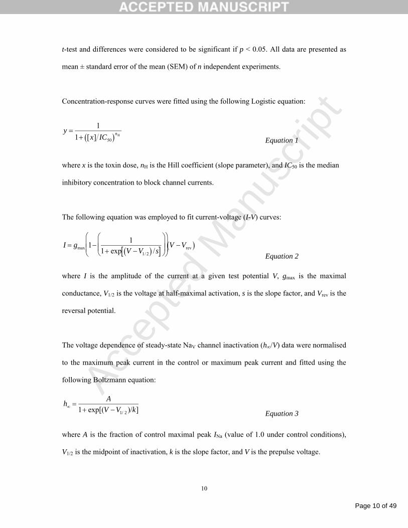
t-test and differences were considered to be significant if p < 0.05. All data are presented as
mean ± standard error of the mean (SEM) of n independent experiments.
Concentration-response curves were fitted using the following Logistic equation:
y 1
1 [x] IC50 nH
Equation 1
where x is the toxin dose, nH is the Hill coefficient (slope parameter), and IC50 is the median
inhibitory concentration to block channel currents.
The following equation was employed to fit current-voltage (I-V) curves:
I gmax 11
1 exp V V1/ 2 /s
V Vrev
Equation 2
where I is the amplitude of the current at a given test potential V, gmax is the maximal
conductance, V1/2 is the voltage at half-maximal activation, s is the slope factor, and Vrev is the
reversal potential.
The voltage dependence of steady-state NaV channel inactivation (h∞/V) data were normalised
to the maximum peak current in the control or maximum peak current and fitted using the
following Boltzmann equation:
h
A
1 exp[(V V1/ 2 )/k] Equation 3
where A is the fraction of control maximal peak INa (value of 1.0 under control conditions),
V1/2 is the midpoint of inactivation, k is the slope factor, and V is the prepulse voltage.

Page 11 of 49
Accep
ted
Man
uscr
ipt
11
2.5.4 Two-electrode voltage-clamp recordings from Xenopus oocytes
To prepare cRNA for oocyte injection, separate plasmids encoding the -subunit of the NaV1
channel from the German cockroach Blatella germanica (BgNaV1; [31]) and the Drosophila
NaV1 auxiliary subunit TipE [32] were linearised with NotI, followed by in vitro transcription
using T7 polymerase (mMESSAGE mMACHINE kit, Life Technologies, CA, USA). After
Xenopus oocytes were co-injected at 1:5 molar ratio with cRNA encoding BgNaV1 and TipE,
they were incubated for 2–3 days at 17°C (in 96 mM NaCl, 2 mM KCl, 5 mM HEPES, 1 mM
MgCl2, 1.8 mM CaCl2, 50 μg/ml gentamycin, pH 7.6) prior to recording BgNaV1-mediated
currents via two-electrode voltage-clamp recording techniques using an OC-725C Oocyte
Clamp Amplifier (Warner Instruments, CT, USA) with a 150-μl recording chamber. Data
were filtered at 4 kHz and digitised at 50 kHz using pCLAMP 10. Microelectrode resistances
were 0.1–1 MΩ when filled with 3 M KCl. The external recording solution contained (in
mM): 96 NaCl, 2 KCl, 5 HEPES, 1 MgCl2 and 1.8 CaCl2, pH 7.6. Experiments were
performed at ambient temperature (~22 °C) and leak and background conductance was
subtracted by blocking the residual sodium current with TTX.
Voltage–activation relationships were obtained by measuring steady-state currents elicited by
stepwise depolarisations of 5 mV from a holding potential of –90 mV and calculating
conductance (G) using G = I/(Vm–Erev) in which G is conductance, I is peak inward
current, Vm is the test potential, and Erev is the reversal potential. Reversal potentials were
individually estimated for each data set [33]. After addition of the toxin to the recording
chamber (150 l), the equilibration between the toxin and the channel was monitored using
weak depolarizations (50-ms test pulse to a voltage near the foot of the G-V curve, ~ –30 mV)
elicited at intervals of 5 s. We recorded voltage–activation relationships in the absence and

Page 12 of 49
Accep
ted
Man
uscr
ipt
12
presence of toxin. Off-line data analysis was performed using Clampfit 10 (Molecular
Devices, USA) and Origin 8 (OriginLab, MA, USA).
2.6 Structure determination
Recombinant 15N/13C-labelled Aps III was dissolved in 20 mM sodium phosphate, pH 6.0 to a
final concentration of 450 μM��5��2H2O was added, then the sample was filtered using a
low-protein-binding Ultrafree-MC centrifugal filter (0.22 m pore size; Millipore, MA, USA)
and 300 μL was added to a susceptibility matched 5 mm outer-diameter microtube (Shigemi
Inc., Japan). NMR data were acquired at 25˚C using a 900 MHz NMR spectrometer (Bruker
BioSpin, Germany) equipped with a cryogenically cooled probe. 3D and 4D data used for
resonance assignments were acquired using non-uniform sampling (NUS). Sampling
schedules that approximated the signal decay in each indirect dimension were generated using
sched3D [34]. NUS data were processed using the Rowland NMR toolkit
(www.rowland.org/rnmrtk/toolkit.html) and maximum entropy parameters were automatically
selected as previously described [35]. 13C- and 15N-edited HSQC-NOESY (mixing time of
200 ms) experiments were acquired using uniform sampling. All experiments were acquired
in H2O except for the 13C-edited HSQC-NOESY, which was acquired in D2O.
Dihedral angles (29 , 30 ψ) were derived from TALOS+ chemical shift analysis [36] and the
restraint range for structure calculations was set to twice the estimated standard deviation.
The Thr6–Pro7 peptide bond was determined to be in the trans conformation on the basis of
characteristic NOEs and the Cα and Cβ chemical shifts of the Pro residue.
Six backbone amide protons were identified as being involved in hydrogen-bonds by
comparison of 2D 1H-15N HSQC spectra acquired either in H2O or 60 min after reconstitution

Page 13 of 49
Accep
ted
Man
uscr
ipt
13
of lyophilised protein in D2O. The presence of intense NOESY crosspeaks for the hydroxyl
proton of Thr 4 indicated that it is also engaged in a hydrogen bond. Hydrogen-bond
acceptors and disulfide-bond partners were identified from preliminary structure calculations,
and hydrogen-bond and disulfide-bond restraints were applied in subsequent structure
calculations as described previously [37]. NOESY spectra were manually peak picked and
integrated, then peaklists were automatically assigned, distance restraints extracted, and an
ensemble of structures calculated using the torsion angle dynamics package CYANA 3.0 [38].
The tolerances used for CYANA 3.0 were 0.025 ppm in the direct 1H dimension, 0.03 ppm in
the indirect 1H dimension, and 0.3 ppm for the heteronucleus (13C/15N). During the automated
NOESY assignment/structure calculation process, CYANA assigned ~86% of all NOESY
crosspeaks (1127 out of 1313).
3. RESULTS
3.1 Production of recombinant Aps III
Recombinant production of venom toxins is often challenging due to the presence of multiple
disulfide bonds, which cannot be formed in the cytoplasm of most prokaryotic and eukaryotic
cells because of the reducing intracellular environment. An alternative approach that has
proved successful for expression of disulfide-rich spider toxins [24, 39, 40] is production in
the periplasm of E. coli, where the enzymes involved in disulfide-bond formation are located
[41]. Thus, we attempted to produce rAps III using an IPTG-inducible construct (Fig. 1A) that
allowed export of a His6-MBP-toxin fusion protein to the E. coli periplasm.
Using this expression system, a significant amount of His6-MBP-toxin fusion protein was
recovered in the soluble cell fraction following IPTG induction (Fig. 1C, lanes 1–4). The
fusion protein was subsequently purified using nickel affinity chromatography (Fig. 1C, lanes
Page 14 of 49
Accep
ted
Man
uscr
ipt
14
5–8) then eluted from the column and cleaved with His6-tagged TEV protease (Fig. 1C, lanes
9–10). The His6-tagged MBP and TEV protease were removed by passage over a nickel
column, then the eluted toxin was further purified using RP-HPLC (Fig. 1D). rAps III eluted
as a single major disulfide-bond isomer with a retention time of ~25 min under the chosen
experimental conditions. The purity of recombinant rAps III following RP-HPLC was >98%
as assessed by SDS-PAGE and MALDI-TOF mass spectrometry (Fig. 1D, inset), and the
final yield was ~1.5 mg of toxin per litre of culture.
3.2 rAps III induces irreversible paralysis in insects
The insecticidal activity of rAps III was tested using the blowfly Lucilia cuprina. This
dipteran pest is the causative agent of flystrike, which results in annual economic losses in
Australia of ~$280 million [42]. rAps III induced flaccid paralysis in adult L. cuprina, and the
PD50 measured 24 h after injection was 700 ± 35 pmol/g (Fig. 1E). The toxicity assay used
does not allow measurement of toxic effects for periods extending beyond 24 h as the survival
rate in control cohorts begins to decrease, possibly due to confinement of the flies in small
tubes. However, flies paralyzed by a high dose of rAps III did not recover but died two days
post-injection. We conclude that rAps III produces an irreversible paralysis in blowflies and
would most likely produce similar effects in related dipterans such as mosquitoes and tsetse
flies that vector human diseases.
3.3 rAps III is a potent blocker of insect NaV channels
The majority of insecticidal spider toxins that have been isolated to date modulate the activity
of voltage-gated ion channels [7, 11, 28, 43, 44]. We therefore used patch-clamp
electrophysiology to examine the ability of rAps III to modulate the activity of a variety of
ion channels in cockroach DUM neurons.

Page 15 of 49
Accep
ted
Man
uscr
ipt
15
NaV channel currents (INa) in DUM neurons were elicited using 50-ms depolarising test pulses
from a holding potential (Vh) of –90 mV to –10 mV every 10 s (0.1 Hz) (Fig. 2D). This
elicited a rapidly activating and inactivating ionic current characteristic of classical INa
observed previously in DUM neurons (Fig. 2A,B) [28, 45, 46]. This current was confirmed to
be mediated by NaV channels following complete INa inhibition with 300 nM TTX (Fig. 2B).
In separate experiments, perfusion with rAps III produced a concentration-dependent
inhibition of peak INa. This occurred in the absence of any significant changes in the time to
peak, time course of inactivation (decay) kinetics, or time course of tail currents at the end of
the depolarising test pulse, as shown in the representative current traces in Fig. 2A. At a
concentration of 30 nM, rAps III reduced peak INa by 29 ± 6% (n = 5 cells, p < 0.05). In
separate experiments, higher concentrations of 300 nM and 1 M rAps III produced a
concentration-dependent reduction in peak INa by 43 ± 5% (n = 9 cells, p < 0.001) and
53 ± 5% (n = 5 cells, p < 0.01), respectively.
The half-maximal inhibitory concentration (IC50) for rAps III on DUM neuron INa was
estimated to be ~540 nM (Fig. 2C). However, the Hill coefficient was significantly less than
unity (shallower slope), which may reflect incomplete block at higher concentrations, as has
been observed with certain µ-conotoxin derivatives [47].
3.4 rAps III is a classical pore blocker
To determine whether toxin inhibition of peak INa was due to a depolarising shift in the
voltage dependence of activation, families of INa (Fig. 3A,B) were elicited using a test pulse
that depolarised the cell from Vh of –90 mV to +70 mV for 50 ms in 10-mV increments
(Fig. 3F). Peak INa were then normalised against the maximum peak INa in the control and
plotted against membrane potential (V) to establish an INa-V curve. Peak INa was then fitted to

Page 16 of 49
Accep
ted
Man
uscr
ipt
16
Equation 2 (Materials and Methods) using non-linear regression analysis. In the absence of
toxin, INa activated around –60 mV. This threshold did not shift in the presence of any
concentration of rAps III tested as shown by the superimposed control and toxin curves
around –60 mV (Fig. 3C,D). The voltage at half maximum NaV channel activation (V1/2) in
control cells was only marginally shifted (4 mV) in the hyperpolarising direction in the
presence of 1 μM rAps III (control V1/2 = –35 ± 1 mV versus toxin V1/2 = –31 ± 3 mV; n = 4
cells, p < 0.005). This is more clearly observed as a lack of any significant shift in the voltage
dependence of activation when currents recorded in the presence of toxin were normalised to
the peak inward control current (Fig. 3D). No significant shifts in V1/2 were observed with
either 30 nM or 300 nM rAps III. Considering the time-dependent hyperpolarising shifts in
V1/2 of around 5 mV over a 10–15 min period that occur in whole-cell patch clamp
configurations, this would indicate that the toxin does not alter the voltage-dependence of
activation. Importantly, only a depolarising shift in the voltage-dependence of NaV channel
activation would reduce INa.
To determine whether toxin-induced block of INa was voltage-dependent, peak INa in the
presence of toxin was calculated as a fraction of the corresponding control INa from the INa-V
relationships. Data were then fitted by linear regression and the slope coefficient determined.
This revealed that the inhibition of NaV channels by 1 µM rAps III was voltage-independent
and the binding of the toxin to the channel was not relieved at increasing membrane potentials
(Fig. 3E). The slope of the line did not significantly deviate from zero (p > 0.05, n = 4 cells).
In separate experiments, the effects of rAps III on the voltage-dependence of steady-state
inactivation (h∞/V) were examined to determine whether the reduction of peak INa was due to
stabilisation of the inactivated (closed) state of the channel, as opposed to a pore blocking

Page 17 of 49
Accep
ted
Man
uscr
ipt
17
mechanism. Accordingly, experiments were conducted using a two-pulse protocol consisting
of a 1 s conditioning pre-pulse (Vprepulse) followed by a 50 ms test pulse (Vtest) to –10 mV. The
conditioning prepulse clamped the membrane potential from –120 mV to 0 mV in 10-mV
increments (see inset to Fig. 4A,B). Due to increasing levels of depolarisation during the
conditioning prepulse, channels eventually accrue in the inactivated state and have
insufficient time to recover from inactivation before the test pulse. Thus, INa amplitude
decreases with increasing prepulse potential (Fig. 4A,C). In the presence of 30 nM rAps III,
INa was inhibited to 79 ± 3% (n = 3 cells) of control amplitude (parameter ‘A’ in Equation 3).
Normalisation of the toxin data to the maximum peak INa during the test pulse revealed that
the curves almost completely overlap (Fig. 4D) with an insignificant 2-mV hyperpolarising
shift in h∞/V from –56 1 mV in controls to –58 1 mV in the presence of rAps III (p > 0.05,
n = 3 cells; Fig. 3D). Thus the 21% reduction in NaV channel current does not appear to be the
result of a reduction in channel availability due to stabilisation of the channels in the
inactivated state. Therefore rAps III appears to be a classical pore blocker.
3.5 rAps III is a weak blocker of insect CaV channels
We next tested the ability of rAps III to modulate the activity of insect CaV channels as
numerous spider-venom peptides have been demonstrated to inhibit this channel [48, 49].
Two distinct CaV channel subtypes have been previously observed in DUM neurons: mid to
low-voltage-activated (M-LVA) and high-voltage activated (HVA) CaV channels [46].
M-LVA and HVA CaV channel barium currents (IBa) were elicited using alternating 100-ms
depolarising test pulses to –30 mV (M-LVA IBa) and +20 mV (HVA IBa) from a Vh of –90 mV
every 7 s. These elicited inward currents characteristic of classical IBa observed previously in
DUM neurons [46]. Perfusion with 1 M rAps III caused weak inhibition of both M-LVA and
HVA CaV channel currents. This occurred in the absence of changes in M-LVA and HVA IBa

Page 18 of 49
Accep
ted
Man
uscr
ipt
18
activation and inactivation kinetics, with no alteration in the time to reach peak or timecourse
of current decay (Fig. 5A). The M-LVA CaV channel currents were reduced by 28 ± 5%
(p < 0.01, n = 5 cells) and HVA CaV channel currents by 31 ± 7% (p < 0.01, n = 6 cells). The
small difference in the block between inhibition of M-LVA and HVA current was statistically
insignificant (unpaired Student’s t-test, p > 0.05). Given the weak effects on CaV channel
currents at 1 M rAps III, experiments were not conducted at 30 nM or 300 nM
concentrations. In an additional smaller number of cells there was a partial recovery from CaV
channel current inhibition. In these cells initial rapid inhibition was followed by partial
recovery to a steady-state level. This resulted in a statistically insignificant block of both
M-LVA CaV channel currents (18 ± 6% block, p > 0.05, n = 4 cells) and HVA CaV channel
currents (19 ± 12%, p > 0.05, n = 3 cells).
To investigate whether the weak block of CaV channels by rAps III was due to a shift in the
threshold of CaV channel activation, the IBa-V relationship was examined. The IBa-V
relationships were established from families of IBa generated by 100-ms depolarising test
potentials from Vh of –90 mV to +40 mV, at 5-mV increments every 7 s. Families of peak
inward IBa were normalised against the maximum control inward peak IBa and plotted against
the membrane potential (Fig. 5Ba). CaV channels activated around –60 mV, and this threshold
was not altered in the presence of 1 M rAps III. Additionally, there were no significant shifts
in the V1/2 of channel activation (p > 0.05, n = 4 cells) and currents were essentially
superimposable when normalised to the peak IBa (Fig. 5Bb). The partial block of CaV
channels by rAps III was also voltage-independent (p > 0.05, n = 4 cells, data not shown).
3.6 rAps III does not modulate the activity of insect KV channels
The major outward KV channel current subtypes present in cockroach DUM neurons include a

Page 19 of 49
Accep
ted
Man
uscr
ipt
19
slowly activating, non-inactivating delayed-rectifier [IK(DR)], transient “A-type” [IK(A)], and
large-conductance Ca2+-activated [IBK(Ca)] KV channel currents [28, 50]. To determine the
effects of 1 M rAps III on KV channels, global KV channel currents (IK) were generated by
100-ms depolarising test pulses to +25 mV from a Vh of –80 mV, every 5 s (0.2 Hz). This
generated a large outward IK that displayed fast activation and partial inactivation, consistent
with global IK previously observed in cockroach DUM neurons [27, 28]. Global IK were
measured at the peak and at the end of the test pulse (100 ms). The early peak global IK results
mainly from the contribution of rapidly activating IK(A) and IK(Ca), while the late global IK
results from the slowly activating IK(DR) and slow inactivating component of the IK(Ca).
Application of 1 M rAps III caused minimal inhibition of global IK. rAps III reduced the
peak global IK by only 2 ± 2% (p > 0.05, n = 4 cells; Fig. 5C) while late global IK were
inhibited only slightly by 5 ± 2% (p < 0.05, n = 4 cells; Fig. 5C). This lack of overt activity
was mirrored by the lack of effect on the voltage-dependence of global IK activation. Global
IK-V relationships for early and late currents were not significantly altered with no marked
differences in V1/2 values between the IK-V for controls (V1/2 = 3 mV for early and –4 mV for
late IK) versus toxin (V1/2 = –4 mV for early and –9 mV for late IK) (Fig. 5D). Due to the lack
of any overt activity of 1 M rAps III on global KV channel currents, the effect of rAps III on
individual KV channel sub-types was not pursued. We conclude that rAps III does not
modulate the activity of delayed-rectifier, ‘A-type’ or BKCa potassium channels that are the
major contributors to the global outward Kv channel current in DUM neurons [51].
3.7 rAps III is a potent blocker of cloned cockroach NaV channels
To further investigate the ability of rAps III to inhibit insect NaV channels, we applied the
toxin to Xenopus oocytes expressing the cloned BgNaV1 channel [31]. At 1 M concentration,

Page 20 of 49
Accep
ted
Man
uscr
ipt
20
rAps III strongly inhibited BgNaV1-mediated sodium currents over a wide voltage range
(Fig. 6A,B). Boltzmann fits of conductance-voltage relationships before (V1/2 = –27 ± 1 mV;
slope factor = 4.1 ± 0.2) and after (V1/2 = –27 ± 1 mV; slope factor = 4.6 ± 0.3; n = 3 cells)
toxin addition as well as the steady-state inactivation relationships (control: V1/2 = –53 ±
1 mV; slope factor = 4.3 ± 0.1 and toxin: V1/2 = –55 ± 1mV; slope factor = 4.3 ± 0.2; n = 3
cells) revealed no significant changes in the midpoints or slope factors (Fig. 6C). The onset of
rAps III action is rapid, and there is a fast and complete recovery of channel current upon
toxin washout (Fig. 6D). In contrast to gating-modifier toxins that inhibit channel opening by
interacting with one or more of the four NaV channel voltage sensors [52], rAps III decreased
INa without shifting the midpoint of activation to more depolarised voltages (Fig. 6B).
Together with the experiments reported above on DUM neurons (Fig. 3), this is consistent
with a pore-blocking activity.
3.8 High-resolution solution structure of rAps III
The development of an efficient bacterial expression system allowed us to produce uniformly
13C/15N-labelled rAps III for structure determination using heteronuclear NMR. 1HN, 15N, 13Cα,
13Cβ, and 13C′ resonance assignments for the toxin were obtained from analysis of amide-
proton strips in 3D HNCACB, CBCA(CO)NH, and HNCO spectra. Sidechain 1H and 13C
chemical shifts were obtained using a 4D HCC(CO)NH-TOCSY experiment, which has the
advantage of providing sidechain 1H-13C connectivities [34]. Complete chemical shift
assignments have been deposited in BioMagResBank (Accession Number 18946).
CYANA was used for automated NOESY assignment and structure calculation [38]. The
disulfide-bond pattern (1–4, 2–5, 3–8, 6–7; see Fig. 1B) was unambiguously determined from
preliminary structures calculated without disulfide-bond restraints [53]; this disulfide

Page 21 of 49
Accep
ted
Man
uscr
ipt
21
framework is notably different from the 1–4, 2–5, 3–6, 7–8 framework predicted in the
UniProt entry for rAps III (P49268). Disulfide-bond and hydrogen-bond restraints were used
in the final round of structure calculations. 200 structures were calculated from random
starting conformations, then the 20 conformers with highest stereochemical quality as judged
by MolProbity [54] were selected to represent the solution structure of rAps III. Coordinates
for the final ensemble of structures are available from the Protein Data Bank (Accession
Number 2M36).
Statistics highlighting the high precision and stereochemical quality of the ensemble of
rAps III structures are shown in Table 1. The average MolProbity score of 1.67 places the
ensemble in the 90th percentile relative to all other structures ranked by MolProbity. The high
stereochemical quality of the ensemble stems from a complete absence of bad close contacts,
very few unfavourable sidechain rotamers (5%), and reasonably high Ramachandran plot
quality (85% of residues in the most favoured region). The structural ensemble is also highly
precise with backbone and heavy-atom RMSD values over all residues of 0.32 ± 0.09 Å and
0.54 ± 0.07 Å, respectively. The ensemble of rAps III structures ranks as “high resolution”
based on these measures of precision and stereochemical quality [55].
Fig. 7A shows a backbone overlay of the ensemble of 20 rAps III structures, while a
schematic of the top-ranked structure highlighting key secondary structure elements is shown
in Fig. 7B. Three of the four disulfide bonds in rAps III form a classical ICK motif in which
the Cys2–16 and Cys9–20 disulfide bonds and the intervening sections of polypeptide
backbone form a 13-residue ring that is pierced by the Cys15–Cys36 disulfide bond (Fig. 7A).
This region forms a highly structured, disulfide-rich core from which emerges an unusual
-hairpin with a very large hairpin loop (Fig. 7B). The two β-strands of the hairpin are formed

Page 22 of 49
Accep
ted
Man
uscr
ipt
22
by residues 19–21 (β2) and 34–37 (β3), while the loop comprises residues 22–33. Residues
7–9 form a third β-strand (β1) and residues 11–14 form a single turn of 310-helix (1)
(Fig. 7B). A summary of the secondary structure of rAps III as judged by PDBsum [56] is
shown in Fig. 7C.
While the compact ICK region of the rAps III structure is similar to previously determined
structures of knottin peptides [57], the protruding β-hairpin loop, which often houses the
pharmacophore in this class of spider toxins [15], is highly unusual. First, this hairpin is
unusually large, comprising 13 residues (Fig. 7A,B). In comparison, the average size of the
β-hairpin loop in the 35 spider-venom ICK toxins listed in ArachnoServer [17, 18] is 4.8 ± 1.8
residues. Second, the hairpin loop contains a glycine triplet (Gly29–Gly30–Gly31; underlined
in Fig. 1B) that is rare in this class of toxins. Third, the glycine-rich portion of the loop is
poorly defined in the ensemble of rAps II structures (Fig. 7A). Gly30 and to a lesser extent
Gly32 consistently fall into unfavorable regions of the Ramachandran plot, suggesting that
this region is highly dynamic in solution. Nevertheless, the dynamics of the triglycine loop is
likely to be limited by the Cys27–Cys32 disulfide bond (Fig. 7A,B) which forms a molecular
staple that serves to isolate this loop from the remainder of the β-hairpin, which in contrast is
well structured.
It should be noted that the disulfide framework has not been experimentally established for
native Aps III. However, the fact that rAps III is insecticidal and that it contains an ICK motif
that is the defining structural characteristic of this class of toxins suggests that the
recombinant peptide has the same disulfide framework and 3D fold as the native peptide.

Page 23 of 49
Accep
ted
Man
uscr
ipt
23
4. DISCUSSION
4.1 rAps III potently inhibits insect NaV channels
In the present study, the effect of recombinantly produced Aps III was examined on three
families of insect voltage-activated ion channels: NaV, CaV and KV channels. The main effect
of rAps III was to produce a concentration-dependent inhibition of insect NaV channels with
an estimated IC50 of 540 nM. The pharmacological properties of rAps III resemble those of
‘pore blocking’ toxins that target NaV channels such as the guanidinium compounds TTX and
saxitoxin as well as -conotoxins from marine cone snail venoms that reduce peak INa [58].
rAps III displays similar activity to -TRTX-Hhn2b (hainantoxin-I) and -TRTX-Hh1a
(huwentoxin-III), depressant neurotoxins from the venom of the Chinese tarantulas
Haplopelma hainanum and H. huwenum, respectively. These toxins have been postulated to
block insect NaV channels via binding to neurotoxin receptor site-1 near the mouth of the
channel [59, 60]. Both toxins block insect NaV channels more potently than vertebrate NaV
channels. For example, -TRTX-Hhn2b blocks Drosophila melanogaster NaV channels
expressed in Xenopus oocytes with an IC50 of 4.5 µM compared with a 15-fold higher IC50 of
68 ± 6 µM for block of rat NaV1.2 channels [59]. -TRTX-Hh1a did not block any of the
vertebrate NaV channel subtypes expressed in rat dorsal root ganglion neurons but it inhibited
insect NaV channel currents in cockroach DUM neurons with an IC50 of 1.1 M [60]. Thus,
although both -TRTX-Hhn2b and -TRTX-Hh1a selectively block insect NaV channels,
rAps III targets the insect channel with higher affinity, consistent with its high level of
lethality (LD50 = 133 pmol/g) in larvae of the tobacco hornworm Manduca sexta [16].
One mechanism that can lead to inhibition of INa is an increase in the number of NaV channels

Page 24 of 49
Accep
ted
Man
uscr
ipt
24
stabilised in the inactivated state. -TMTX-Hme1a (Hm-1) from the venom of the spider
Heriaeus melloteei inhibits mammalian NaV channels expressed in Xenopus oocytes without
an alteration in the activation or inactivation kinetics of the channel [61]. The reduction in
peak INa produced by -TMTX-Hme1a results from a shift in steady-state NaV channel
inactivation in the hyperpolarising direction [61]. Thus, the number of closed channels
available for opening is reduced, resulting in flaccid paralysis. Unlike -TMTX-Hme1a,
neither rAps III nor -TRTX-Hh1a produce a significant shift in the voltage-dependence of
steady-state NaV channel inactivation. Thus rAps III does not share a common mode of action
with -TMTX-Hme1a, despite its ability to inhibit INa.
Interestingly, rAps III displays weak sequence homology with a range of spider -toxins
(41% homology). All -toxins are gating modifiers that delay NaV channel inactivation via an
interaction with the voltage sensor of channel domain IV [62-67]. As a result, cells generate
spontaneous and repetitive action potentials at, or near, the resting membrane potential to
induce contractile paralysis [65]. A similar effect has been noted with spider -toxins that
shift the voltage-dependence of activation in the hyperpolarising direction primarily via an
interaction with the voltage sensor of channel domain II [52, 68, 69]. However, the lack of
any significant shifts in the voltage-dependence of NaV channel activation or slowing of NaV
channel inactivation kinetics indicates that the effect of rAps III on NaV channels is not due a
modification of channel gating. Other toxins, such as the depressant toxin -TRTX-Cm1a
(ceratotoxin-1) from the straight-horned tarantula Ceratogyrus marshalli, can inhibit INa by
shifting the voltage dependence of activation in the depolarising direction to produce a
depressant phenotype [70]. In contrast, rAps III inhibits insect NaV channels conductance in
the absence of any depolarising shift in the voltage-dependence of activation. In summary,
rAps III does not cause a hyperpolarising shift (as seen for excitatory spider -toxins) or a

Page 25 of 49
Accep
ted
Man
uscr
ipt
25
depolarising shift (as observed for depressant spider -toxins) in the voltage-dependence of
NaV channel activation, nor does it slow the kinetics of NaV channel inactivation as observed
for spider -toxins.
Based on the pharmacology described here, Aps III should be renamed -cyrtautoxin-As1a
(-CUTX-As1a) based on the rational nomenclature recently proposed for spider-venom
peptides [71]. This is consistent with its action to induce flaccid paralysis in M. sexta by
inhibiting neuronal excitability [16], in contrast with the spastic paralysis observed with
-toxins and excitatory -toxins.
4.2 Promiscuous activity of rAps III on insect NaV and CaV channels
While rAps III inhibits insect NaV channels with higher potency than other insect-selective
spider neurotoxins such as -TRTX-Hhn2b and -TRTX-Hh1a, the IC50 value (540 nM) is
still relatively high compared to spider toxins that inhibit vertebrate NaV channels [45]. This
suggests that rAps III may also target other voltage-activated channels. Spider toxins can
interact with more than one target often across voltage-activated ion channel families and/or
across channel subtypes to elicit multiple functions. This non-selective activity is due to the
common structural elements shared between voltage-activated ion channels that are
recognised by these toxins [72]. NaV and CaV channels, in particular, are closely related and
contain shared structural motifs and functional domains. Thus it is not surprising to find
toxins with promiscuous activity across these channel families [49, 67]. Promiscuous activity
of spider toxins with high affinity for both NaV and KV, or NaV and CaV, channels is not
without precedence and has been previously demonstrated by vertebrate active spider toxins
including the NaV/KV channel toxins /-TRTX-Hh1a [73] and /-TRTX-Cj1a [74] and the
NaV/CaV channel toxin /-TRTX-Tp2a (ProTx-II) [75]. Moreover, spider toxins targeting

Page 26 of 49
Accep
ted
Man
uscr
ipt
26
CaV channels with additional low affinity for NaV channels have also been described
including -TRTX-Hg1a (SNX482) [76], -agatoxin-Aa4a (-Aga-IVA)[77] and -hexa-
toxin-Ar1a [46].
In addition to its effect on NaV channels, 1 M rAps III produced a modest 30% block of
M-LVA and HVA CaV channel currents without any change in the voltage-dependence of
CaV channel activation or inactivation kinetics. Most spider -toxins block M-LVA and HVA
CaV channels in DUM neurons with IC50 values in the range 270–1000 nM (Table 2). Thus,
CaV channels are unlikely to be the primary target of rAps III, but this pharmacology might
contribute to the depressant phenotype by blocking Ca2+ entry into nerve terminals and thus
inhibiting excitatory neurotransmitter release.
4.3 Advantages of complementary pharmacologies
rAps III produces marked inhibition of NaV channels to inhibit action potential generation and
propagation as well as partial block of CaV channels to inhibit neurotransmitter release. Both
of these pharmacological sensitivities would cause inhibition of neurotransmission, consistent
with the flaccid paralysis induced by rAps III in insect toxicity assays. While the inhibition of
CaV channels by rAps III is not as potent as other spider -toxins with highly selective
actions on a single target, the combined inhibition of both NaV and both CaV channel subtypes
by rAps III is likely responsible for its potent insecticidal activity. It should be noted that
insects have a much smaller repertoire of CaV channels than vertebrates, with only a single
ortholog of the vertebrate CaV1, CaV2 and CaV3 subtypes [48]. Thus, even moderate
inhibition of CaV channels can be lethal to insects [48].
4.4 The unusual structure of rAps III might contribute to its novel pharmacology

Page 27 of 49
Accep
ted
Man
uscr
ipt
27
In addition to its novel mode of action, the 3D structure of rAps III diverges significantly
from typical spider-venom ICK toxins in that it contains an unusually large -hairpin loop.
The upper portion of this loop is stapled together by an additional disulfide bridge that serves
to limit the solution dynamics of the unusual triglycine region of the loop. Although
additional studies will be required to determine the precise mechanism of action and binding
site of rAps III on insect NaV channels, the current data suggests that it might bind in the outer
vestibule of the channel rather than interact with one of the voltage-sensor domains. The large
-hairpin loop increases the footprint of this region of the toxin compared with other ICK-
containing spider-venom toxins, and computer modelling (E. Deplazes and G.F. King,
unpublished) indicates that the -hairpin loop is an ideal size to fit into the turret region of the
recently determined crystal structures of bacterial NaV channels [78]. Thus, in addition to
being a useful bioinsecticide lead, rAps III might prove to be a valuable pharmacological tool
for the study of invertebrate NaV channels.
Acknowledgments
We thank Ke Dong (Michigan State University) for the BgNav1 and TipE clones, Geoff
Brown (Department of Agriculture, Fisheries and Forestry, Brisbane) for the supply of
blowflies, and the Queensland NMR Network for access to the 900 MHz NMR spectrometer
at the University of Queensland. This work was supported by grants from the Australian
Research Council (Discovery Grant DP1095728 to G.F.K.) and the National Institute of
Neurological Disorders And Stroke of the National Institutes of Health (Award Number
R00NS073797 to F.B.).
Conflict of interest
The authors declare that they have no conflicts of interest.

Page 28 of 49
Accep
ted
Man
uscr
ipt
29
References
[1] Tedford HW, Sollod BL, Maggio F, King GF. Australian funnel-web spiders: master insecticide
chemists. Toxicon 2004;43:601–18.
[2] World Health Organization. World Malaria Report 2009. Geneva, Switzerland: WHO Press,
2009.
[3] Oerke EC. Crop losses to pests. J Agric Sci 2006;144:31–43.
[4] Pimental D. Pesticides and pest control. In: Peshin R, Dhawan AK, editors. Integrated pest
management: innovation-development process. Dordrecht: Springer Verlag, 2009. p. 83–7.
[5] Tedford HW, Gilles N, Ménez A, Doering CJ, Zamponi GW, King GF. Scanning mutagenesis
of ω-atracotoxin-Hv1a reveals a spatially restricted epitope that confers selective activity against
invertebrate calcium channels. J Biol Chem 2004;279:44133–40.
[6] Bass C, Field LM. Gene amplification and insecticide resistance. Pest Manag Sci 2011;67:886–
90.
[7] King GF, Hardy MC. Spider-venom peptides: structure, pharmacology, and potential for control
of insect pests. Annual review of entomology 2013;58:475–96.
[8] Gatehouse AM, Ferry N, Edwards MG, Bell HA. Insect-resistant biotech crops and their impacts
on beneficial arthropods. Philos Trans R Soc Lond B – Biol Sci 2011;366:1438–52.
[9] Bates SL, Zhao JZ, Roush RT, Shelton AM. Insect resistance management in GM crops: past,
present and future. Nat Biotechnol 2005;23:57–62.
[10] Maggio F, Sollod BL, Tedford HW, Herzig V, King GF. Spider toxins and their potential for
insect control. In: L.I. G, Gill SS, editors. Insect Pharmacology: Channels, Receptors, Toxins
and Enzymes. London: Academic Press, 2010. p. 101–23.
[11] Kuhn-Nentwig L, Stöcklin R, Nentwig W. Venom composition and strategies in spiders: is
everything possible? In: Casas J, editor. Spider Physiology and Behaviour—Physiology:
Elsevier, 2011. p. 1–86.
[12] Windley MJ, Herzig V, Dziemborowicz SA, Hardy MC, King GF, Nicholson GM. Spider-
venom peptides as bioinsecticides. Toxins 2012;4:191–227.

Page 29 of 49
Accep
ted
Man
uscr
ipt
30
[13] Pallaghy PK, Nielsen KJ, Craik DJ, Norton RS. A common structural motif incorporating a
cystine knot and a triple-stranded β-sheet in toxic and inhibitory polypeptides Protein Sci
1994;3:1833–9.
[14] King GF, Tedford HW, Maggio F. Structure and function of insecticidal neurotoxins from
Australian funnel-web spiders. J Toxicol Toxin Rev 2002;21:359–89.
[15] Saez NJ, Senff S, Jensen JE, Er SY, Herzig V, Rash LD, et al. Spider-venom peptides as
therapeutics. Toxins 2010;2:2851–71.
[16] Skinner WS, Dennis PA, Li JP, Quistad GB. Identification of insecticidal peptides from venom
of the trap-door spider, Aptostichus schlingeri (Ctenizidae). Toxicon 1992;30:1043–50.
[17] Wood DL, Miljenovic T, Cai S, Raven RJ, Kaas Q, Escoubas P, et al. ArachnoServer: a
database of protein toxins from spiders. BMC Genomics 2009;10:375.
[18] Herzig V, Wood DLA, Newell F, Chaumeil P-A, Kaas Q, Binford GJ, et al. ArachnoServer 2.0,
an updated online resource for spider toxin sequences and structures Nucleic Acids Res
2011;39:D653–D7.
[19] Klint JK, Senff S, Rupasinghe DB, Er SY, Herzig V, Nicholson GM, et al. Spider-venom
peptides that target voltage-gated sodium channels: pharmacological tools and potential
therapeutic leads. Toxicon 2012;60:478–91.
[20] Fang L, Jia KZ, Tang YL, Ma DY, Yu M, Hua ZC. An improved strategy for high-level
production of TEV protease in Escherichia coli and its purification and characterization. Protein
expression and purification 2007;51:102–9.
[21] Cabrita LD, Dai W, Bottomley SP. A family of E. coli expression vectors for laboratory scale
and high throughput soluble protein production. BMC Biotechnol 2006;6:12.
[22] Bassford JP. Export of the periplasmic maltose-binding protein of Escherichia coli. J Bioenerg
Biomembr 1990;22:401–39.
[23] Kapust RB, Waugh DS. Escherichia coli maltose-binding protein is uncommonly effective at
promoting the solubility of polypeptides to which it is fused. Protein Sci 1999;8:1668–74.

Page 30 of 49
Accep
ted
Man
uscr
ipt
31
[24] Saez NJ, Mobli M, Bieri M, Chassagnon IR, Malde AK, Gamsjaeger R, et al. A dynamic
pharmacophore drives the interaction between psalmotoxin-1 and the putative drug target acid-
sensing ion channel 1a. Mol Pharmacol 2011;80:796–808.
[25] Eitan M, Fowler E, Herrmann R, Duval A, Pelhate M, Zlotkin E. A scorpion venom neurotoxin
paralytic to insects that affects sodium current inactivation: purification, primary structure, and
mode of action. Biochemistry 1990;29:5941–7.
[26] Herzig V, Hodgson WC. Neurotoxic and insecticidal properties of venom from the Australian
theraphosid spider Selenotholus foelschei. Neurotoxicology 2008;29:471–5.
[27] Gunning SJ, Maggio F, Windley MJ, Valenzuela SM, King GF, Nicholson GM. The Janus-
faced atracotoxins are specific blockers of invertebrate Kca channels. FEBS J 2008;275:4045–59.
[28] Windley MJ, Escoubas P, Valenzuela SM, Nicholson GM. A novel family of insect-selective
peptide neurotoxins targeting insect large-conductance calcium-activated K+ channels isolated
from the venom of the theraphosid spider Eucratoscelus constrictus. Mol Pharmacol 2011;80:1–
13.
[29] Barry PH. JPCalc, a software package for calculating liquid junction potential corrections in
patch-clamp, intracellular, epithelial and bilayer measurements and for correcting junction
potential measurements. J Neurosci Methods 1994;51:107–16.
[30] Wicher D, Penzlin H. Ca2+ currents in central insect neurons: electrophysiological and
pharmacological properties. J Neurophysiol 1997;77:186–99.
[31] Tan J, Liu Z, Nomura Y, Goldin AL, Dong K. Alternative splicing of an insect sodium channel
gene generates pharmacologically distinct sodium channels. J Neurosci 2002;22:5300–9.
[32] Feng G, Deak P, Chopra M, Hall LM. Cloning and functional analysis of TipE, a novel
membrane protein that enhances Drosophila para sodium channel function. Cell 1995;82:1001–
10011.
[33] Zhou W, Goldin AL. Use-dependent potentiation of the NaV1.6 sodium channel. Biophys J
2004;87:3862–72.

Page 31 of 49
Accep
ted
Man
uscr
ipt
32
[34] Mobli M, Stern AS, Bermel W, King GF, Hoch JC. A non-uniformly sampled 4D
HCC(CO)NH-TOCSY experiment processed using maximum entropy for rapid protein
sidechain assignment. J Magn Reson 2010;204:160–4.
[35] Mobli M, Maciejewski MW, Gryk MR, Hoch JC. An automated tool for maximum entropy
reconstruction of biomolecular NMR spectra. Nat Meth 2007;4:467–8.
[36] Shen Y, Delaglio F, Cornilescu G, Bax A. TALOS+: a hybrid method for predicting protein
backbone torsion angles from NMR chemical shifts. J Biomol NMR 2009;44:213–23.
[37] Fletcher JI, Smith R, O'Donoghue SI, Nilges M, Connor M, Howden MEH, et al. The structure
of a novel insecticidal neurotoxin, ω-atracotoxin-HV1, from the venom of an Australian funnel
web spider. Nat Struct Biol 1997;4:559–66.
[38] Güntert P. Automated NMR structure calculation with CYANA. Methods Mol Biol
2004;278:353–78.
[39] Vetter I, Davis JL, Rash LD, Anangi R, Mobli M, Alewood PF, et al. Venomics: a new
paradigm for natural products-based drug discovery. Amino Acids 2011;40:15–28.
[40] Meng E, Cai TF, Li WY, Zhang H, Liu YB, Peng K, et al. Functional expression of spider
neurotoxic peptide huwentoxin-I in E. coli. PLoS One 2011;6:e21608.
[41] Heras B, Shouldice SR, Totsika M, Scanlon MJ, Schembri MA, Martin JL. DSB proteins and
bacterial pathogenicity. Nat Rev Microbiol 2009;7:215–25.
[42] Sackett D, Holmes P, Abbott K, Jephcott S, Barber M. Assessing the economic cost of endemic
disease on the profitability of Australian beef cattle and sheep producers. Sydney: Meat &
Livestock Australia, 2006.
[43] Vassilevski A, Kozlov S, Grishin E. Molecular diversity of spider venom. Biochemistry
(Moscow) 2009;74:1505–34.
[44] Bosmans F, Escoubas P, Nicholson GM. Spider venom peptides as leads for drug and
insecticide design. In: de Lima ME, Pimenta AMC, Martin-Eauclaire M-F, Zingali RB, Rochat
H, editors. Animal Toxins: State of the Art Perspectives In Health and Biotechnology. Belo
Horizonte: Federal University of Minas Gerais Press, 2009. p. 269–90.

Page 32 of 49
Accep
ted
Man
uscr
ipt
33
[45] Yamaji N, Little MJ, Nishio H, Billen B, Villegas E, Nishiuchi Y, et al. Synthesis, solution
structure, and phylum selectivity of a spider δ-toxin that slows inactivation of specific voltage-
gated sodium channel subtypes. J Biol Chem 2009;284:24568–82.
[46] Chong Y, Hayes JL, Sollod B, Wen S, Wilson DT, Hains PG, et al. The ω-atracotoxins:
selective blockers of insect M-LVA and HVA calcium channels. Biochemical pharmacology
2007;74:623–38.
[47] Zhang MM, Han TS, Olivera BM, Bulaj G, Yoshikami D. μ-conotoxin KIIIA derivatives with
divergent affinities versus efficacies in blocking voltage-gated sodium channels. Biochemistry
2010;49:4804–12.
[48] King GF. Modulation of insect Cav channels by peptidic spider toxins. Toxicon 2007;49:513–30.
[49] King GF, Escoubas P, Nicholson GM. Peptide toxins that selectively target insect Nav and Cav
channels. Channels 2008;2:100–16.
[50] Grolleau F, Lapied B. Separation and identification of multiple potassium currents regulating the
pacemaker activity of insect neurosecretory cells (DUM neurons). J Neurophysiol 1995;73:160–
71.
[51] Wicher D, Walther C, Wicher C. Non-synaptic ion channels in insects—basic properties of
currents and their modulation in neurons and skeletal muscles. Prog Neurobiol 2001;64:431–525.
[52] Bosmans F, Swartz KJ. Targeting voltage sensors in sodium channels with spider toxins. Trends
Pharmacol Sci 2010;31:175–82.
[53] Mobli M, King GF. NMR methods for determining disulfide-bond connectivities. Toxicon
2010;56:849–54.
[54] Davis IW, Leaver-Fay A, Chen VB, Block JN, Kapral GJ, Wang X, et al. MolProbity: all-atom
contacts and structure validation for proteins and nucleic acids. Nucl Acids Res 2007;35:W375–
W83.
[55] Kwan AH, Mobli M, Gooley PR, King GF, Mackay JP. Macromolecular NMR spectroscopy for
the non-spectroscopist. FEBS J 2011;278:687–703.
[56] Laskowski RA. PDBsum new things. Nucleic Acids Res 2009;37:D355–D9.

Page 33 of 49
Accep
ted
Man
uscr
ipt
34
[57] Gracy J, Le-Nguyen D, Gelly JC, Kaas Q, Heitz A, Chiche L. KNOTTIN: the knottin or
inhibitor cystine knot scaffold in 2007. Nucleic Acids Res 2008;36:D314–D9.
[58] Shon KJ, Olivera BM, Watkins M, Jacobsen R, Gray W.R, Floresca CZ, et al. μ-Conotoxin
PIIIA, a new peptide for discriminating among tetrodotoxin-sensitive Na channel subtypes. J
Neurosci 1998;18:4473–81.
[59] Li D, Xiao Y, Hu W, Xie J, Bosmans F, Tytgat J, et al. Function and solution structure of
hainantoxin-I, a novel insect sodium channel inhibitor from the Chinese bird spider
Selenocosmia hainana. FEBS Lett 2003;555:616–22.
[60] Wang RL, Yi S, Liang SP. Mechanism of action of two insect toxins huwentoxin-III and
hainantoxin-VI on voltage-gated sodium channels. J Zhejiang Univ Sci B 2010;11:451–7.
[61] Billen B, Vassilevski A, Nikolsky A, Tytgat J, Grishin E. Two novel sodium channel inhibitors
from Heriaeus melloteei spider venom differentially interacting with mammalian channel's
isoforms. Toxicon 2008:309–17.
[62] Fletcher JI, Chapman BE, Mackay JP, Howden MEH, King GF. The structure of versutoxin (δ-
atracotoxin-Hv1) provides insights into the binding of site 3 neurotoxins to the voltage-gated
sodium channel. Structure 1997;5:1525–35.
[63] Little MJ, Wilson H, Zappia C, Cestele S, Tyler MI, Martin-Eauclaire MF, et al. δ-atracotoxins
from Australian funnel-web spiders compete with scorpion α-toxin binding on both rat brain and
insect sodium channels. FEBS Lett 1998:246–52.
[64] Little MJ, Zappia C, Gilles N, Connor M, Tyler MI, Martin-Eauclaire MF, et al. δ-Atracotoxins
from Australian funnel-web spiders compete with scorpion α-toxin binding but differentially
modulate alkaloid toxin activation of voltage-gated sodium channels. J Biol Chem
1998;273:27076–83.
[65] Grolleau F, Stankiewicz M, Birinyi-Strachan L, Wang X, Nicholson GM, Pelhate M, et al.
Electrophysiological analysis of the neurotoxic action of a funnel-web spider toxin, δ-
atracotoxin-Hv1a, on insect voltage-gated Na+ channels. J Exp Biol 2001;204:711–21.

Page 34 of 49
Accep
ted
Man
uscr
ipt
35
[66] Matavel A, Cruz JS, Penaforte CL, Araujo DA, Kalapothakis E, Prado VF, et al.
Electrophysiological characterization and molecular identification of the Phoneutria nigriventer
peptide toxin PnTx2-6. FEBS Lett 2002;523.
[67] Nicholson GM. Insect-selective spider toxins targeting voltage-gated sodium channels. Toxicon
2007;49:490–512.
[68] Zeng X, Deng M, Lin Y, Yuan C, Pi J, Liang S. Isolation and characterization of Jingzhaotoxin-
V, a novel neurotoxin from the venom of the spider Chilobrachys jingzhao. Toxicon
2007;49:388–99.
[69] Bosmans F, Martin-Eauclaire MF, Swartz KJ. Deconstructing voltage sensor function and
pharmacology in sodium channels. Nature 2008;456:202–8.
[70] Bosmans F, Rash L, Zhu S, Diochot S, Lazdunski M, Escoubas P, et al. Four novel tarantula
toxins as selective modulators of voltage-gated sodium channel subtypes. Mol Pharmacol
2006;69:419–29.
[71] King GF, Gentz MC, Escoubas P, Nicholson GM. A rational nomenclature for naming peptide
toxins from spiders and other venomous animals. Toxicon 2008;52:264–76.
[72] Li-Smerin Y, Swartz KJ. Gating modifier toxins reveal a conserved structural motif in voltage-
gated Ca2+ and K+ channels. Proc Natl Acad Sci USA 1998;95:8585–9.
[73] Wang M, Guan X, Liang S. The cross channel activities of spider neurotoxin huwentoxin-I on
rat dorsal root ganglion neurons. Biochem Biophys Res Commun 2007;357:579–83.
[74] Liao Z, Yuan C, Peng K, Xiao Y, Liang S. Solution structure of Jingzhaotoxin-III, a peptide
toxin inhibiting both Nav1.5 and Kv2.1 channels. Toxicon 2007;50:135–43.
[75] Edgerton GB, Blumenthal KM, Hanck DA. Inhibition of the activation pathway of the T-type
calcium channel CaV3.1 by ProTxII. Toxicon 2010;56:624–36.
[76] Arroyo G, Aldea M, Fuentealba J, Albillos A, García AG. SNX482 selectively blocks P/Q
Ca2+channels and delays the inactivation of Na+ channels of chromaffin cells. Eur J Pharmacol
2003;475:11–8.
[77] Wicher D, Penzlin H. ω-Toxins affect Na+ currents in neurosecretory insect neurons. Receptors
& Channels 1998;5:355–66.

Page 35 of 49
Accep
ted
Man
uscr
ipt
36
[78] Catterall WA. Voltage-gated sodium channels at 60: structure, function and pathophysiology. J
Physiol 2012;590:2577–89.
[79] Kubista H, Mafra RA, Nicholson GM, Beirão PS, Cruz JS, Boehm S, et al. CSTX-1, a toxin
from the venom of the hunting spider Cupiennius salei, is a selective blocker of L-type calcium
channels in mammalian neurons. Neuropharmacol 2007;52:1650–62.
Page 36 of 49
Accep
ted
Man
uscr
ipt
37
FIGURE LEGENDS
Fig. 1. Production and functional analysis of recombinant Aps III. (A) Schematic
representation of the pLicC-NSB1 vector used for periplasmic expression of Aps III. The
coding region includes a MalE signal sequence (MalESS) for periplasmic export, a His6
affinity tag, an MBP fusion tag, and a codon-optimised gene encoding Aps III, with a TEV
protease recognition site inserted between the MBP and toxin coding regions. The locations
of key elements of the vector are shown, including the ribosome binding site (RBS). (B)
Primary structure of rAps III. The non-native N-terminal Ser residue is highlighted in grey
and the triglycine sequence is underlined. The disulfide framework of rAps III as determined
in the current study is shown above the amino acid sequence. (C) SDS-PAGE gels illustrating
different steps in the purification of rAps III. Lanes are as follows: M, molecular weight
markers; lane 1, E. coli cell extract prior to IPTG induction; lane 2, E. coli cell extract after
IPTG induction; lane 3, lysate resulting from cell disruption; lane 4, soluble periplasmic
extract; lane 5, Ni-NTA beads after loading the cell lysate (the His6-MBP-Aps III fusion
protein is evident at ~49 kDa); lane 6, eluate from washing Ni-NTA resin with loading buffer;
lane 7, eluate from washing Ni-NTA resin with 10 mM imidazole; lane 8, eluate from
washing Ni-NTA resin with 500 mM imidazole; lane 9, purified fusion protein before TEV
cleavage; lane 10, fusion protein sample after TEV protease cleavage, showing complete
cleavage of fusion protein to His6-MBP. (D) RP-HPLC chromatogram showing the final step
in the purification of rAps III. The asterisk denotes the peak corresponding to correctly folded
rAps III. Inset is a MALDI-TOF MS spectrum showing the [M+H]+ ion for the purified
recombinant toxin (obs. = 3846.58 Da; calc. = 3846.49Da). (E) Dose-response curve for the
paralytic effects of rAps III determined 24 h after injection into sheep blowflies (L. cuprina).
Fig. 2: Effects of rAps III on NaV channels in cockroach DUM neurons. (Aa-c) Typical

Page 37 of 49
Accep
ted
Man
uscr
ipt
38
effects of increasing concentrations of rAps III on INa. Traces show superimposed control
(black) and toxin (grey and shaded) current traces elicited by a 50-ms depolarising test pulse
(Vtest) shown in panel D. The reduction in INa is highlighted in the inset of panels Aa-c. (B)
Complete block of INa by 300 nM TTX, confirming that ionic currents are solely mediated
through NaV channels. Dotted lines represent zero current. (C) Concentration-response
relationship of rAps III to inhibit peak INa. The percentage block at increasing concentrations
of rAps III were fitted with a logistic function (Eq. 1; see Materials and Methods). The
median inhibitory concentration (IC50) was determined to be 540 nM. Data points are the
mean ± SEM of 5–9 cells.
Fig. 3: Effects of rAps III on the voltage-dependence of NaV channel activation in
cockroach DUM neurons. Typical families of INa recorded prior to (A), and following (B),
application of 1 M rAps III. Nav channel currents were elicited by the test pulse protocol
shown in panel F. (C-D) Normalised peak INa-V relationships. Currents recorded in the
presence of 1 M rAps III were normalised to the maximum inward INa in controls (C), or
maximum inward INa (D). Data shows INa before (closed symbols), and after (open symbols
and shaded), application of 1 M rAps III. Data were fitted with Eq. 2 (see Materials and
Methods). As can be seen in panel (D), no significant shifts in the voltage dependence of NaV
channel activation were observed. (E) Fractional block of INa by 1 M rAps III showing lack
of voltage dependence. Normalised peak INa in the presence of toxin were calculated as a
fraction of peak control INa and plotted against the test potential. Data were taken from panel
C and fitted using linear regression. All data are expressed as the mean ± SEM of 4 cells.
Fig. 4: Effects of rAps III on steady-state NaV channel inactivation (h∞). Steady-state
inactivation was determined using a two-pulse protocol (see inset). (A-B) Typical peak INa

Page 38 of 49
Accep
ted
Man
uscr
ipt
39
recorded during the test pulse (Vtest) are shown following 1-s prepulse potentials (Vprepulse) to
–120 mV, –60 mV and –40 mV recorded before (left-hand traces), and following (right-hand
shaded traces), perfusion with 30 nM rAps III. Dotted lines represent zero current. (C-D)
Peak INa, recorded during Vtest were expressed as a fraction of maximum control INa (C), or
normalised to peak INa amplitude (D), and plotted against prepulse potential. Panels show the
proportion of INa that is available for activation under control conditions (closed circles), and
during perfusion with 30 nM rAps III (open circles and shaded). The h∞/V curves were fitted
with Eq. 3 (see Materials and Methods). All data are expressed as the mean ± SEM of 3 cells.
Fig. 5: Effect of rAps III on KV and CaV channel currents in cockroach DUM neurons.
(A) Whole-cell M-LVA and HVA IBa in the absence (black traces), and presence (dark grey
and shaded traces), of 1 M rAps III. M-LVA and HVA IBa were activated by a 100 ms Vtest
to –30 mV and +20 mV, respectively, as shown in the inset in panel C. Perfusion with 1 M
rAps III partially blocked M-LVA (Aa) and HVA (Ab) CaV channel currents. Dotted lines
represent zero current. (B) Effects of 1 M rAps III on the voltage-dependence of CaV
channel activation. Families of CaV channel currents were generated by the Vtest protocol
shown in the inset of panel C. Currents recorded in the presence of 1 M rAps III were
normalised to the maximum inward IBa in control (Ba) or maximum inward IBa in toxin (Bb).
IBa-V relationships show current recorded before (closed circles), and after (open circles and
shaded), perfusion with 1 M rAps III. Normalised I-V relationships were fitted using Eq. 2.
(C) Typical superimposed global IK recorded prior to (black traces), and following (dark grey
and shaded traces), application of 1 M rAps III. Currents were generated by 100-ms
depolarising test pulses (Vtest) as shown in the as shown in the inset in panel C. (D) Effects of
rAps III on the voltage-dependence of global KV channel activation. Families of outward IK
were recorded before (closed circles), and after (open circles and shaded), application of

Page 39 of 49
Accep
ted
Man
uscr
ipt
40
1 M rAps III. Families of IK were generated by the Vtest protocol shown in the inset of panel
C. Global IK-V relationships show effects of the toxin on peak (Da) and late (Db) global IK.
Late currents were measured at 100 ms. All data are expressed as the mean ± SEM of 4 cells.
Fig. 6: rAps III inhibits BgNaV1-mediated sodium currents. (A) Inhibition of BgNaV1-
mediated sodium currents by 1 M rAps III at a depolarization to –20 mV from a holding
voltage of –90 mV. (B) Representative INa-V relationship for BgNaV1 before (black) and after
(grey) addition of 1 M rAps III. Currents were elicited by 5-mV step depolarizations from a
holding voltage of –90 mV. (C) Comparison of the gating properties of BgNaV1 before
(black) and after (grey) addition of 1 M rAps III. Shown are the deduced conductance (G)–
voltage (filled circles) and steady-state inactivation (open circles) relationships. Error bars
denote SEM, with n = 3 cells. (D) Onset of BgNaV1-mediated INa inhibition by 1 M rAps III
at depolarizations to –20 mV (holding voltage was –90 mV) followed by a complete recovery
after toxin washout.
Fig. 7: Three-dimensional structure of rAps III. (A) Stereoview of an overlay of the
ensemble of 20 rAps III structures. Disulfide bonds are highlighted in red and the N- and C-
termini are labelled. The structures are overlaid over the backbone atoms of residues 2–27 and
32–38 in order to highlight the disordered nature of the triglycine loop (residues 27–32)
relative to the well-structured ICK region of the toxin. (B) Ribbon representation of the
rAps III structure highlighting the various secondary structure elements and disulfide bonds.
The views in panels (B) and (C) are related by a ~180˚ rotation around the long axis of the
molecule. (C) Topology map of the secondary structure of rAps III. The N- and C-termini are
labelled.

Page 40 of 49
Accep
ted
Man
uscr
ipt
41
Table 1. Structural statistics for the ensemble of rAps III structures.
Experimental restraints2
Interproton distance restraints
Intraresidue 111
Sequential 159
Medium range (i–j 5) 93
Long range (i–j 5) 165
Hydrogen-bond restraints3 14
Disulfide-bond restraints 12
Dihedral-angle restraints (, ) 59
Total number of restraints per residue 16.1
R.m.s. deviation from mean coordinate structure (Å)
All backbone atoms (residues 1–38) 0.33 ± 0.08
All heavy atoms (residues 1–38) 0.54 ± 0.07
Backbone atoms (residues 2–27, 32–38) 0.13 ± 0.03
Heavy atoms (residues 2–27, 32–38) 0.43 ± 0.06
Stereochemical quality4
Residues in most favored Ramachandran region (%) 84.5 ± 2.8
Ramachandran outliers (%) 2.4 ± 1.4
Unfavorable sidechain rotamers (%) 5.0 ± 2.1
Clashscore, all atoms5 0.0 ± 0.0
Overall MolProbity score 1.67 ± 0.15
1All statistics are given as mean ± S.D.2Only structurally relevant restraints, as defined by CYANA, are included.3Two restraints were used per hydrogen bond.4According to MolProbity (http://molprobity.biochem.duke.edu)5Defined as the number of steric overlaps >0.4 Å per thousand atoms

Page 41 of 49
Accep
ted
Man
uscr
ipt
42
Table 2. Block of cockroach DUM neuron CaV channels by spider neurotoxins.
CaV channel subtype
Toxin M-LVA IC50 (nM) HVA IC50 (nM) Reference
ω-HXTX-Hv1a 279 1080 [46]
ω-HXTX-Ar1a 692 644 [46]
ω-CNTX-Cs1a* 467 274 [79]
*Insecticidal toxin also active on mammalian CaV channels

Page 42 of 49
Accep
ted
Man
uscr
ipt
pLIC-NSB1A
Maltose binding protein (MBP)
His6
MalEss TEV recognition site
Aps III
RBS
SspIKpnI
T7 promoter T7 terminator
lac operator
B
C
10 20 30
Retention time (min)
*1.0
0.0
0.5
A2
14 (
arb
itra
ry u
nit
s) 100
80
60
40
20
0Ins
ec
ts p
ara
lys
ed
(%
)
Toxin dose (mol/g)10–10 10–9 10–8
PD50 =
700 ± 35 pmol/g
E
M 1 2 3 4 5 6 7 8 M 9 10
50
37
25
10075
50
37
25
20
SCNSKGTPCTNADECCGGKCAYNVWNCIGGGCSKTCGY10 20 30 381
D
Figure 1

Page 43 of 49
Accep
ted
Man
uscr
ipt
30 nM Aps III
Control
300 nM Aps III
Control
ControlControl
300 nM TTX
1 μM Aps III
[Aps III] (M)
–90 mV
50 ms
Vtest
–10 mV
Figure 2
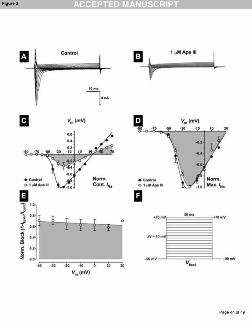
Page 44 of 49
Accep
ted
Man
uscr
ipt
Figure 3

Page 45 of 49
Accep
ted
Man
uscr
ipt
30 nM Aps III
30 nM Aps III
Control
30 nM Aps III
Control
Figure 4
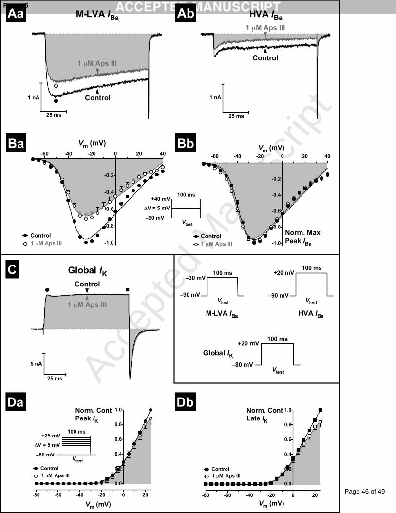
Page 46 of 49
Accep
ted
Man
uscr
ipt
Aps III Aps III
1 μM Aps III
1 μM Aps III
1 μM Aps III
Control
1 μM Aps III
Control
1 μM Aps III
Aa Ab
Ba Bb
C
Da Db
Figure 5
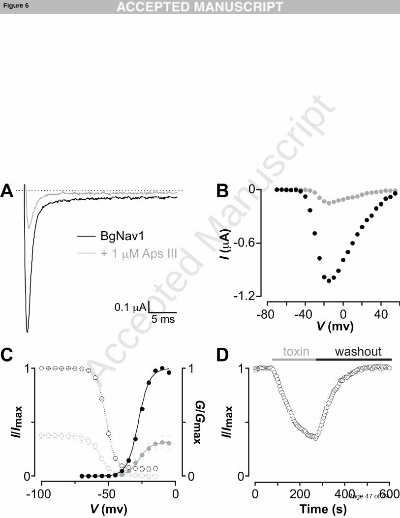
Page 47 of 49
Accep
ted
Man
uscr
ipt
V (mv) Time (s)
V (mv)
BgNav1
+ 1 μM Aps III
5 ms
0.1 μA
A B
I/I m
ax
G/G
ma
x I/I m
ax
I (μ
A)
DC
Figure 6

Page 48 of 49
Accep
ted
Man
uscr
ipt
Figure 7

Page 49 of 49
Accep
ted
Man
uscr
ipt
*Graphical Abstract (for review)