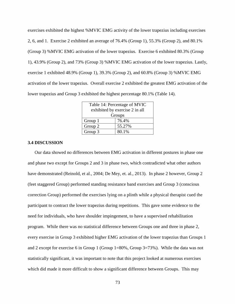
Embed Size (px)
Citation preview

Louisiana State UniversityLSU Digital Commons
LSU Doctoral Dissertations Graduate School
2015
The Influence of the Lower Trapezius Muscle onShoulder Impingement and Scapula DyskinesisChristian Louque CoulonLouisiana State University and Agricultural and Mechanical College c_coulon58yahoocom
Follow this and additional works at httpsdigitalcommonslsuedugradschool_dissertations
Part of the Kinesiology Commons
This Dissertation is brought to you for free and open access by the Graduate School at LSU Digital Commons It has been accepted for inclusion inLSU Doctoral Dissertations by an authorized graduate school editor of LSU Digital Commons For more information please contactgradetdlsuedu
Recommended CitationCoulon Christian Louque The Influence of the Lower Trapezius Muscle on Shoulder Impingement and Scapula Dyskinesis (2015)LSU Doctoral Dissertations 840httpsdigitalcommonslsuedugradschool_dissertations840
THE INFLUENCE OF THE LOWER TRAPEZIUS MUSCLE ON SHOULDER
IMPINGEMENT AND SCAPULAR DYSKINESIS
A Dissertation
Submitted to the Graduate Faculty of the
Louisiana State University and
Agricultural and Mechanical College
in partial fulfillment of the
requirements for the degree of
Doctor of Philosophy
in
The Department of Kinesiology
by
Christian Louque Coulon
BS The University of Louisiana at Lafayette 2005
MS Louisiana State University Health Sciences Center 2007
May 2015
ii
ACKNOWLEDGMENTS
To paraphrase Yogi Berra Irsquod like to thank all the people who made this day
possible Irsquod like to thank Dennis Landin Phil Page Arnold Nelson Laura Stewart Kinesiology
faculty and all of the students from Louisiana State University Kinesiology for all of their
guidance direction and assistance on this project Between recruiting participants marathon
data collections reviewing documents running statistics and overall keeping me on ldquothe
courserdquo I couldnrsquot have done this without you guys Thanks also to my colleges at Baton Rouge
General Medical Center and Peak Performance Physical Therapy for all of the help and support
A special thanks to Phil Page and Theraband Academy for allowing me to use the EMG
equipment for the first two projects and guiding me through the process of collecting
interpreting and analyzing electromyographic data and results And thanks especially to my
committee chair Dennis Landin You were always available to answer questions guide me
through the process and facilitate my further growth
I also wish to thank my family Last but not least (perhaps even most of all) my wife
Brittany Yoursquove always been there to share my good days and cheer me up on the bad ones I
canrsquot possibly thank you enough for all the love support and assistance yoursquove provided along
the way You gave me the strength to persevere to complete this endeavor
iii
PREFACE
Chapters 1 and 2 include the dissertation proposal and literature review as submitted
previously to the Graduate School Chapter 3 and 5 correspond with Study 1 and 2 respectively
In accordance with the wishes of the committee these chapters are formatted as manuscripts to
be submitted for peer-review
iv
TABLE OF CONTENTS
ACKNOWLEDGMENTShelliphelliphelliphelliphelliphelliphelliphelliphelliphelliphelliphelliphelliphelliphelliphelliphelliphelliphelliphelliphelliphelliphelliphelliphelliphelliphellipii
PREFACEhelliphelliphelliphelliphelliphelliphelliphelliphelliphelliphelliphelliphelliphelliphelliphelliphelliphelliphelliphelliphelliphelliphelliphelliphelliphelliphelliphelliphelliphelliphelliphellipv
ABSTRACThelliphelliphelliphelliphelliphelliphelliphelliphelliphelliphelliphelliphelliphelliphelliphelliphelliphelliphelliphelliphelliphelliphelliphelliphelliphelliphelliphelliphelliphelliphelliphellipvi
CHAPTER 1 INTRODUCTIONhelliphelliphelliphelliphelliphelliphelliphelliphelliphelliphelliphelliphelliphelliphelliphelliphelliphelliphelliphelliphelliphelliphellip1
11 SIGNIFICANCE OF DISSERTATIONhelliphelliphelliphelliphelliphelliphelliphelliphelliphelliphelliphelliphelliphelliphelliphellip2
CHAPTER 2 LITERATURE REVIEW4
21 HISTORY INCIDENCE AND EPIDEMIOLOGY OF SHOULDER
IMPINGEMENThelliphelliphelliphelliphelliphelliphelliphelliphelliphelliphelliphelliphelliphelliphelliphelliphelliphelliphelliphelliphelliphelliphelliphelliphellip4
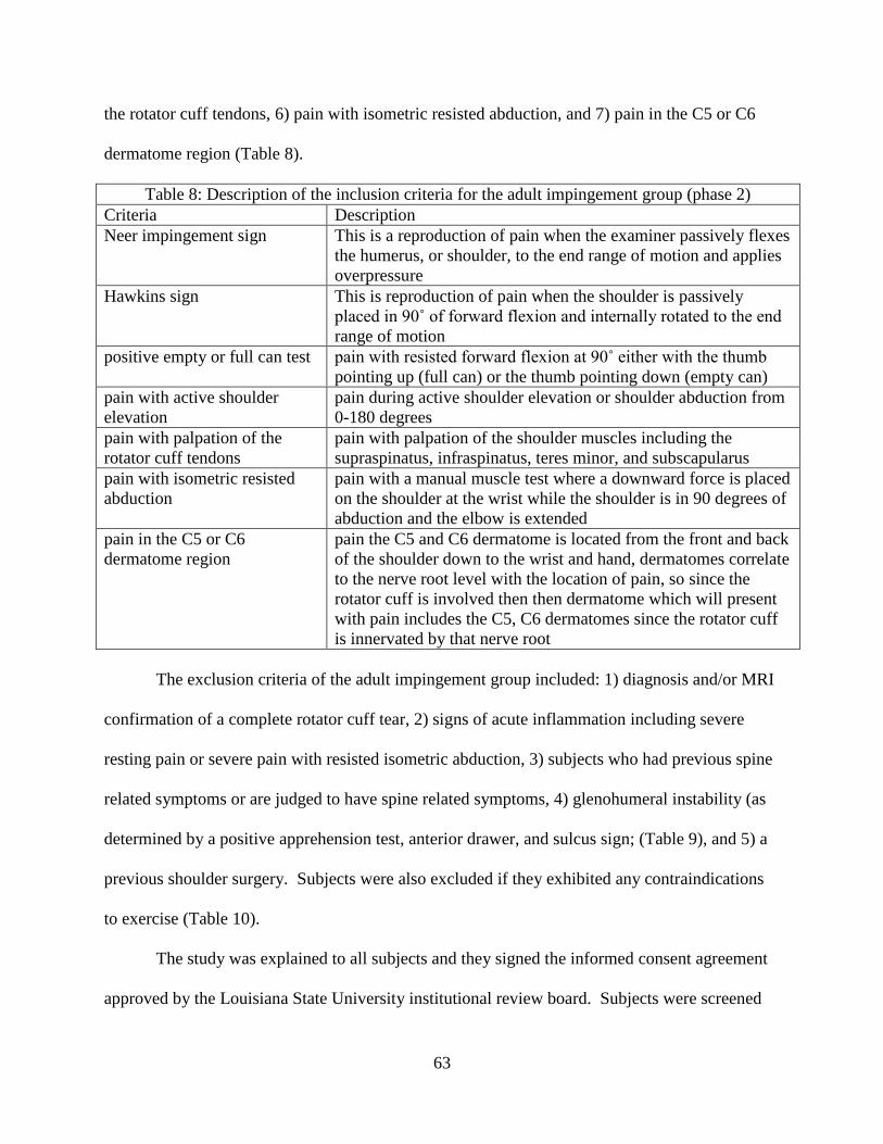
211 Relevant anatomy and pathophysiology of shoulder complexhelliphelliphelliphellip5
22 HISTORY INCIDENCE AND EPIDEMIOLOGY OF SCAPULA DYSKINESIS11
221 Pathophysiology of scapula dyskinesishelliphelliphelliphelliphelliphelliphelliphelliphelliphelliphelliphellip14
23 LIMITATIONS OF STUDYING EMG ON SHOULDER MUSCLES20
24 SHOULDER AND SCAPULAR DYNAMICShelliphelliphelliphelliphelliphelliphelliphelliphelliphelliphelliphelliphellip24
241 Shoulderscapular movementshelliphelliphelliphelliphelliphelliphelliphelliphelliphelliphelliphelliphelliphelliphelliphellip24
242 Loaded vs unloadedhelliphelliphelliphelliphelliphelliphelliphelliphelliphelliphelliphelliphelliphelliphelliphelliphelliphelliphelliphelliphellip28
243 Scapular plane vs other planeshelliphelliphelliphelliphelliphelliphelliphelliphelliphelliphelliphelliphelliphelliphelliphellip29
244 Scapulothoracic EMG activityhelliphelliphelliphelliphelliphelliphelliphelliphelliphelliphelliphelliphelliphelliphelliphellip30
245 Glenohumeral EMG activityhelliphelliphelliphelliphelliphelliphelliphelliphelliphelliphelliphelliphelliphelliphelliphelliphelliphellip32
246 Shoulder EMG activity with impingementhelliphelliphelliphelliphelliphelliphelliphelliphelliphelliphelliphellip32
247 Normal shoulder EMG activityhellip33
248 Abnormal scapulothoracic EMG activityhelliphelliphelliphelliphelliphelliphelliphelliphelliphelliphelliphellip36
249 Abnormal glenohumeralrotator cuff EMG activityhelliphelliphelliphelliphelliphelliphelliphelliphellip40
25 REHABILITATION CONSIDERATIONShelliphelliphelliphelliphelliphelliphelliphelliphelliphelliphelliphelliphelliphellip41
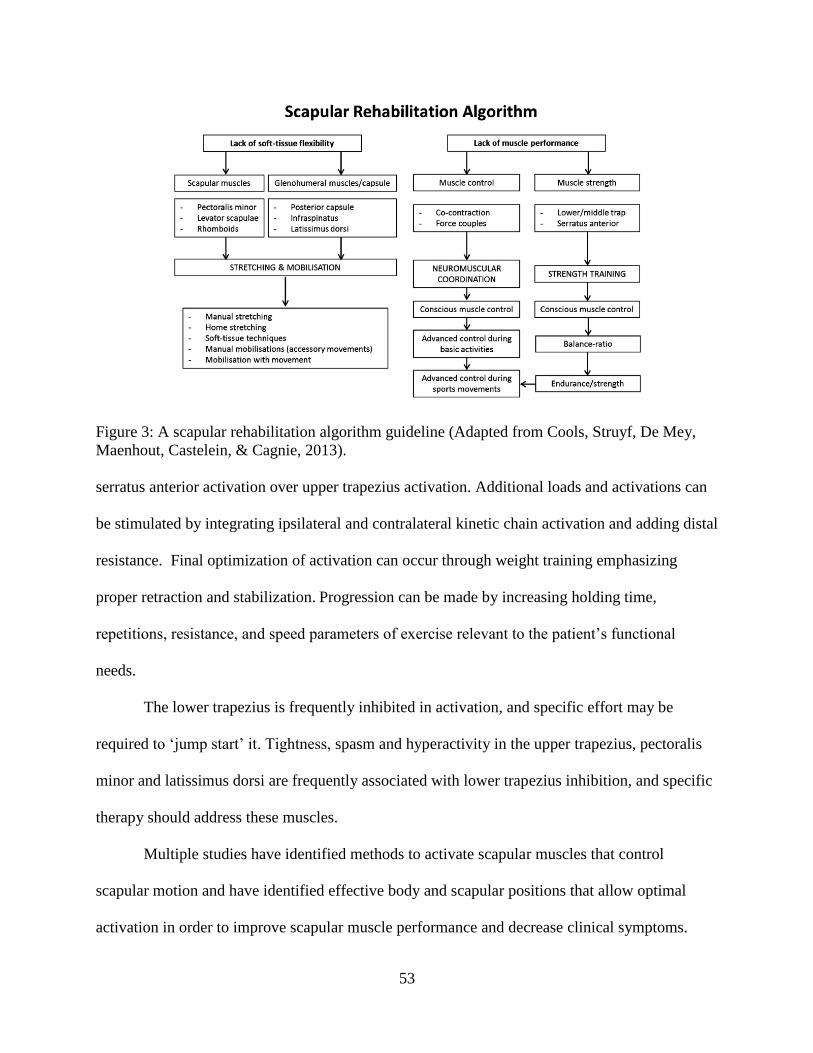
251 Rehabilitation protocols in impingementhelliphelliphelliphelliphelliphelliphelliphelliphelliphelliphelliphellip42
252 Rehabilitation of scapula dyskinesishelliphelliphelliphelliphelliphelliphelliphelliphelliphelliphelliphelliphelliphellip51
253 Effects of rehabilitationhelliphelliphelliphelliphelliphelliphelliphelliphelliphelliphelliphelliphelliphelliphelliphelliphelliphelliphellip54
26 SUMMARYhelliphelliphelliphelliphelliphelliphelliphelliphelliphelliphelliphelliphelliphelliphelliphelliphelliphelliphelliphelliphelliphelliphelliphelliphelliphelliphellip59
CHAPTER 3 THE EFFECT OF VARIOUS POSTURES ON THE SURFACE
ELECTROMYOGRAPHIC ANALYSIS OF THE LOWER TRAPEZIUS DURING SPECIFIC
THERAPEUTIC EXERCISEhelliphelliphelliphelliphelliphelliphelliphelliphelliphelliphelliphelliphelliphelliphelliphelliphelliphelliphelliphelliphelliphelliphelliphelliphellip60
31 INTRODUCTIONhelliphelliphelliphelliphelliphelliphelliphelliphelliphelliphelliphelliphelliphelliphelliphelliphelliphelliphelliphelliphelliphelliphellip60
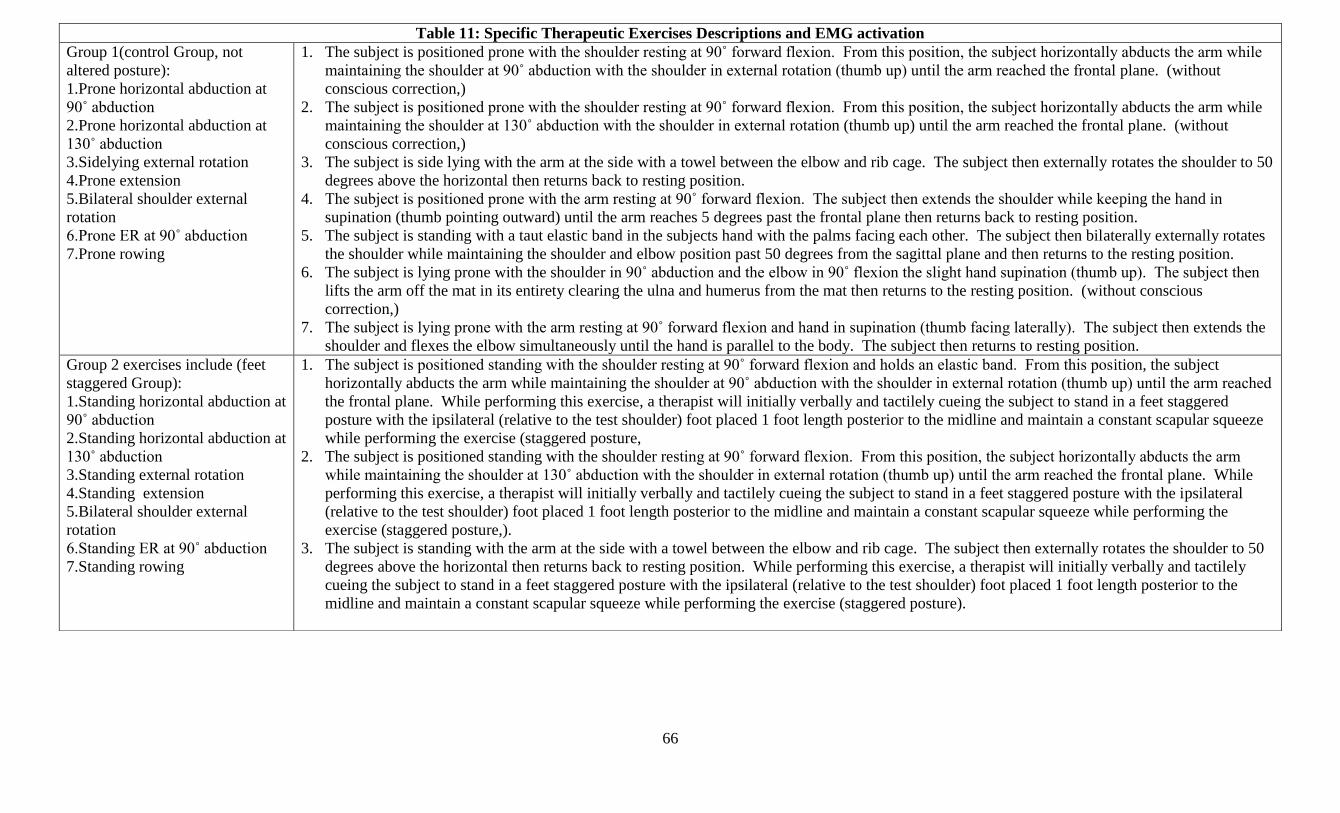
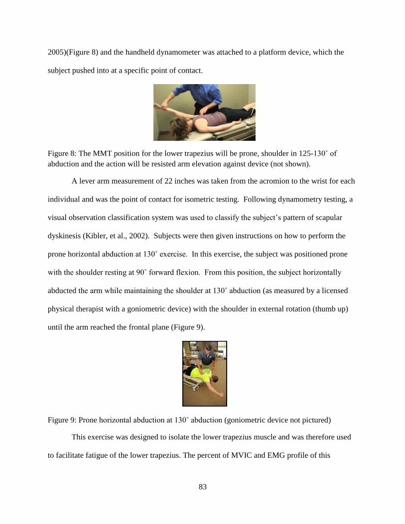
32 METHODShelliphelliphelliphelliphelliphelliphelliphelliphelliphelliphelliphelliphelliphelliphelliphelliphelliphelliphelliphelliphelliphelliphelliphelliphelliphelliphelliphellip62
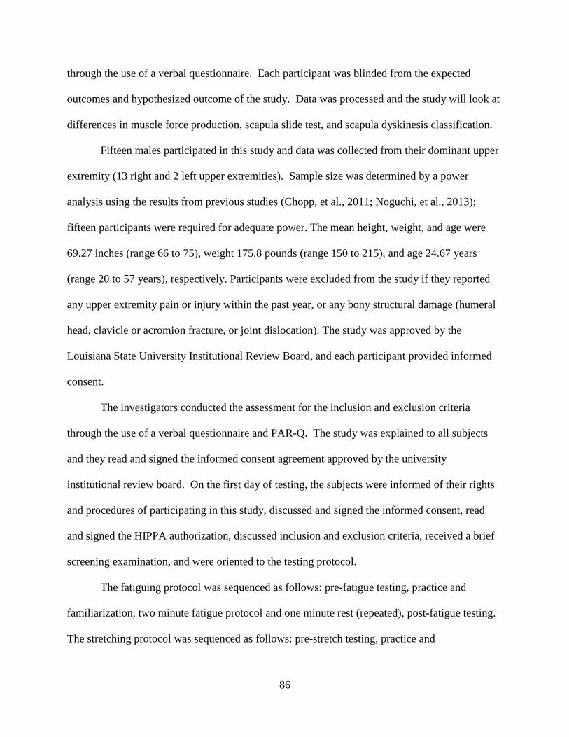
33 RESULTShelliphelliphelliphelliphelliphelliphelliphelliphelliphelliphelliphelliphelliphelliphelliphelliphelliphelliphelliphelliphelliphelliphelliphelliphelliphelliphelliphellip71
34 DISCUSSION helliphelliphelliphelliphelliphelliphelliphelliphelliphelliphelliphelliphelliphelliphelliphelliphelliphelliphelliphelliphelliphelliphelliphelliphelliphellip73
35 CONCLUSIONhelliphelliphelliphelliphelliphelliphelliphelliphelliphelliphelliphelliphelliphelliphelliphelliphelliphelliphelliphelliphelliphelliphelliphelliphellip76
36 ACKNOWLEDGEMENTShelliphelliphelliphelliphelliphelliphelliphelliphelliphelliphelliphelliphelliphelliphelliphelliphelliphelliphelliphelliphellip76
v
CHAPTER 4 THE EFFECT OF LOWER TRAPEZIUS FATIGUE ON SCAPULAR
DYSKINESIS IN INDIVIDUALS WITH A HEALTHY PAIN FREE SHOULDER
COMPLEXhelliphelliphelliphelliphelliphelliphelliphelliphelliphelliphelliphelliphelliphelliphelliphelliphelliphelliphelliphelliphelliphelliphelliphelliphelliphelliphelliphelliphelliphelliphelliphellip77
41 INTRODUCTION helliphelliphelliphelliphelliphelliphelliphelliphelliphelliphelliphelliphelliphelliphelliphelliphelliphelliphelliphelliphelliphelliphelliphellip77
42 METHODShelliphelliphelliphelliphelliphelliphelliphelliphelliphelliphelliphelliphelliphelliphelliphelliphelliphelliphelliphelliphelliphelliphelliphelliphelliphelliphellip81
43 RESULTShelliphelliphelliphelliphelliphelliphelliphelliphelliphelliphelliphelliphelliphelliphelliphelliphelliphelliphelliphelliphelliphelliphelliphelliphelliphelliphelliphellip91
44 DISCUSSIONhelliphelliphelliphelliphelliphelliphelliphelliphelliphelliphelliphelliphelliphelliphelliphelliphelliphelliphelliphelliphelliphelliphelliphelliphellip92
45 CONCLUSIONhelliphelliphelliphelliphelliphelliphelliphelliphelliphelliphelliphelliphelliphelliphelliphelliphelliphelliphelliphelliphelliphelliphelliphelliphellip93
CHAPTER 5 SUMMARY AND CONCLUSIONShelliphelliphelliphelliphelliphelliphelliphelliphelliphelliphelliphelliphelliphelliphellip94
REFERENCES96
APPENDIX A TABLES A-Ghelliphelliphelliphelliphelliphelliphelliphelliphelliphelliphelliphelliphelliphelliphelliphelliphelliphelliphelliphelliphelliphelliphellip109
APPENDIX B IRB INFORMATION STUDY ONE AND TWOhelliphelliphelliphelliphelliphelliphelliphelliphelliphellip116
VITAhelliphelliphelliphelliphelliphelliphelliphelliphelliphelliphelliphelliphelliphelliphelliphelliphelliphelliphelliphelliphelliphelliphelliphelliphelliphelliphelliphelliphelliphelliphelliphelliphelliphelliphellip126
vi
ABSTRACT
This dissertation contains three experiments all conducted in an outpatient physical
therapy setting Shoulder impingement is a common problem seen in overhead athletes and
other individuals and associated changes in muscle activity biomechanics and movement
patterns have been observed in this condition Differentially diagnosing impingement and
specifically addressing the underlying causes is a vital component of any rehabilitation program
and can facilitate the individuals return to normal function and daily living Current
rehabilitation attempts to facilitate healing while promoting proper movement patterns through
therapeutic exercise and understanding each shoulder muscles contribution is vitally important to
treatment of individuals with shoulder impingement This dissertation consisted of two studies
designed to understand how active the lower trapezius muscle will be during common
rehabilitation exercises and the effect lower trapezius fatigue will have on scapula dyskinesis
Study one consisted of two phases and examined muscle activity in healthy individuals and
individuals diagnosed with shoulder impingement Muscle activity was recorded using an
electromyographic (EMG) machine during 7 commonly used rehabilitation exercises performed
in 3 different postures EMG activity of the lower trapezius was recorded and analyzed to
determine which rehabilitation exercise elicited the highest muscle activity and if a change in
posture caused a change in EMG activity The second study took the exercise with the highest
EMG activity of the lower trapezius (prone horizontal abduction at 130˚) and attempted to
compare a fatiguing resistance protocol and a stretching protocol and see if fatigue would elicit
scapula dyskinesis In this study individuals who underwent the fatiguing protocol exhibited
scapula dyskinesis while the stretching group had no change in scapula motion Also of note
both groups exhibited a decrease in force production due to the treatment The scapula
vii
dyskinesis in the fatiguing group implies that lower trapezius function is vitally important to
maintain proper scapula movement patterns and fatigue of this muscle can contribute and even
cause scapula dyskinesis This abnormal scapula motions can cause or increase the risk of injury
in overhead throwing This dissertation provides novel insight about EMG activation during
specific therapeutic exercises and the importance of lower trap function to proper biomechanics
of the scapula
1
CHAPTER 1 INTRODUCTION
The complex human anatomy and biomechanics of the shoulder absorbs a large amount
of stress while performing activities like throwing a baseball swimming overhead material
handling and other repetitive overhead activities The term ldquoshoulder impingementrdquo first
described by Neer (Neer 1972) clarified the etiology pathology and treatment of a common
shoulder disorder Initially patients who were diagnosed with shoulder impingement were
treated with subacromial decompression but Tibone (Tibone et al 1985) demonstrated that
overhead athletes had a success rate of only 43 and only 22 of throwing athletes were able to
return to sport Therefore surgeons sought alternative causes of the overhead throwers pain
Jobe (Jobe Kvitne amp Giangarra 1989) then introduced the concept of instability which would
result in secondary impingement and hypothesized that overhead throwing athletes develop
shoulder instability and this instability in turn led to secondary subacromial impingement Jobe
(Jobe 1996) also later described the phenomenon of ldquointernal impingementrdquo between the
articular side of the posterior rotator cuff and the posterior glenoid labrum while the shoulder is
in abduction and external rotation
From the above stated information it is obvious that shoulder impingement is a common
condition affecting overhead athletes and this condition is further complicated due to the
throwing motion being a high velocity repetitive and skilled movement (Wilk et al 2009
Conte Requa amp Garrick 2001) During the throwing motion an extreme amount of force is
placed on the shoulder including an angular velocity of nearly 7250˚s and distractive or
translatory forces less than or equal to a personrsquos body weight (Wilk et al 2009) For this
reason the glenohumeral joint is the most commonly injured joint in professional baseball
pitchers (Wilk et al 2009) and other overhead athletes (Sorensen amp Jorgensen 2000)
2
Consequently an overhead athletersquos shoulder complex must maintain a high level of muscular
strength adequate joint mobility and enough joint stability to prevent shoulder impingement or
other shoulder pathologies (Wilk et al 2009 Sorensen amp Jorgensen 2000 Heyworth amp
Williams 2009 Forthomme Crielaard amp Croisier 2008)
Once pathology is present typical manifestations include a decrease in throwing
performance strength deficits decreased range of motion joint laxity andor pain (Wilk et al
2009 Forthomme Crielaard amp Croisier 2008) It is important for a clinician to understand the
causes of abnormal shoulder dynamics in overhead athletes with impingement in order to
implement the most effective and appropriate treatment plan and maintain wellness after
pathology Much of the research in shoulder impingement is focused on the kinematics of the
shoulder and scapula muscle activity during these movements static posture and evidence
based exercise prescription to correct deficits Despite the research findings there is uncertainty
as to the link between kinematics and the mechanism of for SIS in overhead athletes The
purpose of this paper is to review the literature on the pathomechanics EMG activity and
clinical considerations in overhead athletes with impingement
11 SIGNIFICANCE OF DISSERTATION
The goal of this project is to investigate the electromyographic (EMG) activity of the
lower trapezius during commonly used therapeutic exercises for individuals with shoulder
impingement and to determine the effect the lower trapezius has on scapular dyskinesis Each
therapeutic exercise has a specific EMG profile and knowing this profile is beneficial to help a
rehabilitation professional determine which exercise dosage and movement pattern to select
muscle rehabilitation In addition the data from study one of this dissertation was used to pick
the specific exercise which exhibited the highest potential to activate and fatigue the lower
3
trapezius From fatiguing the lower trapezius we are able to determine the effect fatigue plays in
inducing scapula dyskinesis and increasing the injury risk of that individual This is important in
preventing devastating shoulder injuries as well as overall shoulder health and wellness and these
studies may shed some light on the mechanism responsible for shoulder impingement and injury
4
CHAPTER 2 LITERATURE REVIEW
This review will begin by discussing the history incidence and epidemiology of shoulder
impingement in Section 10 which will also discuss the relevant anatomy and pathophysiology
of the normal and pathologic shoulder The next section 20 will cover the specific and general
limitations of EMG analysis The following section 30 will discuss shoulder and scapular
movements muscle activation and muscle timing in the healthy and impinged shoulder Finally
section 40 will discuss the clinical implications and the effects of rehabilitation on the overhead
athlete with shoulder impingement
21 HISTORY INCIDENCE AND EPIDEMIOLOGY OF SHOULDER IMPINGEMENT
Shoulder impingement accounts for 44-65 of all cases of shoulder pain (Neer 1972 Van
der Windt Koes de Jong amp Bouter 1995) and is commonly seen in overhead athletes due to the
biomechanics and repetitive nature of overhead motions in sports Commonly the most affected
types of sports activities include throwing athletes racket sports gymnastics swimming and
volleyball (Kirchhoff amp Imhoff 2010)
Subacromial impingement syndrome (SIS) a diagnosis commonly seen in overhead athletes
presenting to rehabilitation is characterized by shoulder pain that is exacerbated with arm
elevation or overhead activities Typically the rotator cuff the long head of the biceps tendon
andor the subacromial bursa are being ldquoimpingedrdquo under the acromion in the subacromial space
causing pain and dysfunction (Ludewig amp Cook 2000 Lukaseiwicz McClure Michener Pratt
amp Sennett 1999 Michener Walsworth amp Burnet 2004 Nyberg Jonsson amp Sundelin 2010)
Factors proposed to contribute to SIS can be classified as either intrinsic or extrinsic and then
further classified based on the cause of the problem into primary secondary or posterior
impingement (Nyberg Jonsson amp Sundelin 2010)
5
211 Relevant anatomy and pathophysiology of shoulder complex
When discussing the relevant anatomy in shoulder impingement it is important to have an
understanding of the glenohumeral and scapula-thoracic musculature subacromial space (SAS)
and soft tissue which can become ldquoimpingedrdquo in the shoulder The primary muscles of the
shoulder complex include the rotator cuff (RTC) (supraspinatus infraspinatus teres minor and
subscapularus) scapular stabilizers (rhomboid major and minor upper trapezius lower trapezius
middle trapezius serratus anterior) deltoid and accessory muscles (latisimmus dorsi biceps
brachii coracobrachialis pectoralis major pectoralis minor) The shoulder also contains
numerous bursae one of which is clinically significant in overhead athletes with impingement
called the subacromial bursae The subacromial bursa is located between the deltoid muscle and
the glenohumeral joint capsule and extends between the acromion and supraspinatus muscle
Often with repetitive overhead activity the subacromial bursae may become inflamed causing a
reduction in the subacromial space (Wilk Reinold amp Andrews 2009) The supraspinatus
tendon lies underneath the subacromial bursae and inserts on the superior facet of the greater
tubercle of the humerus and is the most susceptible to impingement of the RTC muscles The
infraspinatus tendon inserts posterior-inferior to the supraspinatus tendon on the greater tubercle
and may become impinged by the anterior acromion during shoulder movement
The SAS is a 10mm area below the acromial arch in the shoulder (Petersson amp Redlund-
Johnell 1984) and contains numerous soft tissue structures including tendons ligaments and
bursae (Figure 1) These structures can become compressed or ldquoimpingedrdquo in the SAS causing
pain due to excessive humeral head migration scapular dyskinesis muscular weakness and
bony abnormalities Any subtle deviation (1-2 mm) from a normal decrease in the SAS can
contribute to impingement and pain (Allmann et al 1997 Michener McClure amp Karduna
6
2003) Researchers have compared static radiographs of painful and normal shoulders at
numerous positions of glenohumeral range of motion and the findings include 1) humeral head
excursion greater than 15 mm is associated with shoulder pathology (Poppen amp Walker 1976)
2) patientrsquos with impingement demonstrated a 1mm superior humeral head migration (Deutsch
Altchek Schwartz Otis amp Warren 1996) 3) patientrsquos with RTC tears (with and without pain)
demonstrated superior migration of the humeral head with increasing elevation between 60deg-
150deg compared to a normal control (Yamaguchi et al 2000) and 4) in all studies it was
demonstrated that a decrease in SAS was associated with pathology and pain
To maintain the SAS the scapula upwardly rotates which will elevate the lateral acromion
and prevent impingement but the SAS will exhibit a 3mm-39mm decrease in non-pathologic
subjects at 30-120 degrees of abduction (Ludewig amp Cook 2000 Graichen et al 1999)
Scapular posterior tilting also prevents impingement of the RTC tendons by elevating the
anterior acromion and maintaining the SAS
Shoulder impingement believed to contribute to the development of RTC disease
(Ludewig amp Braman 2011 Van der Windt Koes de Jong amp Bouter 1995) is the most
frequently diagnosed shoulder disorder in primary healthcare and despite its reported prevalence
the diagnostic criteria and etiology of SIS are debatable (Ludewig amp Braman 2011) SIS is an
encroachment of soft tissues in the SAS due to narrowing of this space (Figure 1 B) and after
impingement occurs the shoulder soft tissue can and may progress through the 3 stages of lesions
(typically and overhead athlete progresses through these stages more rapidly)(Wilk Reinold
Andrews 2009) Neer described (Neer 1983) three stages of lesions (Table 1) and the higher
the stage the harder to respond to conservative care
7
Table 1 Neer classifications of lesions in impingement syndrome
Stage Characteristics Typical Age of Patient
Stage I edema and hemorrhage of the bursa and cuff
reversible with conservative treatment
lt 25 yo
Stage II irreversible changes such as fibrosis and
tendinitis of the rotator cuff
25-40 yo
Stage III by partial or complete tears of the rotator cuff
and or biceps tendon and acromion andor
AC joint pathology
gt40 yo
SIS can be separated into two main mechanistic theories and two less classic forms of
impingement The two main theories include Neerrsquos (Neer 1972) impingement theory which
focuses on the extrinsic mechanisms (primary impingement) and the second theory focuses on
intrinsic mechanisms (secondary impingement) The less classic forms of shoulder impingement
include internal impingement and coracoid impingement
Primary shoulder impingement results from mechanical abrasion and compression of the
RTC tendons subacromial bursa or long head of the biceps tendon under the anterior
undersurface of the acromion coracoacromial ligament or undersurface of the acromioclavicular
joint during arm elevation (Neer 1972) This type of impingement is typically seen in persons
older than 40 years old and is typically due to degeneration Scapular dyskinesis has been
observed in this population and causes superior translation of the humeral head further
decreasing the SAS (Lukaseiwicz McClure Michener Pratt amp Sennett 1999 Ludewig amp
Cook 2000 de Witte et al 2011)
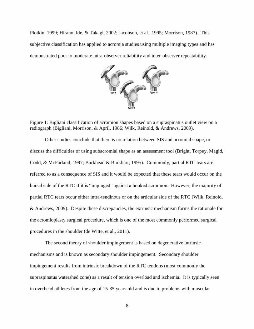
In some studies a correlation between acromial shape (Bigliani classification type II or
type III) (Figure 1) (Bigliani Morrison amp April 1986) and SIS has been observed and it is
presumed that the hooked acromion is a pre-existing anatomic variation or traction spur caused
by repetitive superior translation of the humerus or by tendinopathy (Nordt Garretson amp
8
Plotkin 1999 Hirano Ide amp Takagi 2002 Jacobson et al 1995 Morrison 1987) This
subjective classification has applied to acromia studies using multiple imaging types and has
demonstrated poor to moderate intra-observer reliability and inter-observer repeatability
Figure 1 Bigliani classification of acromion shapes based on a supraspinatus outlet view on a
radiograph (Bigliani Morrison amp April 1986 Wilk Reinold amp Andrews 2009)
Other studies conclude that there is no relation between SIS and acromial shape or
discuss the difficulties of using subacromial shape as an assessment tool (Bright Torpey Magid
Codd amp McFarland 1997 Burkhead amp Burkhart 1995) Commonly partial RTC tears are
referred to as a consequence of SIS and it would be expected that these tears would occur on the
bursal side of the RTC if it is ldquoimpingedrdquo against a hooked acromion However the majority of
partial RTC tears occur either intra-tendinous or on the articular side of the RTC (Wilk Reinold
amp Andrews 2009) Despite these discrepancies the extrinsic mechanism forms the rationale for
the acromioplasty surgical procedure which is one of the most commonly performed surgical
procedures in the shoulder (de Witte et al 2011)
The second theory of shoulder impingement is based on degenerative intrinsic
mechanisms and is known as secondary shoulder impingement Secondary shoulder
impingement results from intrinsic breakdown of the RTC tendons (most commonly the
supraspinatus watershed zone) as a result of tension overload and ischemia It is typically seen
in overhead athletes from the age of 15-35 years old and is due to problems with muscular
9
dynamics and associated shoulder or scapular instability (de Witte et al 2011) Typically this
condition is enhanced by overuse subacromial inflammation tension overload on degenerative
RTC tendons or inadequate RTC function leading to an imbalance in joint stability and mobility
with consequent altered shoulder kinematics (Yamaguchi et al 2000 Mayerhoefer
Breitenseher Wurnig amp Roposch 2009 Uhthoff amp Sano 1997) Instability is generally
classified as traumatic or atraumatic in origin as well as by the direction (anterior posterior
inferior or multidirectional) and amount (grade I- grade III) of instability (Wilk Reinold amp
Andrews 2009) Instability in overhead athletes is typically due to repetitive microtrauma
which can contribute to secondary shoulder impingement (Ludewig amp Reynolds 2009)
Recently internal impingement has been identified and thought to be caused by friction
and mechanical abrasion of the undersurface of the supraspinatus and infraspinatus against the
anterior or posterior glenoid rim or glenoid labrum
This has been seen posteriorly in overhead athletes when the arm is abducted to 90
degrees and externally rotated (Pappas et al 2006) and is usually accompanied with complaints
of posterior shoulder pain during this late cocking phase of throwing when the arm is at the end
range of external rotation (Myers Laudner Pasquale Bradley amp Lephart 2006) Posterior
shoulder tightness (PST) and glenohumeral internal rotation deficit (GIRD) have also been
linked to internal impingement by Burkhart and colleagues (Burkhart Morgan amp Kibler 2003)
Correction of the PST through physical therapy has been shown to lead to resolution of the
symptoms of internal impingement (Tyler Nicholas Lee Mullaney amp Mchugh 2012)
Coracoid impingement is typically associated with anterior shoulder pain at the extreme
ranges of glenohumeral internal rotation (Jobe Coen amp Screnar 2000) This type of
impingement is less commonly discussed but consists of the subscapularis tendon being
10
impinged between the coracoid process and lesser tuberosity of the humerus (Ludewig amp
Braman 2011)
Since the RTC muscles are involved in throwing and overhead activities partial thickness
tears full thickness tears and rotator cuff disease is seen in overhead athletes When this
becomes a chronic condition secondary impingement or internal impingement can result in
primary tensile cuff disease (PTCD) or primary compressive cuff disease (PCCD) PTCD
hypothesized to be a byproduct of internal impingement occurs during the deceleration phase of
throwing in a stable shoulder and is the result of large repetitive eccentric loads placed on the
RTC as it attempts to decelerate the arm resulting in partial undersurface tears in the
supraspinatus and infraspinatus tendons (Andrews amp Angelo 1988 Wilk et al 2009) In
contrast PCCD occurs on the bursal side of the RTC and results in partial thickness tears of the
RTC It is hypothesized that processes that cause a decrease in the SIS increase the risk of this
pathology and this is a byproduct of RTC muscular imbalance and weakness especially during
the deceleration phase of throwing (Andrews amp Angelo 1988) During the late cocking and
early acceleration phases of throwing with the arm at maximal external rotation the rotator cuff
has the potential to become impinged between the humeral head and the posterior-superior
glenoid internal or posterior impingement (Wilk et al 2009) and may cause articular or
undersurface tearing of the RTC in overhead athletes
In conclusion tears of the RTC may be caused by primarily 3 mechanisms in overhead
athletes including internal impingement primary tensile cuff disease (PTCD) or primary
compressive cuff disease (PCCD) (Wilk et al 2009) and the causes of SIS are multifactorial
and variable
11
22 HISTORY INCIDENCE AND EPIDEMIOLOGY OF SCAPULA DYSKINESIS
The scapula and its associated movements are a critical component facilitating normal
functional movements in the shoulder complex while maintaining stability of the shoulder and
acting as an area of force transfer (Kibler amp McMullen 2003) Assessing scapular movement
and position is an important part of the clinical examination (Wright et al 2012) and identifies
the presence or absence of optimal motion in order to guide specific treatment options (Ludwig
amp Reynolds 2009) The literature lacks the ability to identify if altered scapula positions or
motions are specific to shoulder pathology or if these alterations are a normal variation (Wright
et al 2012) Scapula motion abnormalities consist of premature excessive or dysrhythmic
motions during active glenohumeral elevation lowering of the upper extremity or upon bilateral
comparison (Ludwig amp Reynolds 2009 Wright et al 2012) Research has demonstrated that
the scapula upwardly rotates (Ludwig amp Reynolds 2009) posteriorly tilts and externally rotates
to clear the acromion from the humerus in forward elevation Also the scapula synchronously
externally rotates while posteriorly tilting to maintain the glenoid as a congruent socket for the
moving arm and maximize concavity compression of ball and socket kinematics The scapula is
also dynamically stabilized in a position of retraction during arm use to maximize activation and
length tension relationships of all muscles that originate on the scapula (Ludwig amp Reynolds
2009) Finally the scapula is a link in the kinetic chain of integrated segment motions that starts
from the ground and ends at the hand (Kibler Ludewig McClure Michener Bak Sciascia
2013) Because of the important but minimal bony stabilization of the scapula by the clavicle
through the acromioclavicular joint dynamic muscle function is the major method by which the
scapula is stabilized and purposefully moved to accomplish its roles Muscle activation is
coordinated in task specific force couple patterns to allow stabilization of position and control of
12
dynamic coupled motion Also the scapula will assist with acromial elevation to increase
subacromial space for underlying soft tissue clearance (Ludwig amp Reynolds 2009 Wright et al
2012) and for this reason changes in scapular position are important
The clavicle exists to help maintain optimal scapular position during arm motion (Ludwig amp
Reynolds 2009) In this manner it acts as a strut for the shoulder as it attaches the arm to the
axial skeleton via the acromioclavicular and sternoclavicular joints Injury to any of the static
restraints can cause the scapula to become unstable which in turn will negatively affect arm
function (Kibler amp Sciascia 2010)
Previous research has found that changes to scapular positioning or motion were evident in
68 to 100 of patients with shoulder impairments (Warner Micheli Arslanian Kennedy amp
Kennedy 1992) resulting in compensatory motions at distal segments The motions begin
causing a diminished dynamic control of humeral-head deceleration and lead to shoulder
pathologies (Voight Hardin Blackburn Tippett amp Canner 1996 Wilk Meister amp Andrews
2002 McQuade Dawson amp Smidt 1998 Kibler amp McMullen 2003 Warner Micheli
Arslanian Kennedy amp Kennedy 1992 Nadler 2004 Hutchinson amp Ireland 2003) For this
reason the effects of scapular fatigue warrants further research
Scapular upward rotation provides a stable base during overhead activities and previous
research has examined the effect of fatigue on scapula movements and shoulder function
(Suzuki Swanik Bliven Kelly amp Swanik 2006 Birkelo Padua Guskiewicz amp Karas 2003
Su Johnson Gravely amp Karduna 2004 Tsai McClure amp Karduna 2003 McQuade Dawson
amp Smidt 1998 Joshi Thigpen Bunn Karas amp Padua 2011 Tyler Cuoco Schachter Thomas
amp McHugh 2009 Noguchi Chopp Borgs amp Dickerson 2013 Chopp Fischer amp Dickerson
2011 Madsen Bak Jensen amp Welter 2011) Prior studies found no change in scapula upward
13
rotation due to fatigue in healthy individuals (Suzuki Swanik Bliven Kelly amp Swanik 2006)
and healthy overhead athletes (Birkelo Padua Guskiewicz amp Karas 2003 Su Johnson
Gravely amp Karduna 2004) However the results of these studies should be interpreted with
caution and may not be applied to functional movements since one study (Suzuki Swanik
Bliven Kelly amp Swanik 2006) performed seated overhead throwing before and after fatigue
with healthy college age men Since the kinematics and dynamics of overhead throwing cannot
be seen in sitting the authorrsquos results canrsquot draw a comparison to overhead athletes or the
pathological populations since the participants were healthy Also since the scapula is thought
to be involved in the kinetic chain of overhead motion (Kibler Ludewig McClure Michener
Bak amp Sciascia 2013) sitting would limit scapula movements and limit the interpretation of the
resulting scapula motion
Nonetheless several researchers have identified decreased scapular upward rotation in both
healthy subjects and subjects with shoulder pathologies (Su Johnson Gravely amp Karduna
2004 Warner Micheli Arslanian Kennedy amp Kennedy 1992 Lukaseiwicz McClure
Michener Pratt amp Sennett 1999) In addition after shoulder complex fatigue significant
changes in scapular position (decreased upward rotation posterior tilting and external rotation)
have been demonstrated using exercises that induced scapular and glenohumeral muscle fatigue
(Tsai McClure amp Karduna 2003) However this previous research has focused on shoulder
external rotation fatigue and not on scapular musculature fatigue
Lack of agreement in the findings are explained by the nature of measurements used which
differ between static and dynamic movements as well as instrumentation One explanation for
these differences involves the muscles targeted for fatigue For example some studies have
examined shoulder complex fatigue due to a functional activity (Birkelo Padua Guskiewicz amp
14
Karas 2003 Su Johnson Gravely amp Karduna 2004 Madsen Bak Jensen amp Welter 2011)
while others have compared a more isolated scapular-muscle fatigue protocol (McQuade
Dawson amp Smidt 1998 Suzuki Swanik Bliven Kelly amp Swanik 2006 Tyler Cuoco
Schachter Thomas amp McHugh 2009 Chopp Fischer amp Dickerson 2011) and others have
examined shoulder complex fatigue (Tsai McClure amp Karduna 2003 Joshi Thigpen Bunn
Karas amp Padua 2011 Noguchi Chopp Borgs amp Dickerson 2013 Madsen Bak Jensen amp
Welter 2011 Chopp Fischer amp Dickerson 2011) Therefore to date no prior research has
specifically targeted the lower trapezius muscle using a therapeutic exercise with a maximal
activation pattern of the muscle
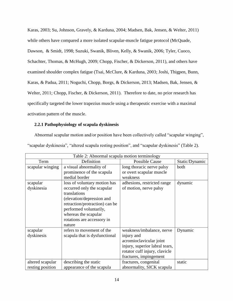
221 Pathophysiology of scapula dyskinesis
Abnormal scapular motion andor position have been collectively called ldquoscapular wingingrdquo
ldquoscapular dyskinesiardquo ldquoaltered scapula resting positionrdquo and ldquoscapular dyskinesisrdquo (Table 2)
Table 2 Abnormal scapula motion terminology
Term Definition Possible Cause StaticDynamic
scapular winging a visual abnormality of
prominence of the scapula
medial border
long thoracic nerve palsy
or overt scapular muscle
weakness
both
scapular
dyskinesia
loss of voluntary motion has
occurred only the scapular
translations
(elevationdepression and
retractionprotraction) can be
performed voluntarily
whereas the scapular
rotations are accessory in
nature
adhesions restricted range
of motion nerve palsy
dynamic
scapular
dyskinesis
refers to movement of the
scapula that is dysfunctional
weaknessimbalance nerve
injury and
acromioclavicular joint
injury superior labral tears
rotator cuff injury clavicle
fractures impingement
Dynamic
altered scapular
resting position
describing the static
appearance of the scapula
fractures congenital
abnormality SICK scapula
static
15
The most appropriate term to refer to dysfunctional dynamic movement of the scapula is the
term scapular dyskinesis (lsquodysrsquomdashalteration of lsquokinesisrsquomdashmovement) When the arm is raised
overhead the generally accepted pattern of scapulothoracic motion is upward rotation external
rotation and posterior tilt of the scapula as well as elevation and retraction of the clavicle
(Ludewig et al 1996 McClure et al 2001) Of the 14 muscles that attach to the scapula the
trapezius and serratus anterior play a critical role in the production and control of scapulothoracic
motion (Ebaugh et al 2005 Inman et al 1944 Ludewig et al 1996) Furthermore scapular
dyskinesis is reported to be more prominent as the arm is lowered from an overhead position and
individuals with shoulder pathology generally report more pain when lowering the arm (Kibler amp
McMullen 2003 Sharman 2002)
Scapular dyskinesis has been identified by a group of experts as (1) abnormal static scapular
position andor dynamic scapular motion characterized by medial border prominence or (2)
inferior angle prominence andor early scapular elevation or shrugging on arm elevation andor
(3) rapid downward rotation during arm lowering (Kibler amp Sciascia 2010) Scapular
dyskinesis is a non-specific response to a painful condition in the shoulder rather than a specific
response to certain glenohumeral pathology and alters the scapulohumeral rhythm Scapular
dyskinesis occurs when the upper trapezius middle trapezius lower trapezius serratus anterior
and latissimus dorsi (stabilizing muscles) are unable to preserve typical scapular movement
(Kibler amp Sciascia 2010) Scapula dyskinesis is potentially harmful when it results in increased
anterior tilting downward rotation and protraction which reorients the acromion and decreases
the subacromial space width (Tsai et al 2003 Borstad et al 2009)
Alterations in static stabilizers (bone) muscle activation patterns or strength in scapula
musculature have contributed to scapula dyskinesis Researchers have shown that injuries to the
16
stabilizing ligaments of the acromioclavicular joint can cause the scapula to displace in a
downward protracted and internally rotated position (Kibler amp Sciascia 2010) With
displacement of the scapula significant functional consequences to shoulder biomechanics occur
including an uncoupling of the scapulohumeral complex inability of the scapular stabilizing
muscles to maintain appropriate positioning of the glenohumeral and acromiohumeral joints and
a subsequent loss of rotator cuff strength and function (Joshi Thigpen Bunn Karas amp Padua
2011)
Scapular dyskinesis is associated with impingement by altering arm motion and scapula
position upon dynamic elevation which is characterized by a loss of acromial upward rotation
excessive scapular internal rotation and excessive scapular anterior tilt (Cools Struyf De Mey
Maenhout Castelein amp Cagnie 2013 Forthomme Crielaard amp Croisier 2008) These
associated alterations cause a decrease in the subacromial space and increase the individualrsquos
impingement risk
Prior research has demonstrated altered activation sequencing patterns and strength of the
stabilizing muscles of the scapula in individuals diagnosed with impingement risk and scapular
dyskinesis (Cools Struyf De Mey Maenhout Castelein amp Cagnie 2013 Kibler amp Sciascia
2010) Each scapula muscle makes a specific contribution to scapular function but the lower
trapezius and serratus anterior appear to play the major role in stabilizing the scapula during arm
movement Weakness fatigue or injury in either of these muscles may cause a disruption of the
dynamic stability which leads to abnormal kinematics and symptoms of impingement In a prior
study (Madsen Bak Jensen amp Welter 2011) the authors demonstrated increased incidence of
scapula dyskinesis in pain-free competitive overhead athletes during increasing training and
17
fatigue The prevalence of scapula dyskinesis seemed to increase with increased training to a
cumulative presence of 82 in pain-free competitive overhead athletes
A classification system which aids in clinical evaluation of scapula dyskinesis has also been
reported in the literature (Kibler Uhl Maddux Brooks Zeller amp McMullen 2002) and
modified to increase sensitivity (Uhl Kibler Gecewich amp Tripp 2009) This method classifies
scapula dyskinesis based on the prominent part of the scapula and includes four types 1) inferior
angle pattern (Type I) 2) medial border pattern (Type II) 3) superior border patters (Type III)
and 4) normal pattern (Type IV) The examiner first predicts if the individual has scapula
dyskinesis (yesno method) then classifies the individual pattern type which has a higher
sensitivity (76) and positive predictive value (74) than any other clinical dyskinesis measure
(Uhl Kibler Gecewich amp Tripp 2009)
Increased upper trapezius activity imbalance of upper trapeziuslower trapezius activation
and decreased serratus anterior activity have been reported in patients with impingement (Cools
Struyf De Mey Maenhout Castelein amp Cagnie 2013 Lawrence Braman Laprade amp
Ludewig 2014) Authors have hypothesized that impingement due to lack of acromial elevation
is caused by increased upper trapezius activity (shrug maneuver) resulting in a type III (upper
medial border prominence) dyskinesis pattern (Kibler amp Sciascia 2010) Frequently lower
trapezius activation is inhibited or is delayed (Cools Struyf De Mey Maenhout Castelein amp
Cagnie 2013) which results in a type IIItype II (entire medial border prominence) dyskinesis
pattern and impingement due to loss of acromial elevation and posterior tilt (Kibler amp Sciascia
2010)
Scapular position and kinematics influence rotator cuff strength (Kibler Ludewig McClure
Michener Bak amp Sciascia 2013) and prior research (Kebaetse McClure amp Pratt 1999) has
18
demonstrated a 23 maximum rotator cuff strength decrease due to excessive scapular
protraction a posture seen frequently in individuals with scapular dyskinesis Another study
(Smith Dietrich Kotajarvi amp Kaufman 2006) indicates that maximal rotator cuff strength is
achieved with a position of lsquoneutral scapular protractionretractionrsquo and the positions of
excessive protraction or retraction demonstrates decreased rotator cuff abduction strength
Lastly research has demonstrated (Kibler Sciascia amp Dome 2006) an increase of 24
supraspinatus strength in a position of scapular retraction in individuals with shoulder pain and
11 increase in individuals without shoulder pain The clinically observable finding in scapular
dyskinesis prominence of the medial scapular border is associated with the biomechanical
position of scapular internal rotation and protraction which is a less than optimal base for muscle
strength (Kibler amp Sciascia 2010)
Table 3 Causes of scapula dyskinesis
Cause Associated pathology
Bony thoracic kyphosis clavicle fracture nonunion clavicle shortened mal-union
scapular fractures
Neurological cervical radiculopathy long thoracic dorsal scapular nerve or spinal accessory
nerve palsy
Joint high grade AC instability AC arthrosis GH joint internal derangement (labral
injury) glenohumeral instability biceps tendinitis
Soft Tissue inflexibility (tightness) or intrinsic muscle problems Inflexibility and stiffness of
the pectoralis minor and biceps short head can create anterior tilt and protraction
due to their pull on the coracoid
soft tissue posterior shoulder inflexibility can lead to glenohumeral internal rotation
deficit (GIRD) shoulder rotation tightness (GIRD and Total Range of Motion
Deficit) and pectoralis minor inflexibility
Muscular periscapular muscle activation serratus anterior activation and strength is decreased
the upper trapeziuslower trapezius force couple may be altered delayed onset of
activation in the lower trapezius
lower trapezius and serratus anterior weakness upper trapezius hyperactivity or
scapular muscle detachment and kinetic chain factors include hipleg weakness and
core weakness
19
Causes of scapula dyskinesis remain multifactorial (Table 3) but altered scapular motion or
position decrease linear measures of the subacromial space (Giphart van der Meijden amp Millett
2012) increase impingement symptoms (Kibler Ludewig McClure Michener Bak amp Sciascia
2013) decrease rotator cuff strength (Kebaetse McClure amp Pratt 1999 Smith Dietrich
Kotajarvi amp Kaufman 2006 Kibler Sciascia amp Dome 2006) and increase the risk of internal
impingement (Kibler amp Sciascia 2010)
However no conclusive study indicating the occurrence of scapular dyskinesis occurring as a
direct result of solely lower trapezius muscle fatigue even though scapular orientation changes
in an impinging direction (downward rotation anterior tilt and protraction) have been reported
with fatigue (Birkelo Padua Guskiewicz amp Karas 2003 Su Johnson Gravely amp Karduna
2004 Madsen Bak Jensen amp Welter 2011 McQuade Dawson amp Smidt 1998 Suzuki
Swanik Bliven Kelly amp Swanik 2006 Tyler Cuoco Schachter Thomas amp McHugh 2009
Chopp Fischer amp Dickerson 2011 Tsai McClure amp Karduna 2003 Joshi Thigpen Bunn
Karas amp Padua 2011 Noguchi Chopp Borgs amp Dickerson 2013 Madsen Bak Jensen amp
Welter 2011 Chopp Fischer amp Dickerson 2011) Determining the effects of upper extremity
muscular fatigue and the associated mechanisms of subacromial space reduction is important
from a prevention and rehabilitation perspective However changes in scapular orientation
following targeted fatigue of scapular stabilizing lower trapezius muscles is currently unverified
but one study (Borstad Szucs amp Navalgund 2009) used a lsquolsquomodified push-up plusrsquorsquo as a
fatiguing protocol which elicited fatigue from the serratus anterior upper and lower trapezius
and the infraspinatus The resulting kinematics from fatigue includes a decrease in posterior tilt
(-38˚) increase in internal rotation (protraction) (+32˚) and no change in upward rotation The
prone rowing exercises in which a patient lies prone on a bench and flexes the elbow from 0˚ to
20
90˚ while the shoulder flexion angle moves from 90˚ to 0˚ using a resistive weight are clinically
recommended to strengthen the scapular stabilizers while minimally activating the rotator cuff
(Escamilla et al 2009 Reinold et al 2004) Research (Noguchi Chopp Borgs amp Dickerson
2013) investigates the ability of this prone rowing task to solely target the scapular stabilizers in
order to help clarify whether scapular dyskinesis is a possible mechanism of fatigue-induced
subacromial impingement risk However the authors (Noguchi Chopp Borgs amp Dickerson
2013) showed no significant changes in 3-Dimensional scapula orientation These results may
be due to the fact that the prone rowing exercise has a moderate to minimal EMG activation
profile of the lower trapezius (45plusmn17MVIC Ekstrom Donatelli amp Soderberg 2003) and
(67plusmn50MVIC Moseley Jobe Pink Perry amp Tibone 1992) Prone rowing has a maximal
activation of the upper trapezius (112plusmn84MVIC Moseley Jobe Pink Perry amp Tibone 1992
and 63plusmn17MVIC Ekstrom Donatelli amp Soderberg 2003) middle trapezius (59plusmn51MVIC
Moseley Jobe Pink Perry amp Tibone 1992 and 79plusmn23MVIC Ekstrom Donatelli amp
Soderberg 2003) and levator scapulae (117plusmn69MVIC Moseley Jobe Pink Perry amp Tibone
1992) Therefore it is difficult to demonstrate significant changes in scapular motion when the
primary scapular stabilizer (lower trapezius) isnrsquot specifically targeted in a fatiguing exercise
Therefore prone rowing or similar exertions intended to highly activate the scapular stabilizing
muscles while minimally activating the rotator cuff failed to do so suggesting that the correct
muscle which contributes to maintain healthy glenohumeral and scapulothoracic kinematics was
not targeted
23 LIMITATIONS OF STUDYING EMG ON SHOULDER MUSCLES
Abnormal muscle activity patterns have been observed in overhead athletes with
impingement (Lukaseiwicz McClure Michener Pratt amp Sennett 1999 Ekstrom Donatelli amp
21
Soderberg 2003 Ludewig amp Cook 2000) and electromyography (EMG) analysis is used to
assess muscle activity in the shoulder (Kelly Backus Warren amp Williams 2002) Fine wire
(fw) EMG and surface (s) EMG have been used to demonstrate changes in muscle activity
(Jaggi et al 2009) and the study of muscle function through EMG helps quantify muscle
activity by recording the electrical activity of the muscle (Solomonow et al 1994) In general
the electrical activity of an individual musclersquos motor unit is measured and therefore the more
active the motor units the greater the electrical activity The choice of electrode type is typically
determined by the size and site of the muscle being investigated with fwEMG used for deep
muscles and sEMG used for superficial muscles (Jaggi et al 2009) It is also important to note
that it can be difficult to test in the exact same area for fwEMG and sEMG since they are both
attached to the skin and the skin can move above the muscle
Jaggi (Jaggi et al 2009) examined the level of agreement in sEMG and fwEMG in the
infraspinatus pectoralis major latissimus dorsi and anterior deltoid of 18 subjects with a
diagnosis of shoulder instability While this study didnrsquot have a control the sEMG and fwEMG
demonstrated a poor level of agreement but the sensitivity and specificity for the infraspinatus
was good (Jaggi et al 2009) However this article demonstrated poor power a lack of a
control group and a possible investigator bias In this article two different investigators
performed the five identical uniplanar movements but at different times the individual
investigator bias may have affected levels of agreement in this study Also the diagnosis of
shoulder instability is a multifactorial diagnosis which may or may not include pain and which
may also contain a secondary pathology like a RTC tear labral tear shoulder impingement and
numerous types of instability (including anterior inferior posterior and superior instability)
22
In a study by Meskers and colleagues (Meskers de Groot Arwert Rozendaal amp Rozing
2004) 12 subjects without shoulder pathology underwent sEMG and fwEMG testing of 12
shoulder muscles while performing various movements of the upper extremity Also some
subjects were retested again at days 7 and 14 and this method demonstrated sufficient accuracy
for intra-individual measurements on different days Therefore this article gives some support
to the use of EMG testing of shoulder musculature before and after interventions
In general sEMG may be more representative of the overall activity of a given muscle
but a disadvantage to this is that some of the measured electrical activity may originate from
other muscles not being studied a phenomenon called crosstalk (Solomonow et al 1994)
Generally sEMG may pick up 5-15 electrical activity from surrounding muscles not being
studied and subcutaneous fat may also influence crosstalk in sEMG amplitudes (Solomonow et
al 1994 Jaggi et al 2009) Inconsistencies in sEMG interpretations arise from differences in
subcutaneous fat layers familiarity with test exercise actual individual strain level during
movement or other physiological factors
Methodological inconsistencies of EMG testing include accuracy of skin preparation
distance between electrodes electrode localization electrode type and orientation and
normalization methods The standard for EMG normalization is the calculation of relative
amplitudes which is referred to as maximum voluntary contraction level (MVC) (Anders
Bretschneider Bernsdorf amp Schneider 2005) However some studies have shown non-linear
amplitudes due to recruitment strategies and the speed of contraction (Anders Bretschneider
Bernsdorf amp Schneider 2005)
Maximum voluntary isometric contraction (MVIC) has also been used in normalization
of EMG data Knutson et al (Knutson Soderberg Ballantyne amp Clarke 2005) found that
23
MVIC method of normalization demonstrates lower variability and higher inter-individual
reliability compared to MVC of dynamic contractions The overall conclusion was that MVIC
was the standard for normalization in the normal and orthopedically impaired population When
comparing EMG between subjects EMG is normalized to MVIC (Ekstrom Soderberg amp
Donatelli 2005)
When testing EMG on healthy and orthopedically impaired overhead athletes muscle
length bone position and muscle contraction can all add variance to final observed measures
Intra-individual errors between movements and between groups (healthy vs pathologic) and
intra-observer variance can also add variance to the results Pain in the pathologic population
may not allow the individual to perform certain movements which is a limitation specific to this
population Also MVIC testing is a static test which may be used for dynamic testing but allows
for between subject comparisons Kelly and colleagues (Kelly Backus Warren amp Williams
2002) have described 3 progressive levels of EMG activity in shoulder patients The authors
suggested that a minimal reading was between 0-39 MVIC a moderate reading was between
40-74 MVIC and a maximal reading was between 75-100 MVIC
When dealing with recording EMG while performing therapeutic exercise changing
muscle length and the speed of contraction is an issue that should be addressed since it may
influence the magnitude of the EMG signal (Ekstrom Donatelli amp Soderberg 2003) This can
be addressed by controlling the speed by which the movement is performed since it has been
demonstrated that a near linear relationship exists between force production and EMG recording
in concentric and eccentric contractions with a constant velocity (Ekstrom Donatelli amp
Soderberg 2003) The use of a metronome has been used in prior studies to address the velocity
of movements and keep a constant rate of speed
24
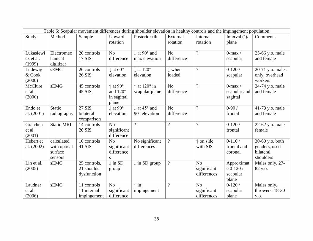
24 SHOULDER AND SCAPULA DYNAMICS
Shoulder dynamics result from the interplay of complex muscular osseous and
supporting structures which provide a range of motion that exceeds that of any other joint in the
body and maintain proper control and stability of all involved joints The glenohumeral joint
resting position and its supporting structures static alignment are influenced by static thoracic
spine alignment humeral bone components scapular bone components clavicular bony
components and the muscular attachments from the thoracic and cervical spine (Wilk Reinold
amp Andrews 2009)
Alterations in shoulder range of motion (ROM) have been associated with shoulder
impingement along with scapular dyskinesis (Lukaseiwicz McClure Michener Pratt Sennett
1999 Ludewig amp Cook 2000 Endo Ikata Katoh amp Takeda 2001) clavicular movement and
increased humeral head translations (Ludewig amp Cook 2002 Laudner Myers Pasquale
Bradley amp Lephart 2006 McClure Michener amp Karduna 2006 Warner Micheli Arslanian
Kennedy amp Kennedy 1992 Deutsch Altchek Schwartz Otis amp Warren 1996 Lin et al
2005) All of these deviations are believed to reduce the subacromial space or approximate the
tendon undersurface to the glenoid labrum creating decreased clearance of the RTC tendons and
other structures under the acromion (Graichen et al 1999) These altered shoulder kinematics
cause alterations in shoulder and scapular muscle activation patterns or altered resting length of
shoulder muscles
241 Shoulderscapular movements
Normal shoulder biomechanics have been studied with EMG during ROM (Ludewig amp
Cook 2000 Kibler amp McMullen 2003 Bagg amp Forrest 1986) cadaver studies (Johnson
Bogduk Nowitzke amp House 1994) patients with nerve injuries (Brunnstrom 1941 Wiater amp
25
Bigliani 1999) and in predictive biomechanical modeling of the arm and muscular function
(Johnson Bogduk Nowitzke amp House 1994 Poppen amp Walker 1978) These approaches have
refined our knowledge about the function and movements of the shoulder and scapula
musculature Understanding muscle adaptation to pathology in the shoulder is important for
developing guidelines for interventions to improve shoulder function These studies have
defined a general consensus on what muscles will be active and when during normal shoulder
range of motion
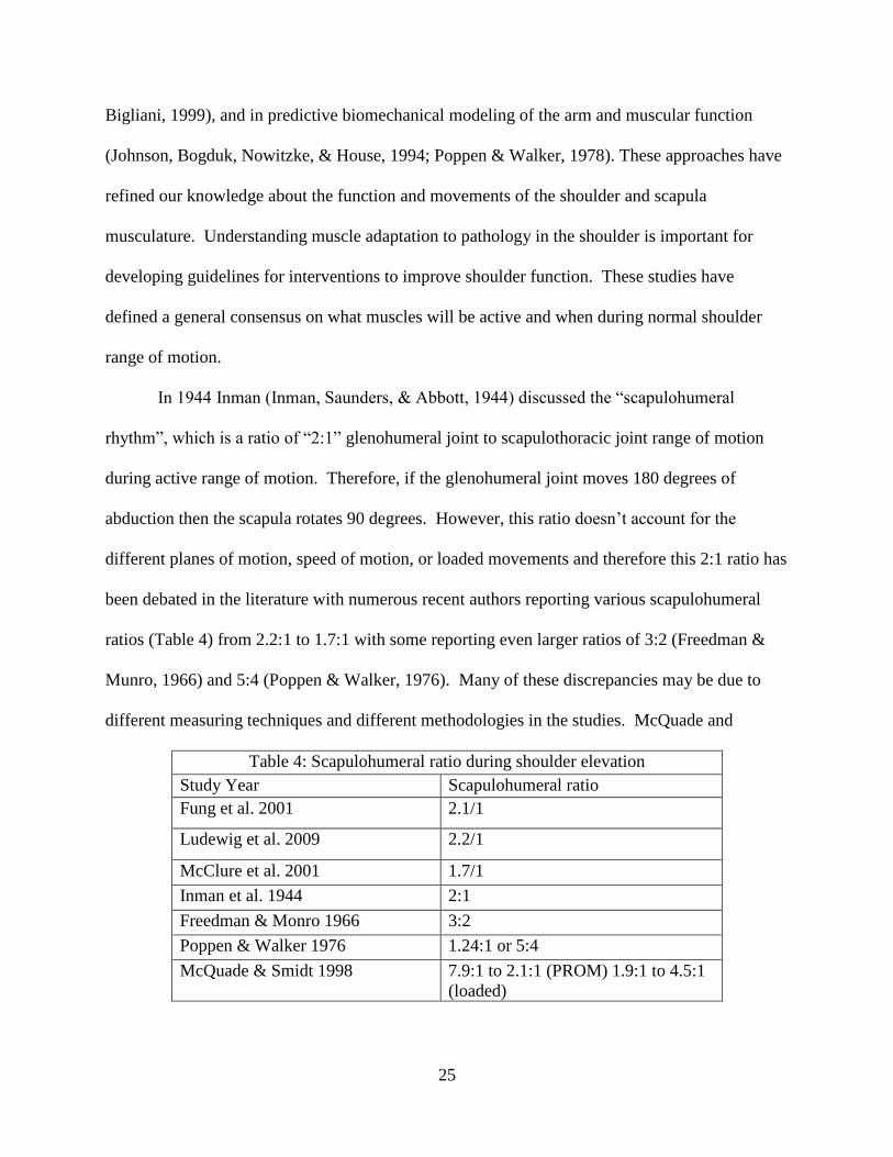
In 1944 Inman (Inman Saunders amp Abbott 1944) discussed the ldquoscapulohumeral
rhythmrdquo which is a ratio of ldquo21rdquo glenohumeral joint to scapulothoracic joint range of motion
during active range of motion Therefore if the glenohumeral joint moves 180 degrees of
abduction then the scapula rotates 90 degrees However this ratio doesnrsquot account for the
different planes of motion speed of motion or loaded movements and therefore this 21 ratio has
been debated in the literature with numerous recent authors reporting various scapulohumeral
ratios (Table 4) from 221 to 171 with some reporting even larger ratios of 32 (Freedman amp
Munro 1966) and 54 (Poppen amp Walker 1976) Many of these discrepancies may be due to
different measuring techniques and different methodologies in the studies McQuade and
Table 4 Scapulohumeral ratio during shoulder elevation
Study Year Scapulohumeral ratio
Fung et al 2001 211
Ludewig et al 2009 221
McClure et al 2001 171
Inman et al 1944 21
Freedman amp Monro 1966 32
Poppen amp Walker 1976 1241 or 54
McQuade amp Smidt 1998 791 to 211 (PROM) 191 to 451
(loaded)
26
colleagues (McQuade amp Smidt 1998) also reported that that the 21 ratio doesnrsquot adequately
explain normal shoulder kinematics However McQuade and colleagues didnrsquot look at
submaximal loaded conditions a pathological population EMG activity during the test but
rather looked at only the concentric phase which will all limit the clinical application of the
research results
There is also disagreement as to when this 21 scapulohumeral ratio occurs even though it
is generally considered to occur in 60 to 120 degrees with 1 degree of scapular movement
occurring for every 2 degrees of elevation movement until 120 degrees and thereafter 1 degree of
scapular movement for every 1 degrees of elevation movement (Reinold Escamilla amp Wilk
2009) Contrary to general considerations some authors have noted the greatest scapular
movement at 30 to 60 degrees while others have found the greatest movement at 80 to 140
degrees but generally these discrepancies are due to different measuring techniques (Bagg amp
Forrest 1986)
Normal scapular movement during glenohumeral elevation helps maintain correct length
tension relationships of the shoulder musculature and prevent the subacromial structures from
being impinged and generally includes upward rotation external rotation and posterior tilting on
the thorax with upward rotation being the dominant motion (McClure et al 2001 Ludewig amp
Reynolds 2009) Overhead athletes generally exhibit increased scapular upward rotation
internal rotation and retraction during elevation and this is hypothesized to be an adaptation to
allow for clearance of subacromial structures during throwing (Wilk Reinold amp Andrews
2009) Generally accepted normal ranges have been observed for scapular upward rotation (45-
55 degrees) posterior tilting (20-40 degrees) and external rotation (15-35 degrees) during
elevation and the scapular muscles are vitally important in maintaining the scapulohumeral
27
kinematic balance since they cause scapular movements (Wilk Reinold amp Andrews 2009
Ludewig amp Reynolds 2009)
However the amount of scapular internal rotation during elevation has shown a great
deal of variability across investigations elevation planes subjects and points in the
glenohumeral range of motion Authors suggest that a slight increase in scapular internal
rotation may be normal early in glenohumeral elevation (McClure Michener Sennett amp
Karduna 2001) and it is also generally accepted (but has limited evidence to support) that end
range elevation involves scapular external rotation (Ludewig amp Reynolds 2009)
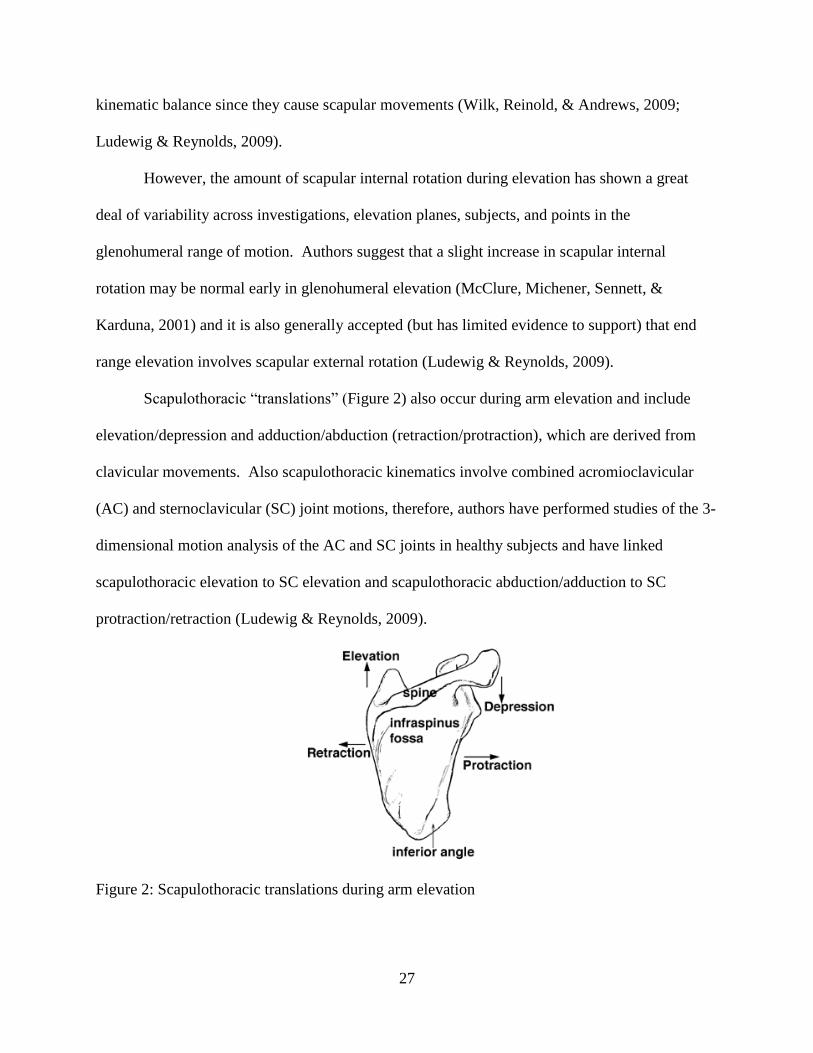
Scapulothoracic ldquotranslationsrdquo (Figure 2) also occur during arm elevation and include
elevationdepression and adductionabduction (retractionprotraction) which are derived from
clavicular movements Also scapulothoracic kinematics involve combined acromioclavicular
(AC) and sternoclavicular (SC) joint motions therefore authors have performed studies of the 3-
dimensional motion analysis of the AC and SC joints in healthy subjects and have linked
scapulothoracic elevation to SC elevation and scapulothoracic abductionadduction to SC
protractionretraction (Ludewig amp Reynolds 2009)
Figure 2 Scapulothoracic translations during arm elevation
28
Despite these numerous scapular movements there remain gaps in the literature and
unanswered questions including 1) which muscles are responsible for internalexternal rotation
or anteriorposterior tilting of the scapula 2) what are normal values for protractionretraction 3)
what are normal values for scapulothoracic elevationdepression 4) how do we measure
scapulothoracic ldquotranslationsrdquo
242 Loaded vs unloaded
The effect of an external load in the hand during elevation remains unclear on scapular
mechanics scapulohumeral ratio and EMG activity of the scapular musculature Adding a 5kg
load in the hand while performing shoulder movements has been shown to increase the EMG
activity of the shoulder musculature In a study of 16 subjects by Antony and Keir (Antony amp
Keir 2010) subjects performed scaption with a 5kg load added to the hand and shoulder
maximum voluntary excitation (MVE) increased by 4 across all postures and velocities Also
when the subjects use a firmer grip on the load a decrease of 2 was demonstrated in the
anterior and middle deltoid and increase of 2 was seen in the posterior deltoid infraspinatus
and trapezius and lastly the biceps increased by 6 MVE While this study gives some evidence
for the use of a loaded exercise with a firmer grip on dumbbells while performing rehabilitation
the study had limited participants and was only performed on a young and healthy population
which limits clinical application of the results
Some researchers have shown no change in scapulothoracic ratio with the addition of
resistance (Freedman amp Munro 1966) while others reported different ratios with addition of
resistance (McQuade amp Smidt 1998) However several limitations are noted in the McQuade amp
Smidt study including 1) submaximal loads were not investigated 2) pathological population
not assessed 3) EMG analysis was not performed and 4) only concentric movements were
29
investigated All of these shortcomings limit the studyrsquos results to a pathological population and
more research is needed on the effect of loads on the scapulohumeral ratio
Witt and colleagues (Witt Talbott amp Kotowski 2011) examined upper middle and
lower trapezius and serratus anterior EMG activity with a 3 pound dumbbell weight and elastic
resistance during diagonal patterns of movement in 21 healthy participants They concluded that
the type of resistance didnrsquot significantly change muscle activity in the diagonal patterns tested
However this study did demonstrate limitations which will alter interpretation including 1) the
study populationrsquos exercisefitness level was not determined 2) the resistance selection
procedure didnrsquot use any form of repetition maximum percentage and 3) there may have been
crosstalk with the sEMG selection
243 Scapular plane vs other planes
The scapular plane is located 30 to 40 degrees anterior to the coronal plane which offers
biomechanical and anatomical features In the scapular plane elevation the joint surfaces have
greater conformity the inferior shoulder capsule ligaments and RTC tendons remain untwisted
and the supraspinatus and deltoid are advantageously aligned for elevation than flexion andor
abduction (Dvir amp Berme 1978) Besides these advantages the scapular plane is where most
functional activities are performed and is also the optimal plane for shoulder strengthening
exercises While performing strengthening exercises in the scapular plane shoulder
rehabilitation is enhanced since unwanted passive tension on the RTC tendons and the
glenohumeral joint capsule are at its lowest point and much lower than in flexion andor
abduction (Wilk Reinold amp Andrews 2009) Scapular upward rotation is also greater in the
scapular plane which will decrease during elevation but will allow for more ldquoclearance in the
subacromial spacerdquo and decrease the risk of impingement
30
244 Scapulothoracic EMG activity
Previous studies have also examined scapulothoracic EMG activity and kinematics
simultaneously to relate the functional status of muscle with scapular mechanics In general
during normal shoulder elevation the scapula will upwardly rotate and posteriorly tilt on the
thorax Scapula internal rotation has also been studied but shows variability across investigations
(Ludwig amp Reynolds 2009)
A general consensus has been established regarding the role of the scapular muscles
during arm movements even with various approaches (different positioning of electrodes on
muscles during EMG analysis [Ludwig amp Cook 2000 Lin et al 2005 Ekstrom Bifulco Lopau
Andersen amp Gough 2004)] different normalization techniques (McLean Chislett Keith
Murphy amp Walton 2003 Ekstrom Soderberg amp Donatelli 2005) varying velocity of
contraction various types of contraction and various muscle length during contraction Though
EMG activity doesnrsquot specify if a muscle is stabilizing translating or rotating a joint it does
demonstrate how active a muscle is during a movement Even with these various approaches and
confounding factors it is generally understood that the trapezius and serratus anterior (middle
and lower) can stabilize and rotate the scapula (Bagg amp Forrest 1986 Johnson Bogduk
Nowitzke amp House 1994 Brunnstrom 1941 Ekstrom Bifulco Lopau Andersen Gough
2004 Inman Saunders amp Abbott 1944) Also during arm elevation the scapulothoracic
muscles produce upward rotation and resist downward rotation acting on the scapula (Dvir amp
Berme 1978) Three muscles including the trapezius (upper middle and lower) the pectoralis
minor and the serratus anterior (middle lower and superior) have been observed using EMG
analysis
31
In prior studies the trapezius has been responsible for stabilizing the scapula since the
middle and lower fibers are perfectly aligned to produce scapula external rotation facilitating
scapular stabilization (Johnson Bogduk Nowitzke amp House 1994) Also the trapezius is more
active during abduction versus flexion (Inman Saunders amp Abbott 1944 Wiedenbauer amp
Mortensen 1952) due to decreased internal rotation of the scapula in scapular plane abduction
The upper trapezius is most active with scapular elevation and is produced through clavicular
elevation The lower trapezius is the only part of the trapezius that can upwardly rotate the
scapula while the middle and lower trapezius are ideally suited for scapular stabilization and
external rotation of the scapula
Another important muscle is the serratus anterior which can be broken into upper
middle and lower groups The middle and lower serratus anterior fibers are oriented in such a
way that they are at a substantial mechanical advantage for scapular upward rotation (Dvir amp
Berme 1978) in combination with the ability to posterior tilt and externally rotate the scapula
Therefore the middle and lower serratus anterior are the primary movers for scapular rotation
during arm elevation and they are the only muscles that can posteriorly tilt the scapula on the
thorax Lastly the upper serratus has been minimally investigated (Ekstrom Bifulco Lopau
Andersen Gough 2004)
The pectoralis minor can produce scapular downward rotation internal rotation and
anterior tilting (Borstad amp Ludewig 2005) opposing upward rotation and posterior tilting during
arm elevation (McClure Michener Sennett amp Karduna 2001) Prior studies (Borstad amp
Ludewig 2005) have demonstrated that decreased length of the pectoralis minor decreases the
posterior tilt and increases the internal rotation during arm elevation which increases
impingement risk
32
245 Glenohumeral EMG activity
Besides the scapulothoracic musculature the glenohumeral musculature including the
deltoid and rotator cuff (supraspinatus infraspinatus subscapularis and teres minor) are
contributors to proper shoulder function The deltoid is the primary mover in elevation and it is
assisted by the supraspinatus initially (Sharkey Marder amp Hanson 1994) The rotator cuff
stabilizes the glenohumeral joint against excessive humeral head translations through a medially
directed compression of the humeral head into the glenoid (Sharkey amp Marder 1995) The
subscapularis infraspinatus and teres minor have an inferiorly directed line of action offsetting
the superior translation component of the deltoid muscle (Sharkey Marder amp Hanson 1994)
Therefore proper balance between increasing and decreasing forces results in (1-2mm) superior
translation of humeral head during elevation Finally the infraspinatus and teres minor produce
humeral head external rotation during arm elevation
246 Shoulder EMG activity with impingement
Besides experiencing pain and other deficits decreased EMG activation of numerous muscles
has been observed in patients with shoulder impingement In patients with shoulder
impingement a decrease in overall serratus anterior activity from 70 to 100 degrees and a
decrease activation of lower serratus anterior from 31 to 120 degrees in scapular plane arm
elevation (Ludwig amp Cook 2000) The upper trapezius has also shown decreased activity
between 40 to 100 degrees and increased activity of the upper and lower trapezius from 61-120
degrees while performing scaption loaded (Ludwig amp Cook 2000 Peat amp Grahame 1977)
Increased upper trap activation is consistent (Ludwig amp Cook 2000 Peat amp Grahame 1977) and
associated with increased clavicular elevation or scapular elevation found in studies (McClure
Michener amp Karduna 2006 Kibler amp McMullen 2003) This increased clavicular elevation at
33
the SC joint may be produced by increased upper trapezius activity (Johnson Bogduk Nowitzke
amp House 1994) and results in scapular anterior tilting causing a potential mechanism to cause
or aggravate impingement symptoms In conclusion middle and lower serratus weakness or
decreased activity contributes to impingement syndrome Increasing function of this muscle may
alleviate pain and dysfunction in shoulder impingement patients
Alterations in rotator cuff muscle activation have been seen in patients with
impingement Decreased activity of the deltoid and rotator cuff is not pronounced in early areas
of motion (Reddy Mohr Pink amp Jobe 2000) However the infraspinatus supraspinatus and
middle deltoid demonstrate decreased activity from 30-60 degrees decreased infraspinatus
activity from 60-90 degrees and no significant difference was seen from 90-120 degrees This
decreased activity is theorized to be related to inadequate humeral head depression (Reddy
Mohr Pink amp Jobe 2000) Another study demonstrated that impingement decreased activity of
the subscapularus supraspinatus and infraspinatus increased middle deltoid activation from 0-
30 degrees decreased coactivation of the supraspinatus and infraspinatus from 30-60 degrees
and increased activation of the infraspinatus subscapularis and supraspinatus from 90-120
degrees (Myers Hwang Pasquale Blackburn amp Lephart 2008) Overall impingement caused
decreased RTC coactivation and increased deltoid activity at the initiation of elevation (Reddy
Mohr Pink amp Jobe 2000 Myers Hwang Pasquale Blackburn amp Lephart 2008)
247 Normal shoulder EMG activity
Normal Shoulder EMG activity will allow for proper shoulder function and maintain
adequate clearance of the subacromial structures during shoulder function and elevation (Table
5) The scapulohumeral muscles are vitally important to provide motion provide dynamic
stabilization and provide proper coordination and sequencing in the glenohumeral complex of
34
overhead athletes due to the complexity and motion needed in overhead sports Since the
glenohumeral and scapulothoracic joints are attached by musculature the muscular activity of
the shoulder complex musculature can be correlated to the maintenance of the scapulothoracic
rhythm and maintenance of the shoulder force couples including 1) Deltoid-rotator cuff 2)
Upper trapezius and serratus anterior and 3) anterior posterior rotator cuff
Table 5 Mean glenohumeral EMG normalized by MVIC during scaption with neutral rotation
(Adapted from Alpert Pink Jobe McMahon amp Mathiyakom 2000)
Interval Anterior
Deltoid
EMG
(MVIC
)
Middle
Deltoid
EMG
(MVIC)
Posterior
Deltoid
EMG
(MVIC)
Supraspin
atus EMG
(MVIC)
Infraspina
tus EMG
(MVIC)
Teres
Minor
EMG
(MVIC)
Subscapul
aris EMG
(MVIC)
0-30˚ 22plusmn10 30plusmn18 2plusmn2 36plusmn21 16plusmn7 9plusmn9 6plusmn7
30-60˚ 53plusmn22 60plusmn27 2plusmn3 49plusmn25 34plusmn14 11plusmn10 14plusmn13
60-90˚ 68plusmn24 69plusmn29 2plusmn3 47plusmn19 37plusmn15 15plusmn14 18plusmn15
90-120˚ 78plusmn27 74plusmn33 2plusmn3 42plusmn14 39plusmn20 19plusmn17 21plusmn19
120-150˚ 90plusmn31 77plusmn35 4plusmn4 40plusmn20 39plusmn29 25plusmn25 23plusmn19
During initial arm elevation the more powerful deltoid exerts an upward and outward
force on the humerus If this force would occur unopposed then superior migration of the
humerus would occur and result in impingement and a 60 pressure increase of the structures
between the greater tuberosity and the acromion when the rotator cuff is not working properly
(Ludewig amp Cook 2002) While the direction of the RTC force vector is debated to be parallel
to the axillary border (Inman et al 1944) or perpendicular to the glenoid (Poppen amp Walker
1978) the overall effect is a force vector which counteracts the deltoid
35
In normal healthy shoulders Matsuki and colleagues (Matsuki et al 2012) demonstrated
21mm of average humeral head superior migration from 0-105˚ of elevation and a 9mm average
inferior translation from 105-180˚ in elevation during fluoroscopic images of the shoulder of 12
male subjects The deltoid-rotator cuff force couple exists when the deltoids superior directed
force is counteracted by an inferior and medially directed force from the infraspinatus
subscapularis and teres minor The supraspinatus also exerts a compressive force on the
humerus onto the glenoid therefore serving an approximating role in the force couple (Inman
Saunders amp Abbott 1944) This RTC helps neutralize the upward shear force reduces
workload on the deltoid through improving mechanical advantage (Sharkey Marder amp Hanson
1994) and assists in stabilization Previous authors have also demonstrated that RTC fatigue or
tears will increase superior migration of the humeral head (Yamaguchi et al 2000)
demonstrating the importance of a correctly functioning force couple
A second force couple a synergistic relation between the upper trapezius and serratus
anterior exists to produce upward rotation of the scapula during shoulder elevation and servers 4
functions 1) allows for rotation of the scapula maintaining the glenoid surface for optimal
positioning 2) maintains efficient length tension relationship for the deltoid 3) prevents
impingement of the rotator cuff from the subacromial structures and 4) provides a stable
scapular base enabling appropriate recruitment of the scapulothoracic muscles The
instantaneous center of rotation starts near the medial border of the scapular spine at lower levels
of elevation and therefore the lower trapezius has a small lever arm due to its distal attachment
being near the center of rotation However during continued elevation the instantaneous center
of rotation moves laterally along the spine toward the acromioclavicular joint and therefore at
higher levels of abduction (ge90˚) the lower trapezius will have a larger lever arm and a greater
36
influence on upward rotation and scapular stabilization along with the serratus anterior (Bagg amp
Forrest 1988)
Overall the position of the scapula is important to center the humeral head on the glenoid
creating a stable foundation for shoulder movements in overhead athletes (Ludwig amp Reynolds
2009) In healthy shoulders the force couple between the serratus anterior and the trapezius
rotates the scapula whereby maintaining the glenoid surface in an optimal position positions the
deltoid muscle in an optimal length tension relationship and provides a stable foundation (Wilk
Reinold amp Andrews 2009) A correctly functioning force couple will prevent impingement of
the subacromial structures on the coracoacromial arch and enable the deltoid and scapulothoracic
muscles to generate more power stability and force (Wilk Reinold amp Andrews 2009) A
muscle imbalance from weakness or shortening can result in an alteration of this force couple
whereby contributing to impaired shoulder stabilization and possibly leading to impingement
The anterior-posterior RTC force couple creates inferior dynamic stability (depressing the
humeral head) and a concavity-compression mechanism (compress humeral head in glenoid) due
to the relationship between the anterior-based subscapularis and the posterior-based teres minor
and infraspinatus Imbalances have been demonstrated in overhead athletes due to overdeveloped
internal rotators and underdeveloped external rotators in the shoulder
248 Abnormal scapulothoracic EMG activity
While no significant change has been noted in resting scapular position of the
impingement population (Ludewig amp Cook 2000 Lukaseiwicz McClure Michener Pratt amp
Sennett 1999) alterations of scapular upward rotation posterior tilting clavicular
elevationretraction scapular internal rotation scapular symmetry and scapulohumeral rhythm
have been observed (Ludewig amp Reynolds 2009 Lukasiewicz McClure Michener Pratt amp
37
Sennett 1999 Ludewig amp Cook 2000 McClure Michener amp Karduna 2006 Endo Ikata
Katoh amp Takeda 2001) Overhead athletes have also demonstrated a relationship between
scapulothoracic muscle imbalance and altered scapular muscle activity has been associated with
SIS (Reinold Escamilla amp Wilk 2009)
SAS has been linked with altered kinematics of the scapula while elevating the arm called
scapular dyskinesis which is defined as observable alterations in the position of the scapula and
the patterns of scapular motion in relation to the thoracic cage JP Warner coined the term
scapular dyskinesis and Ben Kibler described a classification system which outlined 3 primary
scapular dysfunctions which names the condition based on the portion of the scapula most
pronounced or most presently visible when viewed during clinical examination
Burkhart and colleagues (Burkhart Morgan amp Kibler 2003) also coined the term SICK
(Scapular malposition Inferior medial border prominence Coracoid pain and malposition and
dyskinesis of scapular movement) scapula to describe an asymmetrical malposition of the
scapula in throwing athletes
In normal healthy arm elevation the scapula will upwardly rotate posteriorly tilt and
externally rotate and numerous authors have studied the alterations in scapular movements with
SAS (Table 6) The current literature is conflicting in regard to the specific deviations of
scapular motion in the SAS population Researchers have reported a decrease in posterior tilt in
the SAS population (Lukasiewicz McClure Michener Pratt amp Sennett 1999 Ludewig amp
Cook 2000 2002 Endo Ikata Katoh amp Takeda 2001 Lin Hanten Olson Roddey Soto-
quijano Lim et al 2005) while others have demonstrated an increase (McClure Michener amp
Karduna 2006 McClure Michener Sennett amp Karduna 2001 Laudner Myers Pasquale
Bradley amp Lephart 2006) or no difference (Hebert Moffet McFadyen amp Dionne 2002)
38
Table 6 Scapular movement differences during shoulder elevation in healthy controls and the impingement population
Study Method Sample Upward
rotation
Posterior tilt External
rotation
internal
rotation
Interval (˚)
plane
Comments
Lukasiewi
cz et al
(1999)
Electromec
hanical
digitizer
20 controls
17 SIS
No
difference
darr at 90deg and
max elevation
No
difference
0-max
scapular
25-66 yo male
and female
Ludewig
amp Cook
(2000)
sEMG 26 controls
26 SIS
darr at 60deg
elevation
darr at 120deg
elevation
darr when
loaded
0-120
scapular
20-71 yo males
only overhead
workers
McClure
et al
(2006)
sEMG 45 controls
45 SIS
uarr at 90deg
and 120deg
in sagittal
plane
uarr at 120deg in
scapular plane
No
difference
0-max
scapular and
sagittal
24-74 yo male
and female
Endo et
al (2001)
Static
radiographs
27 SIS
bilateral
comparison
darr at 90deg
elevation
darr at 45deg and
90deg elevation
No
difference
0-90
frontal
41-73 yo male
and female
Graichen
et al
(2001)
Static MRI 14 controls
20 SIS
No
significant
difference
0-120
frontal
22-62 yo male
female
Hebert et
al (2002)
calculated
with optical
surface
sensors
10 controls
41 SIS
No
significant
difference
s
No significant
differences
uarr on side
with SIS
0-110
frontal and
coronal
30-60 yo both
genders used
bilateral
shoulders
Lin et al
(2005)
sEMG 25 controls
21 shoulder
dysfunction
darr in SD
group
darr in SD group No
significant
differences
Approximat
e 0-120
scapular
plane
Males only 27-
82 yo
Laudner
et al
(2006)
sEMG 11 controls
11 internal
impingement
No
significant
difference
uarr in
impingement
No
significant
differences
0-120
scapular
plane
Males only
throwers 18-30
yo
39
Similarly Researchers have reported a decrease in upward rotation in the SAS population
(Ludewig amp Cook 2000 2002 Endo Ikata Katoh amp Takeda 2001 Lin Hanten Olson
Roddey Soto-quijano Lim et al 2005) while others have demonstrated an increase (McClure
Michener amp Karduna 2006) or no difference (Lukasiewicz McClure Michener Pratt amp
Sennett 1999 Hebert Moffet McFadyen amp Dionne 2002 Laudner Myers Pasquale Bradley
amp Lephart 2006 Graichen Stammberger Bone Wiedemann Englmeier Reiser amp Eckstein
2001) Lastly researchers have also reported a decrease in external rotation during weighted
elevation (Ludewig amp Cook 2000) while other have shown no difference during unweighted
elevation (Lukasiewicz McClure Michener Pratt amp Sennett 1999 Endo Ikata Katoh amp
Takeda 2001 McClure Michener Sennett amp Karduna 2001) One study has reported an
increase internal rotation (Hebert Moffet McFadyen amp Dionne 2002) while others have shown
no differences (Lin Hanten Olson Roddey Soto-quijano Lim et al 2005 Laudner Myers
Pasquale Bradley amp Lephart 2006) or reported a decrease (Ludewig amp Cook 2000) However
with all these deviations and differences researches seem to agree that athletes with SIS have
decreased upward rotation during elevation (Ludewig amp Cook 2000 2002 Endo Ikata Katoh
amp Takeda 2001 Lin Hanten Olson Roddey Soto-quijano Lim et al 2005) with exception of
one study (McClure Michener amp Karduna 2006)
These conflicting results in the scapular motion literature are likely due to the smaller
measurements of scapular tilt and internalexternal rotation (25˚-30˚) when compared to scapular
upward rotation (50˚) the altered scapular kinematics related to a specific type of impingement
the specific muscular contributions to anteriorposterior tilting and internalexternal rotation are
unclear andor the lack of valid scapular motion measurement techniques in anteriorposterior
tilting and internalexternal rotation compared to upward rotation
40
The scapular muscles have also exhibited altered muscle activation patterns during
elevation in the impingement population including increased activation of the upper trapezius
and decreased activation of the middlelower trapezius and serratus anterior (Cools et al 2007
Cools Witvrouw Declercq Danneels amp Cambier 2003 Wadsworth amp Bullock-Saxton 1997)
In contrast Ludewig amp Cook (Ludewig amp Cook 2000) demonstrated increased activation in
both the upper and lower trapezius in SIS when compared to a control and Lin and colleagues
(Lin et al 2005) demonstrated no change in lower trapezius activity These different results
make the final EMG assessment unclear in the impingement population however there are some
possible explanation for the differences in results including 1) Ludewig amp Cook performed there
experiment weighted in male and female construction workers 2) Lin and colleagues performed
their experiment with numerous shoulder pathologies and in males only 3) Cools and colleagues
used maximal isokinetic testing in abduction in overhead athletes and 4) all of these studies
demonstrated large age ranges in their populations
However there is a lack of reliable studies in the literature pertaining to the EMG activity
changes in overhead throwers with SIS after injurypre-rehabilitation and after injury post-
rehabilitation The inability to detect significant differences between groups by investigators is
primarily due to limited sample sizes limited statistical power for some comparisons the large
variation in the healthy population sEMG signals in studies is altered by skin motion and
limited static imaging in supine
249 Abnormal glenohumeralrotator cuff EMG activity
Abnormal muscle patterns in the deltoid-rotator cuff andor anterior posterior rotator cuff
force couple can contribute to SIS and have been demonstrated in the impingement population
(Myers Hwang Pasquale Blackburn amp Lephart 2008 Reddy Mohr Pink amp Jobe 2000) In
41
general researchers have found decreased deltoid activity (Reddy Mohr Pink amp Jobe 2000)
deltoid atrophy (Leivseth amp Reikeras 1994) and decreased rotator cuff activity (Reddy Mohr
Pink amp Jobe 2000) which can lead to decreased stabilization unopposed deltoid activity and
induce compression of subacromial structures causing a 17mm-21mm humeral head
anteriosuperior migration during 60˚-90˚ of abduction (Sharkey Marder amp Hanson 1994) The
impingement population has demonstrated decreased infraspinatus and subscapularis EMG
activity from 30˚-90˚ elevation when compared to a control (Reddy Mohr Pink amp Jobe 2000)
Myers and colleagues (Myers Hwang Pasquale Blackburn amp Lephart 2009) have
demonstrated with fwEMG analysis decreased rotator cuff coactivation (subscapularis-
infraspinatus and supraspinatus-infraspinatus) and abnormal deltoid activation (increased middle
deltoid activation from 0-30˚) during humeral elevation in 10 subjects with subacromial
impingent when compared to 10 healthy controls and the authors hypothesized this was
contributing to their symptoms
Isokinetic testing has also demonstrated lower protractionretraction ratios in 30 overhead
athletes with chronic shoulder impingement when compared to controls (Cools Witvrouw
Mahieu amp Danneels 2005) Decreased isokinetic force output has also been demonstrated in the
protractor muscles of overhead athletes with impingement (-137 at 60degreess -155 at
180degreess) (Cools Witvrouw Mahieu amp Danneels 2005)
25 REHABILITATION CONSIDERATIONS
Current treatment of impingement generally starts with conservative methods including
arm rest physical therapy nonsteroidal anti-inflammatory drugs (NSAIDs) and subacromial
corticosteroids injections (de Witte et al 2011) While it is beyond the scope of this paper
interventions should be based on a thorough and accurate clinical examination including
42
observations posture evaluation manual muscle testing individual joint evaluation functional
testing and special testing of the shoulder complex Based on this clinical examination and
stage of healing treatments and interventions are prescribed and while each form of treatment is
important this section of the paper will primarily focus on the role of prescribing specific
therapeutic exercise in rehabilitation Also of importance but beyond the scope of this paper is
applying the appropriate exercise progression based on pathology clinical examination and
healing stage
Current treatments in rehabilitation aim to addresses the type of shoulder pathology
involved and present dysfunctions including compensatory patterns of movement poor motor
control shoulder mobilitystability thoracic mobility and finally decrease pain in order to return
the individual to their prior level of function As our knowledge of specific muscular activity
and biomechanics have increased a gradual progression towards more scientifically based
rehabilitation exercises which facilitate recovery while placing minimal strain on healing
tissues have been reported in the literature (Reinold Escamilla amp Wilk 2009) When treating
overhead athletes with impingement the stage of the soft tissue lesion will have an important
impact on the prognosis for conservative treatment and overall recovery Understanding the
previously discussed biomechanical factors of normal shoulder function pathological shoulder
function and the performed exercise is necessary to safely and effectively design and prescribe
appropriate therapeutic exercise programs
251 Rehabilitation protocols in impingement
Typical treatments of impingement in the clinical setting of physical therapy include
specific supervised exercise manual therapy posture education flexibility exercises taping and
modality treatments and are administered based on the phase of treatment (acute intermediate
43
advanced strengthening or return to sport) For the purpose of this paper the focus will be on
specific supervised exercise which refers to addressing individual muscles with therapeutic
exercise geared to address the strength or endurance deficits in that particular muscle The
muscles which are the foci in rehabilitation include the rotator cuff (RTC) (supraspinatus
infraspinatus teres minor and subscapularus) scapular stabilizers (rhomboid major and minor
upper trapezius lower trapezius middle trapezius serratus anterior) deltoid and accessory
muscles (latisimmus dorsi biceps brachii coracobrachialis pectoralis major pectoralis minor)
Recent research has demonstrated strengthening exercises focusing on certain muscles
(serratus anterior trapezius infraspinatus supraspinatus and teres minor) may be more
beneficial for athletes with impingement and exercise prescription should be based on the EMG
activity profile of the exercise (Reinold Escamilla amp Wilk 2009) In order to prescribe the
appropriate exercise based on scientific rationale the muscle EMG activity profile of the
exercise must be known and various authors have found different results with the same exercise
(See APPENDIX) Another important component is focusing on muscles which are known to be
dysfunctional in the shoulder impingement population specifically the lower and middle
trapezius serratus anterior supraspinatus and infraspinatus
Numerous researchers have demonstrated the 3 parts of trapezius generally acting as a
scapular upward rotator and elevator (upper trapezius) a scapular retractor (middle trapezius)
and a downward rotator and depressor (lower trapezius)(Reinold Escamilla amp Wilk 2009) The
lower trapezius has also contributed to scapular posterior tilting and external rotation during
elevation which is hypothesized to decrease impingement risk (Ludewig amp Cook 2000) and
make the lower trapezius vitally important in rehabilitation Upper trapezius EMG activity has
demonstrated a progressive increase from 0-60˚ remain constant from 60-120˚ and increased
44
from 120-180˚ during elevation (Bagg amp Forrest 1986) In contrast the lower trapezius EMG
activity tends to be low during elevation flexion and abduction below 90˚ and then
progressively increases from 90˚-180˚ (Bagg amp Forrest 1986 Ekstrom Donatelli amp Soderberg
2003 Hardwick Beebe McDonnell amp Lang 2006 Moseley Jobe Pink Perry amp Tibone
1992 Smith et al 2006)
Several exercises have been recommended in order to maximally activate the lower
trapezius and the following exercises have demonstrated a high moderate to maximal (65-100)
contraction including 1) prone horizontal abduction at 135˚ with ER (97plusmn16MVIC Ekstrom
Donatelli amp Soderberg 2003) 2) standing ER at 90˚ abduction (88plusmn51MVIC Myers
Pasquale Laudner Sell Bradley amp Lephart 2005) 3) prone ER at 90˚ abduction
(79plusmn21MVIC Ekstrom Donatelli amp Soderberg 2003) 4) prone horizontal abduction at 90˚
abduction with ER (74plusmn21MVIC Ekstrom Donatelli amp Soderberg 2003)(63plusmn41MVIC
Moseley Jobe Pink Perry amp Tibone 1992) 5) abduction above 120˚ with ER (68plusmn53MVIC
Moseley Jobe Pink Perry amp Tibone 1992) and 6) prone rowing (67plusmn50MVIC Moseley
Jobe Pink Perry amp Tibone 1992)
Significantly greater EMG activity has been reported in prone ER at 90˚ when compared
to the empty can exercise (Ballantyne et al 1993) and authors have reported significant EMG
amplitude during prone ER at 90˚ prone full can and prone horizontal abduction at 90˚ with ER
(Ekstrom Donatelli amp Soderberg 2003) Based on these results it appears that obtaining
maximal EMG activity of the lower trapezius in prone exercises requires performing exercises
prone approximately 120-130˚ of abduction may be most beneficial and will fluctuate depending
on body type It is also important to note that these exercises have been performed in prone
instead of standing Typically symptoms of SIS are increased during standing abduction greater
45
than 90˚ therefore this exercise is performed in the scapular plane with shoulder external
rotation in order to clear the subacromial structures from impinging on the acromion and should
not be performed during the acute phase of healing in SIS
It is often clinically beneficial to enhance the ratio of lower trapezius to upper trapezius
in rehabilitation Poor posture and muscle imbalance is often seen in shoulder impingement
along with alterations in the force couple between the upper trapezius and serratus anterior
McCabe and colleagues (McCabe Orishimo McHugh amp Nicholas 2007) demonstrated that
ldquothe press uprdquo (56MVIC) and ldquoscapular retractionrdquo (40MVIC) exercises exhibited
significantly greater lower trapezius sEMG activity than the ldquobilateral shoulder external rotationrdquo
and ldquoscapular depressionrdquo exercise The authors also demonstrated that the ldquobilateral shoulder
external rotationrdquo and ldquothe press uprdquo demonstrated the highest UTLT ratios at 235 and 207
(McCabe Orishimo McHugh amp Nicholas 2007) Even with the authors proposed
interpretation to apply to patient population it is difficult to apply the results to a patient since
the experiment was performed on a healthy population
The middle trapezius has demonstrated high EMG activity during elevation at 90˚ and
gt120˚ (Bagg amp Forrest 1986 Decker Hintermeister Faber amp Hawkins 1999 Ekstrom
Donatelli amp Soderberg 2003) while other authors have shown low EMG activity in the same
exercise (Moseley Jobe Pink Perry amp Tibone 1992)
However several exercises have been recommended in order to maximally activate the
middle trapezius and the following exercises have demonstrated a high moderate to maximal
(65-100) contraction including 1) prone horizontal abduction at 90˚ abduction with IR
(108plusmn63MVIC Moseley Jobe Pink Perry amp Tibone 1992) 2) prone horizontal abduction at
135˚ abduction with ER (101plusmn32MVIC Ekstrom Donatelli amp Soderberg 2003) 3) prone
46
horizontal abduction at 90˚ abduction with ER (87plusmn20MVIC Ekstrom Donatelli amp
Soderberg 2003)(96plusmn73MVIC Moseley Jobe Pink Perry amp Tibone 1992) 4) prone rowing
(79plusmn23MVIC Ekstrom Donatelli amp Soderberg 2003) and 5) prone extension at 90˚ flexion
(77plusmn49MVIC Moseley Jobe Pink Perry amp Tibone 1992) In therdquo prone horizontal
abduction at 90˚ abduction with ERrdquo exercise the authors demonstrated some agreement in
amplitude of EMG activity One author demonstrated 87plusmn20MVIC (Ekstrom Donatelli amp
Soderberg 2003) while a second demonstrated 96plusmn73MVIC (Moseley Jobe Pink Perry amp
Tibone 1992) while these amplitudes are not exact they are both considered maximal EMG
activity
The supraspinatus is also a very important muscle to focus on in rehabilitation of SIS due
to the numerous force couples it is involved in and the potential for injury during SIS Initially
Jobe (Jobe amp Moynes 1982) recommended scapular plane elevation with glenohumeral IR
(empty can) exercises to strengthen the supraspinatus muscle but other authors (Poppen amp
Walker 1978 Reinold et al 2004) have suggested scapular plane elevation with glenohumeral
ER (full can) exercises Recently evidence based therapeutic exercise prescriptions have
avoided the use of the empty can exercise due to the increased deltoid activity potentially
increasing the amount of superior humeral head migration and the inability of a weak RTC to
counteract the force in the impingement population (Reinold Escamilla amp Wilk 2009)
Several exercises have been recommended in order to maximally activate the
supraspinatus and the following exercises have demonstrated a high moderate to maximal (65-
100) contraction including 1) push-up plus (99plusmn36MVIC Decker Tokish Ellis Torry amp
Hawkins 2003) 2) prone horizontal abduction at 100˚ abduction with ER (82plusmn37MVIC
Reinold et al 2004) 3) prone ER at 90˚ abduction (68plusmn33MVIC Reinold et al 2004) 4)
47
military press (80plusmn48MVIC Townsend Jobe Pink amp Perry 1991) 5) scaption above 120˚
with IR (74plusmn33MVIC Townsend Jobe Pink amp Perry 1991) and 6) flexion above 120˚ with
ER (67plusmn14MVIC Townsend Jobe Pink amp Perry 1991)(42plusmn21MVIC Myers Pasquale
Laudner Sell Bradley amp Lephart 2005) Interestingly some of the same exercises showed
different results in the EMG amplitude in different studies For example ldquoflexion above 120˚
with ERrdquo demonstrated 67plusmn14MVIC (Townsend Jobe Pink amp Perry 1991) in one study and
42plusmn21MVIC (Myers Pasquale Laudner Sell Bradley amp Lephart 2005) in another study As
you can see this is a large disparity but potential mechanisms for the difference may be due to the
fact that one study used dumbbellrsquos and the other used resistance tubing Also the participants
werenrsquot given a weight based on a ten repetition maximum
3-D biomechanical model data implies that the infraspinatus is a more effective shoulder
ER at lower angles of abduction (Reinold Escamilla amp Wilk 2009) and numerous studies have
tested this model with conflicting results in exercise selection (Decker Tokish Ellis Torry amp
Hawkins 2003 Myers Pasquale Laudner Sell Bradley amp Lephart 2005 Townsend Jobe
Pink amp Perry 1991 Reinold et al 2004) In general infraspinatus and teres minor activity
progressively decrease as the shoulder moves into the abducted position while the supraspinatus
and deltoid increase activity
Several exercises have been recommended in order to maximally activate the
infraspinatus the following exercises have demonstrated a high moderate to maximal (65-100)
contraction including 1) push-up plus (104plusmn54MVIC Decker Tokish Ellis Torry amp
Hawkins 2003) 2) SL ER at 0˚ abduction (62plusmn13MVIC Reinold et al 2004)
(85plusmn26MVIC Townsend Jobe Pink amp Perry 1991) 3) prone horizontal abduction at 90˚
abduction with ER (88plusmn25MVIC Townsend Jobe Pink amp Perry 1991) 4) prone horizontal
48
abduction at 90˚ abduction with IR (74plusmn32MVIC Townsend Jobe Pink amp Perry 1991) 5)
abduction above 120˚ with ER (74plusmn23MVIC Townsend Jobe Pink amp Perry 1991) and 6)
flexion above 120˚ with ER (66plusmn16MVIC Townsend Jobe Pink amp Perry 1991)
(47plusmn34MVIC Myers Pasquale Laudner Sell Bradley amp Lephart 2005)
Reinold and colleagues (Reinold et al 2004) also examined several exercises
commonly used in rehabilitation used to strengthen the posterior RTC and specifically the
infraspinatus and teres minor The authors determined that 3 exercisersquos demonstrated the best
combined EMG activity and in order include 1) side lying ER (infraspinatus 62MVIC teres
minor 67MVIC) 2) standing ER in scapular plane at 45˚ abduction (infraspinatus 53MVIC
teres minor 55MVIC) and 3) prone ER in the 90˚ abducted position (infraspinatus
50MVIC teres minor 48MVIC) The 90˚ abducted position is commonly used in overhead
athletes to simulate the throwing position in overhead athletes The side lying ER exercise is also
clinically significant since it exerts less capsular strain specifically on the anterior band of the
glenohumeral ligament (Reinold et al 2004) than the more functionally advantageous standing
ER at 90˚ It has also been demonstrated that the application of a towel roll while performing ER
at 0˚ increases EMG activity by approximately 20 when compared to no towel roll (Reinold et
al 2004)
The serratus anterior contributes to scapular posterior tilting upward rotation and
external rotation of the scapula (Ludewig amp Cook 2000 McClure Michener amp Karduna 2006)
and has demonstrated decreased EMG activity in the impingement population (Cools et al
2007 Cools Witvrouw Declercq Danneels amp Cambier 2003 Wadsworth amp Bullock-Saxton
1997) Serratus anterior activity tends to increase as arm elevation increases however increased
elevation may also increase impingement symptoms and risk (Reinold Escamilla amp Wilk
49
2009) Interestingly performing 90˚ shoulder abduction with IR or ER has generated high
serratus anterior activity while initially Jobe (Jobe amp Moynes 1982) recommended IR or ER for
rotator cuff strengthening Serratus anterior activity also increases as the gravitational challenge
increased when comparing the wall push up plus push-up plus on knees and push up plus with
feet elevated (Reinold Escamilla amp Wilk 2009)
Prior authors have recommended the push-up plus dynamic hug and punch exercise to
specifically recruit the serratus anterior (Decker Hintermeister Faber amp Hawkins 1999) while
other authorsrsquo (Ekstrom Donatelli amp Soderberg 2003) data indicated that performing
movements which create scapular upward rotationprotraction (punch at 120˚ abduction) and
diagonal exercises incorporating flexion horizontal abduction and ER
Hardwick and colleges (Hardwick Beebe McDonnell amp Lang 2006) contrary to
previous authors (Ekstrom Donatelli amp Soderberg 2003) demonstrated no statistical difference
in serratus anterior EMG activity during the wall slide push-up plus (only at 90˚) and scapular
plane shoulder elevation in 20 healthy individuals measured at 90˚ 120˚ and 140˚ The study
also demonstrated that the wall slide and scapular plane shoulder elevation EMG activity was
highest at 140˚ (approximately 76MVIC and 82MVIC) However these results should be
interpreted with caution since the methodological issues of limited healthy sample and only the
plus phase of the push up plus exercise was examined in the study
The serratus anterior is important for the acceleration phase of overhead throwing and
several exercises have been recommended to maximally activate this muscle The following
exercises have demonstrated a high moderate to maximal (65-100) contraction including 1)
D1 diagonal pattern flexion horizontal adduction and ER (100plusmn24MVIC Ekstrom Donatelli
amp Soderberg 2003) 2) scaption above 120˚ with ER (96plusmn24MVIC Ekstrom Donatelli amp
50
Soderberg 2003)(91plusmn52MVIC Middle Serratus 84plusmn20MVIC Lower Serratus Moseley
Jobe Pink Perry amp Tibone 1992) 3) supine upward punch (62plusmn19MVIC Ekstrom
Donatelli amp Soderberg 2003) 4) flexion above 120˚ with ER(96plusmn45MVIC Middle Serratus
72plusmn46MVIC Lower Serratus Moseley Jobe Pink Perry amp Tibone 1992) (67plusmn37MVIC
Myers Pasquale Laudner Sell Bradley amp Lephart 2005) 5) abduction above 120˚ with ER
(96plusmn53MVIC Middle Serratus 74plusmn65MVIC Lower Serratus Moseley Jobe Pink Perry amp
Tibone 1992) 7) military press (82plusmn36MVIC Middle Serratus 60plusmn42MVIC Lower
Serratus Moseley Jobe Pink Perry amp Tibone 1992) 7) push-up plus (80plusmn38MVIC Middle
Serratus 73plusmn3MVIC Lower Serratus Moseley Jobe Pink Perry amp Tibone 1992) 8) push-up
with hands separated (57plusmn36MVIC Middle Serratus 69plusmn31MVIC Lower Serratus Moseley
Jobe Pink Perry amp Tibone 1992) 9) standing ER at 90˚ abduction (66plusmn39MVIC Myers
Pasquale Laudner Sell Bradley amp Lephart 2005) and 10) standing forward scapular punch
(67plusmn45MVIC Myers Pasquale Laudner Sell Bradley amp Lephart 2005)
Even though the research has demonstrated exercises which may be more beneficial than
others the lack of statistical analysis lack of data and absence of the significant muscle activity
(including the deltoid) were methodological limitations of these studies Also while performing
exercises with a high EMG activity are the most effective to maximally exercise specific
muscles the stage of rehabilitation may contraindicate the specific exercise recommended For
example it is generally accepted that performing standing exercises below 90˚ elevation is
necessary to avoid exacerbations of impingement symptoms In conclusion the previously
described therapeutic exercises have demonstrated clinical benefit and high EMG activity in the
prior discussed muscles (Table 5)
51
252 Rehabilitation of scapula dyskinesis
Scapular rehabilitation should be based on an accurate and thorough clinical evaluation
performed by an individual licensed to evaluate and treat dysfunction to permit appropriate goal
setting and rehabilitation for the patient A comprehensive initial patient interview is necessary to
ascertain the individualrsquos functional requirements and problematic activities followed by the
physical examination The health care professional should address all possible deficiencies
found on different levels of the kinetic chain and appropriate treatment goals should be set
leading to proper rehabilitation strategies Therefore although considered to be key points in
functional shoulder and neck rehabilitation more proximal links in the kinetic chain such as
thoracic spine mobility and strength core stability and lower limb function will not be addressed
in this manuscript
Treatment of scapular dyskinesis is only successful if the anatomical base is optimal and
the individual does not exhibit problems which require surgery such as nerve injury scapular
muscle detachment severe bony derangement (acromioclavicular separation fractured clavicle)
or soft tissue derangement (labral injury rotator cuff disease glenohumeral instability) (Kibler amp
Sciascia 2010 Wright Wassinger Frank Michener amp Hegedus 2012) The large majorities of
cases of dyskinesis however are caused by muscle weakness inhibition or inflexibility and can
be managed with rehabilitation
Optimal rehabilitation of scapular dyskinesis requires addressing all of the causative
factors that can create the dyskinesis and then restoring the balance of muscle forces that allow
scapular position and motion The emphasis of scapular dyskinesis rehabilitation should start
proximally and end distally with an initial goal of achieving the position of optimal scapular
function (posterior tilt external rotation and upward elevation) The serratus anterior is an
52
important external rotator of the scapula and the lower trapezius is a stabilizer of the acquired
scapular position Scapular stabilization protocols should focus on re-educating these muscles to
act as dynamic scapula stabilizers first by the implementation of short lever kinetic chain
assisted exercises then progress to long lever movements Maximal rotator cuff strength is
achieved off a stabilized retracted scapula and rotator cuff emphasis should be after scapular
control is achieved (Kibler amp Sciascia 2010) An increase in impingement pain when doing
open chain rotator cuff exercises indicates an incorrect protocol emphasis and stage of
rehabilitation A logical progression of exercises (isometric to dynamic) focused on
strengthening the lower trapezius and serratus anterior while minimizing upper trapezius
activation has been described in the literature (Kibler amp Sciascia 2010 Kibler Ludewig
McClure Michener Bak amp Sciascia 2013) and on an algorithm guideline (Figure 3) has been
proposed that is based on restoration of soft tissue inflexibilities and maximizing muscle
performance (Cools Struyf De Mey Maenhout Castelein amp Cagnie 2013)
Several principles guide the progression through the algorithm with the first requirement
being acquisition of flexibility in muscles and joints because tight muscles and joint capsules can
inhibit strength activation Also later protocols in rehabilitation should train functional
movements in sport or activity specific patterns since research has demonstrated maximal
scapular muscle activation when muscles are activated in functional patterns (vs isolated)(ie
when the muscles are activated in specific diagonal patterns using kinetic chain sequencing)
(Kibler amp Sciascia 2010) Using these principles many rehabilitation interventions can be
considered but a reasonable program could start with standing low-loadlow-activation (activate
the scapular retractors gt20 MVIC) exercises with the arm below shoulder level and progress
to prone and side-lying exercises that increase the load but still emphasize lower trapezius and
53
Figure 3 A scapular rehabilitation algorithm guideline (Adapted from Cools Struyf De Mey
Maenhout Castelein amp Cagnie 2013)
serratus anterior activation over upper trapezius activation Additional loads and activations can
be stimulated by integrating ipsilateral and contralateral kinetic chain activation and adding distal
resistance Final optimization of activation can occur through weight training emphasizing
proper retraction and stabilization Progression can be made by increasing holding time
repetitions resistance and speed parameters of exercise relevant to the patientrsquos functional
needs
The lower trapezius is frequently inhibited in activation and specific effort may be
required to lsquojump startrsquo it Tightness spasm and hyperactivity in the upper trapezius pectoralis
minor and latissimus dorsi are frequently associated with lower trapezius inhibition and specific
therapy should address these muscles
Multiple studies have identified methods to activate scapular muscles that control
scapular motion and have identified effective body and scapular positions that allow optimal
activation in order to improve scapular muscle performance and decrease clinical symptoms
54
Only two randomized clinical trials have examined the effects of a scapular focused program by
comparing it to a general shoulder rehabilitation and the findings indicate the use of scapular
exercises results in higher patient-rated outcomes (Başkurt Başkurt Gelecek amp Oumlzkan 2011
Struyf Nijs Mollekens Jeurissen Truijen Mottram amp Meeusen 2013)
Multiple clinical trials have incorporated scapular exercises within their rehabilitation
programs and have found positive patient-rated outcomes in patients with impingement
syndrome (Kromer Tautenhahn de Bie Staal amp Bastiaenen 2009) It appears that it is not only
the scapular exercises but also the inclusion of the scapular exercises as part of a rehabilitation
program that may include the use of the kinetic chain is what achieves positive outcomes When
the scapular exercises are prescribed multiple components must be emphasized including
activation sequencing force couple activation concentriceccentric emphasis strength
endurance and avoidance of unwanted patterns (Cools Struyf De Mey Maenhout Castelein amp
Cagnie 2013)
253 Effects of rehabilitation
Conservative therapy is successful in 42 (Bigliani type III) to 91 (Bigliani type I) (de
Witte et al 2011) and most shoulder injuries in the overhead thrower can be successfully
treated non-operatively (Wilk Obma Simpson Cain Dugas amp Andrews 2009) Evidence
supports the use of thoracic mobilizations (Theisen et al 2010) glenohumeral mobilizations
(Tyler Nicholas Lee Mullaney amp Mchugh 2012 Sauers 2005) supervised shoulder and
scapular muscle strengthening (Fleming Seitz amp Edaugh 2010 Osteras Torstensen amp Osteras
2010 McClure Bialker Neff Williams amp Karduna 2004 Sauers 2005 Bang amp Deyle 2000
Senbursa Baltaci amp Atay 2007) supervised shoulder and scapular muscle strengthening with
manual therapy (Bang amp Deyle 2000 Senbursa Baltaci amp Atay 2007) taping (Lin Hung amp
Yang 2011 Williams Whatman Hume amp Sheerin 2012 Selkowitz Chaney Stuckey amp Vlad
55
2007 Smith Sparkes Busse amp Enright 2009) and laser therapy (Sauers 2005) in decreasing
pain increasing mobility improving function and improving altering muscle activity of shoulder
muscles
In systematic reviews of randomized controlled trials there is a lack of high quality
intervention studies but some studies suggest that therapeutic exercise is as effective as surgery
in SIS (Nyberg Jonsson amp Sundelin 2010 Trampas amp Kitsios 2006) the combination of
manual therapy and exercise is better than exercise alone in SIS (Michener Walsworth amp
Burnet 2004) and high dosage exercise is better than low dosage exercise in SIS (Nyberg
Jonsson amp Sundelin 2010) in reducing pain and improving function In evidence-based clinical
practice guidelines therapeutic exercise is effective in treatment of SIS (Trampas amp Kitsios
2006 Kelly Wrightson amp Meads 2010) and is recommended to be combined with joint
mobilization of the shoulder complex (Tyler Nicholas Lee Mullaney amp Mchugh 2012 Sauers
2005) Joint mobilization techniques have demonstrated increased improvements in symptoms
when applied by experienced physical therapists rather than applied by novice clinicians (Tyler
Nicholas Lee Mullaney amp Mchugh 2012) A course of therapeutic exercise in the SIS
population has also been shown to be more beneficial than no treatment or a placebo treatment
and should be attempted to reduce symptoms and restore function before surgical intervention is
considered (Michener Walsworth amp Burnet 2004)
In a study by McClure and colleagues (McClure Bialker Neff Williams amp Karduna
2004) the authors demonstrated after a 6 week therapeutic exercise program combined with
education significant improvements in pain shoulder function increased passive range of
motion increased ER and IR force and no changes in scapular kinematics in a SIS population
56
However these results should be interpreted with caution since the rate of attrition was 33
there was no control group and numerous clinicians performed the interventions
In a randomized clinical trial by Conroy amp Hayes (Conroy amp Hayes 1998) 14 patients
with SIS underwent either a supervised exercise program or a supervised exercise program with
joint mobilization for 9 sessions over 3 weeks At 3 weeks the supervised exercise program
with joint mobilization had less pain compared to the supervised exercise program group In a
larger randomized clinical trial by Bang amp Deyle (Bang amp Deyle 2000) patientsrsquo with SIS
underwent either an exercise program or an exercise program with manual therapy for 6 sessions
over 3-4 weeks At the end of treatment and at 1 month follow up the exercise program with
manual therapy group had superior gains in strength function and pain compared to the exercise
program group
Recently numerous studies have observed the EMG activity in the shoulder complex
musculature during numerous rehabilitation exercises In exploring evidence-based exercises
while treating SIS the population the following has been shown to be effective to improve
outcome measures for this population 1) serratus anterior strengthening 2) scapular control with
external rotation exercises 3) external rotation exercises with tubing 4) resisted flexion
exercises 5) resisted extension exercises 6) resisted abduction exercise 7) resisted internal
rotation exercise (Dewhurst 2010)
57
Table 7 Therapeutic exercises for the shoulder musculature which is involved in rehabilitation that has demonstrated a moderate to maximal EMG profile for that particular
muscle along with its clinical significance (DB=dumbbell T=Tubing)
Muscle Exercise Clinical Significance
lower
trapeziu
s
1 Prone horizontal abduction at 135˚ with ER (DB)
2 Standing ER at 90˚ (T)
3 Prone ER at 90˚ abd (DB)
4 Prone horizontal abduction at 90˚ with ER (DB)
5 Abd gt 120˚ with ER (DB)
6 Prone rowing (DB)
1 In line with lower trapezius fibers High EMG activity of trapezius effectivegood supraspinatusserratus anterior
2 High EMG activity lower trap rhomboids serratus anterior moderate-maximal EMG activity of RTC
3 Below 90˚ abduction High EMG of lower trapezius
4 Below 90˚ abduction good UTLT ratio moderate to maximal EMG of upper middle and lower trapezius
5 Used later in rehabilitation since gt90˚ abduction can symptoms high serratus anterior EMG moderate upper and lower
trapezius EMG
6 Below 90˚ abduction High EMG of upper middle and lower trapezius
middle
trapeziu
s
1 Prone horizontal abduction at 90˚ with IR (DB)
2 Prone horizontal abduction at 135˚ with ER (DB)
3 Prone horizontal abduction at 90˚ with ER (DB)
4 Prone rowing (DB)
5 Prone extension at 90˚ flexion (DB)
1 IR tension on subacromial structures deltoid activity not for patient with SIS high EMG for all parts of trapezius
2 High EMG activity of all parts of trapezius effective and good for supraspinatus and serratus anterior also
3 Below 90˚ abduction good UTLT ratio moderate to maximal EMG of upper middle and lower trapezius
4 Below 90˚ abduction High EMG of upper middle and lower trapezius
5 Below 90˚ abduction High middle trapezius activity
serratus
anterior
1 D1 diagonal pattern flexion horizontal adduction
and ER (T)
2 Scaption above 120˚ with ER (DB)
3 Supine upward punch (DB)
4 Flexion above 120˚ with ER (DB)
5 Abduction above 120˚ with ER (DB)
6 Military press (DB)
7 Push-up Plus
8 Push-up with hands separated
9 Standing ER at 90˚ abduction (T)
10 Standing forward scapular punch (T)
1 Effective to begin functional movements patterns later in rehabilitation high EMG activity
2 Above 90˚ to be performed after resolution of symptoms
3 Effective and below 90˚
4 Above 90˚ to be performed after resolution of symptoms
5 Used later in rehabilitation since gt90˚ abduction can symptoms high serratus anterior EMG moderate upper and lower
trapezius EMG
6 Perform in advanced strengthening phase since can cause impingement
7 Closed chain exercise below 90˚ high serratus anterior supraspinatus and infraspinatus activity
8 Closed chain exercise
9 High teres minor lower trapezius and rhomboid EMG activity
10 Below 90˚ abduction high subscapularis and teres minor EMG activity
suprasp
inatus
1 Push-up plus
2 Prone horizontal abduction at 100˚ with ER (DB)
3 Prone ER at 90˚ abd (DB)
4 Military press (DB)
5 Scaption above 120˚ with IR (DB)
6 Flexion above 120˚ with ER (DB)
1 Closed chain exercise below 90˚ high serratus anterior supraspinatus and infraspinatus activity
2 High supraspinatus middleposterior deltoid EMG activity
3 Below 90˚ abduction High EMG of lower trapezius also
4 Perform in advanced strengthening phase since can cause impingement
5 IR tension on subacromial structures anteriormiddle deltoid activity not for patient with SIS moderate infraspinatus
EMG activity
6 High anteriormiddle deltoid activity not for patient with SIS moderate infraspinatus and subscapularis EMG activity
58
Table 7 Therapeutic exercises for the shoulder musculature which is involved in rehabilitation that has demonstrated a moderate to maximal EMG profile for that particular
muscle along with its clinical significance (DB=dumbbell T=Tubing)(Continued)
Muscle Exercise Clinical Significance
Infraspi
natus
1 Push-up plus
2 SL ER at 0˚ abduction (DB)
3 Prone horizontal abduction at 90˚ with ER (DB)
4 Prone horizontal abduction at 90˚ with IR (DB)
5 Abduction gt 120˚ with ER (DB)
6 Flexion above 120˚ with ER (DB)
1 Closed chain exercise below 90˚ high serratus anterior supraspinatus and infraspinatus activity
2 Stable shoulder position Most effective exercise to recruit infraspinatus
3 Below 90˚ abduction good UTLT ratio moderate to maximal EMG of upper middle and lower trapezius
4 IR increases tension on subacromial structures increased deltoid activity not for patient with SIS high EMG for all parts
of trapezius
5 Used later in rehabilitation since gt90˚ abduction can increase symptoms high serratus anterior EMG moderate upper and
lower trapezius EMG
6 High anteriormiddle deltoid activity not for patient with SIS moderate infraspinatus and subscapularis EMG activity
Infraspi
natus amp
Teres
minor
1 SL ER at 0˚ abduction (DB)
2 Standing ER in scapular plane at 45˚ abduction
(DB)
3 Prone ER in 90˚ abduction (DB)
1 Stable shoulder position Most effective exercise to recruit infraspinatus
2 High EMG of teres and infraspinatus
3 Below 90˚ abduction High EMG of lower trapezius
59
However no studies have explored whether or not specific rehabilitation exercises
targeting muscles based on EMG profile could correct prior EMG deficits and speed recovery
in patients with shoulder impingement In conclusion there is a need for further well-defined
clinical trials on specific exercise interventions for the treatment of SIS This literature reveals
the need for improved sample sizes improved diagnostic criteria and similar diagnostic criteria
applied between studies longer follow ups studies measuring function and pain and
(specifically in overhead athletes) sooner return to play
26 SUMMARY
Overhead athletes with SIS or shoulder impingement will exhibit muscle imbalances and
tightness in the GH and scapular musculature These dysfunctions can lead to altered shoulder
complex kinematics altered EMG activity and functional limitations which will cause
impingement The exact mechanism of impingement is debated in the literature as well its
relation to scapular kinematic variation Therapeutic exercise has shown to be beneficial in
alleviating dysfunctions and pain in SIS and supervised exercise with manual techniques by an
experienced clinician is an effective treatment It is unknown whether prescribing specific
therapeutic exercise based on EMG profile will speed the recovery time increase force
production resolve scapular dyskinesis or change SAS height in SIS Few research articles
have examined these variables and its association with prescribing specific therapeutic exercise
and there is a general need for further well-defined clinical trials on specific exercise
interventions for the treatment of SIS
60
CHAPTER 3 THE EFFECT OF VARIOUS POSTURES ON THE SURFACE
ELECTROMYOGRAPHIC ANALYSIS OF THE LOWER TRAPEZIUS DURING
SPECIFIC THERAPEUTIC EXERCISE
31 INTRODUCTION
Individuals diagnosed with shoulder impingement exhibit muscle imbalances in the
shoulder complex and specifically in the force couple (lower trapezius upper trapezius and
serratus anterior) which controls scapular movements The deltoid plays an important role in the
muscle force couple since it is the prime mover of the glenohumeral joint Dysfunctions in these
muscles lead to altered shoulder complex kinematics and functional limitations which will cause
an increase in impingement symptoms Therapeutic exercises are beneficial in alleviating
dysfunctions and pain in individuals diagnosed with shoulder impingement However no studies
demonstrate the effect various postures will have on electromyographic (EMG) activity in
healthy adults or in adults with impingement during specific therapeutic exercise The purpose
of the study was to identify the therapeutic exercise and posture which elicits the highest EMG
activity in the lower trapezius shoulder muscle tested This study also tested the exercises and
postures in the healthy population and the shoulder impingement population since very few
studies have correlated specific therapeutic exercises in the shoulder impingement population
Individuals with shoulder impingement exhibit muscle imbalances in the shoulder
complex and specifically in the lower trapezius upper trapezius and serratus anterior all of
which control scapular movements with the deltoid acting as the prime mover of the shoulder
Dysfunctions in these muscles lead to altered kinematics and functional limitations
which cause an increase in impingement symptoms Therapeutic exercise has shown to be
beneficial in alleviating dysfunctions and pain in impingement and the following exercises have
been shown to be effective treatment to improve outcome measures for this diagnosis 1) serratus
61
anterior strengthening 2) scapular control with external rotation exercises 3) external rotation
exercises 4) prone extension 5) press up exercises 6) bilateral shoulder external rotation
exercise and 7) prone horizontal abduction exercises at 135˚ and 90˚ of abduction (Dewhurst
2010 Trampas amp Kitsios 2006 Kelly Wrightson amp Meads 2010 Fleming Seitz amp Edaugh
2010 Osteras Torstensen amp Osteras 2010 McClure Bialker Neff Williams amp Karduna
2004 Sauers 2005 Senbursa Baltaci amp Atay 2007 Bang amp Deyle 2000 Senbursa Baltaci
amp Atay 2007) The therapeutic exercises in this study were derived from specific therapeutic
exercises shown to improve outcomes in the impingement population and of particular
importance are the amount of EMG activity in the lower trapezius since this muscle is directly
responsible for stabilizing the scapula
Evidence based treatment of impingement requires a high dosage of therapeutic exercises
over a low dosage (Nyberg Jonsson amp Sundelin 2010) and applying the exercise EMG profile
to exercise prescription facilitates a speedy recovery However no studies have correlated the
effect various postures will have on the EMG activity of the lower trapezius in healthy adults or
in adults with impingement The purpose of this study was to identify the therapeutic exercise
and posture which elicits the highest EMG activity in the lower trapezius muscle The postures
included in the study include a normal posture with towel roll under the arm (if applicable) a
posture with the feet staggeredscapula retracted and a towel roll under the arm (if applicable)
and a normal posturescapula retracted with a towel roll under the arm (if applicable) with a
physical therapist observing and cueing to maintain the scapula retraction Recent research has
demonstrated that the application of a towel roll increases the EMG activity of the shoulder
muscles by 20 in certain exercises (Reinold Wilk Fleisig Zheng Barrentine Chmielewski
Cody Jameson amp Andrews 2004) thereby increasing the effectiveness of therapeutic exercise
62
However no studies have examined the effect of the towel roll in conjunction with different
postures or the effect of a physical therapist observing the movement and issuing verbal and
tactile cues
This study addressed two current issues First it sought to demonstrate if it is more
beneficial to change posture in order to facilitate increased activity of the lower trapezius in
healthy individuals or individuals diagnosed with shoulder impingement Second it attempts to l
provide more clarity over which therapeutic exercise exhibits the highest percentage of EMG
activity in a healthy and pathologic population Since physical therapists use therapeutic
exercise to target specific weak muscles this study will better help determine which of the
selected exercises help maximally activate the target muscle and allow for better exercise
selection and although it is unknown in research a hypothesized faster recovery time for an
individual with shoulder impingement
32 METHODS
One investigator conducted the assessment for the inclusion and exclusion criteria
through the use of a verbal questionnaire The inclusion criteria for all subjects are 1) 18-50
years old and 2) able to communicate in English The exclusion criteria of the healthy adult
group (phase 1) include 1) recent history (less than 1 year) of a musculoskeletal injury
condition or surgery involving the upper extremity or the cervical spine and 2) a prior history of
a neuromuscular condition pathology or numbness or tingling in either upper extremity The
inclusion criteria for the adult impingement group (phase 2) included 1) recent diagnosis of
shoulder impingement by physician 2) diagnosis confirmed by physical therapist (based on
having at least 4 of the following 7 criteria) 1) a Neer impingement sign 2) a Hawkins sign 3) a
positive empty or full can test 4) pain with active shoulder elevation 5) pain with palpation of
63
the rotator cuff tendons 6) pain with isometric resisted abduction and 7) pain in the C5 or C6
dermatome region (Table 8)
Table 8 Description of the inclusion criteria for the adult impingement group (phase 2)
Criteria Description
Neer impingement sign This is a reproduction of pain when the examiner passively flexes
the humerus or shoulder to the end range of motion and applies
overpressure
Hawkins sign This is reproduction of pain when the shoulder is passively
placed in 90˚ of forward flexion and internally rotated to the end
range of motion
positive empty or full can test pain with resisted forward flexion at 90˚ either with the thumb
pointing up (full can) or the thumb pointing down (empty can)
pain with active shoulder
elevation
pain during active shoulder elevation or shoulder abduction from
0-180 degrees
pain with palpation of the
rotator cuff tendons
pain with palpation of the shoulder muscles including the
supraspinatus infraspinatus teres minor and subscapularus
pain with isometric resisted
abduction
pain with a manual muscle test where a downward force is placed
on the shoulder at the wrist while the shoulder is in 90 degrees of
abduction and the elbow is extended
pain in the C5 or C6
dermatome region
pain the C5 and C6 dermatome is located from the front and back
of the shoulder down to the wrist and hand dermatomes correlate
to the nerve root level with the location of pain so since the
rotator cuff is involved then then dermatome which will present
with pain includes the C5 C6 dermatomes since the rotator cuff
is innervated by that nerve root
The exclusion criteria of the adult impingement group included 1) diagnosis andor MRI
confirmation of a complete rotator cuff tear 2) signs of acute inflammation including severe
resting pain or severe pain with resisted isometric abduction 3) subjects who had previous spine
related symptoms or are judged to have spine related symptoms 4) glenohumeral instability (as
determined by a positive apprehension test anterior drawer and sulcus sign (Table 9) and 5) a
previous shoulder surgery Subjects were also excluded if they exhibited any contraindications
to exercise (Table 10)
The study was explained to all subjects and they signed the informed consent agreement
approved by the Louisiana State University institutional review board Subjects were screened
64
Table 9 Glenohumeral instability tests used in exclusion criteria of the adult impingement group
Test Procedure
apprehension
test
reproduction of pain when an anteriorly directed force is applied to the
proximal humerus in the position of 90˚ of abduction an 90˚ of external
rotation
anterior drawer subject supine and examiner stands facing the affected shoulder and holds it at
80-120deg of abduction 0-20deg of forward flexion and 0-30deg of external rotation
The examiner holds the patients scapula spine forward with his index and
middle fingers the thumb exerts counter pressure on the coracoid The
examiner uses his right hand to grasp the patients relaxed upper arm and draws
it anteriorly with a force The relative movement between the fixed scapula
and the moveable humerus is appreciated and graded An audible click on
forward movement of the humeral head due to labral pathology is a positive
sign
sulcus sign with the subject sitting the elbow is grasped and an inferior traction is applied
the area adjacent to the acromion is observed and if dimpling of the skin is
present then a positive sulcus sign is present
Table 10 Contraindications to exercise
1 a recent change in resting ECG suggesting significant ischemia
2 a recent myocardial infarction (within 7 days)
3 an acute cardiac event
4 unstable angina
5 uncontrolled cardiac dysrhythmias
6 symptomatic severe aortic stenosis
7 uncontrolled symptomatic heart failure
8 acute pulmonary embolus or pulmonary infarction
9 acute myocarditis or pericarditis
10 suspected or known dissecting aneurysm
11 acute systemic infection accompanied by fever body aches or
swollen lymph glands
for latex allergies or current pregnancy Pregnant individuals were excluded from the study and
individuals with latex allergy used the latex free version of the resistance band
Phase 1 participants were recruited from university students pre-physical therapy
students and healthy individuals willing to volunteer Phase 2 participants were recruited from
current physical therapy patients willing to volunteer who are diagnosed by a physician with
shoulder impingement and referred to physical therapy for treatment Participants filled out an
informed consent PAR-Q HIPAA authorization agreement and screened for the inclusion and
65
exclusion criteria through the use of a verbal questionnaire Each phase participants was
randomized into one of three posture groups blinded from the expectedhypothesized outcomes
of the study and all exercises were counterbalanced
Surface electrodes were applied and recorded EMG activity of the lower trapezius during
exercises and various postures in 30 healthy adults and 16 adults with impingement The
healthy subjects (phase 1) were randomized into one of three groups and performed ten
repetitions on each of seven exercises The subjects with impingement (Phase 2) and were
randomized into one of three groups and perform ten repetitions on each of the same exercises
The therapeutic exercises selected are common in rehabilitation of individuals diagnosed
with shoulder impingement and each subject performed ten repetitions of each exercise (Table
11) with the repetition speed regulated by a metronome set to sixty beats per minute (bpm) The
subject performed each concentric or eccentric phase of the exercise during 2 beats of the
metronome The mass determination was based on a standardizing formula based on
anthropometrics and calculated the desired weight from height arm length and weight
measurements
On the day of testing the subjects were informed of their rights procedures of
participating in this study read and signed the informed consent read and signed the HIPPA
authorization discussed inclusion and exclusion criteria with examiner received a brief
screening examination and were oriented to the testing protocol The protocol was sequenced as
follows randomization 10-repetition maximum determination electrode placement practice and
familiarization MVIC testing five minute rest and exercise testing In total the study took one
hour of the individualrsquos time Phase 1 participants (healthy adult subjects) were randomized into
1 of three groups (Table 11) Group 1 consisted of specific therapeutic exercises performed with
66
Table 11 Specific Therapeutic Exercises Descriptions and EMG activation
Group 1(control Group not
altered posture)
1Prone horizontal abduction at
90˚ abduction
2Prone horizontal abduction at
130˚ abduction
3Sidelying external rotation
4Prone extension
5Bilateral shoulder external
rotation
6Prone ER at 90˚ abduction
7Prone rowing
1 The subject is positioned prone with the shoulder resting at 90˚ forward flexion From this position the subject horizontally abducts the arm while
maintaining the shoulder at 90˚ abduction with the shoulder in external rotation (thumb up) until the arm reached the frontal plane (without
conscious correction)
2 The subject is positioned prone with the shoulder resting at 90˚ forward flexion From this position the subject horizontally abducts the arm while
maintaining the shoulder at 130˚ abduction with the shoulder in external rotation (thumb up) until the arm reached the frontal plane (without
conscious correction)
3 The subject is side lying with the arm at the side with a towel between the elbow and rib cage The subject then externally rotates the shoulder to 50
degrees above the horizontal then returns back to resting position
4 The subject is positioned prone with the arm resting at 90˚ forward flexion The subject then extends the shoulder while keeping the hand in
supination (thumb pointing outward) until the arm reaches 5 degrees past the frontal plane then returns back to resting position
5 The subject is standing with a taut elastic band in the subjects hand with the palms facing each other The subject then bilaterally externally rotates
the shoulder while maintaining the shoulder and elbow position past 50 degrees from the sagittal plane and then returns to the resting position
6 The subject is lying prone with the shoulder in 90˚ abduction and the elbow in 90˚ flexion the slight hand supination (thumb up) The subject then
lifts the arm off the mat in its entirety clearing the ulna and humerus from the mat then returns to the resting position (without conscious
correction)
7 The subject is lying prone with the arm resting at 90˚ forward flexion and hand in supination (thumb facing laterally) The subject then extends the
shoulder and flexes the elbow simultaneously until the hand is parallel to the body The subject then returns to resting position
Group 2 exercises include (feet
staggered Group)
1Standing horizontal abduction at
90˚ abduction
2Standing horizontal abduction at
130˚ abduction
3Standing external rotation
4Standing extension
5Bilateral shoulder external
rotation
6Standing ER at 90˚ abduction
7Standing rowing
1 The subject is positioned standing with the shoulder resting at 90˚ forward flexion and holds an elastic band From this position the subject
horizontally abducts the arm while maintaining the shoulder at 90˚ abduction with the shoulder in external rotation (thumb up) until the arm reached
the frontal plane While performing this exercise a therapist will initially verbally and tactilely cueing the subject to stand in a feet staggered
posture with the ipsilateral (relative to the test shoulder) foot placed 1 foot length posterior to the midline and maintain a constant scapular squeeze
while performing the exercise (staggered posture
2 The subject is positioned standing with the shoulder resting at 90˚ forward flexion From this position the subject horizontally abducts the arm
while maintaining the shoulder at 130˚ abduction with the shoulder in external rotation (thumb up) until the arm reached the frontal plane While
performing this exercise a therapist will initially verbally and tactilely cueing the subject to stand in a feet staggered posture with the ipsilateral
(relative to the test shoulder) foot placed 1 foot length posterior to the midline and maintain a constant scapular squeeze while performing the
exercise (staggered posture)
3 The subject is standing with the arm at the side with a towel between the elbow and rib cage The subject then externally rotates the shoulder to 50
degrees above the horizontal then returns back to resting position While performing this exercise a therapist will initially verbally and tactilely
cueing the subject to stand in a feet staggered posture with the ipsilateral (relative to the test shoulder) foot placed 1 foot length posterior to the
midline and maintain a constant scapular squeeze while performing the exercise (staggered posture)
67
Table 11 Specific Therapeutic Exercises Descriptions and EMG activation (continued 1)
4 The subject is positioned standing with the arm resting at 90˚ forward flexion The subject then extends the shoulder while keeping the hand in
supination (thumb pointing outward) until the arm reaches 5 degrees past the frontal plane then returns back to resting position While performing
this exercise a therapist will initially verbally and tactilely cueing the subject to stand in a feet staggered posture with the ipsilateral (relative to the
test shoulder) foot placed 1 foot length posterior to the midline and maintain a constant scapular squeeze while performing the exercise (staggered
posture)
5 The subject is standing with a taut elastic band in the subjects hand with the palms facing each other The subject then bilaterally externally rotates
the shoulder while maintaining the shoulder and elbow position past 50 degrees from the sagittal plane and then returns to the resting position
While performing this exercise a therapist will initially verbally and tactilely cueing the subject to stand in a feet staggered posture with the
ipsilateral (relative to the test shoulder) foot placed 1 foot length posterior to the midline and maintain a constant scapular squeeze while performing
the exercise (staggered posture)
6 The subject is standing with the shoulder in 90˚ abduction and the elbow in 90˚ flexion the slight hand supination (thumb up) The subject then
extends the arm clearing the frontal plane then returns to the resting position While performing this exercise a therapist will initially verbally and
tactilely cueing the subject to stand in a feet staggered posture with the ipsilateral (relative to the test shoulder) foot placed 1 foot length posterior to
the midline and maintain a constant scapular squeeze while performing the exercise (staggered posture)
7 The subject is standing with the arm resting at 90˚ forward flexion and hand in supination (thumb facing laterally) The subject then extends the
shoulder and flexes the elbow simultaneously until the hand is parallel to the body The subject then returns to resting position While performing
this exercise a therapist will initially verbally and tactilely cueing the subject to stand in a feet staggered posture with the ipsilateral (relative to the
test shoulder) foot placed 1 foot length posterior to the midline and maintain a constant scapular squeeze while performing the exercise (staggered
posture)
Group 3 exercises include
(conscious correction Group)
1Prone horizontal abduction at
90˚ abduction
2Prone horizontal abduction at
130˚ abduction
3Sidelying external rotation
4Prone extension
5Bilateral shoulder external
rotation
6Prone ER at 90˚ abduction
7Prone rowing
1 The subject is positioned prone with the shoulder resting at 90˚ forward flexion From this position the subject horizontally abducts the arm while
maintaining the shoulder at 90˚ abduction with the shoulder in external rotation (thumb up) until the arm reached the frontal plane While
performing this exercise a therapist will be verbally and tactilely cueing the subject to contract the lower trapezius (conscious correction)
2 The subject is positioned prone with the shoulder resting at 90˚ forward flexion From this position the subject horizontally abducts the arm while
maintaining the shoulder at 130˚ abduction with the shoulder in external rotation (thumb up) until the arm reached the frontal plane While
performing this exercise a therapist will be verbally and tactilely cueing the subject to contract the lower trapezius (conscious correction)
3 The subject is side lying with the arm at the side with a towel between the elbow and rib cage The subject then externally rotates the shoulder to 50
degrees above the horizontal then returns back to resting position While performing this exercise a therapist will be verbally and tactilely cueing
the subject to contract the lower trapezius (conscious correction)
4 The subject is positioned prone with the arm resting at 90˚ forward flexion The subject then extends the shoulder while keeping the hand in
supination (thumb pointing outward) until the arm reaches 5 degrees past the frontal plane then returns back to resting position While performing
this exercise a therapist will be verbally and tactilely cueing the subject to contract the lower trapezius (conscious correction)
68
Table 11 Specific Therapeutic Exercises Descriptions and EMG activation (continued 2)
5 The subject is standing with a taut elastic band in the subjects hand with the palms facing each other The subject then bilaterally externally rotates
the shoulder while maintaining the shoulder and elbow position past 50 degrees from the sagittal plane and then returns to the resting position
While performing this exercise a therapist will be verbally and tactilely cueing the subject to contract the lower trapezius (conscious correction)
6 The subject is lying prone with the shoulder in 90˚ abduction and the elbow in 90˚ flexion the slight hand supination (thumb up) The subject then
lifts the arm off the mat in its entirety clearing the ulna and humerus from the mat then returns to the resting position While performing this
exercise a therapist will be verbally and tactilely cueing the subject to contract the lower trapezius (conscious correction)
7 The subject is lying prone with the arm resting at 90˚ forward flexion and hand in supination (thumb facing laterally) The subject then extends the
shoulder and flexes the elbow simultaneously until the hand is parallel to the body The subject then returns to resting position While performing
this exercise a therapist will be verbally and tactilely cueing the subject to contract the lower trapezius (conscious correction)
69
a normal posture without conscious correction or a staggered foot posture Group 2 performed
specific therapeutic exercises with a staggered foot posture where the foot ipsilateral to the arm
performing the exercise is placed behind the frontal plane Group 3 was comprised of specific
therapeutic exercises performed with a conscious posture correction by a physical therapist
Phase 2 of the study involved individuals who had been diagnosed with shoulder impingement
and met the inclusion and exclusion criteria Then each subject in phase 2 was randomized into
one of the three groups described above and shown in Table 11
Group 1 exercises included (control Group not altered posture) 1) prone horizontal
abduction at 90˚ abduction 2) prone horizontal abduction at 130˚ abduction 3) side lying
external rotation 4) prone extension 5) bilateral shoulder external rotation 6) prone external
rotation at 90˚ abduction and 7) prone rowing Exercises for Group 2 included (feet staggered
Group) 1) standing horizontal abduction at 90˚ abduction 2) standing horizontal abduction at
130˚ abduction 3) standing external rotation 4) standing extension 5) bilateral shoulder
external rotation 6) standing external rotation at 90˚ abduction and 7) standing rowing The
exercises Group 3 performed were (conscious correction Group) 1) prone horizontal abduction
at 90˚ abduction 2) prone horizontal abduction at 130˚ abduction 3) side lying external rotation
4) prone extension 5) bilateral shoulder external rotation 6) prone external rotation at 90˚
abduction 7) prone rowing (Table 11)
The phase 1 participants included 30 healthy adults (12 males and 18 females) with an
average height of 596 inches (range 52 to 72 inches) average weight of 14937 pounds (range
115 to 220 pounds) and average of 2257 years (range 18-49 years) In phase 2 participants
included 16 adults diagnosed with impingement and having an average height of 653 inches
(range 58 to 70 inches) average weight of 18231 pounds (range 129 to 290 pounds) average
70
age of 4744 years (range 19-65 years) and an average duration of symptoms of 1281 months
(range 20 days to 10 years)
Muscle activity was measured in the dominant shoulderrsquos lower trapezius muscle using
surface electromyography (sEMG) Noraxon AgndashAgCl bipolar surface electrodes (Noraxon
Arizona USA) were placed over the belly of the lower trapezius using published placements
(Basmajian amp DeLuca 1995) The electrode position of the lower trapezius was placed
obliquely upward and laterally along a line between the intersection of the spine of the scapula
with the vertebral border of the scapula and the seventh thoracic spinous process (Figure 4)
Prior to electrode placement the placement area was shaved and cleaned with alcohol to
minimize impedance with a ground electrode placed over the clavicle EMG signals were
collected using a Noraxon MyoSystem 1200 system (Noraxon Arizona USA) 4 channel EMG
to collect data on a processing and analyzing computer program The lower trapezius EMG
activity was collected during therapeutic exercises and the skin was prepared prior to electrode
placement by shaving hair (if necessary) abrading the skin with fine sandpaper and cleaning the
skin with isopropyl alcohol to reduce skin impedance
Figure 4 Surface electrode placement for lower trapezius muscle
Data collection for each subject began by first recording the resting level of EMG
electrical activity Post exercise EMG data was rectified and smoothed within a root mean square
71
in 150ms window and MVIC was normalized over a 500ms window ECG reduction was also
used if ECG rhythm was present in the data
During the protocol EMG data was recorded over a series of three isometric contractions
selected to obtain the maximum voluntary isometric contraction (MVIC) of the lower trapezius
muscle tested and sustained for three seconds in positions specific to the muscle of interest
(Kendall 2005)(Figure 5) The MVIC test consisted of manual resistance provided by the
investigator a physical therapist and a metronome used to control the duration of contraction
Figure 5 The MVIC position for the lower trapezius was prone shoulder in 125˚ of abduction
and the MVIC action will be resisted arm elevation
All analyses were performed using SPSS statistics software (SPSS Science Inc Chicago
Illinois) with significance established at the p le 005 level A 3x7 repeated measures analysis of
variance (ANOVA) was used to test hypothesis Mauchlys tests of sphericity were significant in
phase one and phase two therefore the Huynh-Feldt correction for both phases Tukey post-hoc
tests were used in phase one and phase two and least significant difference adjustment for
multiple comparisons were used in comparison of means
33 RESULTS
Our data revealed no significant difference in EMG activation of the lower trapezius with
varying postures in phase one participants Pairwise comparisons between Group 1 and Group 2
(p = 371) p Group 2 and Group 3 (p = 635 and Group 1 and Group 3 (p = 176 (Table 12)
However statistical differences did exist between exercises All exercises were
72
statistically significant from the others with the exceptions of exercise 1 and 6 for lower
trapezius activation (p=323) exercise 3 and 5 (p=783) and exercise 4 and 7 (p=398) Also
some exercises exhibited the highest EMG activity of the lower trapezius including exercises 2
6 and 1 Exercise 2 exhibited 739 (Group 1) 889 (Group 2) and 736 (Group 3)
MVIC EMG activation of the lower trapezius Exercise 6 exhibited 585 (Group 1) 792
(Group 2) and 479 (Group 3) MVIC EMG activation of the lower trapezius Lastly
exercise 1 exhibited 597 (Group 1) 595 (Group 2) and 574 (Group 3) MVIC EMG
activation of the lower trapezius Overall exercise 2 exhibited the greatest EMG activation of the
lower trapezius
Our data suggests no significant difference in EMG activation of the lower trapezius with
varying postures when comparing Group 1 to Group 2 (p =161) and when comparing Group 3 to
Group 1 (p=304) in phase two participants (Table 13) However a significant difference was
obtained when comparing Group 2 to Group 3 (p=021) In general Group 3 exhibited higher
EMG activity of the lower trapezius in every exercise when compared to Group 2 Also
statistical differences existed between exercises All exercises were statistically significant from
the others for lower trapezius activation with the exceptions of exercise 2 and 6 (p=481)
exercise 3 and 4 (p=270) exercise 3 and 5 (p=408) and exercise 3 and 7 (p=531) Also some
Table 12 Pairwise comparisons of the 3 Groups in phase 1
Comparison Significance
Group 1 v Group 2
Group 3
371
176
Group 2 v Group 3 635
Table 13 Pairwise comparisons of the 3 Groups in phase 2
Comparison Significance
Group 1 v Group 2
Group 3
161
304
Group 2 v Group 3 021
73
exercises exhibited the highest MVIC EMG activity of the lower trapezius including exercises
2 6 and 1 Exercise 2 exhibited an average of 764 (Group 1) 553 (Group 2) and 801
(Group 3) MVIC EMG activation of the lower trapezius Exercise 6 exhibited 803 (Group
1) 439 (Group 2) and 73 (Group 3) MVIC EMG activation of the lower trapezius Lastly
exercise 1 exhibited 489 (Group 1) 393 (Group 2) and 608 (Group 3) MVIC EMG
activation of the lower trapezius Overall exercise 2 exhibited the greatest EMG activation of the
lower trapezius and Group 3 exhibited the highest percentage 801 (Table 14)
Table 14 Percentage of MVIC
exhibited by exercise 2 in all
Groups
Group 1 764
Group 2 5527
Group 3 801
34 DISCUSSION
Our data showed no differences between EMG activation in different postures in phase one
and phase two except for Groups 2 and 3 in phase two which contradicted what other authors
have demonstrated (Reinold et al 2004 De Mey et al 2013) In phase 2 however Group 2
(feet staggered Group) performed standing resistance band exercises and Group 3 (conscious
correction Group) performed the exercises lying on a plinth while a physical therapist cued the
participant to contract the lower trapezius during repetitions This gave some evidence to the
need for individuals who have shoulder impingement to have a supervised rehabilitation
program While there was no statistical difference between Groups one and three in phase 2
every exercise in Group 3 exhibited higher EMG activation of the lower trapezius than Groups 1
and 2 except for exercise 6 in Group 1 (Group 1=80 Group 3=73) While the data was not
statistically significant it was important to note that this project looked at numerous exercises
which did made it more difficult to show a significant difference between Groups This may
74
warrant further research looking at individual exercises with changed posture and the effect on
EMG activation
When looking at the exercises which exhibited the highest EMG activation phase one
exercise 2 exhibited the highest EMG activation in the participants 739 (Group 1) 889
(Group 2) and 736 (Group 3) and there was no statistical difference between Groups Phase
2 participants also exhibited a high EMG activation in the lower trapezius in exercise two 764
(Group 1) 553 (Group 2) and 801 (Group 3) Overall this exercise showed to exhibited
the highest EMG activity of the lower trapezius which demonstrates its importance to activating
the lower trap during therapeutic exercises in rehabilitation patients Prior research has
demonstrated the prone horizontal abduction at 135˚ with external rotation (97plusmn16MVIC
Ekstrom Donatelli amp Soderberg 2003) to exhibit high EMG activity of the lower trapezius
Therefore in both phases the prone horizontal abduction at 130˚ with external rotation exercise
is the optimal exercise to activate the lower trapezius
Exercise 6 also exhibited a high EMG activity of the lower trapezius in both phases In phase
one exercise 6 exhibited 585 (Group 1) 792 (Group 2) and 479 (Group 3) MVIC
EMG activation of the lower trapezius and in phase two exercise 6 exhibited 803 (Group 1)
439 (Group 2) and 73 (Group 3) MVIC EMG activation of the lower trapezius Prior
research has demonstrated standing external rotation at 90˚ abduction (88plusmn51MVIC Myers
Pasquale Laudner Sell Bradle amp Lephart 2005) to have a high EMG activation of the lower
trapezius which was comparable to the Group 2 postures in phase one (792) and two (439)
Both Groups seemed consistent in the findings of prior research on activation of the lower
trapezius
75
Prior research has also demonstrated the prone external rotation at 90˚ abduction
(79plusmn21MVIC Ekstrom Donatelli amp Soderberg 2003) exhibited high EMG activation of the
lower trapezius This was comparable to exercise 6 in Group 1 (585) and Group 3 (479) in
phase one and Group 1 (803) and Group 3 in phase 2 (73) Our results seemed comparable
to prior research on the EMG activation of this exercise Exercise 1 also exhibited high-moderate
lower trapezius activation which was comparable to prior research In phase one exercise 1
exhibited 597 (Group 1) 595 (Group 2) and 574 (Group 3) and in phase two exercise 1
exhibited 489 (Group 1) 393 (Group 2) and 608 (Group 3) EMG activation of the lower
trapezius Prior research has demonstrated prone horizontal abduction at 90˚ abduction with
external rotation (74plusmn21MVIC Ekstrom Donatelli amp Soderberg 2003)(63plusmn41MVIC
Moseley Jobe Pink Perry amp Tibone 1992) exhibited moderate to high EMG activation which
was comparable to phase one Group 1(597) phase one Group 3(574) phase two Group 1
(489) and phase two Group 3(608) Our results seemed comparable to prior research
Inherent limitations existed using surface EMG (sEMG) since the point of attachment was a
mobile skin and the skins mobility made it difficult to test over the same area in different
exercises Another limitation was the possibility that some electrical activity originated from
other muscles not being studied called crosstalk (Solomonow et al 1994) In this study
subjects also had varying amounts of subcutaneous fat which may have may have influenced
crosstalk in the sEMG amplitudes (Solomonow et al 1994 Jaggi et al 2009) Another
limitation included the fact that the phase two participants were currently in physical therapy and
possibly had performed some of the exercises in a rehabilitation program which would have
increased their familiarity with the exercise as compared to phase one participants
76
In weight selection determination a standardization formula was used which calculated the
weight for the individual based on their anthropometrics This limits the amount of
interpretation because individuals were not all performing at the same level of their rep
maximum which may decrease or increase the individuals strain level and alter EMG
interpretation One reason for the lack of statistically significant differences may be due to the
participants were not performing a repetition maximum test and determining the weight to use
from a percentage of the one repetition max This may have yielded higher EMG activation in
certain Groups or individuals Also fatiguing exertion may have caused perspiration or changes
in skin temperature which may have decreased the adhesiveness of electrodes and or skin
markers where by altering EMG signals
Intra-individual errors between movements and between Groups (healthy vs pathologic) and
intra-observer variance can also add variance to the results Even though individuals in phase 2
were screened for pain during the project pain in the pathologic population may not allow the
individual to perform certain movements which is a limitation specific to this population
35 CONCLUSION
In conclusion the prone 130 of abduction with external rotation exercise demonstrated a
maximal MVIC activation profile for the lower trapezius Unfortunately no differences were
displayed in the Groups to correlate a change in posture with an increase in EMG activation of
the lower trapezius however this may warrant further research which examines each exercise
individually
36 ACKNOWLEDGEMENTS
I would like to acknowledge Dennis Landin for his help guidance in this project Phil Page for
providing me with the tools to perform EMG analysis and Peak Performance Physical Therapy
for providing the facilities for this project
77
CHAPTER 4 THE EFFECT OF LOWER TRAPEZIUS FATIGUE ON SCAPULAR
DYSKINESIS IN INDIVIDUALS WITH A HEALTHY PAIN FREE SHOULDER
COMPLEX
41 INTRODUCTION
Subacromial impingement is used to describe a decrease in the distance between the
inferior border of the acromion and superior border of the humeral head and proposed precursors
include altered scapula kinematics or scapula dyskinesis The proposed study examined the
effect of lower trapezius fatigue on scapular dyskinesis in a healthy male adult population with a
pain-free (dominant arm) shoulder complex During the study the subjects were under the
supervision and guidance of a licensed physical therapist while each individual performed a
fatiguing protocol on the lower trapezius a passive stretching protocol on the lower trapezius
and the individual was evaluated for scapular dyskinesis and muscle weakness before and after
the protocols
Subacromial impingement is defined by a decrease in the distance between the inferior
border of the acromion and superior border of the humeral head (Neer 1972) This has been
shown to cause compression and potential damage of the soft tissues including the supraspinatus
tendon subacromial bursa long head of the biceps tendon and the shoulder capsule (Bey et al
2007 Flatow et al 1994 McFarland et al 1999 Michener et al 2003) This impingement
often a precursor to rotator cuff tears have been shown to result from either (1) superior humeral
head translation (2) altered scapular kinematics (Grieve amp Dickerson 2008) or a combination of
the two The first mechanism superior humeral translation has been linked to rotator cuff
fatigue (Chen et al 1999 Chopp et al 2010 Cote et al 2009 Teyhen et al 2008) and
confirmation has been attained radiographically following a generalized rotator cuff fatigue
protocol (Chopp et al 2010) The second previously proposed mechanism for impingement has
78
been altered scapular kinematics during movement Individuals diagnosed with shoulder
impingement have exhibited muscle imbalances in the shoulder complex and specifically in the
force couple responsible for controlled scapular movements The lower trapezius upper
trapezius and serratus anterior have been included as the target muscles in this force couple
(Figure 6)
Figure 6 Trapezius Muscles
During arm elevation in an asymptomatic shoulder upward rotation posterior tilt and
retraction of the scapula have been demonstrated (Michener et al 2003) However for
individuals diagnosed with subacromial impingement or shoulder dysfunction these movements
have been impaired (Endo et al 2001 Lin et al 2005 Ludewig amp Cook 2000) Endo et al
(2001) examined scapular orientation through radiographic assessment in patients with shoulder
impingement and healthy controls taking radiographs at three angles of abduction 0deg 45deg and
90deg Patients with unilateral impingement syndrome had significant decreases in upward rotation
and posterior tilt of the scapula compared to the contralateral arm and these decreases were more
pronounced when the arm was abducted from neutral (0deg) These decreases were absent in both
shoulders of healthy controls thus changes seem related to impingement
79
Prior research has demonstrated that shoulder external rotator muscle fatigue contributed
to altered scapular muscle activation and kinematics (Joshi et al 2011) but to this authors
knowledge no prior articles have examined the effect of fatiguing the lower trapezius The
lower trapezius and serratus anterior have been generally accepted as the scapular stabilizing
muscles which have produced scapular upward rotation posterior tilting and retraction during
arm elevation It has been anticipated that by functionally debilitating these muscles by means of
fatigue changes in scapular orientation similar to impingement should occur In prior shoulder
external rotator fatiguing protocols from pre-fatigue to post-fatigue lower trapezius activation
decreased by 4 and scapular upward rotation motion increased in the ascending phase by 3deg
while serratus activation remained unchanged from pre-fatigue to post-fatigue (Joshi et al
2011) The authors concluded that alterations in the lower trapezius due to shoulder external
rotator muscle fatigue might predispose the shoulder to injury and has contributed to alterations
in scapula movements
Scapular dysfunction or scapular dyskinesis has been defined as abnormal motion or
position of the scapula during motion (McClure et al 2009) These altered kinematics have
been caused by a shoulder injury such as impingement or by alterations in muscle force couples
(Forthomme Crielaard amp Croisier 2008 Kolber amp Corrao 2011 Cools et al 2007) Kibler et
al (2002) published a classification system for scapular dyskinesis for use during clinically
practical visual observation This classification system has included three abnormal patterns and
one normal pattern of scapular motion Type I pattern characterized by inferior angle
prominence has been present when increased prominence or protrusion of the inferior angle
(increased anterior tilting) of the scapula was noted along a horizontal axis parallel to the
scapular spine Type II pattern characterized by medial border prominence has been present
80
when the entire medial border of the scapula was more prominent or protrudes (increased
internal rotation of the scapula) representing excessive motion along the vertical axis parallel to
the spine Type III pattern characterized by superior scapular prominence has been present
when excessive upward motion (elevation) of the scapula was present along an axis in the
sagittal plane Type IV pattern was considered to be normal scapulohumeral motion with no
excess prominence of any portion of the scapula and motion symmetric to the contralateral
extremity (Kibler et al 2002)
According to Burkhart et al scapular dysfunction has been demonstrated in
asymptomatic overhead athletes (Burkhart Morgan amp Kibler 2003) Therefore dyskinesis can
also be the causative factor of a wide array of shoulder injuries not only a result Of particular
importance the lower trapezius has formed and contributed to a force couple with other shoulder
muscles and the general consensus from current research has stated that lower trapezius
weakness has been a predisposing factor to shoulder injury although little data has demonstrated
this theory (Joshi et al 2011 Cools et al 2007) However one study has demonstrated that
scapula dyskinesis can occur in asymptomatic shoulders of competitive swimmers during a
training session (Madsen Bak Jensen amp Welter 2011) Previous authors (Madsen et al 2011)
have demonstrated that training fatigue can induce scapula dyskinesis in healthy adults without
shoulder problems and current research has stated that the lower trapezius can predispose and
individual to injury and scapula dyskinesis However limited data has reinforced this last claim
and current research has lacked information as to what qualifies as weakness or strength
Therefore the purpose of this study was to look at asymptomatic shoulders for lower trapezius
weakness using hand held dynamometry and scapula dyskinesis due to a fatiguing and stretching
protocol
81
Our aim therefore was to determine if strength endurance or stretching of the lower
trapezius will have an effect on inducing scapula dyskinesis The purpose of the study is to
identify if fatigue or stretching can cause scapula dyskinesis in healthy adults and predispose
individuals to shoulder impingement We based a fatiguing protocol on prior research which has
shown to produce known scapula orientation changes (Chopp et al 2010 Tsai et al 2003) and
on prior research and studies which have shown exercises with a high EMG activity profile of
the lower trapezius (Coulon amp Landin 2014) Previous studies have consistently demonstrated
that an acute bout of stretching reduces force generating capacity (Behm et al 2001 Fowles et
al 2000 Kokkonen et al 1998 Nelson et al 2001) which led us in the present investigation
to hypothesize that such reductions would translate to an increase in muscle fatigue
This study has helped address two currently open questions First we have demonstrated
if lower trapezius fatigue can induce scapula dyskinesis in healthy individuals as classified by
Kiblerrsquos classification system Second we have provided more clarity over which mechanism
(superior humeral translation or altered scapular kinematics) dominates changes in the
subacromial space following fatigue Lastly we have determined if there is a difference in
fatigue levels after a stretching protocol or resistance training protocol and if either causes
scapula dyskinesis
42 METHODS
The proposed study examined the effect of lower trapezius fatigue on scapular dyskinesis
in 15 healthy males with a pain-free (dominant arm) shoulder complex During the study the
subjects were under the supervision and guidance of a licensed physical therapist with each
individual performing a fatiguing protocol on the lower trapezius a passive stretching protocol
on the lower trapezius and an individual evaluation for scapular dyskinesis and muscle weakness
before and after the protocols The exercise consisted of an exercise (prone horizontal abduction
82
at 130˚ of abduction) specifically selected since it exhibited high EMG activity in the lower
trapezius from prior work (Coulon amp Landin 2012) and research (Ekstrom Donatelli amp
Soderberg 2003)(Figure 7)
STUDY EMG activation (MVIC)
Coulon amp Landin 2012 801
Ekstrom Donatelli amp Soderberg
2003
97
Figure 7 EMG activation of the lower trapezius during the prone horizontal abduction at 130˚ of
abduction
The stretching protocol consisted of a passive stretch which attempted to increase the
distance from the origin (spinous process T7-T12 vertebrae) to the insertion (spine of the
scapula) as previously described (Moore amp Dalley 2006) There were a minimum of ten days
between protocols if the fatiguing protocol was performed first and three days between protocols
if the stretching protocol was performed first The extended amount of time was given for the
fatiguing protocol since delayed onset muscle soreness has been demonstrated to cause a
detrimental effect of the shoulder complex movements and force production and prior research
has shown these effects have resolved by ten days (Braun amp Dutto 2003 Szymanski 2001
Pettitt et al 2010)
Upon obtaining consent subjects were familiarized with the perceived exertion scale
(PES) and rated their pretest level of fatigue Subjects were instructed to warm up for 5 minutes
at resistance level one on the upper body ergometer (UBE) After the subject completed the
warm up the lower trapezius isometric strength was assessed using a hand held dynamometer
(microFET2 Hoggan Scientific LLC Salt Lake City UT) The isometric hold was assessed 3
times and the average of the 3 trials was used as the pre-fatigue strength score The isometric
hold position used for the lower trapezius has been described in prior research (Kendall et al
83
2005)(Figure 8) and the handheld dynamometer was attached to a platform device which the
subject pushed into at a specific point of contact
Figure 8 The MMT position for the lower trapezius will be prone shoulder in 125-130˚ of
abduction and the action will be resisted arm elevation against device (not shown)
A lever arm measurement of 22 inches was taken from the acromion to the wrist for each
individual and was the point of contact for isometric testing Following dynamometry testing a
visual observation classification system was used to classify the subjectrsquos pattern of scapular
dyskinesis (Kibler et al 2002) Subjects were then given instructions on how to perform the
prone horizontal abduction at 130˚ exercise In this exercise the subject was positioned prone
with the shoulder resting at 90˚ forward flexion From this position the subject horizontally
abducted the arm while maintaining the shoulder at 130˚ abduction (as measured by a licensed
physical therapist with a goniometric device) with the shoulder in external rotation (thumb up)
until the arm reached the frontal plane (Figure 9)
Figure 9 Prone horizontal abduction at 130˚ abduction (goniometric device not pictured)
This exercise was designed to isolate the lower trapezius muscle and was therefore used
to facilitate fatigue of the lower trapezius The percent of MVIC and EMG profile of this
84
exercise is 97 for lower trapezius 101 middle trapezius 78 upper trapezius and 43
serratus anterior (Ekstrom Donatelli amp Soderberg 2003) Data collection for each subject
began with a series of three isometric contractions of which the average was determined and a
scapula classification system and lateral scapular glide test allowed for scapula assessment and
was performed before and after each fatiguing protocol
Once the subjects were comfortable with the lower trapezius exercise they were then
instructed to complete this exercise for two minutes at a rate of 30 repetitions per minute
(metronome assisted) using a dumbbell weight and maintaining a scapular squeeze Each subject
performed repetitions of each exercise with the speed of the repetition regulated by the use of a
metronome set to 60 beats per minute The subject performed each concentric and eccentric
phase of the exercise during two beats The repetition rate was set by a metronome and all
subjects used a weighted resistance 15-20 of their average maximal isometric hold
assessment Subjects were asked to rate their level of fatigue using the PES after the 2 minutes
(Figure 10) and were given max encouragement during the exercise
Figure 10 Perceived Exertion Scale (PES) (Adapted from Borg 1998)
85
The subjects were then given a one minute rest period before performing the exercise for
another two minutes This process was repeated until they could no longer perform the exercise
and reported a 20 on the PES This fatiguing activity is unilateral and once fatigue was reached
the subjectrsquos lower trapezius isometric strength was again assessed using a hand held
dynamometer The isometric hold was assessed three times and the average of the three trials
was used as the post-fatigue strength Then the scapula classification system and lateral scapula
slide test were assessed again
The participants of this study had to meet the inclusionexclusion criteria The inclusion
criteria for all subjects were 1) 18-65 years old and 2) able to communicate in English The
exclusion criteria of the healthy adult Group included 1) recent history (less than 1 year) of a
musculoskeletal injury condition or surgery involving the upper extremity or the cervical spine
and 2) a prior history of a neuromuscular condition pathology or numbness or tingling in either
upper extremity Subjects were also excluded if they exhibited any contraindications to exercise
(Table 15)
Table 15 Contraindications to exercise 1 a recent change in resting ECG suggesting significant ischemia
2 a recent myocardial infarction (within 7 days)
3 an acute cardiac event
4 unstable angina
5 uncontrolled cardiac dysrhythmias
6 symptomatic severe aortic stenosis
7 uncontrolled symptomatic heart failure
8 acute pulmonary embolus or pulmonary infarction
9 acute myocarditis or pericarditis
10 suspected or known dissecting aneurysm
11 acute systemic infection accompanied by fever body aches or
swollen lymph glands
Participants were recruited from Louisiana State University students pre-physical
therapy students and healthy individuals willing to volunteer Participants filled out an informed
consent PAR-Q HIPAA authorization agreement and met the inclusion and exclusion criteria
86
through the use of a verbal questionnaire Each participant was blinded from the expected
outcomes and hypothesized outcome of the study Data was processed and the study will look at
differences in muscle force production scapula slide test and scapula dyskinesis classification
Fifteen males participated in this study and data was collected from their dominant upper
extremity (13 right and 2 left upper extremities) Sample size was determined by a power
analysis using the results from previous studies (Chopp et al 2011 Noguchi et al 2013)
fifteen participants were required for adequate power The mean height weight and age were
6927 inches (range 66 to 75) weight 1758 pounds (range 150 to 215) and age 2467 years
(range 20 to 57 years) respectively Participants were excluded from the study if they reported
any upper extremity pain or injury within the past year or any bony structural damage (humeral
head clavicle or acromion fracture or joint dislocation) The study was approved by the
Louisiana State University Institutional Review Board and each participant provided informed
consent
The investigators conducted the assessment for the inclusion and exclusion criteria
through the use of a verbal questionnaire and PAR-Q The study was explained to all subjects
and they read and signed the informed consent agreement approved by the university
institutional review board On the first day of testing the subjects were informed of their rights
and procedures of participating in this study discussed and signed the informed consent read
and signed the HIPPA authorization discussed inclusion and exclusion criteria received a brief
screening examination and were oriented to the testing protocol
The fatiguing protocol was sequenced as follows pre-fatigue testing practice and
familiarization two minute fatigue protocol and one minute rest (repeated) post-fatigue testing
The stretching protocol was sequenced as follows pre-stretch testing practice and
87
familiarization manually stretch protocol (three stretches for 65 seconds each) one min rest
(after each stretch) and post-stretch testing In total the individual was tested over two test
periods with a minimum of ten days between protocols if the fatiguing protocol was performed
first and three days between protocols if the stretching protocol was performed first The
extended amount of time was given for the fatiguing protocol since delayed onset muscle
soreness may cause a detrimental effect of the shoulder complex movements and force
production and prior research has shown these effects have resolved by ten days (Braun amp Dutto
2003 Szymanski 2001)
The fatiguing protocol consisted of five parts (1) pre-fatigue scapula kinematic
evaluation (2) muscle-specific maximum voluntary contractions used to determine repetition
max and weight selection (3) scaling of a weight used during the fatiguing protocol (4) a prone
horizontal abduction at 130˚ fatiguing task and (5) post-fatigue scapula kinematic evaluation
The stretching protocol consisted of four parts (1) pre-stretch scapula kinematic evaluation (2)
muscle-specific maximum voluntary contractions (3) a manual lower trapezius stretch
performed by a physical therapist performed in prone and (5) post-stretch scapula kinematic
evaluation
Participants performed three repetitions of lower trapezius muscle-specific maximal
voluntary contractions (MVCs) against a stationary device using a hand held dynamometer
(microFET2 Hoggan Scientific LLC Salt Lake City UT) Two minute rest periods were
provided between each exertion to reduce the likelihood of fatigue (Knutson et al 1994 Chopp
et al 2010) and the MVC were preformed prior to and after the stretching and fatigue protocols
During the fatiguing protocol participants held a weight in their hand (determined to be between
15-20 of MVC) with their thumb facing up and a tight grip on the dumbbell
88
Pre-fatigue trials consisted of obtaining MVC test levels during isometric holds and
scapular evaluationorientation measurements at varying humeral elevation angles and during
active elevation Data was later compared to post-fatigue trials To avoid residual fatigue from
MVCs participants were given approximately five minutes of rest prior to the pre-fatigue
measurements
The fatiguing protocol consisted of a repeated voluntary movement of prone horizontal
abduction at 130˚ repeated until exhaustion The task consisted of repetitively lifting a dumbbell
with thumb up and a firm grip on dumbbell weight from 90˚ shoulder flexion with 0˚ elbow
flexion to 180˚ shoulder flexion with 0˚ elbow flexion at a controlled speed of 60 bpm
(controlled by metronome) until fatigued The subject performed each task for two minutes and
the subjects were given a one minute rest period before performing the task for another two
minutes The subject repeated the process until the task could no longer be performed and the
subject reported a 20 on the PES The subject performed the fatiguing activity unilateral and
once fatigue was reached the subjectrsquos lower trapezius isometric strength was assessed using a
hand held dynamometer The isometric hold was assessed three times and the average of the
three trials was used as the post-fatigue strength The subject was also classified with the
scapular dyskinesis classification system and data was analyzed All arm angles during task were
positioned by the experimenter using a manual goniometer
During the protocol verbal coaching and max encouragement were continuously
provided by the researcher to promote scapular retraction and subsequent scapular stabilizer
fatigue Fatigue was monitored using a Borg Perceived Exertion Scale (PES)(Borg 1982) The
participants verbally expressed the PES prior to and after every two minute fatiguing trial during
the fatiguing protocol Participants continued the protocol until ldquofailurerdquo as determined by prior
89
scapular retractor fatigue research (Tyler et al 2009 Noguchi et al 2013) The subject was
considered in failure when the subject verbally indicated exhaustion (PES of 20) the subject
demonstrated and inability to maintain repetitions at 60 bpm the subject demonstrated an
inability to retract the scapula completely before exercise on three consecutive repetitions and
the subject demonstrated the inability to break the frontal plane at the cranial region with the
elbow on three consecutive repetitions
Fifteen healthy male adults without shoulder pathology on their dominant shoulder
performed the stretching protocol Upon obtaining consent subjects were familiarized with the
perceived exertion scale (PES) and asked to rate their pretest level of fatigue Subjects were
instructed to warm up for five minutes at resistance level one on the upper body ergometer
(UBE) After the warm up was completed the examiner assessed the lower trapezius isometric
strength using a hand held dynamometer (microFET2 Hoggan Scientific LLC Salt Lake City
UT) The isometric hold was assessed three times and the average of the three trials indicated the
pre-fatigue strength score The isometric hold position used for the lower trapezius is described
in prior research (Kendall et al 2005) the handheld dynamometer was attached to a platform and
the subject then pushed into the device Prior to dynamometry testing a visual observation
classification system classified the subjectrsquos pattern of scapular dyskinesis (Kibler et al 2002)
Subjects were then manually stretched which attempted to increase the distance from the origin
(spinous process of T7-T12 thoracic vertebrae) to the insertion (spine of the scapula) as
previously described (Moore amp Dalley 2006) The examiner performed three passive stretches
and held each for 65 seconds since only long duration stretches (gt60 s) performed in a pre-
exercise routine have been shown to compromise maximal muscle performance and are
hypothesized to induce scapula dyskinesis The examiner performed the stretching activity
90
unilaterally and once performed the subjectrsquos lower trapezius isometric strength was assessed
using a hand held dynamometer The isometric hold was assessed 3 times and the average of the
3 trials was then used as the post-stretch strength Lastly the subject was classified into the
scapular dyskinesis classification system and all data will be analyzed
Post-fatigue trials were collected using an identical protocol to that described in pre-
fatigue trials In order to prevent fatigue recovery confounding the data the examiner
administered post-fatigue trials immediately after completion of the fatiguing or stretching
protocol
When evaluating the scapula the examiner observed both the resting and dynamic
position and motion patterns of the scapula to determine if aberrant position or motion was
present (Magee 2008 Ludewig amp Reynolds 2009 Wright et al 2012) This classification
system (discussed earlier in this paper) consisted of three abnormal patterns and one normal
pattern of scapular motion (Kibler et al 2002) The examiner used two observational methods
First determining if the individual demonstrated scapula dyskinesis with the YESNO method
and secondary determining what type the individual demonstrated (type I-type IV) The
sensitivity (76) inter-rater agreement (79) and positive predictive value (74) have all been
documented (Kibler et al 2002) The second method used was the lateral scapula slide test a
semi-dynamic test used to evaluate scapular position and scapular stabilizer strength The test is
performed in three positions (arms at side hands-on-hips 90˚ glenohumeral abduction with full
internal rotation) measured (cm) from the inferior angle of the scapula to the spinous process in
direct horizontal line A positive test consisted of greater than 15cm difference between sides
and indicated a deficit in dynamic stabilization or postural adaptations The ICC (84) and inter-
tester reliability (88) have been determined for this test (Kibler 1998)
91
A paired-sample t-test was used to determine differences in lower trapezius muscle
testing and stretching between pre-fatigue and post-fatigue conditions All analyses were
performed using Statistical Package for Social Science Version 120 software (SPSS Inc
Chicago IL) An alpha level of 05 probability was set a priori to be considered statistically
significant
43 RESULTS
Data suggested a statistically significant difference between the fatigue and stretching
Group (p=002) The stretching Group exhibited no scapula dyskinesis pre-stretching protocol
and post-stretching protocol in the scapula classification system or the 3 phases of the scapula
slide test (arms at side hands on hips 90˚ glenohumeral abduction with full humeral internal
rotation) However a statistically significant difference (plt001) was observed in the pre-stretch
MVC test (251556 pounds) and post-stretch MVC test (245556 pounds) This is a 2385
decrease in force production after stretching
In the pre-testing of the pre-fatigue Group all participants exhibited no scapula
dyskinesis in the YesNo classification system and all exhibited type IV scapula movement
pattern prior to fatigue protocol All participants were negative for the three phases of the
scapula slide test (arms at side hands on hips 90˚ glenohumeral abduction with full humeral
internal rotation) with the exception of one participant who had a positive result on the 90˚
glenohumeral abduction with full humeral internal rotation part of the test During testing this
participant did report he had participated in a fitness program prior to coming to his assessment
Our data suggests a statistically significant difference (plt001) in pre-fatigue MVC
(252444 pounds) and post-fatigue MVC (165333 pounds) This is a 345 decrease in force
production and all participants exhibited a decrease in average MVC with a mean of 16533
pounds There was also a statistically significant difference in mean force production pre- and
92
post- fatiguing exercise (p=lt001) demonstrating the individuals exhibited true fatigue In the
post-fatigue trial all but four of the participants were classified as yes (733) for scapula
dyskinesis and the post fatigue dyskinesis types were type I (6 40) type II (5 3333) type
III (0) and type IV (4 2667) All participants were negative for the arms at side phase of the
scapula slide test except for participants 46101112 and 14 (6 40) All participants were
negative for the hands on hips phase of the scapula slide test except participants 4 6 9 and 10
(4 2667) All participants were negative for the 90˚ glenohumeral abduction with full
humeral internal rotation phase of the scapula slide test with the exception of participants 1 2 3
4 7 8 9 10 12 13 and 14 (10 6667)
The average number of fatiguing trials each participant completed was 8466 with the
lowest being four trials and the longest being sixteen trials The average weight used based on
MVC was 46 pounds with the lowest being four pounds and the highest being seven pounds
44 DISCUSSION
In this study the participants exhibited scapula dyskinesis with an exercise specifically
selected to fatigue the lower trapezius The results agreed with prior research which has shown
significant differences in scapula upward rotation and posterior tilt for 0 to 45 degrees and 45 to
90 degrees of elevation (Chopp Fischer amp Dickerson 2010) The presence of scapula
dyskinesis gives some evidence that fatigue of the lower trapezius had a detrimental effect on
shoulder function and possibly leads to shoulder pathology Also these results demonstrated
that proper function and training of the lower trapezius is vitally important for overhead athletes
and shoulder health
With use of the classification system an investigator bias was possible since the same
participants and tester participated in both sessions Also the scapula physical examination test
have demonstrated a moderate level of sensitivity and specificity (Table G in Appendix) with
93
prior research finding sensitivity measurements from 28-96 depending on position and
specificity measurements ranging from 4-58
The results of our study have also demonstrated relevance for shoulder rehabilitation and
injury-prevention programs Fatigue induced through repeated overhead glenohumeral
movements while in external rotation resulted in altered strength and endurance in the lower
trapezius muscle and in scapular dyskinesis and has been linked to many injuries including
subacromial impingement rotator cuff tears and glenohumeral instability Addressing
imbalances in the lower trapezius through appropriate exercises is imperative for establishing
normal shoulder function and health
45 CONCLUSION
In conclusion lower trapezius fatigue appeared to contribute or even caused scapula
dyskinesis after a fatiguing task which could have identified a precursor to injury in repetitive
overhead activities This demonstrated the importance of addressing lower trapezius endurance
especially in overhead athletes and the possibility that lower trapezius is the key muscle in
rehabilitation of scapula dyskinesis
94
CHAPTER 5 SUMMARY AND CONCLUSIONS
In summary shoulder impingement has been identified as a common problem in the
orthopedically impaired population and scapula dyskinesis is involved in this pathology The
literature has been uncertain as to the causative factor of scapula dyskinesis in shoulder
impingement and no links have been demonstrated as to the specific muscle contributing to the
biomechanical abnormality These studies attempted to demonstrate therapeutic exercises which
specifically activate the lower trapezius and use the appropriate exercise to fatigue the lower
trapezius and induce scapula dyskinesis
The first study demonstrated that healthy individuals and individuals diagnosed with
shoulder impingement can maximally activate the lower trapezius with a specific prone shoulder
exercise (prone horizontal abduction at 130˚ with external rotation) This knowledge
demonstrated an important finding in the application of rehabilitation exercise prescription in
shoulder pathology and scapula pathology The results from the second study demonstrated the
importance of the lower trapezius in normal scapula dynamic movements and the important
muscles contribution to scapula dyskinesis Interestingly lower trapezius fatigue was a causative
factor in initiating scapula dyskinesis and possibly increased the risk of injury Applying this
knowledge to clinical practice a clinician might have assumed that lower trapezius endurance
may be a vital component in preventing injuries in overhead athletes This might lead future
injury prevention studies to examine the effect of a lower trapezius endurance program on
shoulder injury prevention
Also the results of this research have allowed further research to specifically target
rehabilitation protocols in scapula dyskinesis which determine if addressing the lower trapezius
may abolish scapula dyskinesis and prevent future shoulder pathology This would be a
groundbreaking discovery since no other studies have demonstrated appropriate rehabilitation
95
protocols for scapula dyskinesis and no research articles have demonstrated a cause effect
relationship to correct the abnormal movement pattern
96
REFERENCES
Alpert S W Pink M M Jobe F W McMahon P J amp Mathiyakom W (2000) Electromyographic analysis of deltoid and rotator cuff function under varying loads and speeds J Shoulder Elbow Surg 9(1) 47-58 Allmann K H Uhl M Gufler H Biebow N Hauer M P Kotter E et al (1997) Cine- MR imaging oh the shoulder Acta Radiol 38(6) 1043-1046 Anders C Bretschneider S Bernsdorf A amp Schneider W (2005) Activation characteristics
of shoulder muscles during maximal and submaximal efforts Eur J Appl Physiol 93 540-546
Andrews J R amp Angelo R L (1988) Shoulder arthroscopy for the throwing athlete Tech Orthop 3 75-82 Andrews J R amp Mazoue C G In Krishnan SG Hawkins RJ Warren RF eds (2004) The shoulder and the overhead athlete Philadelphia PA Lippincott Williams amp Wilkins Antony N T amp Keir P J (2010) Effects of posture movement and hand load on shoulder muscle activity J Electromyogr Kinesiol 20 191-198 Bagg S D amp Forrest W J (1986) Electromyographic study of the scapular rotators during arm abduction in the scapular plane Am J Phys Med 65(3) 111-124 Bagg S D amp Forrest W J (1988) A biomechanical analysis of scapular rotation during arm abduction in the scapular plane Am J Phys Med Rehabil 67(6) 238-245 Ballantyne B T OHare S J Paschall J L Pavia-Smith M M Pitz A M Gillon J F amp Soderberg G L (1993) Electromyographic activity of selected shoulder muscles in commonly used therapeutic exercises PHYS THER 73 668-677 Bang M D amp Deyle G D (2000) Comparison of supervised exercise with and without manual physical therapy for patients with shoulder impingement syndrome J Orthop Sports Phys Ther 30(3) 126-137 Başkurt Z Başkurt F Gelecek N amp H Oumlzkan M (2011) The effectiveness of scapular
stabilization exercise in the patients with subacromial impingement syndrome Journal of back and musculoskeletal rehabilitation 24(3) 173-179
Behm D G Button D amp Butt J (2001) Factors affecting force loss with stretching Canadian Journal of Applied Physiology 26262ndash272 Bigliani L U Morrison D U amp April E W (1986) The morphology of the acromion and its relationship to rotator cuff tears Orthop Trans 10 228 Birkelo J R Padua D A Guskiewicz K M Karas S G (2003) Prolonged overhead
throwing alters scapular kinematics and scapular muscle strength J Athl Train 38S10-S11
Borg G Borgrsquos Perceived Exertion and Pain Scales Champaign IL Human Kinetics 1998
97
Borstad J D amp Ludewig P M (2005) The effect of long versus short pectoralis minor resting length on scapular kinematics in healthy individuals J Orthop Sports Phys Ther 35(4) 227-238 Borstad J D Szucs K amp Navalgund A (2009) Scapula kinematic alterations following a modified push-up plus task Human movement science 28(6) 738-751 Braun W A amp Dutto D J (2003) The effects of a single bout of downhill running and
ensuing delayed onset of muscle soreness on running economy performed 48 h later European Journal of Applied Physiology 90 29-34
Bright A S Torpey B Magid D Codd T amp McFarland E G (1997) Reliability of radiographic evaluation for acromial morphology Skeletal Radiol 26 718-721 Brudvig T J Kulkarni H amp Shah S (2011) The effect of therapeutic exercise and mobilization on patients with shoulder dysfunction a systematic review with meta- analysis J Orthop Sports Phys Ther 41 734-748 Brunnstrom S (1941) Muscle testing around the shoulder girdle A study of the function of shoulder-blade fixators in seventeen cases of shoulder paralysis J Bone Joint Surg 23A 263-272 Burkhead W Z Burkhart S S amp Gerber C (1995) Symposium The rotator cuff Debridement versus repair - Part I 262-271 Burkhart S S Morgan C D amp Kibler W B (2003) The disabled throwing shoulder spectrum of pathology part I pathoanatomy and biomechanics Arthroscopy 19(4) 404- 420 Burkhart S S Morgan C D amp Kibler W B (2003) The disabled throwing shoulder spectrum of pathology part II evaluation and treatment of SLAP lesions in throwers Arthroscopy 19(5) 531-539 Burkhart S S Morgan C D amp Kibler W B (2003) The disabled throwing shoulder spectrum of pathology part III the SICK scapula scapular dyskinesis the kinetic chain and rehabilitation Arthroscopy 19(6) 641-661 Cagnie B Struyf F Cools A Castelein B Danneels L OLeary S (2014) Relevance of
Scapular Dysfunction in Neck Pain A Brief Commentary J Orthop Sports Phys Ther 44(6)435-439 Epub 10 May 2014 doi102519jospt20145038
Chopp JN ONeill JM Hurley K Dickerson CR 2010 Superior humeral head migration occurs following a protocol designed to fatigue the rotator cuff a radiographic analysis J Shoulder Elbow Surg 19(8) 1137ndash1144
Chopp J N Fischer S L amp Dickerson C R (2011) The specificity of fatiguing protocols affects scapular orientation implications for subacromial impingement Clinical Biomechanics 26(1) 40-45
Conroy D E amp Hayes K W (1998) The effect of joint mobilization as a component of comprehensive treatment for primary shoulder impingement syndrome J Orthop Sports Phys Ther 28(1) 3-14
98
Conte S Requa R K amp Garrick J G (2001) Disability days in major league baseball Am J Sports Med 29 431-436 Cools A M Witvrouw E E Declercq G A Danneels L A amp Cambier D C (2003) Scapular muscle recruitment patterns trapezius muscle latency with and without impingement symptoms Am J Sports Med 31 542-549 Cools A M Witvrouw E E Mahieu N N amp Danneels L A (2005) Isokinetic scapular muscle performance in overhead athletes with and without impingement symptoms Journal of Athletic Training 40(2) 104-110 Cools A M Dewitte V Lanszweert F Notebaert D Roets A Soetens B Witvrouw E
E (2007) Rehabilitation of scapular muscle balance which exercises to prescribe Am J Sports Med 35 1744-1751 doi 0363546507303560 [pii]
Cools A M Struyf F De Mey K Maenhout A Castelein B Cagnie B (2013) Rehabilitation of scapular dyskinesis from the office worker to the elite overhead athlete Br J Sports Med 001ndash8 doi101136bjsports-2013-092148
Coulon CL amp Landin D (2014) The Effect of Various Postures on the Surface Electromyographic Analysis of the Trapezius Serratus Anterior and Deltoid during Specific Therapeutic Exercise LSU Kinesiology department
Decker M J Hintermeister R A Faber K J amp Hawkins R J (1999) Serratus anterior muscle activity during selected rehabilitation exercises Am J Sports Med 27(6) 784- 791 Decker M J Tokish J M Ellis H B Torry M R amp Hawkins R J (2003) Subscapularis muscle activity during selected rehabilitation exercises Am J Sports Med 31(1) 126- 134 De Mey K Danneels L Cagnie B Huyghe L Seyns E Cools A M (2013) Conscious
Correction of Scapular Orientation in Overhead Athletes Performing Selected Shoulder Rehabilitation Exercises The Effect on Trapezius Muscle Activation Measured by Surface Electromyography Journal of Orthopaedic amp Sports Physical Therapy 43(1) 3-10 doi102519jospt20134283
Deutsch A Altchek D Schwartz E Otis J C amp Warren R F (1996) Radiologic measurement of superior displacement of humeral head in impingement syndrome J Shoulder Elbow Surg 5(3) 186-193 Dewhurst A (2010) An exploration of evidence-based exercises for shoulder impingement syndrome International Musculoskeletal Medicine 32(3) 111-116 DeWitte P B Nagels J Van Arkel E R Visser C P Nelissen R G amp De Groot J H
(2011) Study protocol subacromial impingement syndrome the identification of pathophysiologic mechanisms (SISTIM) BMC Musculoskelet Disord 14(12) 282
Dvir Z amp Berme N (1978) The shoulder complex in elevation of the arm A mechanism approach J Biomech 11(5) 219-225 Ebaugh D D amp Spinelli B A (2010) Scapulothoracic motion and muscle activity during the
raising and lowering phases of an overhead reaching task Journal of Electromyography and Kinesiology 20 199ndash205
99
Ekstrom R A Bifulco K M Lopau C J Andersen C F amp Gough J R (2004) Comparing the function of the upper and lower parts of the serratus anterior muscle using surface electromyography J Orthop Sports Phys Ther 34(5) 235-243 Ekstrom R A Donatelli R A amp Soderberg G L (2003) Surface electromyographic analysis of exercise for the trapezius and serratus anterior muscles J Orthop Sports Phys Ther 33(5) 247-258 Ekstrom R A Soderberg G L amp Donatelli R A (2005) Normalization procedures using maximum voluntary isometric contractions for the serratus anterior and trapezius muscles during surface EMG analysis J Electromyogr Kinesiol 15(4) 418-428 Endo K Ikata T Katoh S amp Takeda Y (2001) Radiographic assessment of scapular rotational tilt in chronic shoulder impingement syndrome J Orthop Sci 6(1) 3-10 Fleming J A Seitz A L amp Ebaugh D D (2010) Exercise protocol for the treatment of rotator cuff impingement syndrome J Athl Train 45(5) 483-485 doi 1040851062- 6050-455483 Fowles J R Sale D G amp MacDougall J D (2000) Reduced strength after passive stretch of human plantar flexor Journal of Applied Physiology 89 1179ndash1188 Forthomme B Crielaard J M amp Croisier J L (2008) Scapular positioning in athletes shoulder particularities clinical measurements and implications Sports Med 38(5) 369- 386 Freedman L amp Munro R (1966) Abduction of the arm in the scapular plane Scapular and glenohumeral movements Journal of bone and Joint Surgery 48A 1503-1510 Giphart J E van der Meijden O A amp Millett P J (2012) The effects of arm elevation on the
3-dimensional acromiohumeral distance a biplane fluoroscopy study with normative data Journal of Shoulder and Elbow Surgery 21(11) 1593-1600
Graichen H Bonel H Stammberger T Englmeier K H Reiser M amp EcKstein F (1999) Subacromial space width changes during abduction and rotationmdasha 3-D MR imaging study Surg Radiol Anat 21(1) 59-64 Graichen H Bonel H Stammberger T Haubner M Rohrer H Englmeier K H et al (1999) Three-dimensional analysis of the width of the subacromial space in healthy subjects and patients with impingement syndrome Am J Roentgenol 172(4) 1081-1086 Graichen H Stammberger T Bonel H Wiedemann E Englmeier K H Reiser M Eckstein F (2001) Three-dimensional analysis of shoulder girdle and supraspinatus motion patterns in patients with impingement syndrome J Orthop Res 19(6) 1192-1198 Gumina S Carbone S Postacchini F (2009) Scapular dyskinesis and SICK scapula
syndrome in patients with chronic type III acromioclavicular dislocation Arthroscopy 2540ndash5
Hardwick D H Beebe J A McDonnell M K amp Lang C E (2006) A comparison of serratus anterior muscle activation during a wall slide exercise and other traditional exercises J Orthop Sports Phys Ther 36(12) 903-910
100
Hebert L J Moffet H McFadyen B J amp Dionne C E (2002) Scapular behavior in shoulder impingement syndrome Arch Phys Med Rehabil 83(1) 60-69 Hess S A (2000) Functional stability of the glenohumeral joint Man Ther 5 63-71 Hirano M Ide J amp Takagi K (2002) Acromial shapes and extension of rotator cuff tears magnetic resonance imaging evaluation J Shoulder Elbow Surg 11 576-578 Heyworth B E amp Williams R J (2009) Internal impingement of the shoulder Am J Sports Med 37(5) 1024-1037 Hutchinson M R amp Ireland M L (2003) Overuse and throwing injuries in the skeletally immature athlete Instr Course Lect 5225-36 Inman V T Saunders J B amp Abbott L C (1944) Observations on the function of the shoulder joint J Bone Joint Surg 26A 1-30 Jacobson S R et al (1995) Reliability of radiographic assessment of acromial morphology J Shoulder Elbow Surg 4 449-453 Jaggi A Malone A A Cowan J Lambert S Bayley I amp Cairns M C (2009) Prospective blinded comparison of surface versus wire electromyographic analysis of muscle recruitment in shoulder instability Physiother Res Int 14(1) 17-29 Jobe C M (1996) Superior glenoid impingement current concepts Clin Orthop Relat Res 330 98-107 Jobe C M Coen M J amp Screnar P (2000) Evaluation of impingement syndromes in the overhead-throwing athlete Journal of Athletic Training 35(3) 293-299 Jobe F W Kvitne R S amp Giangarra C E (1989) Shoulder pain in the overhand or throwing athlete The relationship of anterior instability and rotator cuff impingement Orthop
Rev 18 963-975
Jobe F W amp Moynes D R (1982) Delineation of diagnostic criteria and a rehabilitation program for rotator cuff injuries Am J Sports Med 10 336-339 Johnson G Bogduk N Nowitzke A amp House D (1994) Anatomy and actions of the trapezius muscle Clin Biomech 9 44-50 Johnson G R amp Pandyan A D (2005) The activity in the three regions of the trapezius under controlled loading conditions an experimental and modeling study Clin Biomech 20(2) 155-161 Joshi M Thigpen C A Bunn K Karas S G Padua D A (2011) Shoulder External
Rotation Fatigue and Scapular Muscle Activation and Kinematics in Overhead Athletes Journal of Athletic Training 46(4)349ndash357
Kay AD (2012) Effect of acute static stretch on maximal muscle performance a systematic review Med Sci Sports Exerc 44(1) 154-64 Kebaetse M McClure P amp Pratt N A (1999) Thoracic position effect on shoulder range of
motion strength and three-dimensional scapular kinematics Archives of physical medicine and rehabilitation 80(8) 945-950
101
Kelly B T Backus S I Warren R F amp Williams R J (2002) Electromyographic analysis and phase definition of the overhead football throw Am J Sports Med 30(6) 837-844 Kelly S M Wrishtson P A amp Meads C A (2010) Clinical outcomes of exercise in the management of subacromial impingement syndrome a systematic review Clinical Rehabilitation24 99-109 Kendall F P (2005) Muscles testing and function with posture and pain (5th ed) Baltimore MD Lippincott Williams amp Wilkins Kibler W B amp McMullen J (2003) Scapular dyskinesis and its relation to shoulder pain J Am Acad Orthop Surg 11(2) 142-151 Kibler W B amp Sciascia A (2010) Current concepts scapular dyskinesis Br J Sports Med 44(5)300-5 doi 101136bjsm2009058834 Epub 2009 Dec 8 Kibler W B Sciascia A amp Dome D (2006) Evaluation of apparent and absolute
supraspinatus strength in patients with shoulder injury using the scapular retraction test The American journal of sports medicine 34(10) 1643-1647
Kibler W B Ludewig P M McClure P W Michener L A Bak K Sciascia A D (2013) Clinical implications of scapular dyskinesis in shoulder injury the 2013 consensus statement from the Scapular Summit Br J Sports Med 47(14)877-85 doi 101136bjsports-2013-092425 Epub 2013 Apr 11
Kibler W B Uhl T L Maddux J W Brooks P V Zeller B McMullen J (2002) Qualitative clinical evaluation of scapular dysfunction a reliability study J Shoulder Elbow Surg 11550ndash556
Kirchhoff C amp Imhoff A B (2010) Posterosuperior and anterosuperior impingement of the shoulder in overhead athletes-evolving concepts Int Orthop 34(7) 1049-1058 Knutson L M Soderberg G L Ballantyne B T amp Clarke W R (1994) A study of various normalization procedures for within day electromyographic data J Electromyogr Kinesiol 4(1)47-59 doi 1010161050-6411(94)90026-4 Kokkonen J Nelson A G amp Cornwell A (1998) Acute muscle strength inhibits maximal strength performance Research Quarterly for Exercise and Sport 69 411ndash415 Kolber M J amp Corrao M (2011) Shoulder joint and muscle characteristics among healthy
female recreational weight training participants J Strength Cond Res 25(1) 231-241 doi 101519JSC0b013e3181fb3fab
Kromer T O Tautenhahn U G de Bie R A Staal J B amp Bastiaenen C H (2009) Effects of physiotherapy in patients with shoulder impingement syndrome a systematic review of the literature Journal of Rehabilitation Medicine 41(11) 870-880
Kuijpers T Van der Windt D A Van der Heijden G J Twisk J W Vergouwe Y amp Bouter L M (2006) A prediction rule for shoulder pain related sick leave a prospective cohort study BMC Musculoskelet Disord 7 97 Laudner K G Myers J B Pasquale M R Bradley J P amp Lephart S M (2006) Scapular dysfunction in throwers with pathologic internal impingement J Orthop Sports Phys Ther 36(7) 485-494
102
Lawrence R L Braman J P Laprade R F amp Ludewig P M (2014) Comparison of 3- Dimensional Shoulder Complex Kinematics in Individuals With and Without Shoulder Pain Part 1 Sternoclavicular Acromioclavicular and Scapulothoracic Joints Journal of Orthopaedic amp Sports Physical Therapy 44(9) 636-A8 doi102519jospt20145339
Leivseth G amp Reikeras O (1994) Changes in muscle fiber cross-sectional area and concentrations of NaK-ATPase in deltoid muscle in patients with impingement syndrome of the shoulder J Orthop Sports Phys Ther 19(3)146-149 Lin J J Hanten W P Olson S L Roddey T S Soto-quijano D A Lim H K et al (2005) Functional activity characteristics of individuals with shoulder dysfunctions J Electromyogr Kinesiol 15(6) 576-586 Lin J J Hung C J amp Yang P L (2011) The effects of scapular taping on electromyographic muscle activity and proprioception feedback in healthy shoulders J Orthop Res 29(1) 53-57 doi 101002jor21146 Ludewig P M amp Braman J P (2011) Shoulder impingement biomechanical considerations in rehabilitation Manual Therapy 16 33-39 Ludewig P M amp Cook T M (2000) Alterations in shoulder kinematics and associated muscle activity in people with symptoms of shoulder impingement Phys Ther 80(3) 276-291 Ludewig P M amp Cook T M (2002) Translations of the humerus in persons with shoulder impingement symptoms J Orthop Sports Phys Ther 32(6) 248-259 Ludwig P M amp Reynolds J F (2009) The association of scapular kinematics and glenohumeral joint pathologies J Orthop Sports Phys Ther 39(2) 90-104 Lukaseiwicz A C McClure P Michener L Pratt N amp Sennett B (1999) Comparison of 3-dimensional scapular position and orientation between subjects with and without shoulder impingement J Orthop Sports Phys Ther 29(10) 574-583 Madsen P H Bak K Jensen S Welter U (2011) Training induces scapular dyskinesis in
pain-free competitive swimmers a reliability and observational study Clin J Sport Med 21(2)109-13 doi 101097JSM0b013e3182041de0
Magee D J (2008) Orthopedic physical assessment Saunders Elsevier Matsuki K Matsuki K O Yamaguchi S Ochiai N Sasho T Sugaya H Toyone T Wada Y Takahashi K amp Banks S A (2012) Dynamic in vivo glenohumeral kinematics during scapular plane abduction in healthy shoulders J Orthop Sports Phys Ther 42(2) 96-104 doi 102519jospt20123584 Mayerhoefer M E Breitenseher M J Wurnig C amp Roposch A (2009) Shoulder impingement relationship of clinical symptoms and imaging criteria Clin J Sport Med 19 83-89 McCabe R A Orishimo K F McHugh M P amp Nicholas S J (2007) Surface electromygraphic analysis of the lower trapezius muscle during exercises performed below ninety degrees of shoulder elevation in healthy subjects N Am J Sports Phys Ther 2(1) 34ndash43
103
McClure P W Bialker J Neff N Williams G amp Karduna A (2004) Shoulder function and 3-dimensional kinematics in people with shoulder impingement syndrome before and after a 6-week exercise program Phys Ther 84(9) 832-848 McClure P W Michener L A amp Karduna A R (2006) Shoulder function and 3- dimensional scapular kinematics in people with and without shoulder impingement syndrome Phys Ther 86(8) 1075-1090 McClure P W Michener L A Sennett B J amp Karduna A R (2001) Direct 3-dimensional measurement of scapular kinematics during dynamic movements in vivo J Shoulder Elbow Surg 10(3) 269-277 McClure P Tate A R Kareha S Irwin D amp Zlupko E (2009) A clinical method for
identifying scapular dyskinesis part 1 reliability J Athl Train 44(2) 160-164 doi 1040851062-6050-442160
McLean L Chislett M Keith M Murphy M amp Walton P (2003) The effect of head position electrode site movement and smoothing window in the determination of a reliable maximum voluntary activation of the upper trapezius muscle J Electromyogr Kinesiol 13(2) 169-180 McQuade K J amp Smidt G L (1998) Dynamic scapulohumeral rhythm the effects of external resistance during elevation of the arm in the scapular plane J Orthop Sports Phys Ther 27(2) 125-133 McQuade K J Dawson J Smidt G L (1998) Scapulothoracic muscle fatigue associated
with alteration in scapulohumeral rhythm kinematics during maximum resistive shoulder elevation J Orthop Sports Phys Ther 2874-80
Meislin R J Sperling J W amp Stitik T P (2005) Persistent shoulder pain epidemiology pathophysiology and diagnosis Am J Orthop 34 5-9 Meskers C G M de Groot J H Arwert H J Rozendaal L A amp Rozing P M (2004) Reliability of force direction dependent EMG parameters of shoulder muscles for clinical measurements Clinical Biomechanics 19 913-920 Michener L A McClure P W amp Karduna A R (2003) Anatomical and biomechanical mechanisms of subacromial impingement syndrome Clin Biomech 18(5) 369-379 Michener L A Walsworth M K amp Burnet E N (2004) Effectiveness of rehabilitation for patients with subacromial impingement syndrome a systematic review J Hand Ther 17(2) 152-164 Moore K L amp Dalley A F (2006) Clinically Oriented Anatomy (5th ed) Baltimore MD Lippincott Williams amp Wilkins Morrison D S (1987) The clinical significance of variation in acromial morphology Orthop Trans 11 234 Moseley J B Jobe F W Pink M Perry J Tibone J (1992) EMG analysis of the scapular muscles during a shoulder rehabilitation program Am J Sports Med 20(2) 128-134
104
Myers J B Hwang J H Pasquale M R Blackburn J T amp Lephart S M (2008) Rotator cuff coactivation ratios in participants with subacromial impingement syndrome J Sci Med Sport 12 603-608 doi101016jjsams200806003 Myers J B Hwang J H Pasquale M R Blackburn J T Lephart S M (2009) Rotator cuff coactivation ratios in participants with subacromial impingement syndrome J Sci Med Sport 12(6) 603-608 doi 101016jjsams200806003 Myers J B Laudner K G Pasquale M R Bradley J P amp Lephart S M (2006) Glenohumeral range of motion deficits and posterior shoulder tightness in throwers with pathologic internal impingement Am J Sports Med 34(3) 385-391 Myers J B Pasquale M R Laudner K G Sell T C Bradley J P Lephart S M (2005) On-the-field resistance-tubing exercises for throwers an electromyographic analysis J Athl Train 40(1) 15-22 Nadler S F (2004) Injury in a throwing athlete understanding the kinetic chain Am J Phys Med Rehabil 8379 Neer C S (1972) Anterior acromioplasty for the chronic impingement syndrome in the shoulder a preliminary report J Bone Joint Surg Am 54(1) 41-50 Neer C S (1983) Impingement lesions Clin Orthop 173 70-77 Nelson A G Allen J D Cornwell A amp Kokkonen J (2001) Inhibition of maximal
voluntary isometric torque production by acute stretching is joint-angle specific Research Quarterly for Exercise and Sport 72 68ndash70
Nordt W E III Garretson R B III amp Plotkin E (1999) The measurement of subacromial contact pressure in patients with impingement syndrome Arthroscopy 15 121-125 Noguchi M Chopp J N Borgs S P Dickerson C R (2013) Scapular orientation following
repetitive prone rowing Implications for potential subacromial impingement mechanisms Journal of Electromyography and Kinesiology 23(6) 1356-1361
Nyberg A Jonsson P amp Sundelin G (2010) Limited scientific evidence supports the use of conservative treatment interventions for pain and function in patients with subacromial impingement syndrome randomized control trials Physical Therapy Reviews 15(6) 436-452 Odom C J Taylor A B Hurd C E Denegar C R (2001) Measurement of scapular
asymetry and assessment of shoulder dysfunction using the Lateral Scapular Slide Test a reliability and validity study Phys Ther 81799ndash809
Osteras H Torstensen T A Osteras B (2010) High-dosage medical exercise therapy in patients with long-term subacromial shoulder pain a randomized controlled trial Physiother Res Int 15(4) 232-242 Pappas G P Blemker S S Beaulieu C F McAdams T R Whalen S T amp Gold G E (2006) In vivo anatomy of the neer and hawkins sign positions for shoulder impingement J Shoulder Elbow Surg 15(1) 40-49 Peat M amp Grahame R E (1997) Electromyographic analysis of soft tissue lesions affecting shoulder function Am J Phys Med 56(5) 223-240
105
Petersson C J amp Redlund-Johnell I (1984) The subacromial space in normal shoulder radiographs Acta Orthop Scand 55(1) 57-58 Pettitt R W Udermann B E Reineke D M Wright G A Battista R A Mayer J M amp Murray S R (2010) Time-course of delayed onset muscle soreness evoked by three intensities of lumbar eccentric exercise Athl Training Sports Health Care 2 171-176 Poppen N K amp Walker P S (1976) Normal and abnormal motion of the shoulder J Bone Joint Surg Am 58(2) 195-201 Poppen N K amp Walker P S (1978) Forces at the glenohumeral joint in abduction Clin Orthop Relat Res 135 165-170 Reddy A S Mohr K J Pink M M amp Jobe F W (2000) Electromyographic analysis of the deltoid and rotator cuff muscles in persons with subacromial impingement J Shoulder Elbow Surg 9(6) 519-523 Reinold M M Escamilla R amp Wilk K E (2009) Current concepts in the scientific and clinical rationale behind exercises for glenohumeral and scapulothoracic musculature J Orthop Sports Phys Ther 39(2) 105-117 Reinold M M Wilk K E Fleisig G S Zheng N Barrentine S W Chmielewski T Cody R C Jameson G G amp Andrews J R (2004) Electromyographic analysis of the rotator cuff and deltoid musculature during common shoulder external rotation exercises J Orthop Sports Phys Ther 34(7) 385-394 Sauers E L (2005) Effectiveness of rehabilitation for patients with subacromial impingement syndrome J Athl Train 40(3) 221ndash223 Senbursa G Baltaci G amp Atay A (2007) Comparison of conservative treatment with and without manual physical therapy for patients with shoulder impingement syndrome a prospective randomized clinical trial Knee Surg Sports Traumatol Arthrosc 15 915- 921 Selkowitz D M Chaney C Stuckey S J amp Vlad G (2007) The effects of scapular taping on the surface electromyographic signal amplitude of shoulder girdle muscles during upper extremity elevation in individuals with suspected shoulder impingement syndrome J Orthop Sports Phys Ther 37(11) 694-702 Shadmehr A Bagheri H Ansari N N Sarafraz H (2010) The reliability measurements of
lateral scapular slide test at three different degrees of shoulder joint abduction Br J Sports Med 201044289ndash93
Sharkey N A amp Marder R A (1995) The rotator cuff opposes superior translation of the humeral head Am J Sports Med 23(3) 270-275 Sharkey N A Marder R A amp Hanson P B (1994) The entire rotator cuff contributes to elevation of the arm J Orthop Res 12(5) 699-708 Smith J Dahm D L Kaufman K R Boon A J Laskowski E R Kotajarvi B R amp Jacofsky D J (2006) Electromyographic activity in the immobilized shoulder girdle musculature during scapulothoracic exercises Arch Phys Med Rehabil 87(7) 923-927
106
Smith J Dietrich C T Kotajarvi B R amp Kaufman K R (2006) The effect of scapular protraction on isometric shoulder rotation strength in normal subjects Journal of shoulder and elbow surgery 15(3) 339-343
Smith M Sparkes V Busse M amp Enright S (2009) Upper and lower trapezius muscle activity in subjects with subacromial impingement symptoms is there imbalance and can taping change it Phys Ther Sport 10(2) 45-50 doi 101016jptsp200812002 Solomonow M et al (1994) Surface and wire EMG crosstalk in neighbouring muscles J Electromyogr Kinesiol 4 131-142 Sorensen A K B amp Jorgensen U (2000) Secondary impingement in the shoulder Scandinavian Journal of Medicine amp Science in Sports 10 266ndash278 doi 101034j1600-08382000010005266x Struyf F Nijs J Mollekens S Jeurissen I Truijen S Mottram S amp Meeusen R (2013)
Scapular-focused treatment in patients with shoulder impingement syndrome a randomized clinical trial Clinical rheumatology 32(1) 73-85
Su K P Johnson M P Gravely E J Karduna A R (2004) Scapular rotation in swimmers with and without impingement syndrome practice effects Med Sci Sports Exerc 361117-1123
Suzuki H Swanik K Bliven K H Kelly J D Swanik C B (2006) Alterations in Upper Extremity Motion After Scapular-Muscle Fatigue J Sport Rehabil 15 71 ndash 88 Szymanski D J (2001) Recommendations for the Avoidance of Delayed-Onset Muscle Soreness Strength amp Conditioning Journal 23(4) 7-13 Tate A R McClure P Kareha S Irwin D Barbe M F (2009) A clinical method for identifying scapular dyskinesis part 2 validity J Athl Train 200944165ndash73 Theisen C van Wagensveld A Timmesfeld N Efe T Heyse T J Fuchs-Winkelmann S amp Schofer M D (2010) Co-occurrence of outlet impingement syndrome of the shoulder and restricted range of motion in the thoracic spine--a prospective study with ultrasound based motion analysis BMC Musculoskelet Disord 11 135 doi 1011861471-2474-11- 135 Thomas S J Swanik K A Swanik C B amp Kelly J D (2010) Internal Rotation and
Scapular Position Differences A Comparison of Collegiate and High School Baseball Players J Athl Train 45(1) 44ndash50 doi 1040851062-6050-45144
Tibone J E Jobe F W Kerlan R K Carter V S Shields C L Lombardo S J amp Yocum L A (1985) Shoulder impingement syndrome in athletes treated by an anterior acromioplasty Clin Orthop Relat Res 198 134-140 Tong C W C Ho H C L amp Chan K M (2003) Shoulder impingement and rotator cuff disorders in the athletic shoulder International SportsMed Journal 4(2) 1-10 Townsend H Jobe F W Pink M amp Perry J (1991) Electromyographic analysis of the glenohumeral muscles during a baseball rehabilitation program Am J Sports Med
19(3) 264-272
107
Trampas A amp Kitsios A (2006) Exercise and manual therapy for the treatment of impingement syndrome of the shoulder a systematic review Physical Therapy Reviews
11 125-142
Tsai N T McClure P W Karduna AR (2003) Effect of muscle fatigue on 3-dimentional scapular kinematics Arch Phys Med Rehabil 841000-1005 Tyler T F Cuoco A Schachter A K Thomas G C amp McHugh M P (2009) The Effect
of Scapular-Retractor Fatigue on External and Internal Rotation in Patients With Internal Impingement Journal of Sport Rehabilitation 18 229-239
Tyler T F Nicholas S J Lee S J Mullaney M amp Mchugh M P (2012) Correction of posterior shoulder tightness is associated with symptom resolution in patients with internal impingement Am J Sports Med 38(1) 114-119 Uhl T L Kibler W B Gecewich B amp Tripp B L (2009) Evaluation of clinical assessment
methods for scapular dyskinesis Arthroscopy The Journal of Arthroscopic amp Related Surgery 25(11) 1240-1248
Uhthoff H K amp Sano H (1997) Pathology of failure of the rotator cuff tendon Orthop Clin North Am 28 31-41 Van der Windt D A amp Bouter L M (2003) Physiotherapy or corticosteroid injection for shoulder pain Ann Rheum Dis 62 385-387 Van der Windt D A Koes B W De Jong B A amp Bouter L M (1995) Shoulder disorders in general practice incidence patient characteristics and management Ann Rheum Dis 54 959-964 Voight M L Hardin J A Blackburn T ATippett S Canner G C (1996) The effects of
muscle fatigue on and the relationship of arm dominance to shoulder proprioception J Orthop Sports Phys Ther 23348-352
Wadsworth D J amp Bullock-Saxton J E (1997) Recruitment patterns of the scapular rotator muscles in freestyle swimmers with subacromial impingement Int J Sports Med 18 618- 624 Warner J J Micheli L J Arslanian L E Kennedy J amp Kennedy R (1992) Scapulothoracic motion in normal shoulders and shoulders with glenohumeral instability and impingement syndrome A study using moire topographic analysis Clin Orthop Rel Res 285 191-199 Wiater J M amp Bigliani L U (1999) Spinal accessory nerve injury Clin Orthop Relat Res 368 5-16 Wiedenbauer M M amp Mortensen O A (1952) An electromyographic study of the trapezius muscle Am J Phys Med 31(5) 363-372 Wilk K E Meister K amp Andrews J R (2002) Current concepts in the rehabilitation of the overhead throwing athlete Am J Sports Med 30136-151 Wilk K E Obma P Simpson C D Cain E L Dugas J amp Andrews J R (2009) Shoulder injuries in the overhead athlete J Orthop Sports Phys Ther 39(2) 38-54
108
Wilk K E Reinold M M amp Andrews J R (2009) The Athletes Shoulder 2nd
ed Philadelphia PA Churchill Livingstone Elsevier Williams S Whatman C Hume P A amp Sheerin K (2012) Kinesio taping in treatment and prevention of sports injuries a meta-analysis of the evidence for its effectiveness Sports Med 42(2) 153-164 doi 10216511594960-000000000-00000 Witt D Talbott N amp Kotowski S (2011) Electromyographic activity of scapular muscles during diagonal patterns using elastic resistance and free weights The international Journal of Sports Physical Therapy 6(4) 322-332 Wright A A Wassinger C A Frank M Michener L A Hegedus E J (2012) Diagnostic
accuracy of scapular physical examination tests for shoulder disorders a systematic review Br J Sports Med 47886ndash892 doi101136bjsports-2012- 091573
Yamaguchi K Sher J S Anderson W K Garretson R Uribe J W Hecktman K et al (2000) Glenohumeral motion in patients with rotator cuff tears a comparison of asymptomatic and symptomatic shoulders J Shoulder Elbow Surg 9(1) 6-11
109
APPENDIX A TABLES A-G
Table A Mean tubing force and EMG activity normalized by MVIC during shoulder exercises with intensity normalized by a ten repetition maximum (Adapted
from Decker Tokish Ellis Torry amp Hawkins 2003)
Exercise Upper subscapularis
EMG (MVIC)
Lower
subscapularis
EMG (MVIC)
Supraspinatus
EMG (MVIC)
Infraspinatus
EMG (MVIC)
Pectoralis Major
EMG (MVIC)
Teres Major
EMG (MVIC)
Latissimus dorsi
EMG (MVIC)
Standing Forward Scapular
Punch
33plusmn28a lt20
abcd 46plusmn24
a 28plusmn12
a 25plusmn12
abcd lt20
a lt20
ad
Standing IR at 90˚ Abduction 58plusmn38a
lt20abcd
40plusmn23a
lt20a lt20
abcd lt20
a lt20
ad
Standing IR at 45˚ abduction 53plusmn40a
26plusmn19 33plusmn25ab
lt20a 39plusmn22
ad lt20
a lt20
ad
Standing IR at 0˚ abduction 50plusmn23a
40plusmn27 lt20
abde lt20
a 51plusmn24
ad lt20
a lt20
ad
Standing scapular dynamic hug 58plusmn32a
38plusmn20 62plusmn31a
lt20a 46plusmn24
ad lt20
a lt20
ad
D2 diagonal pattern extension
horizontal adduction IR
60plusmn34a
39plusmn26 54plusmn35a
lt20a 76plusmn32
lt20
a 21plusmn12
a
Push-up plus 122plusmn22 46plusmn29
99plusmn36
104plusmn54
94plusmn27
47plusmn26
49plusmn25
=gt40 MVIC or moderate level of activity
a=significantly less EMG amplitude compared to push-up plus (plt002)
b= significantly less EMG amplitude compared with standing scapular dynamic hug (plt002)
c= significantly less EMG amplitude compared to standing IR at 0˚ abd (plt002)
d= significantly less EMG amplitude compared to D2 diagonal pattern extension (plt002)
e= significantly less EMG amplitude compared to standing forward scapular punch (plt002)
IR=internal rotation
110
Table B Mean RTC and deltoid EMG normalized by MVIC during shoulder dumbbell exercises with intensity normalized to ten-repetition maximum (Adapted
from Reinold et al 2004)
Exercise Infraspinatus EMG
(MVIC)
Teres Minor EMG
(MVIC)
Supraspinatus EMG
(MVIC)
Middle Deltoid EMG
(MVIC)
Posterior Deltoid EMG
(MVIC)
SL ER at 0˚ abduction 62plusmn13 67plusmn34
51plusmn47
e 36plusmn23
e 52plusmn42
e
Standing ER in scapular plane 53plusmn25 55plusmn30
32plusmn24
ce 38plusmn19 43plusmn30
e
Prone ER at 90˚ abduction 50plusmn23 48plusmn27
68plusmn33
49plusmn15
e 79plusmn31
Standing ER at 90˚ abduction 50plusmn25 39plusmn13
a 57plusmn32
55plusmn23
e 59plusmn33
e
Standing ER at 15˚abduction (towel roll) 50plusmn14 46plusmn41
41plusmn37
ce 11plusmn6
cde 31plusmn27
acde
Standing ER at 0˚ abduction (no towel roll) 40plusmn14a
34plusmn13a 41plusmn38
ce 11plusmn7
cde 27plusmn27
acde
Prone horizontal abduction at 100˚ abduction
with ER
39plusmn17a 44plusmn25
82plusmn37
82plusmn32
88plusmn33
=gt40 MVIC or moderate level of activity
a=significantly less EMG amplitude compared to SL ER at 0˚ abduction (plt05)
b= significantly less EMG amplitude compared to standing ER in scapular plane (plt05)
c= significantly less EMG amplitude compared to prone ER at 90˚ abduction (plt05)
d= significantly less EMG amplitude compared to standing ER at 90˚ abduction (plt05)
e= significantly less EMG amplitude compared to prone horizontal abduction at 100˚ abduction with ER (plt05)
ER=external rotation SL=side-lying
111
Table C Mean trapezius and serratus anterior EMG activity normalized by MVIC during dumbbell shoulder exercises with and intensity normalized by a five
repetition max (Adapted from Ekstrom Donatelli amp Soderberg 2003) 45plusmn17
Exercise Upper Trapezius EMG
(MVIC)
Middle Trapezius EMG
(MVIC)
Lower trapezius EMG
(MVIC)
Serratus Anterior EMG
(MVIC)
Shoulder shrug 119plusmn23 53plusmn25
bcd 21plusmn10bcdfgh 27plusmn17
cefghij
Prone rowing 63plusmn17a 79plusmn23
45plusmn17cdh 14plusmn6
cefghij
Prone horizontal abduction at 135˚ abduction with ER 79plusmn18a 101plusmn32
97plusmn16 43plusmn17
ef
Prone horizontal abduction at 90˚ abduction with ER 66plusmn18a 87plusmn20
74plusmn21c 9plusmn3
cefghij
Prone ER at 90˚ abduction 20plusmn18abcdefg 45plusmn36
bcd 79plusmn21 57plusmn22
ef
D1 diagonal pattern flexion horizontal adduction and ER 66plusmn10a 21plusmn9
abcdfgh 39plusmn15bcdfgh 100plusmn24
Scaption above 120˚ with ER 79plusmn19a 49plusmn16
bcd 61plusmn19c 96plusmn24
Scaption below 80˚ with ER 72plusmn19a 47plusmn16
bcd 50plusmn21ch 62plusmn18
ef
Supine scapular protraction with shoulders horizontally flexed 45˚ and
elbows flexed 45˚
7plusmn5abcdefgh 7plusmn3
abcdfgh 5plusmn2bcdfgh 53plusmn28
ef
Supine upward punch 7plusmn3abcdefgh 12plusmn10
bcd 11plusmn5bcdfgh 62plusmn19
ef
=gt40 MVIC or moderate level of activity
a= significantly less EMG amplitude compared to shoulder shrug (plt05)
b= significantly less EMG amplitude compared to prone rowing (plt05)
c= significantly less EMG amplitude compared to Prone horizontal abduction at 135˚ abduction with ER (plt05)
d= significantly less EMG amplitude compared to Prone horizontal abduction at 90˚ abduction with ER (plt05)
e= significantly less EMG amplitude compared to D1 diagonal pattern flexion horizontal adduction and ER (plt05)
f= significantly less EMG amplitude compared to Scaption above 120˚ with ER (plt05)
g= significantly less EMG amplitude compared to Scaption below 80˚ with ER (plt05)
h= significantly less EMG amplitude compared to Prone ER at 90˚ abduction (plt05)
i= significantly less EMG amplitude compared to Supine scapular protraction with shoulders horizontally flexed 45˚ and elbows flexed 45˚ (plt05)
j= significantly less EMG amplitude compared to Supine upward punch (plt05)
ER=external rotation
112
Table D Peak EMG activity normalized by MVIC over 30˚ arc of movement during dumbbell shoulder exercises (Adapted from Townsend Jobe Pink amp
Perry 1991)
Exercise Anterior
Deltoid EMG
(MVIC)
Middle
Deltoid EMG
(MVIC)
Posterior
Deltoid EMG
(MVIC)
Supraspinatus
EMG
(MVIC)
Subscapularis
EMG
(MVIC)
Infraspinatus
EMG
(MVIC)
Teres Minor
EMG
(MVIC)
Pectoralis
Major EMG
(MVIC)
Latissimus
dorsi EMG
(MVIC)
Flexion above 120˚ with ER 69plusmn24 73plusmn16 le50 67plusmn14 52plusmn42 66plusmn16 le50 le50 le50
Abduction above 120˚ with ER 62plusmn28 64plusmn13 le50 le50 50plusmn44 74plusmn23 le50 le50 le50
Scaption above 120˚ with IR 72plusmn23 83plusmn13 le50 74plusmn33 62plusmn33 le50 le50 le50 le50
Scaption above 120˚ with ER 71plusmn39 72plusmn13 le50 64plusmn28 le50 60plusmn21 le50 le50 le50
Military press 62plusmn26 72plusmn24 le50 80plusmn48 56plusmn46 le50 le50 le50 le50
Prone horizontal abduction at 90˚
abduction with IR le50 80plusmn23 93plusmn45 le50 le50 74plusmn32 68plusmn28 le50 le50
Prone horizontal abduction at 90˚
abduction with ER le50 79plusmn20 92plusmn49 le50 le50 88plusmn25 74plusmn28 le50 le50
Press-up le50 le50 le50 le50 le50 le50 le50 84plusmn42 55plusmn27
Prone Rowing le50 92plusmn20 88plusmn40 le50 le50 le50 le50 le50 le50
SL ER at 0˚ abduction le50 le50 64plusmn62 le50 le50 85plusmn26 80plusmn14 le50 le50
SL eccentric control of 0-135˚ horizontal
adduction (throwing deceleration) le50 58plusmn20 63plusmn28 le50 le50 57plusmn17 le50 le50 le50
ER=external rotation IR=internal rotation BOLD=gt50MVIC
113
Table E Peak scapular muscle EMG normalized to MVIC over a 30˚ arc of movement during shoulder dumbbell exercises with intensity normalized by a ten-
repetition maximum (Moseley Jobe Pink Perry amp Tibone 1992)
Exercise Upper
Trapezius
EMG
(MVIC)
Middle
Trapezius
EMG
(MVIC)
Lower
Trapezius
EMG
(MVIC)
Levator
Scapulae
EMG
(MVIC)
Rhomboids
EMG
(MVIC)
Middle
Serratus
EMG
(MVIC)
Lower
Serratus
EMG
(MVIC)
Pectoralis
Major EMG
(MVIC)
Flexion above 120˚ with ER le50 le50 60plusmn18 le50 le50 96plusmn45 72plusmn46 le50
Abduction above 120˚ with ER 52plusmn30 le50 68plusmn53 le50 64plusmn53 96plusmn53 74plusmn65 le50
Scaption above 120˚ with ER 54plusmn16 le50 60plusmn22 69plusmn49 65plusmn79 91plusmn52 84plusmn20 le50
Military press 64plusmn26 le50 le50 le50 le50 82plusmn36 60plusmn42 le50
Prone horizontal abduction at 90˚
abduction with IR 62plusmn53 108plusmn63 56plusmn24 96plusmn57 66plusmn38 le50 le50 le50
Prone horizontal abduction at 90˚
abduction with ER 75plusmn27 96plusmn73 63plusmn41 87plusmn66 le50 le50 le50 le50
Press-up le50 le50 le50 le50 le50 le50 le50 89plusmn62
Prone Rowing 112plusmn84 59plusmn51 67plusmn50 117plusmn69 56plusmn46 le50 le50 le50
Prone extension at 90˚ flexion le50 77plusmn49 le50 81plusmn76 le50 le50 le50 le50
Push-up Plus le50 le50 le50 le50 le50 80plusmn38 73plusmn3 58plusmn45
Push-up with hands separated le50 le50 le50 le50 le50 57plusmn36 69plusmn31 55plusmn34
ER=external rotation IR=internal rotation BOLD=gt50MVIC
114
Table F Mean shoulder muscle EMG normalized to MVIC during shoulder tubing exercises (Myers Pasquale Laudner Sell Bradley amp Lephart 2005)
Exercise Anterior Deltoid
EMG
(MVIC)
Middle Deltoid
EMG
(MVIC)
Subscapularis EMG
(MVIC)
Supraspinatus EMG
(MVIC)
Teres Minor
EMG
(MVIC)
Infraspinatus EMG
(MVIC)
Pectoralis Major
EMG
(MVIC)
Latissimus dorsi
EMG
(MVIC)
Biceps Brachii
EMG
(MVIC)
Triceps brachii
EMG
(MVIC)
Lower Trapezius
EMG
(MVIC)
Rhomboids EMG
(MVIC)
Serratus Anterior
EMG
(MVIC)
D2 diagonal pattern extension
horizontal adduction IR 27plusmn20 22plusmn12 94plusmn54 36plusmn32 89plusmn57 33plusmn22 36plusmn30 26plusmn37 6plusmn4 32plusmn15 54plusmn46 82plusmn82 56plusmn36
Eccentric arm control portion of D2
diagonal pattern flexion abduction
ER
30plusmn17 44plusmn16 69plusmn48 64plusmn33 90plusmn50 45plusmn21 22plusmn28 35plusmn48 11plusmn7 22plusmn16 63plusmn42 86plusmn49 48plusmn32
Standing ER at 0˚ abduction 6plusmn6 8plusmn7 72plusmn55 20plusmn13 84plusmn39 46plusmn20 10plusmn9 33plusmn29 7plusmn4 22plusmn17 48plusmn25 66plusmn49 18plusmn19
Standing ER at 90˚ abduction 22plusmn12 50plusmn22 57plusmn50 50plusmn21 89plusmn47 51plusmn30 34plusmn65 19plusmn16 10plusmn8 15plusmn11 88plusmn51 77plusmn53 66plusmn39
Standing IR at 0˚ abduction 6plusmn6 4plusmn3 74plusmn47 10plusmn6 93plusmn41 32plusmn51 36plusmn31 34plusmn34 11plusmn7 21plusmn19 44plusmn31 41plusmn34 21plusmn14
Standing IR at 90˚ abduction 28plusmn16 41plusmn21 71plusmn43 41plusmn30 63plusmn38 24plusmn21 18plusmn23 22plusmn48 9plusmn6 13plusmn12 54plusmn39 65plusmn59 54plusmn32
Standing extension from 90-0˚ 19plusmn15 27plusmn16 97plusmn55 30plusmn21 96plusmn50 50plusmn57 22plusmn37 64plusmn53 10plusmn27 67plusmn45 53plusmn40 66plusmn48 30plusmn21
Flexion above 120˚ with ER 61plusmn41 32plusmn14 99plusmn38 42plusmn22 112plusmn62 47plusmn34 19plusmn13 33plusmn34 22plusmn15 22plusmn12 49plusmn35 52plusmn54 67plusmn37
Standing high scapular rows at 135˚ flexion
31plusmn25 34plusmn17 74plusmn53 42plusmn28 101plusmn47 31plusmn15 29plusmn56 36plusmn36 7plusmn4 19plusmn8 51plusmn34 59plusmn40 38plusmn26
Standing mid scapular rows at 90˚
flexion 18plusmn10 26plusmn16 81plusmn65 40plusmn26 98plusmn74 27plusmn17 18plusmn34 40plusmn42 17plusmn32 21plusmn22 39plusmn27 59plusmn44 24plusmn20
Standing low scapular rows at 45˚
flexion 19plusmn13 34plusmn23 69plusmn50 46plusmn38 109plusmn58 29plusmn16 17plusmn32 35plusmn26 21plusmn50 21plusmn13 44plusmn32 57plusmn38 22plusmn14
Standing forward scapular punch 45plusmn36 36plusmn24 69plusmn47 46plusmn31 69plusmn40 35plusmn17 19plusmn33 32plusmn35 12plusmn9 27plusmn28 39plusmn32 52plusmn43 67plusmn45
ER=external rotation IR=Internal rotation BOLD=MVICgt45
115
Table G Scapula physical examination tests
List of scapula physical examination tests (Wright et al 2013)
Test Name Pathology Lead Author Specificity Sensitivity +LR -LR
Lateral Scapula Slide test (15cm
threshold) 0˚ abduction
Shoulder Dysfunction Odom et al 2001 53 28 6 136
Lateral Scapula Slide test (15cm
threshold) 45˚ abduction
Shoulder Dysfunction Odom et al 2001 58 50 119 86
Lateral Scapula Slide test (15cm
threshold) 90˚ abduction
Shoulder Dysfunction Odom et al 2001 52 34 71 127
Lateral Scapula Slide test (15cm
threshold) 0˚ abduction
Shoulder Pathology Shadmehr et al
2010
12-26 90-96 102-13 15-83
Lateral Scapula Slide test (15cm
threshold) 45˚ abduction
Shoulder Pathology Shadmehr et al
2010
15-26 83-93 98-126 27-113
Lateral Scapula Slide test (15cm
threshold) 90˚ abduction
Shoulder Pathology Shadmehr et al
2010
4-19 80-90 83-111 52-50
Scapula Dyskinesis Test Shoulder Pain gt310 Tate et al 2009 71 24 83 107
Scapula Dyskinesis Test Shoulder Pain gt610 Tate et al 2009 72 21 75 110
Scapula Dyskinesis Test Acromioclavicular
dislocation
Gumina et al 2009 NT 71 - -
SICK scapula Acromioclavicular
dislocation
Gumina et al 2009 NT 41 - -
116
APPENDIX B IRB INFORMATION STUDY ONE AND TWO
HIPAA authorization agreement This NOTICE DESCRIBES HOW MEDICAL INFORMATION ABOUT YOU MAY BE USED DISCLOSED AND HOW YOU CAN GET ACCESS INFROMATION PLEASE REVIEW IT CAREFULLY NOTICE OF PRIVACY PRACTICE PURSUANT TO
45 CFR164520
OUR DUTIES We are required by law to maintain the privacy of your protected health information (ldquoProtected Health information ldquo) we must also provide you with notice of our legal duties and privacy practices with respect to protected Health information We are required to abide by the terms of our Notice of privacy Practices currently in effect However we reserve the right to change our privacy practices in regard to protected health Information and make new privacy policies effective form all protected Health information that we maintain We will provide you with a copy of any current privacy policy upon your written request addressed or our privacy officer At our correct address Yoursquore Complaints You may complain to us and to the secretary of the department of health and human services if you believe that your privacy rights have been violated You may file a complaint with us by sending a certified letter addressed to privacy officer at our current address stating what Protected Health Information you belie e has been used or disclosed improperly You will not be retaliated against for making a complaint For further information you may contact our privacy officer at telephone number (337) 303-8150 Description and Examples of uses and Disclosures of Protected Health Information Here are some examples of how we may use or disclose your Protect Health Information In connection with research we will for example allow a health care provider associated with us to use your medical history symptoms injuries or diseases to determine if you are eligible for the study We will treat your protected Health Information as confidential Uses and Disclosures Not Requiring Your Written Authorization The privacy regulation give us the right to use and disclose your Protected Health Information if ( ) you are an inmate in a correctional institution we have a direct or indirect treatment relationship with you we are so required or authorized by law The purposed for which we might use your Protected Health information would be to carry out procedures related to research and health care operations similar to those described in Paragraph 1 Uses of Protected Health Information to Contact You We may use your Protected Health Information to contact you regarding scheduled appointment reminders or to contact you with information about the research you are involved in Disclosures for Directory and notification purposes If you are incapacitated or not present at the time we may disclose your protected health information (a) for use in a facility directory (b) to notify family of other appropriate persons of your location or condition and to inform family friend or caregivers of information relevant to their involvement in your care or involved research If you are present and not incapacitated we will make the above disclosures as well as disclose any other information to anyone you have identified only upon your signed consent your verbal agreement or the reasonable belief that you would not object to disclosures Individual Rights You may request us to restrict the uses and disclosures of our Protected Health Information but we do not have to agree to your request You have the right to request that we but we communicate with you regarding your Protected Health Information in a confidential manner or pursuant to an alternative means such as by a sealed envelope rather than a postcard or by communicating to an alternative means such as by a sealed to a specific phone number or by sending mail to a specific address We are required to accommodate all reasonable request in this regard You have the right to request that you be allowed to inspect and copy your Protected Health Information as long as it is kept as a designated record set Certain records are exempt from inspection and cannot be
117
inspected and copied Certain records are exempt from inspection and cannot be inspected and copied so each request will be reviewed in accordance with the stands published in 45 CFR 164524 You have the right to amend your protected Health Information for as long as the Protected Health Information is maintained in the designated record set We may deny your request for an amendment if the protected Health Information was not created by us or is not part of the designated record set or would not be available for inspection as described under 45 CFR 164524 or if the Protected Health Information is already accurate and complete without regard to the amendment You also have a right to receive a copy of this Notice upon request By signing this agreement you are authorizing us to perform research collect data and possibly publish research on the results of the study Your individual health information will be kept confidential Effective Date The effective date of this Notice is __________________________________________________ I hereby acknowledge that I have received a copy of this notice Signature__________________________________________________________________________ Date______________________________________________________________________________
118
Physical Activity Readiness Questionnaire (PAR-Q)
For most people physical activity should not pose any problem or hazard This questionnaire has been designed to identify the small number of adults for whom physical activity might be inappropriate or those who should have medical advice concerning the suitable type of activity
1 Has your doctor ever said you have heart trouble Yes No
2 Do you frequently suffer from chest pains Yes No
3 Do you often feel faint or have spells of severe dizziness Yes No
4 Has a doctor ever said your blood pressure was too high Yes No
5 Has a doctor ever told you that you have a bone or joint problem such as arthritis that has been aggravated by or might be made worse with exercise
Yes No
6 Is there any other good physical reason why you should not
follow an activity program even if you want to Yes No
7 Are you 65 and not accustomed to vigorous exercise Yes No
If you answer yes to any question vigorous exercise or exercise testing should be postponed Medical clearance may be necessary
I have read this questionnaire I understand it does not provide a medical assessment in lieu of a physical examination by a physician
Participants signature _ _ _ _ _ _ _ _ _ _ _ _ _ _ _ _ _ _ _ _ Date ----------
lnvestigatorsignature _ _ _ _ _ _ _ _ _ _ _ _ _ _ _ _ _ _ _ _ Date_ _ _ _ _ _ _ _ _ _ _
Adapted from PAR-Q Validation Report British Columbia Department of Health June 19
75 Reference Hafen B Q amp Hoeger W W K (1994) Wellness Guidelines for a Healthy Lifestyle
Morton Publishing Co Englewood CO
119
120
121
122
123
124
125
126
VITA
Christian Coulon is a native of Louisiana and a practicing physical therapist He
specializes in shoulder pathology and rehabilitation of orthopedic injuries He began his pursuit
of this degree in order to better his education and understanding of shoulder pathology In
completion of this degree he has become a published author performed clinical research and
advanced his knowledge and understanding of the shoulder

THE INFLUENCE OF THE LOWER TRAPEZIUS MUSCLE ON SHOULDER
IMPINGEMENT AND SCAPULAR DYSKINESIS
A Dissertation
Submitted to the Graduate Faculty of the
Louisiana State University and
Agricultural and Mechanical College
in partial fulfillment of the
requirements for the degree of
Doctor of Philosophy
in
The Department of Kinesiology
by
Christian Louque Coulon
BS The University of Louisiana at Lafayette 2005
MS Louisiana State University Health Sciences Center 2007
May 2015
ii
ACKNOWLEDGMENTS
To paraphrase Yogi Berra Irsquod like to thank all the people who made this day
possible Irsquod like to thank Dennis Landin Phil Page Arnold Nelson Laura Stewart Kinesiology
faculty and all of the students from Louisiana State University Kinesiology for all of their
guidance direction and assistance on this project Between recruiting participants marathon
data collections reviewing documents running statistics and overall keeping me on ldquothe
courserdquo I couldnrsquot have done this without you guys Thanks also to my colleges at Baton Rouge
General Medical Center and Peak Performance Physical Therapy for all of the help and support
A special thanks to Phil Page and Theraband Academy for allowing me to use the EMG
equipment for the first two projects and guiding me through the process of collecting
interpreting and analyzing electromyographic data and results And thanks especially to my
committee chair Dennis Landin You were always available to answer questions guide me
through the process and facilitate my further growth
I also wish to thank my family Last but not least (perhaps even most of all) my wife
Brittany Yoursquove always been there to share my good days and cheer me up on the bad ones I
canrsquot possibly thank you enough for all the love support and assistance yoursquove provided along
the way You gave me the strength to persevere to complete this endeavor
iii
PREFACE
Chapters 1 and 2 include the dissertation proposal and literature review as submitted
previously to the Graduate School Chapter 3 and 5 correspond with Study 1 and 2 respectively
In accordance with the wishes of the committee these chapters are formatted as manuscripts to
be submitted for peer-review
iv
TABLE OF CONTENTS
ACKNOWLEDGMENTShelliphelliphelliphelliphelliphelliphelliphelliphelliphelliphelliphelliphelliphelliphelliphelliphelliphelliphelliphelliphelliphelliphelliphelliphelliphelliphellipii
PREFACEhelliphelliphelliphelliphelliphelliphelliphelliphelliphelliphelliphelliphelliphelliphelliphelliphelliphelliphelliphelliphelliphelliphelliphelliphelliphelliphelliphelliphelliphelliphelliphellipv
ABSTRACThelliphelliphelliphelliphelliphelliphelliphelliphelliphelliphelliphelliphelliphelliphelliphelliphelliphelliphelliphelliphelliphelliphelliphelliphelliphelliphelliphelliphelliphelliphelliphellipvi
CHAPTER 1 INTRODUCTIONhelliphelliphelliphelliphelliphelliphelliphelliphelliphelliphelliphelliphelliphelliphelliphelliphelliphelliphelliphelliphelliphelliphellip1
11 SIGNIFICANCE OF DISSERTATIONhelliphelliphelliphelliphelliphelliphelliphelliphelliphelliphelliphelliphelliphelliphelliphellip2
CHAPTER 2 LITERATURE REVIEW4
21 HISTORY INCIDENCE AND EPIDEMIOLOGY OF SHOULDER
IMPINGEMENThelliphelliphelliphelliphelliphelliphelliphelliphelliphelliphelliphelliphelliphelliphelliphelliphelliphelliphelliphelliphelliphelliphelliphelliphellip4
211 Relevant anatomy and pathophysiology of shoulder complexhelliphelliphelliphellip5
22 HISTORY INCIDENCE AND EPIDEMIOLOGY OF SCAPULA DYSKINESIS11
221 Pathophysiology of scapula dyskinesishelliphelliphelliphelliphelliphelliphelliphelliphelliphelliphelliphellip14
23 LIMITATIONS OF STUDYING EMG ON SHOULDER MUSCLES20
24 SHOULDER AND SCAPULAR DYNAMICShelliphelliphelliphelliphelliphelliphelliphelliphelliphelliphelliphelliphellip24
241 Shoulderscapular movementshelliphelliphelliphelliphelliphelliphelliphelliphelliphelliphelliphelliphelliphelliphelliphellip24
242 Loaded vs unloadedhelliphelliphelliphelliphelliphelliphelliphelliphelliphelliphelliphelliphelliphelliphelliphelliphelliphelliphelliphelliphellip28
243 Scapular plane vs other planeshelliphelliphelliphelliphelliphelliphelliphelliphelliphelliphelliphelliphelliphelliphelliphellip29
244 Scapulothoracic EMG activityhelliphelliphelliphelliphelliphelliphelliphelliphelliphelliphelliphelliphelliphelliphelliphellip30
245 Glenohumeral EMG activityhelliphelliphelliphelliphelliphelliphelliphelliphelliphelliphelliphelliphelliphelliphelliphelliphelliphellip32
246 Shoulder EMG activity with impingementhelliphelliphelliphelliphelliphelliphelliphelliphelliphelliphelliphellip32
247 Normal shoulder EMG activityhellip33
248 Abnormal scapulothoracic EMG activityhelliphelliphelliphelliphelliphelliphelliphelliphelliphelliphelliphellip36
249 Abnormal glenohumeralrotator cuff EMG activityhelliphelliphelliphelliphelliphelliphelliphelliphellip40
25 REHABILITATION CONSIDERATIONShelliphelliphelliphelliphelliphelliphelliphelliphelliphelliphelliphelliphelliphellip41
251 Rehabilitation protocols in impingementhelliphelliphelliphelliphelliphelliphelliphelliphelliphelliphelliphellip42
252 Rehabilitation of scapula dyskinesishelliphelliphelliphelliphelliphelliphelliphelliphelliphelliphelliphelliphelliphellip51
253 Effects of rehabilitationhelliphelliphelliphelliphelliphelliphelliphelliphelliphelliphelliphelliphelliphelliphelliphelliphelliphelliphellip54
26 SUMMARYhelliphelliphelliphelliphelliphelliphelliphelliphelliphelliphelliphelliphelliphelliphelliphelliphelliphelliphelliphelliphelliphelliphelliphelliphelliphelliphellip59
CHAPTER 3 THE EFFECT OF VARIOUS POSTURES ON THE SURFACE
ELECTROMYOGRAPHIC ANALYSIS OF THE LOWER TRAPEZIUS DURING SPECIFIC
THERAPEUTIC EXERCISEhelliphelliphelliphelliphelliphelliphelliphelliphelliphelliphelliphelliphelliphelliphelliphelliphelliphelliphelliphelliphelliphelliphelliphelliphellip60
31 INTRODUCTIONhelliphelliphelliphelliphelliphelliphelliphelliphelliphelliphelliphelliphelliphelliphelliphelliphelliphelliphelliphelliphelliphelliphellip60
32 METHODShelliphelliphelliphelliphelliphelliphelliphelliphelliphelliphelliphelliphelliphelliphelliphelliphelliphelliphelliphelliphelliphelliphelliphelliphelliphelliphelliphellip62
33 RESULTShelliphelliphelliphelliphelliphelliphelliphelliphelliphelliphelliphelliphelliphelliphelliphelliphelliphelliphelliphelliphelliphelliphelliphelliphelliphelliphelliphellip71
34 DISCUSSION helliphelliphelliphelliphelliphelliphelliphelliphelliphelliphelliphelliphelliphelliphelliphelliphelliphelliphelliphelliphelliphelliphelliphelliphelliphellip73
35 CONCLUSIONhelliphelliphelliphelliphelliphelliphelliphelliphelliphelliphelliphelliphelliphelliphelliphelliphelliphelliphelliphelliphelliphelliphelliphelliphellip76
36 ACKNOWLEDGEMENTShelliphelliphelliphelliphelliphelliphelliphelliphelliphelliphelliphelliphelliphelliphelliphelliphelliphelliphelliphelliphellip76
v
CHAPTER 4 THE EFFECT OF LOWER TRAPEZIUS FATIGUE ON SCAPULAR
DYSKINESIS IN INDIVIDUALS WITH A HEALTHY PAIN FREE SHOULDER
COMPLEXhelliphelliphelliphelliphelliphelliphelliphelliphelliphelliphelliphelliphelliphelliphelliphelliphelliphelliphelliphelliphelliphelliphelliphelliphelliphelliphelliphelliphelliphelliphelliphellip77
41 INTRODUCTION helliphelliphelliphelliphelliphelliphelliphelliphelliphelliphelliphelliphelliphelliphelliphelliphelliphelliphelliphelliphelliphelliphelliphellip77
42 METHODShelliphelliphelliphelliphelliphelliphelliphelliphelliphelliphelliphelliphelliphelliphelliphelliphelliphelliphelliphelliphelliphelliphelliphelliphelliphelliphellip81
43 RESULTShelliphelliphelliphelliphelliphelliphelliphelliphelliphelliphelliphelliphelliphelliphelliphelliphelliphelliphelliphelliphelliphelliphelliphelliphelliphelliphelliphellip91
44 DISCUSSIONhelliphelliphelliphelliphelliphelliphelliphelliphelliphelliphelliphelliphelliphelliphelliphelliphelliphelliphelliphelliphelliphelliphelliphelliphellip92
45 CONCLUSIONhelliphelliphelliphelliphelliphelliphelliphelliphelliphelliphelliphelliphelliphelliphelliphelliphelliphelliphelliphelliphelliphelliphelliphelliphellip93
CHAPTER 5 SUMMARY AND CONCLUSIONShelliphelliphelliphelliphelliphelliphelliphelliphelliphelliphelliphelliphelliphelliphellip94
REFERENCES96
APPENDIX A TABLES A-Ghelliphelliphelliphelliphelliphelliphelliphelliphelliphelliphelliphelliphelliphelliphelliphelliphelliphelliphelliphelliphelliphelliphellip109
APPENDIX B IRB INFORMATION STUDY ONE AND TWOhelliphelliphelliphelliphelliphelliphelliphelliphelliphellip116
VITAhelliphelliphelliphelliphelliphelliphelliphelliphelliphelliphelliphelliphelliphelliphelliphelliphelliphelliphelliphelliphelliphelliphelliphelliphelliphelliphelliphelliphelliphelliphelliphelliphelliphelliphellip126
vi
ABSTRACT
This dissertation contains three experiments all conducted in an outpatient physical
therapy setting Shoulder impingement is a common problem seen in overhead athletes and
other individuals and associated changes in muscle activity biomechanics and movement
patterns have been observed in this condition Differentially diagnosing impingement and
specifically addressing the underlying causes is a vital component of any rehabilitation program
and can facilitate the individuals return to normal function and daily living Current
rehabilitation attempts to facilitate healing while promoting proper movement patterns through
therapeutic exercise and understanding each shoulder muscles contribution is vitally important to
treatment of individuals with shoulder impingement This dissertation consisted of two studies
designed to understand how active the lower trapezius muscle will be during common
rehabilitation exercises and the effect lower trapezius fatigue will have on scapula dyskinesis
Study one consisted of two phases and examined muscle activity in healthy individuals and
individuals diagnosed with shoulder impingement Muscle activity was recorded using an
electromyographic (EMG) machine during 7 commonly used rehabilitation exercises performed
in 3 different postures EMG activity of the lower trapezius was recorded and analyzed to
determine which rehabilitation exercise elicited the highest muscle activity and if a change in
posture caused a change in EMG activity The second study took the exercise with the highest
EMG activity of the lower trapezius (prone horizontal abduction at 130˚) and attempted to
compare a fatiguing resistance protocol and a stretching protocol and see if fatigue would elicit
scapula dyskinesis In this study individuals who underwent the fatiguing protocol exhibited
scapula dyskinesis while the stretching group had no change in scapula motion Also of note
both groups exhibited a decrease in force production due to the treatment The scapula
vii
dyskinesis in the fatiguing group implies that lower trapezius function is vitally important to
maintain proper scapula movement patterns and fatigue of this muscle can contribute and even
cause scapula dyskinesis This abnormal scapula motions can cause or increase the risk of injury
in overhead throwing This dissertation provides novel insight about EMG activation during
specific therapeutic exercises and the importance of lower trap function to proper biomechanics
of the scapula
1
CHAPTER 1 INTRODUCTION
The complex human anatomy and biomechanics of the shoulder absorbs a large amount
of stress while performing activities like throwing a baseball swimming overhead material
handling and other repetitive overhead activities The term ldquoshoulder impingementrdquo first
described by Neer (Neer 1972) clarified the etiology pathology and treatment of a common
shoulder disorder Initially patients who were diagnosed with shoulder impingement were
treated with subacromial decompression but Tibone (Tibone et al 1985) demonstrated that
overhead athletes had a success rate of only 43 and only 22 of throwing athletes were able to
return to sport Therefore surgeons sought alternative causes of the overhead throwers pain
Jobe (Jobe Kvitne amp Giangarra 1989) then introduced the concept of instability which would
result in secondary impingement and hypothesized that overhead throwing athletes develop
shoulder instability and this instability in turn led to secondary subacromial impingement Jobe
(Jobe 1996) also later described the phenomenon of ldquointernal impingementrdquo between the
articular side of the posterior rotator cuff and the posterior glenoid labrum while the shoulder is
in abduction and external rotation
From the above stated information it is obvious that shoulder impingement is a common
condition affecting overhead athletes and this condition is further complicated due to the
throwing motion being a high velocity repetitive and skilled movement (Wilk et al 2009
Conte Requa amp Garrick 2001) During the throwing motion an extreme amount of force is
placed on the shoulder including an angular velocity of nearly 7250˚s and distractive or
translatory forces less than or equal to a personrsquos body weight (Wilk et al 2009) For this
reason the glenohumeral joint is the most commonly injured joint in professional baseball
pitchers (Wilk et al 2009) and other overhead athletes (Sorensen amp Jorgensen 2000)
2
Consequently an overhead athletersquos shoulder complex must maintain a high level of muscular
strength adequate joint mobility and enough joint stability to prevent shoulder impingement or
other shoulder pathologies (Wilk et al 2009 Sorensen amp Jorgensen 2000 Heyworth amp
Williams 2009 Forthomme Crielaard amp Croisier 2008)
Once pathology is present typical manifestations include a decrease in throwing
performance strength deficits decreased range of motion joint laxity andor pain (Wilk et al
2009 Forthomme Crielaard amp Croisier 2008) It is important for a clinician to understand the
causes of abnormal shoulder dynamics in overhead athletes with impingement in order to
implement the most effective and appropriate treatment plan and maintain wellness after
pathology Much of the research in shoulder impingement is focused on the kinematics of the
shoulder and scapula muscle activity during these movements static posture and evidence
based exercise prescription to correct deficits Despite the research findings there is uncertainty
as to the link between kinematics and the mechanism of for SIS in overhead athletes The
purpose of this paper is to review the literature on the pathomechanics EMG activity and
clinical considerations in overhead athletes with impingement
11 SIGNIFICANCE OF DISSERTATION
The goal of this project is to investigate the electromyographic (EMG) activity of the
lower trapezius during commonly used therapeutic exercises for individuals with shoulder
impingement and to determine the effect the lower trapezius has on scapular dyskinesis Each
therapeutic exercise has a specific EMG profile and knowing this profile is beneficial to help a
rehabilitation professional determine which exercise dosage and movement pattern to select
muscle rehabilitation In addition the data from study one of this dissertation was used to pick
the specific exercise which exhibited the highest potential to activate and fatigue the lower
3
trapezius From fatiguing the lower trapezius we are able to determine the effect fatigue plays in
inducing scapula dyskinesis and increasing the injury risk of that individual This is important in
preventing devastating shoulder injuries as well as overall shoulder health and wellness and these
studies may shed some light on the mechanism responsible for shoulder impingement and injury
4
CHAPTER 2 LITERATURE REVIEW
This review will begin by discussing the history incidence and epidemiology of shoulder
impingement in Section 10 which will also discuss the relevant anatomy and pathophysiology
of the normal and pathologic shoulder The next section 20 will cover the specific and general
limitations of EMG analysis The following section 30 will discuss shoulder and scapular
movements muscle activation and muscle timing in the healthy and impinged shoulder Finally
section 40 will discuss the clinical implications and the effects of rehabilitation on the overhead
athlete with shoulder impingement
21 HISTORY INCIDENCE AND EPIDEMIOLOGY OF SHOULDER IMPINGEMENT
Shoulder impingement accounts for 44-65 of all cases of shoulder pain (Neer 1972 Van
der Windt Koes de Jong amp Bouter 1995) and is commonly seen in overhead athletes due to the
biomechanics and repetitive nature of overhead motions in sports Commonly the most affected
types of sports activities include throwing athletes racket sports gymnastics swimming and
volleyball (Kirchhoff amp Imhoff 2010)
Subacromial impingement syndrome (SIS) a diagnosis commonly seen in overhead athletes
presenting to rehabilitation is characterized by shoulder pain that is exacerbated with arm
elevation or overhead activities Typically the rotator cuff the long head of the biceps tendon
andor the subacromial bursa are being ldquoimpingedrdquo under the acromion in the subacromial space
causing pain and dysfunction (Ludewig amp Cook 2000 Lukaseiwicz McClure Michener Pratt
amp Sennett 1999 Michener Walsworth amp Burnet 2004 Nyberg Jonsson amp Sundelin 2010)
Factors proposed to contribute to SIS can be classified as either intrinsic or extrinsic and then
further classified based on the cause of the problem into primary secondary or posterior
impingement (Nyberg Jonsson amp Sundelin 2010)
5
211 Relevant anatomy and pathophysiology of shoulder complex
When discussing the relevant anatomy in shoulder impingement it is important to have an
understanding of the glenohumeral and scapula-thoracic musculature subacromial space (SAS)
and soft tissue which can become ldquoimpingedrdquo in the shoulder The primary muscles of the
shoulder complex include the rotator cuff (RTC) (supraspinatus infraspinatus teres minor and
subscapularus) scapular stabilizers (rhomboid major and minor upper trapezius lower trapezius
middle trapezius serratus anterior) deltoid and accessory muscles (latisimmus dorsi biceps
brachii coracobrachialis pectoralis major pectoralis minor) The shoulder also contains
numerous bursae one of which is clinically significant in overhead athletes with impingement
called the subacromial bursae The subacromial bursa is located between the deltoid muscle and
the glenohumeral joint capsule and extends between the acromion and supraspinatus muscle
Often with repetitive overhead activity the subacromial bursae may become inflamed causing a
reduction in the subacromial space (Wilk Reinold amp Andrews 2009) The supraspinatus
tendon lies underneath the subacromial bursae and inserts on the superior facet of the greater
tubercle of the humerus and is the most susceptible to impingement of the RTC muscles The
infraspinatus tendon inserts posterior-inferior to the supraspinatus tendon on the greater tubercle
and may become impinged by the anterior acromion during shoulder movement
The SAS is a 10mm area below the acromial arch in the shoulder (Petersson amp Redlund-
Johnell 1984) and contains numerous soft tissue structures including tendons ligaments and
bursae (Figure 1) These structures can become compressed or ldquoimpingedrdquo in the SAS causing
pain due to excessive humeral head migration scapular dyskinesis muscular weakness and
bony abnormalities Any subtle deviation (1-2 mm) from a normal decrease in the SAS can
contribute to impingement and pain (Allmann et al 1997 Michener McClure amp Karduna
6
2003) Researchers have compared static radiographs of painful and normal shoulders at
numerous positions of glenohumeral range of motion and the findings include 1) humeral head
excursion greater than 15 mm is associated with shoulder pathology (Poppen amp Walker 1976)
2) patientrsquos with impingement demonstrated a 1mm superior humeral head migration (Deutsch
Altchek Schwartz Otis amp Warren 1996) 3) patientrsquos with RTC tears (with and without pain)
demonstrated superior migration of the humeral head with increasing elevation between 60deg-
150deg compared to a normal control (Yamaguchi et al 2000) and 4) in all studies it was
demonstrated that a decrease in SAS was associated with pathology and pain
To maintain the SAS the scapula upwardly rotates which will elevate the lateral acromion
and prevent impingement but the SAS will exhibit a 3mm-39mm decrease in non-pathologic
subjects at 30-120 degrees of abduction (Ludewig amp Cook 2000 Graichen et al 1999)
Scapular posterior tilting also prevents impingement of the RTC tendons by elevating the
anterior acromion and maintaining the SAS
Shoulder impingement believed to contribute to the development of RTC disease
(Ludewig amp Braman 2011 Van der Windt Koes de Jong amp Bouter 1995) is the most
frequently diagnosed shoulder disorder in primary healthcare and despite its reported prevalence
the diagnostic criteria and etiology of SIS are debatable (Ludewig amp Braman 2011) SIS is an
encroachment of soft tissues in the SAS due to narrowing of this space (Figure 1 B) and after
impingement occurs the shoulder soft tissue can and may progress through the 3 stages of lesions
(typically and overhead athlete progresses through these stages more rapidly)(Wilk Reinold
Andrews 2009) Neer described (Neer 1983) three stages of lesions (Table 1) and the higher
the stage the harder to respond to conservative care
7
Table 1 Neer classifications of lesions in impingement syndrome
Stage Characteristics Typical Age of Patient
Stage I edema and hemorrhage of the bursa and cuff
reversible with conservative treatment
lt 25 yo
Stage II irreversible changes such as fibrosis and
tendinitis of the rotator cuff
25-40 yo
Stage III by partial or complete tears of the rotator cuff
and or biceps tendon and acromion andor
AC joint pathology
gt40 yo
SIS can be separated into two main mechanistic theories and two less classic forms of
impingement The two main theories include Neerrsquos (Neer 1972) impingement theory which
focuses on the extrinsic mechanisms (primary impingement) and the second theory focuses on
intrinsic mechanisms (secondary impingement) The less classic forms of shoulder impingement
include internal impingement and coracoid impingement
Primary shoulder impingement results from mechanical abrasion and compression of the
RTC tendons subacromial bursa or long head of the biceps tendon under the anterior
undersurface of the acromion coracoacromial ligament or undersurface of the acromioclavicular
joint during arm elevation (Neer 1972) This type of impingement is typically seen in persons
older than 40 years old and is typically due to degeneration Scapular dyskinesis has been
observed in this population and causes superior translation of the humeral head further
decreasing the SAS (Lukaseiwicz McClure Michener Pratt amp Sennett 1999 Ludewig amp
Cook 2000 de Witte et al 2011)
In some studies a correlation between acromial shape (Bigliani classification type II or
type III) (Figure 1) (Bigliani Morrison amp April 1986) and SIS has been observed and it is
presumed that the hooked acromion is a pre-existing anatomic variation or traction spur caused
by repetitive superior translation of the humerus or by tendinopathy (Nordt Garretson amp
8
Plotkin 1999 Hirano Ide amp Takagi 2002 Jacobson et al 1995 Morrison 1987) This
subjective classification has applied to acromia studies using multiple imaging types and has
demonstrated poor to moderate intra-observer reliability and inter-observer repeatability
Figure 1 Bigliani classification of acromion shapes based on a supraspinatus outlet view on a
radiograph (Bigliani Morrison amp April 1986 Wilk Reinold amp Andrews 2009)
Other studies conclude that there is no relation between SIS and acromial shape or
discuss the difficulties of using subacromial shape as an assessment tool (Bright Torpey Magid
Codd amp McFarland 1997 Burkhead amp Burkhart 1995) Commonly partial RTC tears are
referred to as a consequence of SIS and it would be expected that these tears would occur on the
bursal side of the RTC if it is ldquoimpingedrdquo against a hooked acromion However the majority of
partial RTC tears occur either intra-tendinous or on the articular side of the RTC (Wilk Reinold
amp Andrews 2009) Despite these discrepancies the extrinsic mechanism forms the rationale for
the acromioplasty surgical procedure which is one of the most commonly performed surgical
procedures in the shoulder (de Witte et al 2011)
The second theory of shoulder impingement is based on degenerative intrinsic
mechanisms and is known as secondary shoulder impingement Secondary shoulder
impingement results from intrinsic breakdown of the RTC tendons (most commonly the
supraspinatus watershed zone) as a result of tension overload and ischemia It is typically seen
in overhead athletes from the age of 15-35 years old and is due to problems with muscular
9
dynamics and associated shoulder or scapular instability (de Witte et al 2011) Typically this
condition is enhanced by overuse subacromial inflammation tension overload on degenerative
RTC tendons or inadequate RTC function leading to an imbalance in joint stability and mobility
with consequent altered shoulder kinematics (Yamaguchi et al 2000 Mayerhoefer
Breitenseher Wurnig amp Roposch 2009 Uhthoff amp Sano 1997) Instability is generally
classified as traumatic or atraumatic in origin as well as by the direction (anterior posterior
inferior or multidirectional) and amount (grade I- grade III) of instability (Wilk Reinold amp
Andrews 2009) Instability in overhead athletes is typically due to repetitive microtrauma
which can contribute to secondary shoulder impingement (Ludewig amp Reynolds 2009)
Recently internal impingement has been identified and thought to be caused by friction
and mechanical abrasion of the undersurface of the supraspinatus and infraspinatus against the
anterior or posterior glenoid rim or glenoid labrum
This has been seen posteriorly in overhead athletes when the arm is abducted to 90
degrees and externally rotated (Pappas et al 2006) and is usually accompanied with complaints
of posterior shoulder pain during this late cocking phase of throwing when the arm is at the end
range of external rotation (Myers Laudner Pasquale Bradley amp Lephart 2006) Posterior
shoulder tightness (PST) and glenohumeral internal rotation deficit (GIRD) have also been
linked to internal impingement by Burkhart and colleagues (Burkhart Morgan amp Kibler 2003)
Correction of the PST through physical therapy has been shown to lead to resolution of the
symptoms of internal impingement (Tyler Nicholas Lee Mullaney amp Mchugh 2012)
Coracoid impingement is typically associated with anterior shoulder pain at the extreme
ranges of glenohumeral internal rotation (Jobe Coen amp Screnar 2000) This type of
impingement is less commonly discussed but consists of the subscapularis tendon being
10
impinged between the coracoid process and lesser tuberosity of the humerus (Ludewig amp
Braman 2011)
Since the RTC muscles are involved in throwing and overhead activities partial thickness
tears full thickness tears and rotator cuff disease is seen in overhead athletes When this
becomes a chronic condition secondary impingement or internal impingement can result in
primary tensile cuff disease (PTCD) or primary compressive cuff disease (PCCD) PTCD
hypothesized to be a byproduct of internal impingement occurs during the deceleration phase of
throwing in a stable shoulder and is the result of large repetitive eccentric loads placed on the
RTC as it attempts to decelerate the arm resulting in partial undersurface tears in the
supraspinatus and infraspinatus tendons (Andrews amp Angelo 1988 Wilk et al 2009) In
contrast PCCD occurs on the bursal side of the RTC and results in partial thickness tears of the
RTC It is hypothesized that processes that cause a decrease in the SIS increase the risk of this
pathology and this is a byproduct of RTC muscular imbalance and weakness especially during
the deceleration phase of throwing (Andrews amp Angelo 1988) During the late cocking and
early acceleration phases of throwing with the arm at maximal external rotation the rotator cuff
has the potential to become impinged between the humeral head and the posterior-superior
glenoid internal or posterior impingement (Wilk et al 2009) and may cause articular or
undersurface tearing of the RTC in overhead athletes
In conclusion tears of the RTC may be caused by primarily 3 mechanisms in overhead
athletes including internal impingement primary tensile cuff disease (PTCD) or primary
compressive cuff disease (PCCD) (Wilk et al 2009) and the causes of SIS are multifactorial
and variable
11
22 HISTORY INCIDENCE AND EPIDEMIOLOGY OF SCAPULA DYSKINESIS
The scapula and its associated movements are a critical component facilitating normal
functional movements in the shoulder complex while maintaining stability of the shoulder and
acting as an area of force transfer (Kibler amp McMullen 2003) Assessing scapular movement
and position is an important part of the clinical examination (Wright et al 2012) and identifies
the presence or absence of optimal motion in order to guide specific treatment options (Ludwig
amp Reynolds 2009) The literature lacks the ability to identify if altered scapula positions or
motions are specific to shoulder pathology or if these alterations are a normal variation (Wright
et al 2012) Scapula motion abnormalities consist of premature excessive or dysrhythmic
motions during active glenohumeral elevation lowering of the upper extremity or upon bilateral
comparison (Ludwig amp Reynolds 2009 Wright et al 2012) Research has demonstrated that
the scapula upwardly rotates (Ludwig amp Reynolds 2009) posteriorly tilts and externally rotates
to clear the acromion from the humerus in forward elevation Also the scapula synchronously
externally rotates while posteriorly tilting to maintain the glenoid as a congruent socket for the
moving arm and maximize concavity compression of ball and socket kinematics The scapula is
also dynamically stabilized in a position of retraction during arm use to maximize activation and
length tension relationships of all muscles that originate on the scapula (Ludwig amp Reynolds
2009) Finally the scapula is a link in the kinetic chain of integrated segment motions that starts
from the ground and ends at the hand (Kibler Ludewig McClure Michener Bak Sciascia
2013) Because of the important but minimal bony stabilization of the scapula by the clavicle
through the acromioclavicular joint dynamic muscle function is the major method by which the
scapula is stabilized and purposefully moved to accomplish its roles Muscle activation is
coordinated in task specific force couple patterns to allow stabilization of position and control of
12
dynamic coupled motion Also the scapula will assist with acromial elevation to increase
subacromial space for underlying soft tissue clearance (Ludwig amp Reynolds 2009 Wright et al
2012) and for this reason changes in scapular position are important
The clavicle exists to help maintain optimal scapular position during arm motion (Ludwig amp
Reynolds 2009) In this manner it acts as a strut for the shoulder as it attaches the arm to the
axial skeleton via the acromioclavicular and sternoclavicular joints Injury to any of the static
restraints can cause the scapula to become unstable which in turn will negatively affect arm
function (Kibler amp Sciascia 2010)
Previous research has found that changes to scapular positioning or motion were evident in
68 to 100 of patients with shoulder impairments (Warner Micheli Arslanian Kennedy amp
Kennedy 1992) resulting in compensatory motions at distal segments The motions begin
causing a diminished dynamic control of humeral-head deceleration and lead to shoulder
pathologies (Voight Hardin Blackburn Tippett amp Canner 1996 Wilk Meister amp Andrews
2002 McQuade Dawson amp Smidt 1998 Kibler amp McMullen 2003 Warner Micheli
Arslanian Kennedy amp Kennedy 1992 Nadler 2004 Hutchinson amp Ireland 2003) For this
reason the effects of scapular fatigue warrants further research
Scapular upward rotation provides a stable base during overhead activities and previous
research has examined the effect of fatigue on scapula movements and shoulder function
(Suzuki Swanik Bliven Kelly amp Swanik 2006 Birkelo Padua Guskiewicz amp Karas 2003
Su Johnson Gravely amp Karduna 2004 Tsai McClure amp Karduna 2003 McQuade Dawson
amp Smidt 1998 Joshi Thigpen Bunn Karas amp Padua 2011 Tyler Cuoco Schachter Thomas
amp McHugh 2009 Noguchi Chopp Borgs amp Dickerson 2013 Chopp Fischer amp Dickerson
2011 Madsen Bak Jensen amp Welter 2011) Prior studies found no change in scapula upward
13
rotation due to fatigue in healthy individuals (Suzuki Swanik Bliven Kelly amp Swanik 2006)
and healthy overhead athletes (Birkelo Padua Guskiewicz amp Karas 2003 Su Johnson
Gravely amp Karduna 2004) However the results of these studies should be interpreted with
caution and may not be applied to functional movements since one study (Suzuki Swanik
Bliven Kelly amp Swanik 2006) performed seated overhead throwing before and after fatigue
with healthy college age men Since the kinematics and dynamics of overhead throwing cannot
be seen in sitting the authorrsquos results canrsquot draw a comparison to overhead athletes or the
pathological populations since the participants were healthy Also since the scapula is thought
to be involved in the kinetic chain of overhead motion (Kibler Ludewig McClure Michener
Bak amp Sciascia 2013) sitting would limit scapula movements and limit the interpretation of the
resulting scapula motion
Nonetheless several researchers have identified decreased scapular upward rotation in both
healthy subjects and subjects with shoulder pathologies (Su Johnson Gravely amp Karduna
2004 Warner Micheli Arslanian Kennedy amp Kennedy 1992 Lukaseiwicz McClure
Michener Pratt amp Sennett 1999) In addition after shoulder complex fatigue significant
changes in scapular position (decreased upward rotation posterior tilting and external rotation)
have been demonstrated using exercises that induced scapular and glenohumeral muscle fatigue
(Tsai McClure amp Karduna 2003) However this previous research has focused on shoulder
external rotation fatigue and not on scapular musculature fatigue
Lack of agreement in the findings are explained by the nature of measurements used which
differ between static and dynamic movements as well as instrumentation One explanation for
these differences involves the muscles targeted for fatigue For example some studies have
examined shoulder complex fatigue due to a functional activity (Birkelo Padua Guskiewicz amp
14
Karas 2003 Su Johnson Gravely amp Karduna 2004 Madsen Bak Jensen amp Welter 2011)
while others have compared a more isolated scapular-muscle fatigue protocol (McQuade
Dawson amp Smidt 1998 Suzuki Swanik Bliven Kelly amp Swanik 2006 Tyler Cuoco
Schachter Thomas amp McHugh 2009 Chopp Fischer amp Dickerson 2011) and others have
examined shoulder complex fatigue (Tsai McClure amp Karduna 2003 Joshi Thigpen Bunn
Karas amp Padua 2011 Noguchi Chopp Borgs amp Dickerson 2013 Madsen Bak Jensen amp
Welter 2011 Chopp Fischer amp Dickerson 2011) Therefore to date no prior research has
specifically targeted the lower trapezius muscle using a therapeutic exercise with a maximal
activation pattern of the muscle
221 Pathophysiology of scapula dyskinesis
Abnormal scapular motion andor position have been collectively called ldquoscapular wingingrdquo
ldquoscapular dyskinesiardquo ldquoaltered scapula resting positionrdquo and ldquoscapular dyskinesisrdquo (Table 2)
Table 2 Abnormal scapula motion terminology
Term Definition Possible Cause StaticDynamic
scapular winging a visual abnormality of
prominence of the scapula
medial border
long thoracic nerve palsy
or overt scapular muscle
weakness
both
scapular
dyskinesia
loss of voluntary motion has
occurred only the scapular
translations
(elevationdepression and
retractionprotraction) can be
performed voluntarily
whereas the scapular
rotations are accessory in
nature
adhesions restricted range
of motion nerve palsy
dynamic
scapular
dyskinesis
refers to movement of the
scapula that is dysfunctional
weaknessimbalance nerve
injury and
acromioclavicular joint
injury superior labral tears
rotator cuff injury clavicle
fractures impingement
Dynamic
altered scapular
resting position
describing the static
appearance of the scapula
fractures congenital
abnormality SICK scapula
static
15
The most appropriate term to refer to dysfunctional dynamic movement of the scapula is the
term scapular dyskinesis (lsquodysrsquomdashalteration of lsquokinesisrsquomdashmovement) When the arm is raised
overhead the generally accepted pattern of scapulothoracic motion is upward rotation external
rotation and posterior tilt of the scapula as well as elevation and retraction of the clavicle
(Ludewig et al 1996 McClure et al 2001) Of the 14 muscles that attach to the scapula the
trapezius and serratus anterior play a critical role in the production and control of scapulothoracic
motion (Ebaugh et al 2005 Inman et al 1944 Ludewig et al 1996) Furthermore scapular
dyskinesis is reported to be more prominent as the arm is lowered from an overhead position and
individuals with shoulder pathology generally report more pain when lowering the arm (Kibler amp
McMullen 2003 Sharman 2002)
Scapular dyskinesis has been identified by a group of experts as (1) abnormal static scapular
position andor dynamic scapular motion characterized by medial border prominence or (2)
inferior angle prominence andor early scapular elevation or shrugging on arm elevation andor
(3) rapid downward rotation during arm lowering (Kibler amp Sciascia 2010) Scapular
dyskinesis is a non-specific response to a painful condition in the shoulder rather than a specific
response to certain glenohumeral pathology and alters the scapulohumeral rhythm Scapular
dyskinesis occurs when the upper trapezius middle trapezius lower trapezius serratus anterior
and latissimus dorsi (stabilizing muscles) are unable to preserve typical scapular movement
(Kibler amp Sciascia 2010) Scapula dyskinesis is potentially harmful when it results in increased
anterior tilting downward rotation and protraction which reorients the acromion and decreases
the subacromial space width (Tsai et al 2003 Borstad et al 2009)
Alterations in static stabilizers (bone) muscle activation patterns or strength in scapula
musculature have contributed to scapula dyskinesis Researchers have shown that injuries to the
16
stabilizing ligaments of the acromioclavicular joint can cause the scapula to displace in a
downward protracted and internally rotated position (Kibler amp Sciascia 2010) With
displacement of the scapula significant functional consequences to shoulder biomechanics occur
including an uncoupling of the scapulohumeral complex inability of the scapular stabilizing
muscles to maintain appropriate positioning of the glenohumeral and acromiohumeral joints and
a subsequent loss of rotator cuff strength and function (Joshi Thigpen Bunn Karas amp Padua
2011)
Scapular dyskinesis is associated with impingement by altering arm motion and scapula
position upon dynamic elevation which is characterized by a loss of acromial upward rotation
excessive scapular internal rotation and excessive scapular anterior tilt (Cools Struyf De Mey
Maenhout Castelein amp Cagnie 2013 Forthomme Crielaard amp Croisier 2008) These
associated alterations cause a decrease in the subacromial space and increase the individualrsquos
impingement risk
Prior research has demonstrated altered activation sequencing patterns and strength of the
stabilizing muscles of the scapula in individuals diagnosed with impingement risk and scapular
dyskinesis (Cools Struyf De Mey Maenhout Castelein amp Cagnie 2013 Kibler amp Sciascia
2010) Each scapula muscle makes a specific contribution to scapular function but the lower
trapezius and serratus anterior appear to play the major role in stabilizing the scapula during arm
movement Weakness fatigue or injury in either of these muscles may cause a disruption of the
dynamic stability which leads to abnormal kinematics and symptoms of impingement In a prior
study (Madsen Bak Jensen amp Welter 2011) the authors demonstrated increased incidence of
scapula dyskinesis in pain-free competitive overhead athletes during increasing training and
17
fatigue The prevalence of scapula dyskinesis seemed to increase with increased training to a
cumulative presence of 82 in pain-free competitive overhead athletes
A classification system which aids in clinical evaluation of scapula dyskinesis has also been
reported in the literature (Kibler Uhl Maddux Brooks Zeller amp McMullen 2002) and
modified to increase sensitivity (Uhl Kibler Gecewich amp Tripp 2009) This method classifies
scapula dyskinesis based on the prominent part of the scapula and includes four types 1) inferior
angle pattern (Type I) 2) medial border pattern (Type II) 3) superior border patters (Type III)
and 4) normal pattern (Type IV) The examiner first predicts if the individual has scapula
dyskinesis (yesno method) then classifies the individual pattern type which has a higher
sensitivity (76) and positive predictive value (74) than any other clinical dyskinesis measure
(Uhl Kibler Gecewich amp Tripp 2009)
Increased upper trapezius activity imbalance of upper trapeziuslower trapezius activation
and decreased serratus anterior activity have been reported in patients with impingement (Cools
Struyf De Mey Maenhout Castelein amp Cagnie 2013 Lawrence Braman Laprade amp
Ludewig 2014) Authors have hypothesized that impingement due to lack of acromial elevation
is caused by increased upper trapezius activity (shrug maneuver) resulting in a type III (upper
medial border prominence) dyskinesis pattern (Kibler amp Sciascia 2010) Frequently lower
trapezius activation is inhibited or is delayed (Cools Struyf De Mey Maenhout Castelein amp
Cagnie 2013) which results in a type IIItype II (entire medial border prominence) dyskinesis
pattern and impingement due to loss of acromial elevation and posterior tilt (Kibler amp Sciascia
2010)
Scapular position and kinematics influence rotator cuff strength (Kibler Ludewig McClure
Michener Bak amp Sciascia 2013) and prior research (Kebaetse McClure amp Pratt 1999) has
18
demonstrated a 23 maximum rotator cuff strength decrease due to excessive scapular
protraction a posture seen frequently in individuals with scapular dyskinesis Another study
(Smith Dietrich Kotajarvi amp Kaufman 2006) indicates that maximal rotator cuff strength is
achieved with a position of lsquoneutral scapular protractionretractionrsquo and the positions of
excessive protraction or retraction demonstrates decreased rotator cuff abduction strength
Lastly research has demonstrated (Kibler Sciascia amp Dome 2006) an increase of 24
supraspinatus strength in a position of scapular retraction in individuals with shoulder pain and
11 increase in individuals without shoulder pain The clinically observable finding in scapular
dyskinesis prominence of the medial scapular border is associated with the biomechanical
position of scapular internal rotation and protraction which is a less than optimal base for muscle
strength (Kibler amp Sciascia 2010)
Table 3 Causes of scapula dyskinesis
Cause Associated pathology
Bony thoracic kyphosis clavicle fracture nonunion clavicle shortened mal-union
scapular fractures
Neurological cervical radiculopathy long thoracic dorsal scapular nerve or spinal accessory
nerve palsy
Joint high grade AC instability AC arthrosis GH joint internal derangement (labral
injury) glenohumeral instability biceps tendinitis
Soft Tissue inflexibility (tightness) or intrinsic muscle problems Inflexibility and stiffness of
the pectoralis minor and biceps short head can create anterior tilt and protraction
due to their pull on the coracoid
soft tissue posterior shoulder inflexibility can lead to glenohumeral internal rotation
deficit (GIRD) shoulder rotation tightness (GIRD and Total Range of Motion
Deficit) and pectoralis minor inflexibility
Muscular periscapular muscle activation serratus anterior activation and strength is decreased
the upper trapeziuslower trapezius force couple may be altered delayed onset of
activation in the lower trapezius
lower trapezius and serratus anterior weakness upper trapezius hyperactivity or
scapular muscle detachment and kinetic chain factors include hipleg weakness and
core weakness
19
Causes of scapula dyskinesis remain multifactorial (Table 3) but altered scapular motion or
position decrease linear measures of the subacromial space (Giphart van der Meijden amp Millett
2012) increase impingement symptoms (Kibler Ludewig McClure Michener Bak amp Sciascia
2013) decrease rotator cuff strength (Kebaetse McClure amp Pratt 1999 Smith Dietrich
Kotajarvi amp Kaufman 2006 Kibler Sciascia amp Dome 2006) and increase the risk of internal
impingement (Kibler amp Sciascia 2010)
However no conclusive study indicating the occurrence of scapular dyskinesis occurring as a
direct result of solely lower trapezius muscle fatigue even though scapular orientation changes
in an impinging direction (downward rotation anterior tilt and protraction) have been reported
with fatigue (Birkelo Padua Guskiewicz amp Karas 2003 Su Johnson Gravely amp Karduna
2004 Madsen Bak Jensen amp Welter 2011 McQuade Dawson amp Smidt 1998 Suzuki
Swanik Bliven Kelly amp Swanik 2006 Tyler Cuoco Schachter Thomas amp McHugh 2009
Chopp Fischer amp Dickerson 2011 Tsai McClure amp Karduna 2003 Joshi Thigpen Bunn
Karas amp Padua 2011 Noguchi Chopp Borgs amp Dickerson 2013 Madsen Bak Jensen amp
Welter 2011 Chopp Fischer amp Dickerson 2011) Determining the effects of upper extremity
muscular fatigue and the associated mechanisms of subacromial space reduction is important
from a prevention and rehabilitation perspective However changes in scapular orientation
following targeted fatigue of scapular stabilizing lower trapezius muscles is currently unverified
but one study (Borstad Szucs amp Navalgund 2009) used a lsquolsquomodified push-up plusrsquorsquo as a
fatiguing protocol which elicited fatigue from the serratus anterior upper and lower trapezius
and the infraspinatus The resulting kinematics from fatigue includes a decrease in posterior tilt
(-38˚) increase in internal rotation (protraction) (+32˚) and no change in upward rotation The
prone rowing exercises in which a patient lies prone on a bench and flexes the elbow from 0˚ to
20
90˚ while the shoulder flexion angle moves from 90˚ to 0˚ using a resistive weight are clinically
recommended to strengthen the scapular stabilizers while minimally activating the rotator cuff
(Escamilla et al 2009 Reinold et al 2004) Research (Noguchi Chopp Borgs amp Dickerson
2013) investigates the ability of this prone rowing task to solely target the scapular stabilizers in
order to help clarify whether scapular dyskinesis is a possible mechanism of fatigue-induced
subacromial impingement risk However the authors (Noguchi Chopp Borgs amp Dickerson
2013) showed no significant changes in 3-Dimensional scapula orientation These results may
be due to the fact that the prone rowing exercise has a moderate to minimal EMG activation
profile of the lower trapezius (45plusmn17MVIC Ekstrom Donatelli amp Soderberg 2003) and
(67plusmn50MVIC Moseley Jobe Pink Perry amp Tibone 1992) Prone rowing has a maximal
activation of the upper trapezius (112plusmn84MVIC Moseley Jobe Pink Perry amp Tibone 1992
and 63plusmn17MVIC Ekstrom Donatelli amp Soderberg 2003) middle trapezius (59plusmn51MVIC
Moseley Jobe Pink Perry amp Tibone 1992 and 79plusmn23MVIC Ekstrom Donatelli amp
Soderberg 2003) and levator scapulae (117plusmn69MVIC Moseley Jobe Pink Perry amp Tibone
1992) Therefore it is difficult to demonstrate significant changes in scapular motion when the
primary scapular stabilizer (lower trapezius) isnrsquot specifically targeted in a fatiguing exercise
Therefore prone rowing or similar exertions intended to highly activate the scapular stabilizing
muscles while minimally activating the rotator cuff failed to do so suggesting that the correct
muscle which contributes to maintain healthy glenohumeral and scapulothoracic kinematics was
not targeted
23 LIMITATIONS OF STUDYING EMG ON SHOULDER MUSCLES
Abnormal muscle activity patterns have been observed in overhead athletes with
impingement (Lukaseiwicz McClure Michener Pratt amp Sennett 1999 Ekstrom Donatelli amp
21
Soderberg 2003 Ludewig amp Cook 2000) and electromyography (EMG) analysis is used to
assess muscle activity in the shoulder (Kelly Backus Warren amp Williams 2002) Fine wire
(fw) EMG and surface (s) EMG have been used to demonstrate changes in muscle activity
(Jaggi et al 2009) and the study of muscle function through EMG helps quantify muscle
activity by recording the electrical activity of the muscle (Solomonow et al 1994) In general
the electrical activity of an individual musclersquos motor unit is measured and therefore the more
active the motor units the greater the electrical activity The choice of electrode type is typically
determined by the size and site of the muscle being investigated with fwEMG used for deep
muscles and sEMG used for superficial muscles (Jaggi et al 2009) It is also important to note
that it can be difficult to test in the exact same area for fwEMG and sEMG since they are both
attached to the skin and the skin can move above the muscle
Jaggi (Jaggi et al 2009) examined the level of agreement in sEMG and fwEMG in the
infraspinatus pectoralis major latissimus dorsi and anterior deltoid of 18 subjects with a
diagnosis of shoulder instability While this study didnrsquot have a control the sEMG and fwEMG
demonstrated a poor level of agreement but the sensitivity and specificity for the infraspinatus
was good (Jaggi et al 2009) However this article demonstrated poor power a lack of a
control group and a possible investigator bias In this article two different investigators
performed the five identical uniplanar movements but at different times the individual
investigator bias may have affected levels of agreement in this study Also the diagnosis of
shoulder instability is a multifactorial diagnosis which may or may not include pain and which
may also contain a secondary pathology like a RTC tear labral tear shoulder impingement and
numerous types of instability (including anterior inferior posterior and superior instability)
22
In a study by Meskers and colleagues (Meskers de Groot Arwert Rozendaal amp Rozing
2004) 12 subjects without shoulder pathology underwent sEMG and fwEMG testing of 12
shoulder muscles while performing various movements of the upper extremity Also some
subjects were retested again at days 7 and 14 and this method demonstrated sufficient accuracy
for intra-individual measurements on different days Therefore this article gives some support
to the use of EMG testing of shoulder musculature before and after interventions
In general sEMG may be more representative of the overall activity of a given muscle
but a disadvantage to this is that some of the measured electrical activity may originate from
other muscles not being studied a phenomenon called crosstalk (Solomonow et al 1994)
Generally sEMG may pick up 5-15 electrical activity from surrounding muscles not being
studied and subcutaneous fat may also influence crosstalk in sEMG amplitudes (Solomonow et
al 1994 Jaggi et al 2009) Inconsistencies in sEMG interpretations arise from differences in
subcutaneous fat layers familiarity with test exercise actual individual strain level during
movement or other physiological factors
Methodological inconsistencies of EMG testing include accuracy of skin preparation
distance between electrodes electrode localization electrode type and orientation and
normalization methods The standard for EMG normalization is the calculation of relative
amplitudes which is referred to as maximum voluntary contraction level (MVC) (Anders
Bretschneider Bernsdorf amp Schneider 2005) However some studies have shown non-linear
amplitudes due to recruitment strategies and the speed of contraction (Anders Bretschneider
Bernsdorf amp Schneider 2005)
Maximum voluntary isometric contraction (MVIC) has also been used in normalization
of EMG data Knutson et al (Knutson Soderberg Ballantyne amp Clarke 2005) found that
23
MVIC method of normalization demonstrates lower variability and higher inter-individual
reliability compared to MVC of dynamic contractions The overall conclusion was that MVIC
was the standard for normalization in the normal and orthopedically impaired population When
comparing EMG between subjects EMG is normalized to MVIC (Ekstrom Soderberg amp
Donatelli 2005)
When testing EMG on healthy and orthopedically impaired overhead athletes muscle
length bone position and muscle contraction can all add variance to final observed measures
Intra-individual errors between movements and between groups (healthy vs pathologic) and
intra-observer variance can also add variance to the results Pain in the pathologic population
may not allow the individual to perform certain movements which is a limitation specific to this
population Also MVIC testing is a static test which may be used for dynamic testing but allows
for between subject comparisons Kelly and colleagues (Kelly Backus Warren amp Williams
2002) have described 3 progressive levels of EMG activity in shoulder patients The authors
suggested that a minimal reading was between 0-39 MVIC a moderate reading was between
40-74 MVIC and a maximal reading was between 75-100 MVIC
When dealing with recording EMG while performing therapeutic exercise changing
muscle length and the speed of contraction is an issue that should be addressed since it may
influence the magnitude of the EMG signal (Ekstrom Donatelli amp Soderberg 2003) This can
be addressed by controlling the speed by which the movement is performed since it has been
demonstrated that a near linear relationship exists between force production and EMG recording
in concentric and eccentric contractions with a constant velocity (Ekstrom Donatelli amp
Soderberg 2003) The use of a metronome has been used in prior studies to address the velocity
of movements and keep a constant rate of speed
24
24 SHOULDER AND SCAPULA DYNAMICS
Shoulder dynamics result from the interplay of complex muscular osseous and
supporting structures which provide a range of motion that exceeds that of any other joint in the
body and maintain proper control and stability of all involved joints The glenohumeral joint
resting position and its supporting structures static alignment are influenced by static thoracic
spine alignment humeral bone components scapular bone components clavicular bony
components and the muscular attachments from the thoracic and cervical spine (Wilk Reinold
amp Andrews 2009)
Alterations in shoulder range of motion (ROM) have been associated with shoulder
impingement along with scapular dyskinesis (Lukaseiwicz McClure Michener Pratt Sennett
1999 Ludewig amp Cook 2000 Endo Ikata Katoh amp Takeda 2001) clavicular movement and
increased humeral head translations (Ludewig amp Cook 2002 Laudner Myers Pasquale
Bradley amp Lephart 2006 McClure Michener amp Karduna 2006 Warner Micheli Arslanian
Kennedy amp Kennedy 1992 Deutsch Altchek Schwartz Otis amp Warren 1996 Lin et al
2005) All of these deviations are believed to reduce the subacromial space or approximate the
tendon undersurface to the glenoid labrum creating decreased clearance of the RTC tendons and
other structures under the acromion (Graichen et al 1999) These altered shoulder kinematics
cause alterations in shoulder and scapular muscle activation patterns or altered resting length of
shoulder muscles
241 Shoulderscapular movements
Normal shoulder biomechanics have been studied with EMG during ROM (Ludewig amp
Cook 2000 Kibler amp McMullen 2003 Bagg amp Forrest 1986) cadaver studies (Johnson
Bogduk Nowitzke amp House 1994) patients with nerve injuries (Brunnstrom 1941 Wiater amp
25
Bigliani 1999) and in predictive biomechanical modeling of the arm and muscular function
(Johnson Bogduk Nowitzke amp House 1994 Poppen amp Walker 1978) These approaches have
refined our knowledge about the function and movements of the shoulder and scapula
musculature Understanding muscle adaptation to pathology in the shoulder is important for
developing guidelines for interventions to improve shoulder function These studies have
defined a general consensus on what muscles will be active and when during normal shoulder
range of motion
In 1944 Inman (Inman Saunders amp Abbott 1944) discussed the ldquoscapulohumeral
rhythmrdquo which is a ratio of ldquo21rdquo glenohumeral joint to scapulothoracic joint range of motion
during active range of motion Therefore if the glenohumeral joint moves 180 degrees of
abduction then the scapula rotates 90 degrees However this ratio doesnrsquot account for the
different planes of motion speed of motion or loaded movements and therefore this 21 ratio has
been debated in the literature with numerous recent authors reporting various scapulohumeral
ratios (Table 4) from 221 to 171 with some reporting even larger ratios of 32 (Freedman amp
Munro 1966) and 54 (Poppen amp Walker 1976) Many of these discrepancies may be due to
different measuring techniques and different methodologies in the studies McQuade and
Table 4 Scapulohumeral ratio during shoulder elevation
Study Year Scapulohumeral ratio
Fung et al 2001 211
Ludewig et al 2009 221
McClure et al 2001 171
Inman et al 1944 21
Freedman amp Monro 1966 32
Poppen amp Walker 1976 1241 or 54
McQuade amp Smidt 1998 791 to 211 (PROM) 191 to 451
(loaded)
26
colleagues (McQuade amp Smidt 1998) also reported that that the 21 ratio doesnrsquot adequately
explain normal shoulder kinematics However McQuade and colleagues didnrsquot look at
submaximal loaded conditions a pathological population EMG activity during the test but
rather looked at only the concentric phase which will all limit the clinical application of the
research results
There is also disagreement as to when this 21 scapulohumeral ratio occurs even though it
is generally considered to occur in 60 to 120 degrees with 1 degree of scapular movement
occurring for every 2 degrees of elevation movement until 120 degrees and thereafter 1 degree of
scapular movement for every 1 degrees of elevation movement (Reinold Escamilla amp Wilk
2009) Contrary to general considerations some authors have noted the greatest scapular
movement at 30 to 60 degrees while others have found the greatest movement at 80 to 140
degrees but generally these discrepancies are due to different measuring techniques (Bagg amp
Forrest 1986)
Normal scapular movement during glenohumeral elevation helps maintain correct length
tension relationships of the shoulder musculature and prevent the subacromial structures from
being impinged and generally includes upward rotation external rotation and posterior tilting on
the thorax with upward rotation being the dominant motion (McClure et al 2001 Ludewig amp
Reynolds 2009) Overhead athletes generally exhibit increased scapular upward rotation
internal rotation and retraction during elevation and this is hypothesized to be an adaptation to
allow for clearance of subacromial structures during throwing (Wilk Reinold amp Andrews
2009) Generally accepted normal ranges have been observed for scapular upward rotation (45-
55 degrees) posterior tilting (20-40 degrees) and external rotation (15-35 degrees) during
elevation and the scapular muscles are vitally important in maintaining the scapulohumeral
27
kinematic balance since they cause scapular movements (Wilk Reinold amp Andrews 2009
Ludewig amp Reynolds 2009)
However the amount of scapular internal rotation during elevation has shown a great
deal of variability across investigations elevation planes subjects and points in the
glenohumeral range of motion Authors suggest that a slight increase in scapular internal
rotation may be normal early in glenohumeral elevation (McClure Michener Sennett amp
Karduna 2001) and it is also generally accepted (but has limited evidence to support) that end
range elevation involves scapular external rotation (Ludewig amp Reynolds 2009)
Scapulothoracic ldquotranslationsrdquo (Figure 2) also occur during arm elevation and include
elevationdepression and adductionabduction (retractionprotraction) which are derived from
clavicular movements Also scapulothoracic kinematics involve combined acromioclavicular
(AC) and sternoclavicular (SC) joint motions therefore authors have performed studies of the 3-
dimensional motion analysis of the AC and SC joints in healthy subjects and have linked
scapulothoracic elevation to SC elevation and scapulothoracic abductionadduction to SC
protractionretraction (Ludewig amp Reynolds 2009)
Figure 2 Scapulothoracic translations during arm elevation
28
Despite these numerous scapular movements there remain gaps in the literature and
unanswered questions including 1) which muscles are responsible for internalexternal rotation
or anteriorposterior tilting of the scapula 2) what are normal values for protractionretraction 3)
what are normal values for scapulothoracic elevationdepression 4) how do we measure
scapulothoracic ldquotranslationsrdquo
242 Loaded vs unloaded
The effect of an external load in the hand during elevation remains unclear on scapular
mechanics scapulohumeral ratio and EMG activity of the scapular musculature Adding a 5kg
load in the hand while performing shoulder movements has been shown to increase the EMG
activity of the shoulder musculature In a study of 16 subjects by Antony and Keir (Antony amp
Keir 2010) subjects performed scaption with a 5kg load added to the hand and shoulder
maximum voluntary excitation (MVE) increased by 4 across all postures and velocities Also
when the subjects use a firmer grip on the load a decrease of 2 was demonstrated in the
anterior and middle deltoid and increase of 2 was seen in the posterior deltoid infraspinatus
and trapezius and lastly the biceps increased by 6 MVE While this study gives some evidence
for the use of a loaded exercise with a firmer grip on dumbbells while performing rehabilitation
the study had limited participants and was only performed on a young and healthy population
which limits clinical application of the results
Some researchers have shown no change in scapulothoracic ratio with the addition of
resistance (Freedman amp Munro 1966) while others reported different ratios with addition of
resistance (McQuade amp Smidt 1998) However several limitations are noted in the McQuade amp
Smidt study including 1) submaximal loads were not investigated 2) pathological population
not assessed 3) EMG analysis was not performed and 4) only concentric movements were
29
investigated All of these shortcomings limit the studyrsquos results to a pathological population and
more research is needed on the effect of loads on the scapulohumeral ratio
Witt and colleagues (Witt Talbott amp Kotowski 2011) examined upper middle and
lower trapezius and serratus anterior EMG activity with a 3 pound dumbbell weight and elastic
resistance during diagonal patterns of movement in 21 healthy participants They concluded that
the type of resistance didnrsquot significantly change muscle activity in the diagonal patterns tested
However this study did demonstrate limitations which will alter interpretation including 1) the
study populationrsquos exercisefitness level was not determined 2) the resistance selection
procedure didnrsquot use any form of repetition maximum percentage and 3) there may have been
crosstalk with the sEMG selection
243 Scapular plane vs other planes
The scapular plane is located 30 to 40 degrees anterior to the coronal plane which offers
biomechanical and anatomical features In the scapular plane elevation the joint surfaces have
greater conformity the inferior shoulder capsule ligaments and RTC tendons remain untwisted
and the supraspinatus and deltoid are advantageously aligned for elevation than flexion andor
abduction (Dvir amp Berme 1978) Besides these advantages the scapular plane is where most
functional activities are performed and is also the optimal plane for shoulder strengthening
exercises While performing strengthening exercises in the scapular plane shoulder
rehabilitation is enhanced since unwanted passive tension on the RTC tendons and the
glenohumeral joint capsule are at its lowest point and much lower than in flexion andor
abduction (Wilk Reinold amp Andrews 2009) Scapular upward rotation is also greater in the
scapular plane which will decrease during elevation but will allow for more ldquoclearance in the
subacromial spacerdquo and decrease the risk of impingement
30
244 Scapulothoracic EMG activity
Previous studies have also examined scapulothoracic EMG activity and kinematics
simultaneously to relate the functional status of muscle with scapular mechanics In general
during normal shoulder elevation the scapula will upwardly rotate and posteriorly tilt on the
thorax Scapula internal rotation has also been studied but shows variability across investigations
(Ludwig amp Reynolds 2009)
A general consensus has been established regarding the role of the scapular muscles
during arm movements even with various approaches (different positioning of electrodes on
muscles during EMG analysis [Ludwig amp Cook 2000 Lin et al 2005 Ekstrom Bifulco Lopau
Andersen amp Gough 2004)] different normalization techniques (McLean Chislett Keith
Murphy amp Walton 2003 Ekstrom Soderberg amp Donatelli 2005) varying velocity of
contraction various types of contraction and various muscle length during contraction Though
EMG activity doesnrsquot specify if a muscle is stabilizing translating or rotating a joint it does
demonstrate how active a muscle is during a movement Even with these various approaches and
confounding factors it is generally understood that the trapezius and serratus anterior (middle
and lower) can stabilize and rotate the scapula (Bagg amp Forrest 1986 Johnson Bogduk
Nowitzke amp House 1994 Brunnstrom 1941 Ekstrom Bifulco Lopau Andersen Gough
2004 Inman Saunders amp Abbott 1944) Also during arm elevation the scapulothoracic
muscles produce upward rotation and resist downward rotation acting on the scapula (Dvir amp
Berme 1978) Three muscles including the trapezius (upper middle and lower) the pectoralis
minor and the serratus anterior (middle lower and superior) have been observed using EMG
analysis
31
In prior studies the trapezius has been responsible for stabilizing the scapula since the
middle and lower fibers are perfectly aligned to produce scapula external rotation facilitating
scapular stabilization (Johnson Bogduk Nowitzke amp House 1994) Also the trapezius is more
active during abduction versus flexion (Inman Saunders amp Abbott 1944 Wiedenbauer amp
Mortensen 1952) due to decreased internal rotation of the scapula in scapular plane abduction
The upper trapezius is most active with scapular elevation and is produced through clavicular
elevation The lower trapezius is the only part of the trapezius that can upwardly rotate the
scapula while the middle and lower trapezius are ideally suited for scapular stabilization and
external rotation of the scapula
Another important muscle is the serratus anterior which can be broken into upper
middle and lower groups The middle and lower serratus anterior fibers are oriented in such a
way that they are at a substantial mechanical advantage for scapular upward rotation (Dvir amp
Berme 1978) in combination with the ability to posterior tilt and externally rotate the scapula
Therefore the middle and lower serratus anterior are the primary movers for scapular rotation
during arm elevation and they are the only muscles that can posteriorly tilt the scapula on the
thorax Lastly the upper serratus has been minimally investigated (Ekstrom Bifulco Lopau
Andersen Gough 2004)
The pectoralis minor can produce scapular downward rotation internal rotation and
anterior tilting (Borstad amp Ludewig 2005) opposing upward rotation and posterior tilting during
arm elevation (McClure Michener Sennett amp Karduna 2001) Prior studies (Borstad amp
Ludewig 2005) have demonstrated that decreased length of the pectoralis minor decreases the
posterior tilt and increases the internal rotation during arm elevation which increases
impingement risk
32
245 Glenohumeral EMG activity
Besides the scapulothoracic musculature the glenohumeral musculature including the
deltoid and rotator cuff (supraspinatus infraspinatus subscapularis and teres minor) are
contributors to proper shoulder function The deltoid is the primary mover in elevation and it is
assisted by the supraspinatus initially (Sharkey Marder amp Hanson 1994) The rotator cuff
stabilizes the glenohumeral joint against excessive humeral head translations through a medially
directed compression of the humeral head into the glenoid (Sharkey amp Marder 1995) The
subscapularis infraspinatus and teres minor have an inferiorly directed line of action offsetting
the superior translation component of the deltoid muscle (Sharkey Marder amp Hanson 1994)
Therefore proper balance between increasing and decreasing forces results in (1-2mm) superior
translation of humeral head during elevation Finally the infraspinatus and teres minor produce
humeral head external rotation during arm elevation
246 Shoulder EMG activity with impingement
Besides experiencing pain and other deficits decreased EMG activation of numerous muscles
has been observed in patients with shoulder impingement In patients with shoulder
impingement a decrease in overall serratus anterior activity from 70 to 100 degrees and a
decrease activation of lower serratus anterior from 31 to 120 degrees in scapular plane arm
elevation (Ludwig amp Cook 2000) The upper trapezius has also shown decreased activity
between 40 to 100 degrees and increased activity of the upper and lower trapezius from 61-120
degrees while performing scaption loaded (Ludwig amp Cook 2000 Peat amp Grahame 1977)
Increased upper trap activation is consistent (Ludwig amp Cook 2000 Peat amp Grahame 1977) and
associated with increased clavicular elevation or scapular elevation found in studies (McClure
Michener amp Karduna 2006 Kibler amp McMullen 2003) This increased clavicular elevation at
33
the SC joint may be produced by increased upper trapezius activity (Johnson Bogduk Nowitzke
amp House 1994) and results in scapular anterior tilting causing a potential mechanism to cause
or aggravate impingement symptoms In conclusion middle and lower serratus weakness or
decreased activity contributes to impingement syndrome Increasing function of this muscle may
alleviate pain and dysfunction in shoulder impingement patients
Alterations in rotator cuff muscle activation have been seen in patients with
impingement Decreased activity of the deltoid and rotator cuff is not pronounced in early areas
of motion (Reddy Mohr Pink amp Jobe 2000) However the infraspinatus supraspinatus and
middle deltoid demonstrate decreased activity from 30-60 degrees decreased infraspinatus
activity from 60-90 degrees and no significant difference was seen from 90-120 degrees This
decreased activity is theorized to be related to inadequate humeral head depression (Reddy
Mohr Pink amp Jobe 2000) Another study demonstrated that impingement decreased activity of
the subscapularus supraspinatus and infraspinatus increased middle deltoid activation from 0-
30 degrees decreased coactivation of the supraspinatus and infraspinatus from 30-60 degrees
and increased activation of the infraspinatus subscapularis and supraspinatus from 90-120
degrees (Myers Hwang Pasquale Blackburn amp Lephart 2008) Overall impingement caused
decreased RTC coactivation and increased deltoid activity at the initiation of elevation (Reddy
Mohr Pink amp Jobe 2000 Myers Hwang Pasquale Blackburn amp Lephart 2008)
247 Normal shoulder EMG activity
Normal Shoulder EMG activity will allow for proper shoulder function and maintain
adequate clearance of the subacromial structures during shoulder function and elevation (Table
5) The scapulohumeral muscles are vitally important to provide motion provide dynamic
stabilization and provide proper coordination and sequencing in the glenohumeral complex of
34
overhead athletes due to the complexity and motion needed in overhead sports Since the
glenohumeral and scapulothoracic joints are attached by musculature the muscular activity of
the shoulder complex musculature can be correlated to the maintenance of the scapulothoracic
rhythm and maintenance of the shoulder force couples including 1) Deltoid-rotator cuff 2)
Upper trapezius and serratus anterior and 3) anterior posterior rotator cuff
Table 5 Mean glenohumeral EMG normalized by MVIC during scaption with neutral rotation
(Adapted from Alpert Pink Jobe McMahon amp Mathiyakom 2000)
Interval Anterior
Deltoid
EMG
(MVIC
)
Middle
Deltoid
EMG
(MVIC)
Posterior
Deltoid
EMG
(MVIC)
Supraspin
atus EMG
(MVIC)
Infraspina
tus EMG
(MVIC)
Teres
Minor
EMG
(MVIC)
Subscapul
aris EMG
(MVIC)
0-30˚ 22plusmn10 30plusmn18 2plusmn2 36plusmn21 16plusmn7 9plusmn9 6plusmn7
30-60˚ 53plusmn22 60plusmn27 2plusmn3 49plusmn25 34plusmn14 11plusmn10 14plusmn13
60-90˚ 68plusmn24 69plusmn29 2plusmn3 47plusmn19 37plusmn15 15plusmn14 18plusmn15
90-120˚ 78plusmn27 74plusmn33 2plusmn3 42plusmn14 39plusmn20 19plusmn17 21plusmn19
120-150˚ 90plusmn31 77plusmn35 4plusmn4 40plusmn20 39plusmn29 25plusmn25 23plusmn19
During initial arm elevation the more powerful deltoid exerts an upward and outward
force on the humerus If this force would occur unopposed then superior migration of the
humerus would occur and result in impingement and a 60 pressure increase of the structures
between the greater tuberosity and the acromion when the rotator cuff is not working properly
(Ludewig amp Cook 2002) While the direction of the RTC force vector is debated to be parallel
to the axillary border (Inman et al 1944) or perpendicular to the glenoid (Poppen amp Walker
1978) the overall effect is a force vector which counteracts the deltoid
35
In normal healthy shoulders Matsuki and colleagues (Matsuki et al 2012) demonstrated
21mm of average humeral head superior migration from 0-105˚ of elevation and a 9mm average
inferior translation from 105-180˚ in elevation during fluoroscopic images of the shoulder of 12
male subjects The deltoid-rotator cuff force couple exists when the deltoids superior directed
force is counteracted by an inferior and medially directed force from the infraspinatus
subscapularis and teres minor The supraspinatus also exerts a compressive force on the
humerus onto the glenoid therefore serving an approximating role in the force couple (Inman
Saunders amp Abbott 1944) This RTC helps neutralize the upward shear force reduces
workload on the deltoid through improving mechanical advantage (Sharkey Marder amp Hanson
1994) and assists in stabilization Previous authors have also demonstrated that RTC fatigue or
tears will increase superior migration of the humeral head (Yamaguchi et al 2000)
demonstrating the importance of a correctly functioning force couple
A second force couple a synergistic relation between the upper trapezius and serratus
anterior exists to produce upward rotation of the scapula during shoulder elevation and servers 4
functions 1) allows for rotation of the scapula maintaining the glenoid surface for optimal
positioning 2) maintains efficient length tension relationship for the deltoid 3) prevents
impingement of the rotator cuff from the subacromial structures and 4) provides a stable
scapular base enabling appropriate recruitment of the scapulothoracic muscles The
instantaneous center of rotation starts near the medial border of the scapular spine at lower levels
of elevation and therefore the lower trapezius has a small lever arm due to its distal attachment
being near the center of rotation However during continued elevation the instantaneous center
of rotation moves laterally along the spine toward the acromioclavicular joint and therefore at
higher levels of abduction (ge90˚) the lower trapezius will have a larger lever arm and a greater
36
influence on upward rotation and scapular stabilization along with the serratus anterior (Bagg amp
Forrest 1988)
Overall the position of the scapula is important to center the humeral head on the glenoid
creating a stable foundation for shoulder movements in overhead athletes (Ludwig amp Reynolds
2009) In healthy shoulders the force couple between the serratus anterior and the trapezius
rotates the scapula whereby maintaining the glenoid surface in an optimal position positions the
deltoid muscle in an optimal length tension relationship and provides a stable foundation (Wilk
Reinold amp Andrews 2009) A correctly functioning force couple will prevent impingement of
the subacromial structures on the coracoacromial arch and enable the deltoid and scapulothoracic
muscles to generate more power stability and force (Wilk Reinold amp Andrews 2009) A
muscle imbalance from weakness or shortening can result in an alteration of this force couple
whereby contributing to impaired shoulder stabilization and possibly leading to impingement
The anterior-posterior RTC force couple creates inferior dynamic stability (depressing the
humeral head) and a concavity-compression mechanism (compress humeral head in glenoid) due
to the relationship between the anterior-based subscapularis and the posterior-based teres minor
and infraspinatus Imbalances have been demonstrated in overhead athletes due to overdeveloped
internal rotators and underdeveloped external rotators in the shoulder
248 Abnormal scapulothoracic EMG activity
While no significant change has been noted in resting scapular position of the
impingement population (Ludewig amp Cook 2000 Lukaseiwicz McClure Michener Pratt amp
Sennett 1999) alterations of scapular upward rotation posterior tilting clavicular
elevationretraction scapular internal rotation scapular symmetry and scapulohumeral rhythm
have been observed (Ludewig amp Reynolds 2009 Lukasiewicz McClure Michener Pratt amp
37
Sennett 1999 Ludewig amp Cook 2000 McClure Michener amp Karduna 2006 Endo Ikata
Katoh amp Takeda 2001) Overhead athletes have also demonstrated a relationship between
scapulothoracic muscle imbalance and altered scapular muscle activity has been associated with
SIS (Reinold Escamilla amp Wilk 2009)
SAS has been linked with altered kinematics of the scapula while elevating the arm called
scapular dyskinesis which is defined as observable alterations in the position of the scapula and
the patterns of scapular motion in relation to the thoracic cage JP Warner coined the term
scapular dyskinesis and Ben Kibler described a classification system which outlined 3 primary
scapular dysfunctions which names the condition based on the portion of the scapula most
pronounced or most presently visible when viewed during clinical examination
Burkhart and colleagues (Burkhart Morgan amp Kibler 2003) also coined the term SICK
(Scapular malposition Inferior medial border prominence Coracoid pain and malposition and
dyskinesis of scapular movement) scapula to describe an asymmetrical malposition of the
scapula in throwing athletes
In normal healthy arm elevation the scapula will upwardly rotate posteriorly tilt and
externally rotate and numerous authors have studied the alterations in scapular movements with
SAS (Table 6) The current literature is conflicting in regard to the specific deviations of
scapular motion in the SAS population Researchers have reported a decrease in posterior tilt in
the SAS population (Lukasiewicz McClure Michener Pratt amp Sennett 1999 Ludewig amp
Cook 2000 2002 Endo Ikata Katoh amp Takeda 2001 Lin Hanten Olson Roddey Soto-
quijano Lim et al 2005) while others have demonstrated an increase (McClure Michener amp
Karduna 2006 McClure Michener Sennett amp Karduna 2001 Laudner Myers Pasquale
Bradley amp Lephart 2006) or no difference (Hebert Moffet McFadyen amp Dionne 2002)
38
Table 6 Scapular movement differences during shoulder elevation in healthy controls and the impingement population
Study Method Sample Upward
rotation
Posterior tilt External
rotation
internal
rotation
Interval (˚)
plane
Comments
Lukasiewi
cz et al
(1999)
Electromec
hanical
digitizer
20 controls
17 SIS
No
difference
darr at 90deg and
max elevation
No
difference
0-max
scapular
25-66 yo male
and female
Ludewig
amp Cook
(2000)
sEMG 26 controls
26 SIS
darr at 60deg
elevation
darr at 120deg
elevation
darr when
loaded
0-120
scapular
20-71 yo males
only overhead
workers
McClure
et al
(2006)
sEMG 45 controls
45 SIS
uarr at 90deg
and 120deg
in sagittal
plane
uarr at 120deg in
scapular plane
No
difference
0-max
scapular and
sagittal
24-74 yo male
and female
Endo et
al (2001)
Static
radiographs
27 SIS
bilateral
comparison
darr at 90deg
elevation
darr at 45deg and
90deg elevation
No
difference
0-90
frontal
41-73 yo male
and female
Graichen
et al
(2001)
Static MRI 14 controls
20 SIS
No
significant
difference
0-120
frontal
22-62 yo male
female
Hebert et
al (2002)
calculated
with optical
surface
sensors
10 controls
41 SIS
No
significant
difference
s
No significant
differences
uarr on side
with SIS
0-110
frontal and
coronal
30-60 yo both
genders used
bilateral
shoulders
Lin et al
(2005)
sEMG 25 controls
21 shoulder
dysfunction
darr in SD
group
darr in SD group No
significant
differences
Approximat
e 0-120
scapular
plane
Males only 27-
82 yo
Laudner
et al
(2006)
sEMG 11 controls
11 internal
impingement
No
significant
difference
uarr in
impingement
No
significant
differences
0-120
scapular
plane
Males only
throwers 18-30
yo
39
Similarly Researchers have reported a decrease in upward rotation in the SAS population
(Ludewig amp Cook 2000 2002 Endo Ikata Katoh amp Takeda 2001 Lin Hanten Olson
Roddey Soto-quijano Lim et al 2005) while others have demonstrated an increase (McClure
Michener amp Karduna 2006) or no difference (Lukasiewicz McClure Michener Pratt amp
Sennett 1999 Hebert Moffet McFadyen amp Dionne 2002 Laudner Myers Pasquale Bradley
amp Lephart 2006 Graichen Stammberger Bone Wiedemann Englmeier Reiser amp Eckstein
2001) Lastly researchers have also reported a decrease in external rotation during weighted
elevation (Ludewig amp Cook 2000) while other have shown no difference during unweighted
elevation (Lukasiewicz McClure Michener Pratt amp Sennett 1999 Endo Ikata Katoh amp
Takeda 2001 McClure Michener Sennett amp Karduna 2001) One study has reported an
increase internal rotation (Hebert Moffet McFadyen amp Dionne 2002) while others have shown
no differences (Lin Hanten Olson Roddey Soto-quijano Lim et al 2005 Laudner Myers
Pasquale Bradley amp Lephart 2006) or reported a decrease (Ludewig amp Cook 2000) However
with all these deviations and differences researches seem to agree that athletes with SIS have
decreased upward rotation during elevation (Ludewig amp Cook 2000 2002 Endo Ikata Katoh
amp Takeda 2001 Lin Hanten Olson Roddey Soto-quijano Lim et al 2005) with exception of
one study (McClure Michener amp Karduna 2006)
These conflicting results in the scapular motion literature are likely due to the smaller
measurements of scapular tilt and internalexternal rotation (25˚-30˚) when compared to scapular
upward rotation (50˚) the altered scapular kinematics related to a specific type of impingement
the specific muscular contributions to anteriorposterior tilting and internalexternal rotation are
unclear andor the lack of valid scapular motion measurement techniques in anteriorposterior
tilting and internalexternal rotation compared to upward rotation
40
The scapular muscles have also exhibited altered muscle activation patterns during
elevation in the impingement population including increased activation of the upper trapezius
and decreased activation of the middlelower trapezius and serratus anterior (Cools et al 2007
Cools Witvrouw Declercq Danneels amp Cambier 2003 Wadsworth amp Bullock-Saxton 1997)
In contrast Ludewig amp Cook (Ludewig amp Cook 2000) demonstrated increased activation in
both the upper and lower trapezius in SIS when compared to a control and Lin and colleagues
(Lin et al 2005) demonstrated no change in lower trapezius activity These different results
make the final EMG assessment unclear in the impingement population however there are some
possible explanation for the differences in results including 1) Ludewig amp Cook performed there
experiment weighted in male and female construction workers 2) Lin and colleagues performed
their experiment with numerous shoulder pathologies and in males only 3) Cools and colleagues
used maximal isokinetic testing in abduction in overhead athletes and 4) all of these studies
demonstrated large age ranges in their populations
However there is a lack of reliable studies in the literature pertaining to the EMG activity
changes in overhead throwers with SIS after injurypre-rehabilitation and after injury post-
rehabilitation The inability to detect significant differences between groups by investigators is
primarily due to limited sample sizes limited statistical power for some comparisons the large
variation in the healthy population sEMG signals in studies is altered by skin motion and
limited static imaging in supine
249 Abnormal glenohumeralrotator cuff EMG activity
Abnormal muscle patterns in the deltoid-rotator cuff andor anterior posterior rotator cuff
force couple can contribute to SIS and have been demonstrated in the impingement population
(Myers Hwang Pasquale Blackburn amp Lephart 2008 Reddy Mohr Pink amp Jobe 2000) In
41
general researchers have found decreased deltoid activity (Reddy Mohr Pink amp Jobe 2000)
deltoid atrophy (Leivseth amp Reikeras 1994) and decreased rotator cuff activity (Reddy Mohr
Pink amp Jobe 2000) which can lead to decreased stabilization unopposed deltoid activity and
induce compression of subacromial structures causing a 17mm-21mm humeral head
anteriosuperior migration during 60˚-90˚ of abduction (Sharkey Marder amp Hanson 1994) The
impingement population has demonstrated decreased infraspinatus and subscapularis EMG
activity from 30˚-90˚ elevation when compared to a control (Reddy Mohr Pink amp Jobe 2000)
Myers and colleagues (Myers Hwang Pasquale Blackburn amp Lephart 2009) have
demonstrated with fwEMG analysis decreased rotator cuff coactivation (subscapularis-
infraspinatus and supraspinatus-infraspinatus) and abnormal deltoid activation (increased middle
deltoid activation from 0-30˚) during humeral elevation in 10 subjects with subacromial
impingent when compared to 10 healthy controls and the authors hypothesized this was
contributing to their symptoms
Isokinetic testing has also demonstrated lower protractionretraction ratios in 30 overhead
athletes with chronic shoulder impingement when compared to controls (Cools Witvrouw
Mahieu amp Danneels 2005) Decreased isokinetic force output has also been demonstrated in the
protractor muscles of overhead athletes with impingement (-137 at 60degreess -155 at
180degreess) (Cools Witvrouw Mahieu amp Danneels 2005)
25 REHABILITATION CONSIDERATIONS
Current treatment of impingement generally starts with conservative methods including
arm rest physical therapy nonsteroidal anti-inflammatory drugs (NSAIDs) and subacromial
corticosteroids injections (de Witte et al 2011) While it is beyond the scope of this paper
interventions should be based on a thorough and accurate clinical examination including
42
observations posture evaluation manual muscle testing individual joint evaluation functional
testing and special testing of the shoulder complex Based on this clinical examination and
stage of healing treatments and interventions are prescribed and while each form of treatment is
important this section of the paper will primarily focus on the role of prescribing specific
therapeutic exercise in rehabilitation Also of importance but beyond the scope of this paper is
applying the appropriate exercise progression based on pathology clinical examination and
healing stage
Current treatments in rehabilitation aim to addresses the type of shoulder pathology
involved and present dysfunctions including compensatory patterns of movement poor motor
control shoulder mobilitystability thoracic mobility and finally decrease pain in order to return
the individual to their prior level of function As our knowledge of specific muscular activity
and biomechanics have increased a gradual progression towards more scientifically based
rehabilitation exercises which facilitate recovery while placing minimal strain on healing
tissues have been reported in the literature (Reinold Escamilla amp Wilk 2009) When treating
overhead athletes with impingement the stage of the soft tissue lesion will have an important
impact on the prognosis for conservative treatment and overall recovery Understanding the
previously discussed biomechanical factors of normal shoulder function pathological shoulder
function and the performed exercise is necessary to safely and effectively design and prescribe
appropriate therapeutic exercise programs
251 Rehabilitation protocols in impingement
Typical treatments of impingement in the clinical setting of physical therapy include
specific supervised exercise manual therapy posture education flexibility exercises taping and
modality treatments and are administered based on the phase of treatment (acute intermediate
43
advanced strengthening or return to sport) For the purpose of this paper the focus will be on
specific supervised exercise which refers to addressing individual muscles with therapeutic
exercise geared to address the strength or endurance deficits in that particular muscle The
muscles which are the foci in rehabilitation include the rotator cuff (RTC) (supraspinatus
infraspinatus teres minor and subscapularus) scapular stabilizers (rhomboid major and minor
upper trapezius lower trapezius middle trapezius serratus anterior) deltoid and accessory
muscles (latisimmus dorsi biceps brachii coracobrachialis pectoralis major pectoralis minor)
Recent research has demonstrated strengthening exercises focusing on certain muscles
(serratus anterior trapezius infraspinatus supraspinatus and teres minor) may be more
beneficial for athletes with impingement and exercise prescription should be based on the EMG
activity profile of the exercise (Reinold Escamilla amp Wilk 2009) In order to prescribe the
appropriate exercise based on scientific rationale the muscle EMG activity profile of the
exercise must be known and various authors have found different results with the same exercise
(See APPENDIX) Another important component is focusing on muscles which are known to be
dysfunctional in the shoulder impingement population specifically the lower and middle
trapezius serratus anterior supraspinatus and infraspinatus
Numerous researchers have demonstrated the 3 parts of trapezius generally acting as a
scapular upward rotator and elevator (upper trapezius) a scapular retractor (middle trapezius)
and a downward rotator and depressor (lower trapezius)(Reinold Escamilla amp Wilk 2009) The
lower trapezius has also contributed to scapular posterior tilting and external rotation during
elevation which is hypothesized to decrease impingement risk (Ludewig amp Cook 2000) and
make the lower trapezius vitally important in rehabilitation Upper trapezius EMG activity has
demonstrated a progressive increase from 0-60˚ remain constant from 60-120˚ and increased
44
from 120-180˚ during elevation (Bagg amp Forrest 1986) In contrast the lower trapezius EMG
activity tends to be low during elevation flexion and abduction below 90˚ and then
progressively increases from 90˚-180˚ (Bagg amp Forrest 1986 Ekstrom Donatelli amp Soderberg
2003 Hardwick Beebe McDonnell amp Lang 2006 Moseley Jobe Pink Perry amp Tibone
1992 Smith et al 2006)
Several exercises have been recommended in order to maximally activate the lower
trapezius and the following exercises have demonstrated a high moderate to maximal (65-100)
contraction including 1) prone horizontal abduction at 135˚ with ER (97plusmn16MVIC Ekstrom
Donatelli amp Soderberg 2003) 2) standing ER at 90˚ abduction (88plusmn51MVIC Myers
Pasquale Laudner Sell Bradley amp Lephart 2005) 3) prone ER at 90˚ abduction
(79plusmn21MVIC Ekstrom Donatelli amp Soderberg 2003) 4) prone horizontal abduction at 90˚
abduction with ER (74plusmn21MVIC Ekstrom Donatelli amp Soderberg 2003)(63plusmn41MVIC
Moseley Jobe Pink Perry amp Tibone 1992) 5) abduction above 120˚ with ER (68plusmn53MVIC
Moseley Jobe Pink Perry amp Tibone 1992) and 6) prone rowing (67plusmn50MVIC Moseley
Jobe Pink Perry amp Tibone 1992)
Significantly greater EMG activity has been reported in prone ER at 90˚ when compared
to the empty can exercise (Ballantyne et al 1993) and authors have reported significant EMG
amplitude during prone ER at 90˚ prone full can and prone horizontal abduction at 90˚ with ER
(Ekstrom Donatelli amp Soderberg 2003) Based on these results it appears that obtaining
maximal EMG activity of the lower trapezius in prone exercises requires performing exercises
prone approximately 120-130˚ of abduction may be most beneficial and will fluctuate depending
on body type It is also important to note that these exercises have been performed in prone
instead of standing Typically symptoms of SIS are increased during standing abduction greater
45
than 90˚ therefore this exercise is performed in the scapular plane with shoulder external
rotation in order to clear the subacromial structures from impinging on the acromion and should
not be performed during the acute phase of healing in SIS
It is often clinically beneficial to enhance the ratio of lower trapezius to upper trapezius
in rehabilitation Poor posture and muscle imbalance is often seen in shoulder impingement
along with alterations in the force couple between the upper trapezius and serratus anterior
McCabe and colleagues (McCabe Orishimo McHugh amp Nicholas 2007) demonstrated that
ldquothe press uprdquo (56MVIC) and ldquoscapular retractionrdquo (40MVIC) exercises exhibited
significantly greater lower trapezius sEMG activity than the ldquobilateral shoulder external rotationrdquo
and ldquoscapular depressionrdquo exercise The authors also demonstrated that the ldquobilateral shoulder
external rotationrdquo and ldquothe press uprdquo demonstrated the highest UTLT ratios at 235 and 207
(McCabe Orishimo McHugh amp Nicholas 2007) Even with the authors proposed
interpretation to apply to patient population it is difficult to apply the results to a patient since
the experiment was performed on a healthy population
The middle trapezius has demonstrated high EMG activity during elevation at 90˚ and
gt120˚ (Bagg amp Forrest 1986 Decker Hintermeister Faber amp Hawkins 1999 Ekstrom
Donatelli amp Soderberg 2003) while other authors have shown low EMG activity in the same
exercise (Moseley Jobe Pink Perry amp Tibone 1992)
However several exercises have been recommended in order to maximally activate the
middle trapezius and the following exercises have demonstrated a high moderate to maximal
(65-100) contraction including 1) prone horizontal abduction at 90˚ abduction with IR
(108plusmn63MVIC Moseley Jobe Pink Perry amp Tibone 1992) 2) prone horizontal abduction at
135˚ abduction with ER (101plusmn32MVIC Ekstrom Donatelli amp Soderberg 2003) 3) prone
46
horizontal abduction at 90˚ abduction with ER (87plusmn20MVIC Ekstrom Donatelli amp
Soderberg 2003)(96plusmn73MVIC Moseley Jobe Pink Perry amp Tibone 1992) 4) prone rowing
(79plusmn23MVIC Ekstrom Donatelli amp Soderberg 2003) and 5) prone extension at 90˚ flexion
(77plusmn49MVIC Moseley Jobe Pink Perry amp Tibone 1992) In therdquo prone horizontal
abduction at 90˚ abduction with ERrdquo exercise the authors demonstrated some agreement in
amplitude of EMG activity One author demonstrated 87plusmn20MVIC (Ekstrom Donatelli amp
Soderberg 2003) while a second demonstrated 96plusmn73MVIC (Moseley Jobe Pink Perry amp
Tibone 1992) while these amplitudes are not exact they are both considered maximal EMG
activity
The supraspinatus is also a very important muscle to focus on in rehabilitation of SIS due
to the numerous force couples it is involved in and the potential for injury during SIS Initially
Jobe (Jobe amp Moynes 1982) recommended scapular plane elevation with glenohumeral IR
(empty can) exercises to strengthen the supraspinatus muscle but other authors (Poppen amp
Walker 1978 Reinold et al 2004) have suggested scapular plane elevation with glenohumeral
ER (full can) exercises Recently evidence based therapeutic exercise prescriptions have
avoided the use of the empty can exercise due to the increased deltoid activity potentially
increasing the amount of superior humeral head migration and the inability of a weak RTC to
counteract the force in the impingement population (Reinold Escamilla amp Wilk 2009)
Several exercises have been recommended in order to maximally activate the
supraspinatus and the following exercises have demonstrated a high moderate to maximal (65-
100) contraction including 1) push-up plus (99plusmn36MVIC Decker Tokish Ellis Torry amp
Hawkins 2003) 2) prone horizontal abduction at 100˚ abduction with ER (82plusmn37MVIC
Reinold et al 2004) 3) prone ER at 90˚ abduction (68plusmn33MVIC Reinold et al 2004) 4)
47
military press (80plusmn48MVIC Townsend Jobe Pink amp Perry 1991) 5) scaption above 120˚
with IR (74plusmn33MVIC Townsend Jobe Pink amp Perry 1991) and 6) flexion above 120˚ with
ER (67plusmn14MVIC Townsend Jobe Pink amp Perry 1991)(42plusmn21MVIC Myers Pasquale
Laudner Sell Bradley amp Lephart 2005) Interestingly some of the same exercises showed
different results in the EMG amplitude in different studies For example ldquoflexion above 120˚
with ERrdquo demonstrated 67plusmn14MVIC (Townsend Jobe Pink amp Perry 1991) in one study and
42plusmn21MVIC (Myers Pasquale Laudner Sell Bradley amp Lephart 2005) in another study As
you can see this is a large disparity but potential mechanisms for the difference may be due to the
fact that one study used dumbbellrsquos and the other used resistance tubing Also the participants
werenrsquot given a weight based on a ten repetition maximum
3-D biomechanical model data implies that the infraspinatus is a more effective shoulder
ER at lower angles of abduction (Reinold Escamilla amp Wilk 2009) and numerous studies have
tested this model with conflicting results in exercise selection (Decker Tokish Ellis Torry amp
Hawkins 2003 Myers Pasquale Laudner Sell Bradley amp Lephart 2005 Townsend Jobe
Pink amp Perry 1991 Reinold et al 2004) In general infraspinatus and teres minor activity
progressively decrease as the shoulder moves into the abducted position while the supraspinatus
and deltoid increase activity
Several exercises have been recommended in order to maximally activate the
infraspinatus the following exercises have demonstrated a high moderate to maximal (65-100)
contraction including 1) push-up plus (104plusmn54MVIC Decker Tokish Ellis Torry amp
Hawkins 2003) 2) SL ER at 0˚ abduction (62plusmn13MVIC Reinold et al 2004)
(85plusmn26MVIC Townsend Jobe Pink amp Perry 1991) 3) prone horizontal abduction at 90˚
abduction with ER (88plusmn25MVIC Townsend Jobe Pink amp Perry 1991) 4) prone horizontal
48
abduction at 90˚ abduction with IR (74plusmn32MVIC Townsend Jobe Pink amp Perry 1991) 5)
abduction above 120˚ with ER (74plusmn23MVIC Townsend Jobe Pink amp Perry 1991) and 6)
flexion above 120˚ with ER (66plusmn16MVIC Townsend Jobe Pink amp Perry 1991)
(47plusmn34MVIC Myers Pasquale Laudner Sell Bradley amp Lephart 2005)
Reinold and colleagues (Reinold et al 2004) also examined several exercises
commonly used in rehabilitation used to strengthen the posterior RTC and specifically the
infraspinatus and teres minor The authors determined that 3 exercisersquos demonstrated the best
combined EMG activity and in order include 1) side lying ER (infraspinatus 62MVIC teres
minor 67MVIC) 2) standing ER in scapular plane at 45˚ abduction (infraspinatus 53MVIC
teres minor 55MVIC) and 3) prone ER in the 90˚ abducted position (infraspinatus
50MVIC teres minor 48MVIC) The 90˚ abducted position is commonly used in overhead
athletes to simulate the throwing position in overhead athletes The side lying ER exercise is also
clinically significant since it exerts less capsular strain specifically on the anterior band of the
glenohumeral ligament (Reinold et al 2004) than the more functionally advantageous standing
ER at 90˚ It has also been demonstrated that the application of a towel roll while performing ER
at 0˚ increases EMG activity by approximately 20 when compared to no towel roll (Reinold et
al 2004)
The serratus anterior contributes to scapular posterior tilting upward rotation and
external rotation of the scapula (Ludewig amp Cook 2000 McClure Michener amp Karduna 2006)
and has demonstrated decreased EMG activity in the impingement population (Cools et al
2007 Cools Witvrouw Declercq Danneels amp Cambier 2003 Wadsworth amp Bullock-Saxton
1997) Serratus anterior activity tends to increase as arm elevation increases however increased
elevation may also increase impingement symptoms and risk (Reinold Escamilla amp Wilk
49
2009) Interestingly performing 90˚ shoulder abduction with IR or ER has generated high
serratus anterior activity while initially Jobe (Jobe amp Moynes 1982) recommended IR or ER for
rotator cuff strengthening Serratus anterior activity also increases as the gravitational challenge
increased when comparing the wall push up plus push-up plus on knees and push up plus with
feet elevated (Reinold Escamilla amp Wilk 2009)
Prior authors have recommended the push-up plus dynamic hug and punch exercise to
specifically recruit the serratus anterior (Decker Hintermeister Faber amp Hawkins 1999) while
other authorsrsquo (Ekstrom Donatelli amp Soderberg 2003) data indicated that performing
movements which create scapular upward rotationprotraction (punch at 120˚ abduction) and
diagonal exercises incorporating flexion horizontal abduction and ER
Hardwick and colleges (Hardwick Beebe McDonnell amp Lang 2006) contrary to
previous authors (Ekstrom Donatelli amp Soderberg 2003) demonstrated no statistical difference
in serratus anterior EMG activity during the wall slide push-up plus (only at 90˚) and scapular
plane shoulder elevation in 20 healthy individuals measured at 90˚ 120˚ and 140˚ The study
also demonstrated that the wall slide and scapular plane shoulder elevation EMG activity was
highest at 140˚ (approximately 76MVIC and 82MVIC) However these results should be
interpreted with caution since the methodological issues of limited healthy sample and only the
plus phase of the push up plus exercise was examined in the study
The serratus anterior is important for the acceleration phase of overhead throwing and
several exercises have been recommended to maximally activate this muscle The following
exercises have demonstrated a high moderate to maximal (65-100) contraction including 1)
D1 diagonal pattern flexion horizontal adduction and ER (100plusmn24MVIC Ekstrom Donatelli
amp Soderberg 2003) 2) scaption above 120˚ with ER (96plusmn24MVIC Ekstrom Donatelli amp
50
Soderberg 2003)(91plusmn52MVIC Middle Serratus 84plusmn20MVIC Lower Serratus Moseley
Jobe Pink Perry amp Tibone 1992) 3) supine upward punch (62plusmn19MVIC Ekstrom
Donatelli amp Soderberg 2003) 4) flexion above 120˚ with ER(96plusmn45MVIC Middle Serratus
72plusmn46MVIC Lower Serratus Moseley Jobe Pink Perry amp Tibone 1992) (67plusmn37MVIC
Myers Pasquale Laudner Sell Bradley amp Lephart 2005) 5) abduction above 120˚ with ER
(96plusmn53MVIC Middle Serratus 74plusmn65MVIC Lower Serratus Moseley Jobe Pink Perry amp
Tibone 1992) 7) military press (82plusmn36MVIC Middle Serratus 60plusmn42MVIC Lower
Serratus Moseley Jobe Pink Perry amp Tibone 1992) 7) push-up plus (80plusmn38MVIC Middle
Serratus 73plusmn3MVIC Lower Serratus Moseley Jobe Pink Perry amp Tibone 1992) 8) push-up
with hands separated (57plusmn36MVIC Middle Serratus 69plusmn31MVIC Lower Serratus Moseley
Jobe Pink Perry amp Tibone 1992) 9) standing ER at 90˚ abduction (66plusmn39MVIC Myers
Pasquale Laudner Sell Bradley amp Lephart 2005) and 10) standing forward scapular punch
(67plusmn45MVIC Myers Pasquale Laudner Sell Bradley amp Lephart 2005)
Even though the research has demonstrated exercises which may be more beneficial than
others the lack of statistical analysis lack of data and absence of the significant muscle activity
(including the deltoid) were methodological limitations of these studies Also while performing
exercises with a high EMG activity are the most effective to maximally exercise specific
muscles the stage of rehabilitation may contraindicate the specific exercise recommended For
example it is generally accepted that performing standing exercises below 90˚ elevation is
necessary to avoid exacerbations of impingement symptoms In conclusion the previously
described therapeutic exercises have demonstrated clinical benefit and high EMG activity in the
prior discussed muscles (Table 5)
51
252 Rehabilitation of scapula dyskinesis
Scapular rehabilitation should be based on an accurate and thorough clinical evaluation
performed by an individual licensed to evaluate and treat dysfunction to permit appropriate goal
setting and rehabilitation for the patient A comprehensive initial patient interview is necessary to
ascertain the individualrsquos functional requirements and problematic activities followed by the
physical examination The health care professional should address all possible deficiencies
found on different levels of the kinetic chain and appropriate treatment goals should be set
leading to proper rehabilitation strategies Therefore although considered to be key points in
functional shoulder and neck rehabilitation more proximal links in the kinetic chain such as
thoracic spine mobility and strength core stability and lower limb function will not be addressed
in this manuscript
Treatment of scapular dyskinesis is only successful if the anatomical base is optimal and
the individual does not exhibit problems which require surgery such as nerve injury scapular
muscle detachment severe bony derangement (acromioclavicular separation fractured clavicle)
or soft tissue derangement (labral injury rotator cuff disease glenohumeral instability) (Kibler amp
Sciascia 2010 Wright Wassinger Frank Michener amp Hegedus 2012) The large majorities of
cases of dyskinesis however are caused by muscle weakness inhibition or inflexibility and can
be managed with rehabilitation
Optimal rehabilitation of scapular dyskinesis requires addressing all of the causative
factors that can create the dyskinesis and then restoring the balance of muscle forces that allow
scapular position and motion The emphasis of scapular dyskinesis rehabilitation should start
proximally and end distally with an initial goal of achieving the position of optimal scapular
function (posterior tilt external rotation and upward elevation) The serratus anterior is an
52
important external rotator of the scapula and the lower trapezius is a stabilizer of the acquired
scapular position Scapular stabilization protocols should focus on re-educating these muscles to
act as dynamic scapula stabilizers first by the implementation of short lever kinetic chain
assisted exercises then progress to long lever movements Maximal rotator cuff strength is
achieved off a stabilized retracted scapula and rotator cuff emphasis should be after scapular
control is achieved (Kibler amp Sciascia 2010) An increase in impingement pain when doing
open chain rotator cuff exercises indicates an incorrect protocol emphasis and stage of
rehabilitation A logical progression of exercises (isometric to dynamic) focused on
strengthening the lower trapezius and serratus anterior while minimizing upper trapezius
activation has been described in the literature (Kibler amp Sciascia 2010 Kibler Ludewig
McClure Michener Bak amp Sciascia 2013) and on an algorithm guideline (Figure 3) has been
proposed that is based on restoration of soft tissue inflexibilities and maximizing muscle
performance (Cools Struyf De Mey Maenhout Castelein amp Cagnie 2013)
Several principles guide the progression through the algorithm with the first requirement
being acquisition of flexibility in muscles and joints because tight muscles and joint capsules can
inhibit strength activation Also later protocols in rehabilitation should train functional
movements in sport or activity specific patterns since research has demonstrated maximal
scapular muscle activation when muscles are activated in functional patterns (vs isolated)(ie
when the muscles are activated in specific diagonal patterns using kinetic chain sequencing)
(Kibler amp Sciascia 2010) Using these principles many rehabilitation interventions can be
considered but a reasonable program could start with standing low-loadlow-activation (activate
the scapular retractors gt20 MVIC) exercises with the arm below shoulder level and progress
to prone and side-lying exercises that increase the load but still emphasize lower trapezius and
53
Figure 3 A scapular rehabilitation algorithm guideline (Adapted from Cools Struyf De Mey
Maenhout Castelein amp Cagnie 2013)
serratus anterior activation over upper trapezius activation Additional loads and activations can
be stimulated by integrating ipsilateral and contralateral kinetic chain activation and adding distal
resistance Final optimization of activation can occur through weight training emphasizing
proper retraction and stabilization Progression can be made by increasing holding time
repetitions resistance and speed parameters of exercise relevant to the patientrsquos functional
needs
The lower trapezius is frequently inhibited in activation and specific effort may be
required to lsquojump startrsquo it Tightness spasm and hyperactivity in the upper trapezius pectoralis
minor and latissimus dorsi are frequently associated with lower trapezius inhibition and specific
therapy should address these muscles
Multiple studies have identified methods to activate scapular muscles that control
scapular motion and have identified effective body and scapular positions that allow optimal
activation in order to improve scapular muscle performance and decrease clinical symptoms
54
Only two randomized clinical trials have examined the effects of a scapular focused program by
comparing it to a general shoulder rehabilitation and the findings indicate the use of scapular
exercises results in higher patient-rated outcomes (Başkurt Başkurt Gelecek amp Oumlzkan 2011
Struyf Nijs Mollekens Jeurissen Truijen Mottram amp Meeusen 2013)
Multiple clinical trials have incorporated scapular exercises within their rehabilitation
programs and have found positive patient-rated outcomes in patients with impingement
syndrome (Kromer Tautenhahn de Bie Staal amp Bastiaenen 2009) It appears that it is not only
the scapular exercises but also the inclusion of the scapular exercises as part of a rehabilitation
program that may include the use of the kinetic chain is what achieves positive outcomes When
the scapular exercises are prescribed multiple components must be emphasized including
activation sequencing force couple activation concentriceccentric emphasis strength
endurance and avoidance of unwanted patterns (Cools Struyf De Mey Maenhout Castelein amp
Cagnie 2013)
253 Effects of rehabilitation
Conservative therapy is successful in 42 (Bigliani type III) to 91 (Bigliani type I) (de
Witte et al 2011) and most shoulder injuries in the overhead thrower can be successfully
treated non-operatively (Wilk Obma Simpson Cain Dugas amp Andrews 2009) Evidence
supports the use of thoracic mobilizations (Theisen et al 2010) glenohumeral mobilizations
(Tyler Nicholas Lee Mullaney amp Mchugh 2012 Sauers 2005) supervised shoulder and
scapular muscle strengthening (Fleming Seitz amp Edaugh 2010 Osteras Torstensen amp Osteras
2010 McClure Bialker Neff Williams amp Karduna 2004 Sauers 2005 Bang amp Deyle 2000
Senbursa Baltaci amp Atay 2007) supervised shoulder and scapular muscle strengthening with
manual therapy (Bang amp Deyle 2000 Senbursa Baltaci amp Atay 2007) taping (Lin Hung amp
Yang 2011 Williams Whatman Hume amp Sheerin 2012 Selkowitz Chaney Stuckey amp Vlad
55
2007 Smith Sparkes Busse amp Enright 2009) and laser therapy (Sauers 2005) in decreasing
pain increasing mobility improving function and improving altering muscle activity of shoulder
muscles
In systematic reviews of randomized controlled trials there is a lack of high quality
intervention studies but some studies suggest that therapeutic exercise is as effective as surgery
in SIS (Nyberg Jonsson amp Sundelin 2010 Trampas amp Kitsios 2006) the combination of
manual therapy and exercise is better than exercise alone in SIS (Michener Walsworth amp
Burnet 2004) and high dosage exercise is better than low dosage exercise in SIS (Nyberg
Jonsson amp Sundelin 2010) in reducing pain and improving function In evidence-based clinical
practice guidelines therapeutic exercise is effective in treatment of SIS (Trampas amp Kitsios
2006 Kelly Wrightson amp Meads 2010) and is recommended to be combined with joint
mobilization of the shoulder complex (Tyler Nicholas Lee Mullaney amp Mchugh 2012 Sauers
2005) Joint mobilization techniques have demonstrated increased improvements in symptoms
when applied by experienced physical therapists rather than applied by novice clinicians (Tyler
Nicholas Lee Mullaney amp Mchugh 2012) A course of therapeutic exercise in the SIS
population has also been shown to be more beneficial than no treatment or a placebo treatment
and should be attempted to reduce symptoms and restore function before surgical intervention is
considered (Michener Walsworth amp Burnet 2004)
In a study by McClure and colleagues (McClure Bialker Neff Williams amp Karduna
2004) the authors demonstrated after a 6 week therapeutic exercise program combined with
education significant improvements in pain shoulder function increased passive range of
motion increased ER and IR force and no changes in scapular kinematics in a SIS population
56
However these results should be interpreted with caution since the rate of attrition was 33
there was no control group and numerous clinicians performed the interventions
In a randomized clinical trial by Conroy amp Hayes (Conroy amp Hayes 1998) 14 patients
with SIS underwent either a supervised exercise program or a supervised exercise program with
joint mobilization for 9 sessions over 3 weeks At 3 weeks the supervised exercise program
with joint mobilization had less pain compared to the supervised exercise program group In a
larger randomized clinical trial by Bang amp Deyle (Bang amp Deyle 2000) patientsrsquo with SIS
underwent either an exercise program or an exercise program with manual therapy for 6 sessions
over 3-4 weeks At the end of treatment and at 1 month follow up the exercise program with
manual therapy group had superior gains in strength function and pain compared to the exercise
program group
Recently numerous studies have observed the EMG activity in the shoulder complex
musculature during numerous rehabilitation exercises In exploring evidence-based exercises
while treating SIS the population the following has been shown to be effective to improve
outcome measures for this population 1) serratus anterior strengthening 2) scapular control with
external rotation exercises 3) external rotation exercises with tubing 4) resisted flexion
exercises 5) resisted extension exercises 6) resisted abduction exercise 7) resisted internal
rotation exercise (Dewhurst 2010)
57
Table 7 Therapeutic exercises for the shoulder musculature which is involved in rehabilitation that has demonstrated a moderate to maximal EMG profile for that particular
muscle along with its clinical significance (DB=dumbbell T=Tubing)
Muscle Exercise Clinical Significance
lower
trapeziu
s
1 Prone horizontal abduction at 135˚ with ER (DB)
2 Standing ER at 90˚ (T)
3 Prone ER at 90˚ abd (DB)
4 Prone horizontal abduction at 90˚ with ER (DB)
5 Abd gt 120˚ with ER (DB)
6 Prone rowing (DB)
1 In line with lower trapezius fibers High EMG activity of trapezius effectivegood supraspinatusserratus anterior
2 High EMG activity lower trap rhomboids serratus anterior moderate-maximal EMG activity of RTC
3 Below 90˚ abduction High EMG of lower trapezius
4 Below 90˚ abduction good UTLT ratio moderate to maximal EMG of upper middle and lower trapezius
5 Used later in rehabilitation since gt90˚ abduction can symptoms high serratus anterior EMG moderate upper and lower
trapezius EMG
6 Below 90˚ abduction High EMG of upper middle and lower trapezius
middle
trapeziu
s
1 Prone horizontal abduction at 90˚ with IR (DB)
2 Prone horizontal abduction at 135˚ with ER (DB)
3 Prone horizontal abduction at 90˚ with ER (DB)
4 Prone rowing (DB)
5 Prone extension at 90˚ flexion (DB)
1 IR tension on subacromial structures deltoid activity not for patient with SIS high EMG for all parts of trapezius
2 High EMG activity of all parts of trapezius effective and good for supraspinatus and serratus anterior also
3 Below 90˚ abduction good UTLT ratio moderate to maximal EMG of upper middle and lower trapezius
4 Below 90˚ abduction High EMG of upper middle and lower trapezius
5 Below 90˚ abduction High middle trapezius activity
serratus
anterior
1 D1 diagonal pattern flexion horizontal adduction
and ER (T)
2 Scaption above 120˚ with ER (DB)
3 Supine upward punch (DB)
4 Flexion above 120˚ with ER (DB)
5 Abduction above 120˚ with ER (DB)
6 Military press (DB)
7 Push-up Plus
8 Push-up with hands separated
9 Standing ER at 90˚ abduction (T)
10 Standing forward scapular punch (T)
1 Effective to begin functional movements patterns later in rehabilitation high EMG activity
2 Above 90˚ to be performed after resolution of symptoms
3 Effective and below 90˚
4 Above 90˚ to be performed after resolution of symptoms
5 Used later in rehabilitation since gt90˚ abduction can symptoms high serratus anterior EMG moderate upper and lower
trapezius EMG
6 Perform in advanced strengthening phase since can cause impingement
7 Closed chain exercise below 90˚ high serratus anterior supraspinatus and infraspinatus activity
8 Closed chain exercise
9 High teres minor lower trapezius and rhomboid EMG activity
10 Below 90˚ abduction high subscapularis and teres minor EMG activity
suprasp
inatus
1 Push-up plus
2 Prone horizontal abduction at 100˚ with ER (DB)
3 Prone ER at 90˚ abd (DB)
4 Military press (DB)
5 Scaption above 120˚ with IR (DB)
6 Flexion above 120˚ with ER (DB)
1 Closed chain exercise below 90˚ high serratus anterior supraspinatus and infraspinatus activity
2 High supraspinatus middleposterior deltoid EMG activity
3 Below 90˚ abduction High EMG of lower trapezius also
4 Perform in advanced strengthening phase since can cause impingement
5 IR tension on subacromial structures anteriormiddle deltoid activity not for patient with SIS moderate infraspinatus
EMG activity
6 High anteriormiddle deltoid activity not for patient with SIS moderate infraspinatus and subscapularis EMG activity
58
Table 7 Therapeutic exercises for the shoulder musculature which is involved in rehabilitation that has demonstrated a moderate to maximal EMG profile for that particular
muscle along with its clinical significance (DB=dumbbell T=Tubing)(Continued)
Muscle Exercise Clinical Significance
Infraspi
natus
1 Push-up plus
2 SL ER at 0˚ abduction (DB)
3 Prone horizontal abduction at 90˚ with ER (DB)
4 Prone horizontal abduction at 90˚ with IR (DB)
5 Abduction gt 120˚ with ER (DB)
6 Flexion above 120˚ with ER (DB)
1 Closed chain exercise below 90˚ high serratus anterior supraspinatus and infraspinatus activity
2 Stable shoulder position Most effective exercise to recruit infraspinatus
3 Below 90˚ abduction good UTLT ratio moderate to maximal EMG of upper middle and lower trapezius
4 IR increases tension on subacromial structures increased deltoid activity not for patient with SIS high EMG for all parts
of trapezius
5 Used later in rehabilitation since gt90˚ abduction can increase symptoms high serratus anterior EMG moderate upper and
lower trapezius EMG
6 High anteriormiddle deltoid activity not for patient with SIS moderate infraspinatus and subscapularis EMG activity
Infraspi
natus amp
Teres
minor
1 SL ER at 0˚ abduction (DB)
2 Standing ER in scapular plane at 45˚ abduction
(DB)
3 Prone ER in 90˚ abduction (DB)
1 Stable shoulder position Most effective exercise to recruit infraspinatus
2 High EMG of teres and infraspinatus
3 Below 90˚ abduction High EMG of lower trapezius
59
However no studies have explored whether or not specific rehabilitation exercises
targeting muscles based on EMG profile could correct prior EMG deficits and speed recovery
in patients with shoulder impingement In conclusion there is a need for further well-defined
clinical trials on specific exercise interventions for the treatment of SIS This literature reveals
the need for improved sample sizes improved diagnostic criteria and similar diagnostic criteria
applied between studies longer follow ups studies measuring function and pain and
(specifically in overhead athletes) sooner return to play
26 SUMMARY
Overhead athletes with SIS or shoulder impingement will exhibit muscle imbalances and
tightness in the GH and scapular musculature These dysfunctions can lead to altered shoulder
complex kinematics altered EMG activity and functional limitations which will cause
impingement The exact mechanism of impingement is debated in the literature as well its
relation to scapular kinematic variation Therapeutic exercise has shown to be beneficial in
alleviating dysfunctions and pain in SIS and supervised exercise with manual techniques by an
experienced clinician is an effective treatment It is unknown whether prescribing specific
therapeutic exercise based on EMG profile will speed the recovery time increase force
production resolve scapular dyskinesis or change SAS height in SIS Few research articles
have examined these variables and its association with prescribing specific therapeutic exercise
and there is a general need for further well-defined clinical trials on specific exercise
interventions for the treatment of SIS
60
CHAPTER 3 THE EFFECT OF VARIOUS POSTURES ON THE SURFACE
ELECTROMYOGRAPHIC ANALYSIS OF THE LOWER TRAPEZIUS DURING
SPECIFIC THERAPEUTIC EXERCISE
31 INTRODUCTION
Individuals diagnosed with shoulder impingement exhibit muscle imbalances in the
shoulder complex and specifically in the force couple (lower trapezius upper trapezius and
serratus anterior) which controls scapular movements The deltoid plays an important role in the
muscle force couple since it is the prime mover of the glenohumeral joint Dysfunctions in these
muscles lead to altered shoulder complex kinematics and functional limitations which will cause
an increase in impingement symptoms Therapeutic exercises are beneficial in alleviating
dysfunctions and pain in individuals diagnosed with shoulder impingement However no studies
demonstrate the effect various postures will have on electromyographic (EMG) activity in
healthy adults or in adults with impingement during specific therapeutic exercise The purpose
of the study was to identify the therapeutic exercise and posture which elicits the highest EMG
activity in the lower trapezius shoulder muscle tested This study also tested the exercises and
postures in the healthy population and the shoulder impingement population since very few
studies have correlated specific therapeutic exercises in the shoulder impingement population
Individuals with shoulder impingement exhibit muscle imbalances in the shoulder
complex and specifically in the lower trapezius upper trapezius and serratus anterior all of
which control scapular movements with the deltoid acting as the prime mover of the shoulder
Dysfunctions in these muscles lead to altered kinematics and functional limitations
which cause an increase in impingement symptoms Therapeutic exercise has shown to be
beneficial in alleviating dysfunctions and pain in impingement and the following exercises have
been shown to be effective treatment to improve outcome measures for this diagnosis 1) serratus
61
anterior strengthening 2) scapular control with external rotation exercises 3) external rotation
exercises 4) prone extension 5) press up exercises 6) bilateral shoulder external rotation
exercise and 7) prone horizontal abduction exercises at 135˚ and 90˚ of abduction (Dewhurst
2010 Trampas amp Kitsios 2006 Kelly Wrightson amp Meads 2010 Fleming Seitz amp Edaugh
2010 Osteras Torstensen amp Osteras 2010 McClure Bialker Neff Williams amp Karduna
2004 Sauers 2005 Senbursa Baltaci amp Atay 2007 Bang amp Deyle 2000 Senbursa Baltaci
amp Atay 2007) The therapeutic exercises in this study were derived from specific therapeutic
exercises shown to improve outcomes in the impingement population and of particular
importance are the amount of EMG activity in the lower trapezius since this muscle is directly
responsible for stabilizing the scapula
Evidence based treatment of impingement requires a high dosage of therapeutic exercises
over a low dosage (Nyberg Jonsson amp Sundelin 2010) and applying the exercise EMG profile
to exercise prescription facilitates a speedy recovery However no studies have correlated the
effect various postures will have on the EMG activity of the lower trapezius in healthy adults or
in adults with impingement The purpose of this study was to identify the therapeutic exercise
and posture which elicits the highest EMG activity in the lower trapezius muscle The postures
included in the study include a normal posture with towel roll under the arm (if applicable) a
posture with the feet staggeredscapula retracted and a towel roll under the arm (if applicable)
and a normal posturescapula retracted with a towel roll under the arm (if applicable) with a
physical therapist observing and cueing to maintain the scapula retraction Recent research has
demonstrated that the application of a towel roll increases the EMG activity of the shoulder
muscles by 20 in certain exercises (Reinold Wilk Fleisig Zheng Barrentine Chmielewski
Cody Jameson amp Andrews 2004) thereby increasing the effectiveness of therapeutic exercise
62
However no studies have examined the effect of the towel roll in conjunction with different
postures or the effect of a physical therapist observing the movement and issuing verbal and
tactile cues
This study addressed two current issues First it sought to demonstrate if it is more
beneficial to change posture in order to facilitate increased activity of the lower trapezius in
healthy individuals or individuals diagnosed with shoulder impingement Second it attempts to l
provide more clarity over which therapeutic exercise exhibits the highest percentage of EMG
activity in a healthy and pathologic population Since physical therapists use therapeutic
exercise to target specific weak muscles this study will better help determine which of the
selected exercises help maximally activate the target muscle and allow for better exercise
selection and although it is unknown in research a hypothesized faster recovery time for an
individual with shoulder impingement
32 METHODS
One investigator conducted the assessment for the inclusion and exclusion criteria
through the use of a verbal questionnaire The inclusion criteria for all subjects are 1) 18-50
years old and 2) able to communicate in English The exclusion criteria of the healthy adult
group (phase 1) include 1) recent history (less than 1 year) of a musculoskeletal injury
condition or surgery involving the upper extremity or the cervical spine and 2) a prior history of
a neuromuscular condition pathology or numbness or tingling in either upper extremity The
inclusion criteria for the adult impingement group (phase 2) included 1) recent diagnosis of
shoulder impingement by physician 2) diagnosis confirmed by physical therapist (based on
having at least 4 of the following 7 criteria) 1) a Neer impingement sign 2) a Hawkins sign 3) a
positive empty or full can test 4) pain with active shoulder elevation 5) pain with palpation of
63
the rotator cuff tendons 6) pain with isometric resisted abduction and 7) pain in the C5 or C6
dermatome region (Table 8)
Table 8 Description of the inclusion criteria for the adult impingement group (phase 2)
Criteria Description
Neer impingement sign This is a reproduction of pain when the examiner passively flexes
the humerus or shoulder to the end range of motion and applies
overpressure
Hawkins sign This is reproduction of pain when the shoulder is passively
placed in 90˚ of forward flexion and internally rotated to the end
range of motion
positive empty or full can test pain with resisted forward flexion at 90˚ either with the thumb
pointing up (full can) or the thumb pointing down (empty can)
pain with active shoulder
elevation
pain during active shoulder elevation or shoulder abduction from
0-180 degrees
pain with palpation of the
rotator cuff tendons
pain with palpation of the shoulder muscles including the
supraspinatus infraspinatus teres minor and subscapularus
pain with isometric resisted
abduction
pain with a manual muscle test where a downward force is placed
on the shoulder at the wrist while the shoulder is in 90 degrees of
abduction and the elbow is extended
pain in the C5 or C6
dermatome region
pain the C5 and C6 dermatome is located from the front and back
of the shoulder down to the wrist and hand dermatomes correlate
to the nerve root level with the location of pain so since the
rotator cuff is involved then then dermatome which will present
with pain includes the C5 C6 dermatomes since the rotator cuff
is innervated by that nerve root
The exclusion criteria of the adult impingement group included 1) diagnosis andor MRI
confirmation of a complete rotator cuff tear 2) signs of acute inflammation including severe
resting pain or severe pain with resisted isometric abduction 3) subjects who had previous spine
related symptoms or are judged to have spine related symptoms 4) glenohumeral instability (as
determined by a positive apprehension test anterior drawer and sulcus sign (Table 9) and 5) a
previous shoulder surgery Subjects were also excluded if they exhibited any contraindications
to exercise (Table 10)
The study was explained to all subjects and they signed the informed consent agreement
approved by the Louisiana State University institutional review board Subjects were screened
64
Table 9 Glenohumeral instability tests used in exclusion criteria of the adult impingement group
Test Procedure
apprehension
test
reproduction of pain when an anteriorly directed force is applied to the
proximal humerus in the position of 90˚ of abduction an 90˚ of external
rotation
anterior drawer subject supine and examiner stands facing the affected shoulder and holds it at
80-120deg of abduction 0-20deg of forward flexion and 0-30deg of external rotation
The examiner holds the patients scapula spine forward with his index and
middle fingers the thumb exerts counter pressure on the coracoid The
examiner uses his right hand to grasp the patients relaxed upper arm and draws
it anteriorly with a force The relative movement between the fixed scapula
and the moveable humerus is appreciated and graded An audible click on
forward movement of the humeral head due to labral pathology is a positive
sign
sulcus sign with the subject sitting the elbow is grasped and an inferior traction is applied
the area adjacent to the acromion is observed and if dimpling of the skin is
present then a positive sulcus sign is present
Table 10 Contraindications to exercise
1 a recent change in resting ECG suggesting significant ischemia
2 a recent myocardial infarction (within 7 days)
3 an acute cardiac event
4 unstable angina
5 uncontrolled cardiac dysrhythmias
6 symptomatic severe aortic stenosis
7 uncontrolled symptomatic heart failure
8 acute pulmonary embolus or pulmonary infarction
9 acute myocarditis or pericarditis
10 suspected or known dissecting aneurysm
11 acute systemic infection accompanied by fever body aches or
swollen lymph glands
for latex allergies or current pregnancy Pregnant individuals were excluded from the study and
individuals with latex allergy used the latex free version of the resistance band
Phase 1 participants were recruited from university students pre-physical therapy
students and healthy individuals willing to volunteer Phase 2 participants were recruited from
current physical therapy patients willing to volunteer who are diagnosed by a physician with
shoulder impingement and referred to physical therapy for treatment Participants filled out an
informed consent PAR-Q HIPAA authorization agreement and screened for the inclusion and
65
exclusion criteria through the use of a verbal questionnaire Each phase participants was
randomized into one of three posture groups blinded from the expectedhypothesized outcomes
of the study and all exercises were counterbalanced
Surface electrodes were applied and recorded EMG activity of the lower trapezius during
exercises and various postures in 30 healthy adults and 16 adults with impingement The
healthy subjects (phase 1) were randomized into one of three groups and performed ten
repetitions on each of seven exercises The subjects with impingement (Phase 2) and were
randomized into one of three groups and perform ten repetitions on each of the same exercises
The therapeutic exercises selected are common in rehabilitation of individuals diagnosed
with shoulder impingement and each subject performed ten repetitions of each exercise (Table
11) with the repetition speed regulated by a metronome set to sixty beats per minute (bpm) The
subject performed each concentric or eccentric phase of the exercise during 2 beats of the
metronome The mass determination was based on a standardizing formula based on
anthropometrics and calculated the desired weight from height arm length and weight
measurements
On the day of testing the subjects were informed of their rights procedures of
participating in this study read and signed the informed consent read and signed the HIPPA
authorization discussed inclusion and exclusion criteria with examiner received a brief
screening examination and were oriented to the testing protocol The protocol was sequenced as
follows randomization 10-repetition maximum determination electrode placement practice and
familiarization MVIC testing five minute rest and exercise testing In total the study took one
hour of the individualrsquos time Phase 1 participants (healthy adult subjects) were randomized into
1 of three groups (Table 11) Group 1 consisted of specific therapeutic exercises performed with
66
Table 11 Specific Therapeutic Exercises Descriptions and EMG activation
Group 1(control Group not
altered posture)
1Prone horizontal abduction at
90˚ abduction
2Prone horizontal abduction at
130˚ abduction
3Sidelying external rotation
4Prone extension
5Bilateral shoulder external
rotation
6Prone ER at 90˚ abduction
7Prone rowing
1 The subject is positioned prone with the shoulder resting at 90˚ forward flexion From this position the subject horizontally abducts the arm while
maintaining the shoulder at 90˚ abduction with the shoulder in external rotation (thumb up) until the arm reached the frontal plane (without
conscious correction)
2 The subject is positioned prone with the shoulder resting at 90˚ forward flexion From this position the subject horizontally abducts the arm while
maintaining the shoulder at 130˚ abduction with the shoulder in external rotation (thumb up) until the arm reached the frontal plane (without
conscious correction)
3 The subject is side lying with the arm at the side with a towel between the elbow and rib cage The subject then externally rotates the shoulder to 50
degrees above the horizontal then returns back to resting position
4 The subject is positioned prone with the arm resting at 90˚ forward flexion The subject then extends the shoulder while keeping the hand in
supination (thumb pointing outward) until the arm reaches 5 degrees past the frontal plane then returns back to resting position
5 The subject is standing with a taut elastic band in the subjects hand with the palms facing each other The subject then bilaterally externally rotates
the shoulder while maintaining the shoulder and elbow position past 50 degrees from the sagittal plane and then returns to the resting position
6 The subject is lying prone with the shoulder in 90˚ abduction and the elbow in 90˚ flexion the slight hand supination (thumb up) The subject then
lifts the arm off the mat in its entirety clearing the ulna and humerus from the mat then returns to the resting position (without conscious
correction)
7 The subject is lying prone with the arm resting at 90˚ forward flexion and hand in supination (thumb facing laterally) The subject then extends the
shoulder and flexes the elbow simultaneously until the hand is parallel to the body The subject then returns to resting position
Group 2 exercises include (feet
staggered Group)
1Standing horizontal abduction at
90˚ abduction
2Standing horizontal abduction at
130˚ abduction
3Standing external rotation
4Standing extension
5Bilateral shoulder external
rotation
6Standing ER at 90˚ abduction
7Standing rowing
1 The subject is positioned standing with the shoulder resting at 90˚ forward flexion and holds an elastic band From this position the subject
horizontally abducts the arm while maintaining the shoulder at 90˚ abduction with the shoulder in external rotation (thumb up) until the arm reached
the frontal plane While performing this exercise a therapist will initially verbally and tactilely cueing the subject to stand in a feet staggered
posture with the ipsilateral (relative to the test shoulder) foot placed 1 foot length posterior to the midline and maintain a constant scapular squeeze
while performing the exercise (staggered posture
2 The subject is positioned standing with the shoulder resting at 90˚ forward flexion From this position the subject horizontally abducts the arm
while maintaining the shoulder at 130˚ abduction with the shoulder in external rotation (thumb up) until the arm reached the frontal plane While
performing this exercise a therapist will initially verbally and tactilely cueing the subject to stand in a feet staggered posture with the ipsilateral
(relative to the test shoulder) foot placed 1 foot length posterior to the midline and maintain a constant scapular squeeze while performing the
exercise (staggered posture)
3 The subject is standing with the arm at the side with a towel between the elbow and rib cage The subject then externally rotates the shoulder to 50
degrees above the horizontal then returns back to resting position While performing this exercise a therapist will initially verbally and tactilely
cueing the subject to stand in a feet staggered posture with the ipsilateral (relative to the test shoulder) foot placed 1 foot length posterior to the
midline and maintain a constant scapular squeeze while performing the exercise (staggered posture)
67
Table 11 Specific Therapeutic Exercises Descriptions and EMG activation (continued 1)
4 The subject is positioned standing with the arm resting at 90˚ forward flexion The subject then extends the shoulder while keeping the hand in
supination (thumb pointing outward) until the arm reaches 5 degrees past the frontal plane then returns back to resting position While performing
this exercise a therapist will initially verbally and tactilely cueing the subject to stand in a feet staggered posture with the ipsilateral (relative to the
test shoulder) foot placed 1 foot length posterior to the midline and maintain a constant scapular squeeze while performing the exercise (staggered
posture)
5 The subject is standing with a taut elastic band in the subjects hand with the palms facing each other The subject then bilaterally externally rotates
the shoulder while maintaining the shoulder and elbow position past 50 degrees from the sagittal plane and then returns to the resting position
While performing this exercise a therapist will initially verbally and tactilely cueing the subject to stand in a feet staggered posture with the
ipsilateral (relative to the test shoulder) foot placed 1 foot length posterior to the midline and maintain a constant scapular squeeze while performing
the exercise (staggered posture)
6 The subject is standing with the shoulder in 90˚ abduction and the elbow in 90˚ flexion the slight hand supination (thumb up) The subject then
extends the arm clearing the frontal plane then returns to the resting position While performing this exercise a therapist will initially verbally and
tactilely cueing the subject to stand in a feet staggered posture with the ipsilateral (relative to the test shoulder) foot placed 1 foot length posterior to
the midline and maintain a constant scapular squeeze while performing the exercise (staggered posture)
7 The subject is standing with the arm resting at 90˚ forward flexion and hand in supination (thumb facing laterally) The subject then extends the
shoulder and flexes the elbow simultaneously until the hand is parallel to the body The subject then returns to resting position While performing
this exercise a therapist will initially verbally and tactilely cueing the subject to stand in a feet staggered posture with the ipsilateral (relative to the
test shoulder) foot placed 1 foot length posterior to the midline and maintain a constant scapular squeeze while performing the exercise (staggered
posture)
Group 3 exercises include
(conscious correction Group)
1Prone horizontal abduction at
90˚ abduction
2Prone horizontal abduction at
130˚ abduction
3Sidelying external rotation
4Prone extension
5Bilateral shoulder external
rotation
6Prone ER at 90˚ abduction
7Prone rowing
1 The subject is positioned prone with the shoulder resting at 90˚ forward flexion From this position the subject horizontally abducts the arm while
maintaining the shoulder at 90˚ abduction with the shoulder in external rotation (thumb up) until the arm reached the frontal plane While
performing this exercise a therapist will be verbally and tactilely cueing the subject to contract the lower trapezius (conscious correction)
2 The subject is positioned prone with the shoulder resting at 90˚ forward flexion From this position the subject horizontally abducts the arm while
maintaining the shoulder at 130˚ abduction with the shoulder in external rotation (thumb up) until the arm reached the frontal plane While
performing this exercise a therapist will be verbally and tactilely cueing the subject to contract the lower trapezius (conscious correction)
3 The subject is side lying with the arm at the side with a towel between the elbow and rib cage The subject then externally rotates the shoulder to 50
degrees above the horizontal then returns back to resting position While performing this exercise a therapist will be verbally and tactilely cueing
the subject to contract the lower trapezius (conscious correction)
4 The subject is positioned prone with the arm resting at 90˚ forward flexion The subject then extends the shoulder while keeping the hand in
supination (thumb pointing outward) until the arm reaches 5 degrees past the frontal plane then returns back to resting position While performing
this exercise a therapist will be verbally and tactilely cueing the subject to contract the lower trapezius (conscious correction)
68
Table 11 Specific Therapeutic Exercises Descriptions and EMG activation (continued 2)
5 The subject is standing with a taut elastic band in the subjects hand with the palms facing each other The subject then bilaterally externally rotates
the shoulder while maintaining the shoulder and elbow position past 50 degrees from the sagittal plane and then returns to the resting position
While performing this exercise a therapist will be verbally and tactilely cueing the subject to contract the lower trapezius (conscious correction)
6 The subject is lying prone with the shoulder in 90˚ abduction and the elbow in 90˚ flexion the slight hand supination (thumb up) The subject then
lifts the arm off the mat in its entirety clearing the ulna and humerus from the mat then returns to the resting position While performing this
exercise a therapist will be verbally and tactilely cueing the subject to contract the lower trapezius (conscious correction)
7 The subject is lying prone with the arm resting at 90˚ forward flexion and hand in supination (thumb facing laterally) The subject then extends the
shoulder and flexes the elbow simultaneously until the hand is parallel to the body The subject then returns to resting position While performing
this exercise a therapist will be verbally and tactilely cueing the subject to contract the lower trapezius (conscious correction)
69
a normal posture without conscious correction or a staggered foot posture Group 2 performed
specific therapeutic exercises with a staggered foot posture where the foot ipsilateral to the arm
performing the exercise is placed behind the frontal plane Group 3 was comprised of specific
therapeutic exercises performed with a conscious posture correction by a physical therapist
Phase 2 of the study involved individuals who had been diagnosed with shoulder impingement
and met the inclusion and exclusion criteria Then each subject in phase 2 was randomized into
one of the three groups described above and shown in Table 11
Group 1 exercises included (control Group not altered posture) 1) prone horizontal
abduction at 90˚ abduction 2) prone horizontal abduction at 130˚ abduction 3) side lying
external rotation 4) prone extension 5) bilateral shoulder external rotation 6) prone external
rotation at 90˚ abduction and 7) prone rowing Exercises for Group 2 included (feet staggered
Group) 1) standing horizontal abduction at 90˚ abduction 2) standing horizontal abduction at
130˚ abduction 3) standing external rotation 4) standing extension 5) bilateral shoulder
external rotation 6) standing external rotation at 90˚ abduction and 7) standing rowing The
exercises Group 3 performed were (conscious correction Group) 1) prone horizontal abduction
at 90˚ abduction 2) prone horizontal abduction at 130˚ abduction 3) side lying external rotation
4) prone extension 5) bilateral shoulder external rotation 6) prone external rotation at 90˚
abduction 7) prone rowing (Table 11)
The phase 1 participants included 30 healthy adults (12 males and 18 females) with an
average height of 596 inches (range 52 to 72 inches) average weight of 14937 pounds (range
115 to 220 pounds) and average of 2257 years (range 18-49 years) In phase 2 participants
included 16 adults diagnosed with impingement and having an average height of 653 inches
(range 58 to 70 inches) average weight of 18231 pounds (range 129 to 290 pounds) average
70
age of 4744 years (range 19-65 years) and an average duration of symptoms of 1281 months
(range 20 days to 10 years)
Muscle activity was measured in the dominant shoulderrsquos lower trapezius muscle using
surface electromyography (sEMG) Noraxon AgndashAgCl bipolar surface electrodes (Noraxon
Arizona USA) were placed over the belly of the lower trapezius using published placements
(Basmajian amp DeLuca 1995) The electrode position of the lower trapezius was placed
obliquely upward and laterally along a line between the intersection of the spine of the scapula
with the vertebral border of the scapula and the seventh thoracic spinous process (Figure 4)
Prior to electrode placement the placement area was shaved and cleaned with alcohol to
minimize impedance with a ground electrode placed over the clavicle EMG signals were
collected using a Noraxon MyoSystem 1200 system (Noraxon Arizona USA) 4 channel EMG
to collect data on a processing and analyzing computer program The lower trapezius EMG
activity was collected during therapeutic exercises and the skin was prepared prior to electrode
placement by shaving hair (if necessary) abrading the skin with fine sandpaper and cleaning the
skin with isopropyl alcohol to reduce skin impedance
Figure 4 Surface electrode placement for lower trapezius muscle
Data collection for each subject began by first recording the resting level of EMG
electrical activity Post exercise EMG data was rectified and smoothed within a root mean square
71
in 150ms window and MVIC was normalized over a 500ms window ECG reduction was also
used if ECG rhythm was present in the data
During the protocol EMG data was recorded over a series of three isometric contractions
selected to obtain the maximum voluntary isometric contraction (MVIC) of the lower trapezius
muscle tested and sustained for three seconds in positions specific to the muscle of interest
(Kendall 2005)(Figure 5) The MVIC test consisted of manual resistance provided by the
investigator a physical therapist and a metronome used to control the duration of contraction
Figure 5 The MVIC position for the lower trapezius was prone shoulder in 125˚ of abduction
and the MVIC action will be resisted arm elevation
All analyses were performed using SPSS statistics software (SPSS Science Inc Chicago
Illinois) with significance established at the p le 005 level A 3x7 repeated measures analysis of
variance (ANOVA) was used to test hypothesis Mauchlys tests of sphericity were significant in
phase one and phase two therefore the Huynh-Feldt correction for both phases Tukey post-hoc
tests were used in phase one and phase two and least significant difference adjustment for
multiple comparisons were used in comparison of means
33 RESULTS
Our data revealed no significant difference in EMG activation of the lower trapezius with
varying postures in phase one participants Pairwise comparisons between Group 1 and Group 2
(p = 371) p Group 2 and Group 3 (p = 635 and Group 1 and Group 3 (p = 176 (Table 12)
However statistical differences did exist between exercises All exercises were
72
statistically significant from the others with the exceptions of exercise 1 and 6 for lower
trapezius activation (p=323) exercise 3 and 5 (p=783) and exercise 4 and 7 (p=398) Also
some exercises exhibited the highest EMG activity of the lower trapezius including exercises 2
6 and 1 Exercise 2 exhibited 739 (Group 1) 889 (Group 2) and 736 (Group 3)
MVIC EMG activation of the lower trapezius Exercise 6 exhibited 585 (Group 1) 792
(Group 2) and 479 (Group 3) MVIC EMG activation of the lower trapezius Lastly
exercise 1 exhibited 597 (Group 1) 595 (Group 2) and 574 (Group 3) MVIC EMG
activation of the lower trapezius Overall exercise 2 exhibited the greatest EMG activation of the
lower trapezius
Our data suggests no significant difference in EMG activation of the lower trapezius with
varying postures when comparing Group 1 to Group 2 (p =161) and when comparing Group 3 to
Group 1 (p=304) in phase two participants (Table 13) However a significant difference was
obtained when comparing Group 2 to Group 3 (p=021) In general Group 3 exhibited higher
EMG activity of the lower trapezius in every exercise when compared to Group 2 Also
statistical differences existed between exercises All exercises were statistically significant from
the others for lower trapezius activation with the exceptions of exercise 2 and 6 (p=481)
exercise 3 and 4 (p=270) exercise 3 and 5 (p=408) and exercise 3 and 7 (p=531) Also some
Table 12 Pairwise comparisons of the 3 Groups in phase 1
Comparison Significance
Group 1 v Group 2
Group 3
371
176
Group 2 v Group 3 635
Table 13 Pairwise comparisons of the 3 Groups in phase 2
Comparison Significance
Group 1 v Group 2
Group 3
161
304
Group 2 v Group 3 021
73
exercises exhibited the highest MVIC EMG activity of the lower trapezius including exercises
2 6 and 1 Exercise 2 exhibited an average of 764 (Group 1) 553 (Group 2) and 801
(Group 3) MVIC EMG activation of the lower trapezius Exercise 6 exhibited 803 (Group
1) 439 (Group 2) and 73 (Group 3) MVIC EMG activation of the lower trapezius Lastly
exercise 1 exhibited 489 (Group 1) 393 (Group 2) and 608 (Group 3) MVIC EMG
activation of the lower trapezius Overall exercise 2 exhibited the greatest EMG activation of the
lower trapezius and Group 3 exhibited the highest percentage 801 (Table 14)
Table 14 Percentage of MVIC
exhibited by exercise 2 in all
Groups
Group 1 764
Group 2 5527
Group 3 801
34 DISCUSSION
Our data showed no differences between EMG activation in different postures in phase one
and phase two except for Groups 2 and 3 in phase two which contradicted what other authors
have demonstrated (Reinold et al 2004 De Mey et al 2013) In phase 2 however Group 2
(feet staggered Group) performed standing resistance band exercises and Group 3 (conscious
correction Group) performed the exercises lying on a plinth while a physical therapist cued the
participant to contract the lower trapezius during repetitions This gave some evidence to the
need for individuals who have shoulder impingement to have a supervised rehabilitation
program While there was no statistical difference between Groups one and three in phase 2
every exercise in Group 3 exhibited higher EMG activation of the lower trapezius than Groups 1
and 2 except for exercise 6 in Group 1 (Group 1=80 Group 3=73) While the data was not
statistically significant it was important to note that this project looked at numerous exercises
which did made it more difficult to show a significant difference between Groups This may
74
warrant further research looking at individual exercises with changed posture and the effect on
EMG activation
When looking at the exercises which exhibited the highest EMG activation phase one
exercise 2 exhibited the highest EMG activation in the participants 739 (Group 1) 889
(Group 2) and 736 (Group 3) and there was no statistical difference between Groups Phase
2 participants also exhibited a high EMG activation in the lower trapezius in exercise two 764
(Group 1) 553 (Group 2) and 801 (Group 3) Overall this exercise showed to exhibited
the highest EMG activity of the lower trapezius which demonstrates its importance to activating
the lower trap during therapeutic exercises in rehabilitation patients Prior research has
demonstrated the prone horizontal abduction at 135˚ with external rotation (97plusmn16MVIC
Ekstrom Donatelli amp Soderberg 2003) to exhibit high EMG activity of the lower trapezius
Therefore in both phases the prone horizontal abduction at 130˚ with external rotation exercise
is the optimal exercise to activate the lower trapezius
Exercise 6 also exhibited a high EMG activity of the lower trapezius in both phases In phase
one exercise 6 exhibited 585 (Group 1) 792 (Group 2) and 479 (Group 3) MVIC
EMG activation of the lower trapezius and in phase two exercise 6 exhibited 803 (Group 1)
439 (Group 2) and 73 (Group 3) MVIC EMG activation of the lower trapezius Prior
research has demonstrated standing external rotation at 90˚ abduction (88plusmn51MVIC Myers
Pasquale Laudner Sell Bradle amp Lephart 2005) to have a high EMG activation of the lower
trapezius which was comparable to the Group 2 postures in phase one (792) and two (439)
Both Groups seemed consistent in the findings of prior research on activation of the lower
trapezius
75
Prior research has also demonstrated the prone external rotation at 90˚ abduction
(79plusmn21MVIC Ekstrom Donatelli amp Soderberg 2003) exhibited high EMG activation of the
lower trapezius This was comparable to exercise 6 in Group 1 (585) and Group 3 (479) in
phase one and Group 1 (803) and Group 3 in phase 2 (73) Our results seemed comparable
to prior research on the EMG activation of this exercise Exercise 1 also exhibited high-moderate
lower trapezius activation which was comparable to prior research In phase one exercise 1
exhibited 597 (Group 1) 595 (Group 2) and 574 (Group 3) and in phase two exercise 1
exhibited 489 (Group 1) 393 (Group 2) and 608 (Group 3) EMG activation of the lower
trapezius Prior research has demonstrated prone horizontal abduction at 90˚ abduction with
external rotation (74plusmn21MVIC Ekstrom Donatelli amp Soderberg 2003)(63plusmn41MVIC
Moseley Jobe Pink Perry amp Tibone 1992) exhibited moderate to high EMG activation which
was comparable to phase one Group 1(597) phase one Group 3(574) phase two Group 1
(489) and phase two Group 3(608) Our results seemed comparable to prior research
Inherent limitations existed using surface EMG (sEMG) since the point of attachment was a
mobile skin and the skins mobility made it difficult to test over the same area in different
exercises Another limitation was the possibility that some electrical activity originated from
other muscles not being studied called crosstalk (Solomonow et al 1994) In this study
subjects also had varying amounts of subcutaneous fat which may have may have influenced
crosstalk in the sEMG amplitudes (Solomonow et al 1994 Jaggi et al 2009) Another
limitation included the fact that the phase two participants were currently in physical therapy and
possibly had performed some of the exercises in a rehabilitation program which would have
increased their familiarity with the exercise as compared to phase one participants
76
In weight selection determination a standardization formula was used which calculated the
weight for the individual based on their anthropometrics This limits the amount of
interpretation because individuals were not all performing at the same level of their rep
maximum which may decrease or increase the individuals strain level and alter EMG
interpretation One reason for the lack of statistically significant differences may be due to the
participants were not performing a repetition maximum test and determining the weight to use
from a percentage of the one repetition max This may have yielded higher EMG activation in
certain Groups or individuals Also fatiguing exertion may have caused perspiration or changes
in skin temperature which may have decreased the adhesiveness of electrodes and or skin
markers where by altering EMG signals
Intra-individual errors between movements and between Groups (healthy vs pathologic) and
intra-observer variance can also add variance to the results Even though individuals in phase 2
were screened for pain during the project pain in the pathologic population may not allow the
individual to perform certain movements which is a limitation specific to this population
35 CONCLUSION
In conclusion the prone 130 of abduction with external rotation exercise demonstrated a
maximal MVIC activation profile for the lower trapezius Unfortunately no differences were
displayed in the Groups to correlate a change in posture with an increase in EMG activation of
the lower trapezius however this may warrant further research which examines each exercise
individually
36 ACKNOWLEDGEMENTS
I would like to acknowledge Dennis Landin for his help guidance in this project Phil Page for
providing me with the tools to perform EMG analysis and Peak Performance Physical Therapy
for providing the facilities for this project
77
CHAPTER 4 THE EFFECT OF LOWER TRAPEZIUS FATIGUE ON SCAPULAR
DYSKINESIS IN INDIVIDUALS WITH A HEALTHY PAIN FREE SHOULDER
COMPLEX
41 INTRODUCTION
Subacromial impingement is used to describe a decrease in the distance between the
inferior border of the acromion and superior border of the humeral head and proposed precursors
include altered scapula kinematics or scapula dyskinesis The proposed study examined the
effect of lower trapezius fatigue on scapular dyskinesis in a healthy male adult population with a
pain-free (dominant arm) shoulder complex During the study the subjects were under the
supervision and guidance of a licensed physical therapist while each individual performed a
fatiguing protocol on the lower trapezius a passive stretching protocol on the lower trapezius
and the individual was evaluated for scapular dyskinesis and muscle weakness before and after
the protocols
Subacromial impingement is defined by a decrease in the distance between the inferior
border of the acromion and superior border of the humeral head (Neer 1972) This has been
shown to cause compression and potential damage of the soft tissues including the supraspinatus
tendon subacromial bursa long head of the biceps tendon and the shoulder capsule (Bey et al
2007 Flatow et al 1994 McFarland et al 1999 Michener et al 2003) This impingement
often a precursor to rotator cuff tears have been shown to result from either (1) superior humeral
head translation (2) altered scapular kinematics (Grieve amp Dickerson 2008) or a combination of
the two The first mechanism superior humeral translation has been linked to rotator cuff
fatigue (Chen et al 1999 Chopp et al 2010 Cote et al 2009 Teyhen et al 2008) and
confirmation has been attained radiographically following a generalized rotator cuff fatigue
protocol (Chopp et al 2010) The second previously proposed mechanism for impingement has
78
been altered scapular kinematics during movement Individuals diagnosed with shoulder
impingement have exhibited muscle imbalances in the shoulder complex and specifically in the
force couple responsible for controlled scapular movements The lower trapezius upper
trapezius and serratus anterior have been included as the target muscles in this force couple
(Figure 6)
Figure 6 Trapezius Muscles
During arm elevation in an asymptomatic shoulder upward rotation posterior tilt and
retraction of the scapula have been demonstrated (Michener et al 2003) However for
individuals diagnosed with subacromial impingement or shoulder dysfunction these movements
have been impaired (Endo et al 2001 Lin et al 2005 Ludewig amp Cook 2000) Endo et al
(2001) examined scapular orientation through radiographic assessment in patients with shoulder
impingement and healthy controls taking radiographs at three angles of abduction 0deg 45deg and
90deg Patients with unilateral impingement syndrome had significant decreases in upward rotation
and posterior tilt of the scapula compared to the contralateral arm and these decreases were more
pronounced when the arm was abducted from neutral (0deg) These decreases were absent in both
shoulders of healthy controls thus changes seem related to impingement
79
Prior research has demonstrated that shoulder external rotator muscle fatigue contributed
to altered scapular muscle activation and kinematics (Joshi et al 2011) but to this authors
knowledge no prior articles have examined the effect of fatiguing the lower trapezius The
lower trapezius and serratus anterior have been generally accepted as the scapular stabilizing
muscles which have produced scapular upward rotation posterior tilting and retraction during
arm elevation It has been anticipated that by functionally debilitating these muscles by means of
fatigue changes in scapular orientation similar to impingement should occur In prior shoulder
external rotator fatiguing protocols from pre-fatigue to post-fatigue lower trapezius activation
decreased by 4 and scapular upward rotation motion increased in the ascending phase by 3deg
while serratus activation remained unchanged from pre-fatigue to post-fatigue (Joshi et al
2011) The authors concluded that alterations in the lower trapezius due to shoulder external
rotator muscle fatigue might predispose the shoulder to injury and has contributed to alterations
in scapula movements
Scapular dysfunction or scapular dyskinesis has been defined as abnormal motion or
position of the scapula during motion (McClure et al 2009) These altered kinematics have
been caused by a shoulder injury such as impingement or by alterations in muscle force couples
(Forthomme Crielaard amp Croisier 2008 Kolber amp Corrao 2011 Cools et al 2007) Kibler et
al (2002) published a classification system for scapular dyskinesis for use during clinically
practical visual observation This classification system has included three abnormal patterns and
one normal pattern of scapular motion Type I pattern characterized by inferior angle
prominence has been present when increased prominence or protrusion of the inferior angle
(increased anterior tilting) of the scapula was noted along a horizontal axis parallel to the
scapular spine Type II pattern characterized by medial border prominence has been present
80
when the entire medial border of the scapula was more prominent or protrudes (increased
internal rotation of the scapula) representing excessive motion along the vertical axis parallel to
the spine Type III pattern characterized by superior scapular prominence has been present
when excessive upward motion (elevation) of the scapula was present along an axis in the
sagittal plane Type IV pattern was considered to be normal scapulohumeral motion with no
excess prominence of any portion of the scapula and motion symmetric to the contralateral
extremity (Kibler et al 2002)
According to Burkhart et al scapular dysfunction has been demonstrated in
asymptomatic overhead athletes (Burkhart Morgan amp Kibler 2003) Therefore dyskinesis can
also be the causative factor of a wide array of shoulder injuries not only a result Of particular
importance the lower trapezius has formed and contributed to a force couple with other shoulder
muscles and the general consensus from current research has stated that lower trapezius
weakness has been a predisposing factor to shoulder injury although little data has demonstrated
this theory (Joshi et al 2011 Cools et al 2007) However one study has demonstrated that
scapula dyskinesis can occur in asymptomatic shoulders of competitive swimmers during a
training session (Madsen Bak Jensen amp Welter 2011) Previous authors (Madsen et al 2011)
have demonstrated that training fatigue can induce scapula dyskinesis in healthy adults without
shoulder problems and current research has stated that the lower trapezius can predispose and
individual to injury and scapula dyskinesis However limited data has reinforced this last claim
and current research has lacked information as to what qualifies as weakness or strength
Therefore the purpose of this study was to look at asymptomatic shoulders for lower trapezius
weakness using hand held dynamometry and scapula dyskinesis due to a fatiguing and stretching
protocol
81
Our aim therefore was to determine if strength endurance or stretching of the lower
trapezius will have an effect on inducing scapula dyskinesis The purpose of the study is to
identify if fatigue or stretching can cause scapula dyskinesis in healthy adults and predispose
individuals to shoulder impingement We based a fatiguing protocol on prior research which has
shown to produce known scapula orientation changes (Chopp et al 2010 Tsai et al 2003) and
on prior research and studies which have shown exercises with a high EMG activity profile of
the lower trapezius (Coulon amp Landin 2014) Previous studies have consistently demonstrated
that an acute bout of stretching reduces force generating capacity (Behm et al 2001 Fowles et
al 2000 Kokkonen et al 1998 Nelson et al 2001) which led us in the present investigation
to hypothesize that such reductions would translate to an increase in muscle fatigue
This study has helped address two currently open questions First we have demonstrated
if lower trapezius fatigue can induce scapula dyskinesis in healthy individuals as classified by
Kiblerrsquos classification system Second we have provided more clarity over which mechanism
(superior humeral translation or altered scapular kinematics) dominates changes in the
subacromial space following fatigue Lastly we have determined if there is a difference in
fatigue levels after a stretching protocol or resistance training protocol and if either causes
scapula dyskinesis
42 METHODS
The proposed study examined the effect of lower trapezius fatigue on scapular dyskinesis
in 15 healthy males with a pain-free (dominant arm) shoulder complex During the study the
subjects were under the supervision and guidance of a licensed physical therapist with each
individual performing a fatiguing protocol on the lower trapezius a passive stretching protocol
on the lower trapezius and an individual evaluation for scapular dyskinesis and muscle weakness
before and after the protocols The exercise consisted of an exercise (prone horizontal abduction
82
at 130˚ of abduction) specifically selected since it exhibited high EMG activity in the lower
trapezius from prior work (Coulon amp Landin 2012) and research (Ekstrom Donatelli amp
Soderberg 2003)(Figure 7)
STUDY EMG activation (MVIC)
Coulon amp Landin 2012 801
Ekstrom Donatelli amp Soderberg
2003
97
Figure 7 EMG activation of the lower trapezius during the prone horizontal abduction at 130˚ of
abduction
The stretching protocol consisted of a passive stretch which attempted to increase the
distance from the origin (spinous process T7-T12 vertebrae) to the insertion (spine of the
scapula) as previously described (Moore amp Dalley 2006) There were a minimum of ten days
between protocols if the fatiguing protocol was performed first and three days between protocols
if the stretching protocol was performed first The extended amount of time was given for the
fatiguing protocol since delayed onset muscle soreness has been demonstrated to cause a
detrimental effect of the shoulder complex movements and force production and prior research
has shown these effects have resolved by ten days (Braun amp Dutto 2003 Szymanski 2001
Pettitt et al 2010)
Upon obtaining consent subjects were familiarized with the perceived exertion scale
(PES) and rated their pretest level of fatigue Subjects were instructed to warm up for 5 minutes
at resistance level one on the upper body ergometer (UBE) After the subject completed the
warm up the lower trapezius isometric strength was assessed using a hand held dynamometer
(microFET2 Hoggan Scientific LLC Salt Lake City UT) The isometric hold was assessed 3
times and the average of the 3 trials was used as the pre-fatigue strength score The isometric
hold position used for the lower trapezius has been described in prior research (Kendall et al
83
2005)(Figure 8) and the handheld dynamometer was attached to a platform device which the
subject pushed into at a specific point of contact
Figure 8 The MMT position for the lower trapezius will be prone shoulder in 125-130˚ of
abduction and the action will be resisted arm elevation against device (not shown)
A lever arm measurement of 22 inches was taken from the acromion to the wrist for each
individual and was the point of contact for isometric testing Following dynamometry testing a
visual observation classification system was used to classify the subjectrsquos pattern of scapular
dyskinesis (Kibler et al 2002) Subjects were then given instructions on how to perform the
prone horizontal abduction at 130˚ exercise In this exercise the subject was positioned prone
with the shoulder resting at 90˚ forward flexion From this position the subject horizontally
abducted the arm while maintaining the shoulder at 130˚ abduction (as measured by a licensed
physical therapist with a goniometric device) with the shoulder in external rotation (thumb up)
until the arm reached the frontal plane (Figure 9)
Figure 9 Prone horizontal abduction at 130˚ abduction (goniometric device not pictured)
This exercise was designed to isolate the lower trapezius muscle and was therefore used
to facilitate fatigue of the lower trapezius The percent of MVIC and EMG profile of this
84
exercise is 97 for lower trapezius 101 middle trapezius 78 upper trapezius and 43
serratus anterior (Ekstrom Donatelli amp Soderberg 2003) Data collection for each subject
began with a series of three isometric contractions of which the average was determined and a
scapula classification system and lateral scapular glide test allowed for scapula assessment and
was performed before and after each fatiguing protocol
Once the subjects were comfortable with the lower trapezius exercise they were then
instructed to complete this exercise for two minutes at a rate of 30 repetitions per minute
(metronome assisted) using a dumbbell weight and maintaining a scapular squeeze Each subject
performed repetitions of each exercise with the speed of the repetition regulated by the use of a
metronome set to 60 beats per minute The subject performed each concentric and eccentric
phase of the exercise during two beats The repetition rate was set by a metronome and all
subjects used a weighted resistance 15-20 of their average maximal isometric hold
assessment Subjects were asked to rate their level of fatigue using the PES after the 2 minutes
(Figure 10) and were given max encouragement during the exercise
Figure 10 Perceived Exertion Scale (PES) (Adapted from Borg 1998)
85
The subjects were then given a one minute rest period before performing the exercise for
another two minutes This process was repeated until they could no longer perform the exercise
and reported a 20 on the PES This fatiguing activity is unilateral and once fatigue was reached
the subjectrsquos lower trapezius isometric strength was again assessed using a hand held
dynamometer The isometric hold was assessed three times and the average of the three trials
was used as the post-fatigue strength Then the scapula classification system and lateral scapula
slide test were assessed again
The participants of this study had to meet the inclusionexclusion criteria The inclusion
criteria for all subjects were 1) 18-65 years old and 2) able to communicate in English The
exclusion criteria of the healthy adult Group included 1) recent history (less than 1 year) of a
musculoskeletal injury condition or surgery involving the upper extremity or the cervical spine
and 2) a prior history of a neuromuscular condition pathology or numbness or tingling in either
upper extremity Subjects were also excluded if they exhibited any contraindications to exercise
(Table 15)
Table 15 Contraindications to exercise 1 a recent change in resting ECG suggesting significant ischemia
2 a recent myocardial infarction (within 7 days)
3 an acute cardiac event
4 unstable angina
5 uncontrolled cardiac dysrhythmias
6 symptomatic severe aortic stenosis
7 uncontrolled symptomatic heart failure
8 acute pulmonary embolus or pulmonary infarction
9 acute myocarditis or pericarditis
10 suspected or known dissecting aneurysm
11 acute systemic infection accompanied by fever body aches or
swollen lymph glands
Participants were recruited from Louisiana State University students pre-physical
therapy students and healthy individuals willing to volunteer Participants filled out an informed
consent PAR-Q HIPAA authorization agreement and met the inclusion and exclusion criteria
86
through the use of a verbal questionnaire Each participant was blinded from the expected
outcomes and hypothesized outcome of the study Data was processed and the study will look at
differences in muscle force production scapula slide test and scapula dyskinesis classification
Fifteen males participated in this study and data was collected from their dominant upper
extremity (13 right and 2 left upper extremities) Sample size was determined by a power
analysis using the results from previous studies (Chopp et al 2011 Noguchi et al 2013)
fifteen participants were required for adequate power The mean height weight and age were
6927 inches (range 66 to 75) weight 1758 pounds (range 150 to 215) and age 2467 years
(range 20 to 57 years) respectively Participants were excluded from the study if they reported
any upper extremity pain or injury within the past year or any bony structural damage (humeral
head clavicle or acromion fracture or joint dislocation) The study was approved by the
Louisiana State University Institutional Review Board and each participant provided informed
consent
The investigators conducted the assessment for the inclusion and exclusion criteria
through the use of a verbal questionnaire and PAR-Q The study was explained to all subjects
and they read and signed the informed consent agreement approved by the university
institutional review board On the first day of testing the subjects were informed of their rights
and procedures of participating in this study discussed and signed the informed consent read
and signed the HIPPA authorization discussed inclusion and exclusion criteria received a brief
screening examination and were oriented to the testing protocol
The fatiguing protocol was sequenced as follows pre-fatigue testing practice and
familiarization two minute fatigue protocol and one minute rest (repeated) post-fatigue testing
The stretching protocol was sequenced as follows pre-stretch testing practice and
87
familiarization manually stretch protocol (three stretches for 65 seconds each) one min rest
(after each stretch) and post-stretch testing In total the individual was tested over two test
periods with a minimum of ten days between protocols if the fatiguing protocol was performed
first and three days between protocols if the stretching protocol was performed first The
extended amount of time was given for the fatiguing protocol since delayed onset muscle
soreness may cause a detrimental effect of the shoulder complex movements and force
production and prior research has shown these effects have resolved by ten days (Braun amp Dutto
2003 Szymanski 2001)
The fatiguing protocol consisted of five parts (1) pre-fatigue scapula kinematic
evaluation (2) muscle-specific maximum voluntary contractions used to determine repetition
max and weight selection (3) scaling of a weight used during the fatiguing protocol (4) a prone
horizontal abduction at 130˚ fatiguing task and (5) post-fatigue scapula kinematic evaluation
The stretching protocol consisted of four parts (1) pre-stretch scapula kinematic evaluation (2)
muscle-specific maximum voluntary contractions (3) a manual lower trapezius stretch
performed by a physical therapist performed in prone and (5) post-stretch scapula kinematic
evaluation
Participants performed three repetitions of lower trapezius muscle-specific maximal
voluntary contractions (MVCs) against a stationary device using a hand held dynamometer
(microFET2 Hoggan Scientific LLC Salt Lake City UT) Two minute rest periods were
provided between each exertion to reduce the likelihood of fatigue (Knutson et al 1994 Chopp
et al 2010) and the MVC were preformed prior to and after the stretching and fatigue protocols
During the fatiguing protocol participants held a weight in their hand (determined to be between
15-20 of MVC) with their thumb facing up and a tight grip on the dumbbell
88
Pre-fatigue trials consisted of obtaining MVC test levels during isometric holds and
scapular evaluationorientation measurements at varying humeral elevation angles and during
active elevation Data was later compared to post-fatigue trials To avoid residual fatigue from
MVCs participants were given approximately five minutes of rest prior to the pre-fatigue
measurements
The fatiguing protocol consisted of a repeated voluntary movement of prone horizontal
abduction at 130˚ repeated until exhaustion The task consisted of repetitively lifting a dumbbell
with thumb up and a firm grip on dumbbell weight from 90˚ shoulder flexion with 0˚ elbow
flexion to 180˚ shoulder flexion with 0˚ elbow flexion at a controlled speed of 60 bpm
(controlled by metronome) until fatigued The subject performed each task for two minutes and
the subjects were given a one minute rest period before performing the task for another two
minutes The subject repeated the process until the task could no longer be performed and the
subject reported a 20 on the PES The subject performed the fatiguing activity unilateral and
once fatigue was reached the subjectrsquos lower trapezius isometric strength was assessed using a
hand held dynamometer The isometric hold was assessed three times and the average of the
three trials was used as the post-fatigue strength The subject was also classified with the
scapular dyskinesis classification system and data was analyzed All arm angles during task were
positioned by the experimenter using a manual goniometer
During the protocol verbal coaching and max encouragement were continuously
provided by the researcher to promote scapular retraction and subsequent scapular stabilizer
fatigue Fatigue was monitored using a Borg Perceived Exertion Scale (PES)(Borg 1982) The
participants verbally expressed the PES prior to and after every two minute fatiguing trial during
the fatiguing protocol Participants continued the protocol until ldquofailurerdquo as determined by prior
89
scapular retractor fatigue research (Tyler et al 2009 Noguchi et al 2013) The subject was
considered in failure when the subject verbally indicated exhaustion (PES of 20) the subject
demonstrated and inability to maintain repetitions at 60 bpm the subject demonstrated an
inability to retract the scapula completely before exercise on three consecutive repetitions and
the subject demonstrated the inability to break the frontal plane at the cranial region with the
elbow on three consecutive repetitions
Fifteen healthy male adults without shoulder pathology on their dominant shoulder
performed the stretching protocol Upon obtaining consent subjects were familiarized with the
perceived exertion scale (PES) and asked to rate their pretest level of fatigue Subjects were
instructed to warm up for five minutes at resistance level one on the upper body ergometer
(UBE) After the warm up was completed the examiner assessed the lower trapezius isometric
strength using a hand held dynamometer (microFET2 Hoggan Scientific LLC Salt Lake City
UT) The isometric hold was assessed three times and the average of the three trials indicated the
pre-fatigue strength score The isometric hold position used for the lower trapezius is described
in prior research (Kendall et al 2005) the handheld dynamometer was attached to a platform and
the subject then pushed into the device Prior to dynamometry testing a visual observation
classification system classified the subjectrsquos pattern of scapular dyskinesis (Kibler et al 2002)
Subjects were then manually stretched which attempted to increase the distance from the origin
(spinous process of T7-T12 thoracic vertebrae) to the insertion (spine of the scapula) as
previously described (Moore amp Dalley 2006) The examiner performed three passive stretches
and held each for 65 seconds since only long duration stretches (gt60 s) performed in a pre-
exercise routine have been shown to compromise maximal muscle performance and are
hypothesized to induce scapula dyskinesis The examiner performed the stretching activity
90
unilaterally and once performed the subjectrsquos lower trapezius isometric strength was assessed
using a hand held dynamometer The isometric hold was assessed 3 times and the average of the
3 trials was then used as the post-stretch strength Lastly the subject was classified into the
scapular dyskinesis classification system and all data will be analyzed
Post-fatigue trials were collected using an identical protocol to that described in pre-
fatigue trials In order to prevent fatigue recovery confounding the data the examiner
administered post-fatigue trials immediately after completion of the fatiguing or stretching
protocol
When evaluating the scapula the examiner observed both the resting and dynamic
position and motion patterns of the scapula to determine if aberrant position or motion was
present (Magee 2008 Ludewig amp Reynolds 2009 Wright et al 2012) This classification
system (discussed earlier in this paper) consisted of three abnormal patterns and one normal
pattern of scapular motion (Kibler et al 2002) The examiner used two observational methods
First determining if the individual demonstrated scapula dyskinesis with the YESNO method
and secondary determining what type the individual demonstrated (type I-type IV) The
sensitivity (76) inter-rater agreement (79) and positive predictive value (74) have all been
documented (Kibler et al 2002) The second method used was the lateral scapula slide test a
semi-dynamic test used to evaluate scapular position and scapular stabilizer strength The test is
performed in three positions (arms at side hands-on-hips 90˚ glenohumeral abduction with full
internal rotation) measured (cm) from the inferior angle of the scapula to the spinous process in
direct horizontal line A positive test consisted of greater than 15cm difference between sides
and indicated a deficit in dynamic stabilization or postural adaptations The ICC (84) and inter-
tester reliability (88) have been determined for this test (Kibler 1998)
91
A paired-sample t-test was used to determine differences in lower trapezius muscle
testing and stretching between pre-fatigue and post-fatigue conditions All analyses were
performed using Statistical Package for Social Science Version 120 software (SPSS Inc
Chicago IL) An alpha level of 05 probability was set a priori to be considered statistically
significant
43 RESULTS
Data suggested a statistically significant difference between the fatigue and stretching
Group (p=002) The stretching Group exhibited no scapula dyskinesis pre-stretching protocol
and post-stretching protocol in the scapula classification system or the 3 phases of the scapula
slide test (arms at side hands on hips 90˚ glenohumeral abduction with full humeral internal
rotation) However a statistically significant difference (plt001) was observed in the pre-stretch
MVC test (251556 pounds) and post-stretch MVC test (245556 pounds) This is a 2385
decrease in force production after stretching
In the pre-testing of the pre-fatigue Group all participants exhibited no scapula
dyskinesis in the YesNo classification system and all exhibited type IV scapula movement
pattern prior to fatigue protocol All participants were negative for the three phases of the
scapula slide test (arms at side hands on hips 90˚ glenohumeral abduction with full humeral
internal rotation) with the exception of one participant who had a positive result on the 90˚
glenohumeral abduction with full humeral internal rotation part of the test During testing this
participant did report he had participated in a fitness program prior to coming to his assessment
Our data suggests a statistically significant difference (plt001) in pre-fatigue MVC
(252444 pounds) and post-fatigue MVC (165333 pounds) This is a 345 decrease in force
production and all participants exhibited a decrease in average MVC with a mean of 16533
pounds There was also a statistically significant difference in mean force production pre- and
92
post- fatiguing exercise (p=lt001) demonstrating the individuals exhibited true fatigue In the
post-fatigue trial all but four of the participants were classified as yes (733) for scapula
dyskinesis and the post fatigue dyskinesis types were type I (6 40) type II (5 3333) type
III (0) and type IV (4 2667) All participants were negative for the arms at side phase of the
scapula slide test except for participants 46101112 and 14 (6 40) All participants were
negative for the hands on hips phase of the scapula slide test except participants 4 6 9 and 10
(4 2667) All participants were negative for the 90˚ glenohumeral abduction with full
humeral internal rotation phase of the scapula slide test with the exception of participants 1 2 3
4 7 8 9 10 12 13 and 14 (10 6667)
The average number of fatiguing trials each participant completed was 8466 with the
lowest being four trials and the longest being sixteen trials The average weight used based on
MVC was 46 pounds with the lowest being four pounds and the highest being seven pounds
44 DISCUSSION
In this study the participants exhibited scapula dyskinesis with an exercise specifically
selected to fatigue the lower trapezius The results agreed with prior research which has shown
significant differences in scapula upward rotation and posterior tilt for 0 to 45 degrees and 45 to
90 degrees of elevation (Chopp Fischer amp Dickerson 2010) The presence of scapula
dyskinesis gives some evidence that fatigue of the lower trapezius had a detrimental effect on
shoulder function and possibly leads to shoulder pathology Also these results demonstrated
that proper function and training of the lower trapezius is vitally important for overhead athletes
and shoulder health
With use of the classification system an investigator bias was possible since the same
participants and tester participated in both sessions Also the scapula physical examination test
have demonstrated a moderate level of sensitivity and specificity (Table G in Appendix) with
93
prior research finding sensitivity measurements from 28-96 depending on position and
specificity measurements ranging from 4-58
The results of our study have also demonstrated relevance for shoulder rehabilitation and
injury-prevention programs Fatigue induced through repeated overhead glenohumeral
movements while in external rotation resulted in altered strength and endurance in the lower
trapezius muscle and in scapular dyskinesis and has been linked to many injuries including
subacromial impingement rotator cuff tears and glenohumeral instability Addressing
imbalances in the lower trapezius through appropriate exercises is imperative for establishing
normal shoulder function and health
45 CONCLUSION
In conclusion lower trapezius fatigue appeared to contribute or even caused scapula
dyskinesis after a fatiguing task which could have identified a precursor to injury in repetitive
overhead activities This demonstrated the importance of addressing lower trapezius endurance
especially in overhead athletes and the possibility that lower trapezius is the key muscle in
rehabilitation of scapula dyskinesis
94
CHAPTER 5 SUMMARY AND CONCLUSIONS
In summary shoulder impingement has been identified as a common problem in the
orthopedically impaired population and scapula dyskinesis is involved in this pathology The
literature has been uncertain as to the causative factor of scapula dyskinesis in shoulder
impingement and no links have been demonstrated as to the specific muscle contributing to the
biomechanical abnormality These studies attempted to demonstrate therapeutic exercises which
specifically activate the lower trapezius and use the appropriate exercise to fatigue the lower
trapezius and induce scapula dyskinesis
The first study demonstrated that healthy individuals and individuals diagnosed with
shoulder impingement can maximally activate the lower trapezius with a specific prone shoulder
exercise (prone horizontal abduction at 130˚ with external rotation) This knowledge
demonstrated an important finding in the application of rehabilitation exercise prescription in
shoulder pathology and scapula pathology The results from the second study demonstrated the
importance of the lower trapezius in normal scapula dynamic movements and the important
muscles contribution to scapula dyskinesis Interestingly lower trapezius fatigue was a causative
factor in initiating scapula dyskinesis and possibly increased the risk of injury Applying this
knowledge to clinical practice a clinician might have assumed that lower trapezius endurance
may be a vital component in preventing injuries in overhead athletes This might lead future
injury prevention studies to examine the effect of a lower trapezius endurance program on
shoulder injury prevention
Also the results of this research have allowed further research to specifically target
rehabilitation protocols in scapula dyskinesis which determine if addressing the lower trapezius
may abolish scapula dyskinesis and prevent future shoulder pathology This would be a
groundbreaking discovery since no other studies have demonstrated appropriate rehabilitation
95
protocols for scapula dyskinesis and no research articles have demonstrated a cause effect
relationship to correct the abnormal movement pattern
96
REFERENCES
Alpert S W Pink M M Jobe F W McMahon P J amp Mathiyakom W (2000) Electromyographic analysis of deltoid and rotator cuff function under varying loads and speeds J Shoulder Elbow Surg 9(1) 47-58 Allmann K H Uhl M Gufler H Biebow N Hauer M P Kotter E et al (1997) Cine- MR imaging oh the shoulder Acta Radiol 38(6) 1043-1046 Anders C Bretschneider S Bernsdorf A amp Schneider W (2005) Activation characteristics
of shoulder muscles during maximal and submaximal efforts Eur J Appl Physiol 93 540-546
Andrews J R amp Angelo R L (1988) Shoulder arthroscopy for the throwing athlete Tech Orthop 3 75-82 Andrews J R amp Mazoue C G In Krishnan SG Hawkins RJ Warren RF eds (2004) The shoulder and the overhead athlete Philadelphia PA Lippincott Williams amp Wilkins Antony N T amp Keir P J (2010) Effects of posture movement and hand load on shoulder muscle activity J Electromyogr Kinesiol 20 191-198 Bagg S D amp Forrest W J (1986) Electromyographic study of the scapular rotators during arm abduction in the scapular plane Am J Phys Med 65(3) 111-124 Bagg S D amp Forrest W J (1988) A biomechanical analysis of scapular rotation during arm abduction in the scapular plane Am J Phys Med Rehabil 67(6) 238-245 Ballantyne B T OHare S J Paschall J L Pavia-Smith M M Pitz A M Gillon J F amp Soderberg G L (1993) Electromyographic activity of selected shoulder muscles in commonly used therapeutic exercises PHYS THER 73 668-677 Bang M D amp Deyle G D (2000) Comparison of supervised exercise with and without manual physical therapy for patients with shoulder impingement syndrome J Orthop Sports Phys Ther 30(3) 126-137 Başkurt Z Başkurt F Gelecek N amp H Oumlzkan M (2011) The effectiveness of scapular
stabilization exercise in the patients with subacromial impingement syndrome Journal of back and musculoskeletal rehabilitation 24(3) 173-179
Behm D G Button D amp Butt J (2001) Factors affecting force loss with stretching Canadian Journal of Applied Physiology 26262ndash272 Bigliani L U Morrison D U amp April E W (1986) The morphology of the acromion and its relationship to rotator cuff tears Orthop Trans 10 228 Birkelo J R Padua D A Guskiewicz K M Karas S G (2003) Prolonged overhead
throwing alters scapular kinematics and scapular muscle strength J Athl Train 38S10-S11
Borg G Borgrsquos Perceived Exertion and Pain Scales Champaign IL Human Kinetics 1998
97
Borstad J D amp Ludewig P M (2005) The effect of long versus short pectoralis minor resting length on scapular kinematics in healthy individuals J Orthop Sports Phys Ther 35(4) 227-238 Borstad J D Szucs K amp Navalgund A (2009) Scapula kinematic alterations following a modified push-up plus task Human movement science 28(6) 738-751 Braun W A amp Dutto D J (2003) The effects of a single bout of downhill running and
ensuing delayed onset of muscle soreness on running economy performed 48 h later European Journal of Applied Physiology 90 29-34
Bright A S Torpey B Magid D Codd T amp McFarland E G (1997) Reliability of radiographic evaluation for acromial morphology Skeletal Radiol 26 718-721 Brudvig T J Kulkarni H amp Shah S (2011) The effect of therapeutic exercise and mobilization on patients with shoulder dysfunction a systematic review with meta- analysis J Orthop Sports Phys Ther 41 734-748 Brunnstrom S (1941) Muscle testing around the shoulder girdle A study of the function of shoulder-blade fixators in seventeen cases of shoulder paralysis J Bone Joint Surg 23A 263-272 Burkhead W Z Burkhart S S amp Gerber C (1995) Symposium The rotator cuff Debridement versus repair - Part I 262-271 Burkhart S S Morgan C D amp Kibler W B (2003) The disabled throwing shoulder spectrum of pathology part I pathoanatomy and biomechanics Arthroscopy 19(4) 404- 420 Burkhart S S Morgan C D amp Kibler W B (2003) The disabled throwing shoulder spectrum of pathology part II evaluation and treatment of SLAP lesions in throwers Arthroscopy 19(5) 531-539 Burkhart S S Morgan C D amp Kibler W B (2003) The disabled throwing shoulder spectrum of pathology part III the SICK scapula scapular dyskinesis the kinetic chain and rehabilitation Arthroscopy 19(6) 641-661 Cagnie B Struyf F Cools A Castelein B Danneels L OLeary S (2014) Relevance of
Scapular Dysfunction in Neck Pain A Brief Commentary J Orthop Sports Phys Ther 44(6)435-439 Epub 10 May 2014 doi102519jospt20145038
Chopp JN ONeill JM Hurley K Dickerson CR 2010 Superior humeral head migration occurs following a protocol designed to fatigue the rotator cuff a radiographic analysis J Shoulder Elbow Surg 19(8) 1137ndash1144
Chopp J N Fischer S L amp Dickerson C R (2011) The specificity of fatiguing protocols affects scapular orientation implications for subacromial impingement Clinical Biomechanics 26(1) 40-45
Conroy D E amp Hayes K W (1998) The effect of joint mobilization as a component of comprehensive treatment for primary shoulder impingement syndrome J Orthop Sports Phys Ther 28(1) 3-14
98
Conte S Requa R K amp Garrick J G (2001) Disability days in major league baseball Am J Sports Med 29 431-436 Cools A M Witvrouw E E Declercq G A Danneels L A amp Cambier D C (2003) Scapular muscle recruitment patterns trapezius muscle latency with and without impingement symptoms Am J Sports Med 31 542-549 Cools A M Witvrouw E E Mahieu N N amp Danneels L A (2005) Isokinetic scapular muscle performance in overhead athletes with and without impingement symptoms Journal of Athletic Training 40(2) 104-110 Cools A M Dewitte V Lanszweert F Notebaert D Roets A Soetens B Witvrouw E
E (2007) Rehabilitation of scapular muscle balance which exercises to prescribe Am J Sports Med 35 1744-1751 doi 0363546507303560 [pii]
Cools A M Struyf F De Mey K Maenhout A Castelein B Cagnie B (2013) Rehabilitation of scapular dyskinesis from the office worker to the elite overhead athlete Br J Sports Med 001ndash8 doi101136bjsports-2013-092148
Coulon CL amp Landin D (2014) The Effect of Various Postures on the Surface Electromyographic Analysis of the Trapezius Serratus Anterior and Deltoid during Specific Therapeutic Exercise LSU Kinesiology department
Decker M J Hintermeister R A Faber K J amp Hawkins R J (1999) Serratus anterior muscle activity during selected rehabilitation exercises Am J Sports Med 27(6) 784- 791 Decker M J Tokish J M Ellis H B Torry M R amp Hawkins R J (2003) Subscapularis muscle activity during selected rehabilitation exercises Am J Sports Med 31(1) 126- 134 De Mey K Danneels L Cagnie B Huyghe L Seyns E Cools A M (2013) Conscious
Correction of Scapular Orientation in Overhead Athletes Performing Selected Shoulder Rehabilitation Exercises The Effect on Trapezius Muscle Activation Measured by Surface Electromyography Journal of Orthopaedic amp Sports Physical Therapy 43(1) 3-10 doi102519jospt20134283
Deutsch A Altchek D Schwartz E Otis J C amp Warren R F (1996) Radiologic measurement of superior displacement of humeral head in impingement syndrome J Shoulder Elbow Surg 5(3) 186-193 Dewhurst A (2010) An exploration of evidence-based exercises for shoulder impingement syndrome International Musculoskeletal Medicine 32(3) 111-116 DeWitte P B Nagels J Van Arkel E R Visser C P Nelissen R G amp De Groot J H
(2011) Study protocol subacromial impingement syndrome the identification of pathophysiologic mechanisms (SISTIM) BMC Musculoskelet Disord 14(12) 282
Dvir Z amp Berme N (1978) The shoulder complex in elevation of the arm A mechanism approach J Biomech 11(5) 219-225 Ebaugh D D amp Spinelli B A (2010) Scapulothoracic motion and muscle activity during the
raising and lowering phases of an overhead reaching task Journal of Electromyography and Kinesiology 20 199ndash205
99
Ekstrom R A Bifulco K M Lopau C J Andersen C F amp Gough J R (2004) Comparing the function of the upper and lower parts of the serratus anterior muscle using surface electromyography J Orthop Sports Phys Ther 34(5) 235-243 Ekstrom R A Donatelli R A amp Soderberg G L (2003) Surface electromyographic analysis of exercise for the trapezius and serratus anterior muscles J Orthop Sports Phys Ther 33(5) 247-258 Ekstrom R A Soderberg G L amp Donatelli R A (2005) Normalization procedures using maximum voluntary isometric contractions for the serratus anterior and trapezius muscles during surface EMG analysis J Electromyogr Kinesiol 15(4) 418-428 Endo K Ikata T Katoh S amp Takeda Y (2001) Radiographic assessment of scapular rotational tilt in chronic shoulder impingement syndrome J Orthop Sci 6(1) 3-10 Fleming J A Seitz A L amp Ebaugh D D (2010) Exercise protocol for the treatment of rotator cuff impingement syndrome J Athl Train 45(5) 483-485 doi 1040851062- 6050-455483 Fowles J R Sale D G amp MacDougall J D (2000) Reduced strength after passive stretch of human plantar flexor Journal of Applied Physiology 89 1179ndash1188 Forthomme B Crielaard J M amp Croisier J L (2008) Scapular positioning in athletes shoulder particularities clinical measurements and implications Sports Med 38(5) 369- 386 Freedman L amp Munro R (1966) Abduction of the arm in the scapular plane Scapular and glenohumeral movements Journal of bone and Joint Surgery 48A 1503-1510 Giphart J E van der Meijden O A amp Millett P J (2012) The effects of arm elevation on the
3-dimensional acromiohumeral distance a biplane fluoroscopy study with normative data Journal of Shoulder and Elbow Surgery 21(11) 1593-1600
Graichen H Bonel H Stammberger T Englmeier K H Reiser M amp EcKstein F (1999) Subacromial space width changes during abduction and rotationmdasha 3-D MR imaging study Surg Radiol Anat 21(1) 59-64 Graichen H Bonel H Stammberger T Haubner M Rohrer H Englmeier K H et al (1999) Three-dimensional analysis of the width of the subacromial space in healthy subjects and patients with impingement syndrome Am J Roentgenol 172(4) 1081-1086 Graichen H Stammberger T Bonel H Wiedemann E Englmeier K H Reiser M Eckstein F (2001) Three-dimensional analysis of shoulder girdle and supraspinatus motion patterns in patients with impingement syndrome J Orthop Res 19(6) 1192-1198 Gumina S Carbone S Postacchini F (2009) Scapular dyskinesis and SICK scapula
syndrome in patients with chronic type III acromioclavicular dislocation Arthroscopy 2540ndash5
Hardwick D H Beebe J A McDonnell M K amp Lang C E (2006) A comparison of serratus anterior muscle activation during a wall slide exercise and other traditional exercises J Orthop Sports Phys Ther 36(12) 903-910
100
Hebert L J Moffet H McFadyen B J amp Dionne C E (2002) Scapular behavior in shoulder impingement syndrome Arch Phys Med Rehabil 83(1) 60-69 Hess S A (2000) Functional stability of the glenohumeral joint Man Ther 5 63-71 Hirano M Ide J amp Takagi K (2002) Acromial shapes and extension of rotator cuff tears magnetic resonance imaging evaluation J Shoulder Elbow Surg 11 576-578 Heyworth B E amp Williams R J (2009) Internal impingement of the shoulder Am J Sports Med 37(5) 1024-1037 Hutchinson M R amp Ireland M L (2003) Overuse and throwing injuries in the skeletally immature athlete Instr Course Lect 5225-36 Inman V T Saunders J B amp Abbott L C (1944) Observations on the function of the shoulder joint J Bone Joint Surg 26A 1-30 Jacobson S R et al (1995) Reliability of radiographic assessment of acromial morphology J Shoulder Elbow Surg 4 449-453 Jaggi A Malone A A Cowan J Lambert S Bayley I amp Cairns M C (2009) Prospective blinded comparison of surface versus wire electromyographic analysis of muscle recruitment in shoulder instability Physiother Res Int 14(1) 17-29 Jobe C M (1996) Superior glenoid impingement current concepts Clin Orthop Relat Res 330 98-107 Jobe C M Coen M J amp Screnar P (2000) Evaluation of impingement syndromes in the overhead-throwing athlete Journal of Athletic Training 35(3) 293-299 Jobe F W Kvitne R S amp Giangarra C E (1989) Shoulder pain in the overhand or throwing athlete The relationship of anterior instability and rotator cuff impingement Orthop
Rev 18 963-975
Jobe F W amp Moynes D R (1982) Delineation of diagnostic criteria and a rehabilitation program for rotator cuff injuries Am J Sports Med 10 336-339 Johnson G Bogduk N Nowitzke A amp House D (1994) Anatomy and actions of the trapezius muscle Clin Biomech 9 44-50 Johnson G R amp Pandyan A D (2005) The activity in the three regions of the trapezius under controlled loading conditions an experimental and modeling study Clin Biomech 20(2) 155-161 Joshi M Thigpen C A Bunn K Karas S G Padua D A (2011) Shoulder External
Rotation Fatigue and Scapular Muscle Activation and Kinematics in Overhead Athletes Journal of Athletic Training 46(4)349ndash357
Kay AD (2012) Effect of acute static stretch on maximal muscle performance a systematic review Med Sci Sports Exerc 44(1) 154-64 Kebaetse M McClure P amp Pratt N A (1999) Thoracic position effect on shoulder range of
motion strength and three-dimensional scapular kinematics Archives of physical medicine and rehabilitation 80(8) 945-950
101
Kelly B T Backus S I Warren R F amp Williams R J (2002) Electromyographic analysis and phase definition of the overhead football throw Am J Sports Med 30(6) 837-844 Kelly S M Wrishtson P A amp Meads C A (2010) Clinical outcomes of exercise in the management of subacromial impingement syndrome a systematic review Clinical Rehabilitation24 99-109 Kendall F P (2005) Muscles testing and function with posture and pain (5th ed) Baltimore MD Lippincott Williams amp Wilkins Kibler W B amp McMullen J (2003) Scapular dyskinesis and its relation to shoulder pain J Am Acad Orthop Surg 11(2) 142-151 Kibler W B amp Sciascia A (2010) Current concepts scapular dyskinesis Br J Sports Med 44(5)300-5 doi 101136bjsm2009058834 Epub 2009 Dec 8 Kibler W B Sciascia A amp Dome D (2006) Evaluation of apparent and absolute
supraspinatus strength in patients with shoulder injury using the scapular retraction test The American journal of sports medicine 34(10) 1643-1647
Kibler W B Ludewig P M McClure P W Michener L A Bak K Sciascia A D (2013) Clinical implications of scapular dyskinesis in shoulder injury the 2013 consensus statement from the Scapular Summit Br J Sports Med 47(14)877-85 doi 101136bjsports-2013-092425 Epub 2013 Apr 11
Kibler W B Uhl T L Maddux J W Brooks P V Zeller B McMullen J (2002) Qualitative clinical evaluation of scapular dysfunction a reliability study J Shoulder Elbow Surg 11550ndash556
Kirchhoff C amp Imhoff A B (2010) Posterosuperior and anterosuperior impingement of the shoulder in overhead athletes-evolving concepts Int Orthop 34(7) 1049-1058 Knutson L M Soderberg G L Ballantyne B T amp Clarke W R (1994) A study of various normalization procedures for within day electromyographic data J Electromyogr Kinesiol 4(1)47-59 doi 1010161050-6411(94)90026-4 Kokkonen J Nelson A G amp Cornwell A (1998) Acute muscle strength inhibits maximal strength performance Research Quarterly for Exercise and Sport 69 411ndash415 Kolber M J amp Corrao M (2011) Shoulder joint and muscle characteristics among healthy
female recreational weight training participants J Strength Cond Res 25(1) 231-241 doi 101519JSC0b013e3181fb3fab
Kromer T O Tautenhahn U G de Bie R A Staal J B amp Bastiaenen C H (2009) Effects of physiotherapy in patients with shoulder impingement syndrome a systematic review of the literature Journal of Rehabilitation Medicine 41(11) 870-880
Kuijpers T Van der Windt D A Van der Heijden G J Twisk J W Vergouwe Y amp Bouter L M (2006) A prediction rule for shoulder pain related sick leave a prospective cohort study BMC Musculoskelet Disord 7 97 Laudner K G Myers J B Pasquale M R Bradley J P amp Lephart S M (2006) Scapular dysfunction in throwers with pathologic internal impingement J Orthop Sports Phys Ther 36(7) 485-494
102
Lawrence R L Braman J P Laprade R F amp Ludewig P M (2014) Comparison of 3- Dimensional Shoulder Complex Kinematics in Individuals With and Without Shoulder Pain Part 1 Sternoclavicular Acromioclavicular and Scapulothoracic Joints Journal of Orthopaedic amp Sports Physical Therapy 44(9) 636-A8 doi102519jospt20145339
Leivseth G amp Reikeras O (1994) Changes in muscle fiber cross-sectional area and concentrations of NaK-ATPase in deltoid muscle in patients with impingement syndrome of the shoulder J Orthop Sports Phys Ther 19(3)146-149 Lin J J Hanten W P Olson S L Roddey T S Soto-quijano D A Lim H K et al (2005) Functional activity characteristics of individuals with shoulder dysfunctions J Electromyogr Kinesiol 15(6) 576-586 Lin J J Hung C J amp Yang P L (2011) The effects of scapular taping on electromyographic muscle activity and proprioception feedback in healthy shoulders J Orthop Res 29(1) 53-57 doi 101002jor21146 Ludewig P M amp Braman J P (2011) Shoulder impingement biomechanical considerations in rehabilitation Manual Therapy 16 33-39 Ludewig P M amp Cook T M (2000) Alterations in shoulder kinematics and associated muscle activity in people with symptoms of shoulder impingement Phys Ther 80(3) 276-291 Ludewig P M amp Cook T M (2002) Translations of the humerus in persons with shoulder impingement symptoms J Orthop Sports Phys Ther 32(6) 248-259 Ludwig P M amp Reynolds J F (2009) The association of scapular kinematics and glenohumeral joint pathologies J Orthop Sports Phys Ther 39(2) 90-104 Lukaseiwicz A C McClure P Michener L Pratt N amp Sennett B (1999) Comparison of 3-dimensional scapular position and orientation between subjects with and without shoulder impingement J Orthop Sports Phys Ther 29(10) 574-583 Madsen P H Bak K Jensen S Welter U (2011) Training induces scapular dyskinesis in
pain-free competitive swimmers a reliability and observational study Clin J Sport Med 21(2)109-13 doi 101097JSM0b013e3182041de0
Magee D J (2008) Orthopedic physical assessment Saunders Elsevier Matsuki K Matsuki K O Yamaguchi S Ochiai N Sasho T Sugaya H Toyone T Wada Y Takahashi K amp Banks S A (2012) Dynamic in vivo glenohumeral kinematics during scapular plane abduction in healthy shoulders J Orthop Sports Phys Ther 42(2) 96-104 doi 102519jospt20123584 Mayerhoefer M E Breitenseher M J Wurnig C amp Roposch A (2009) Shoulder impingement relationship of clinical symptoms and imaging criteria Clin J Sport Med 19 83-89 McCabe R A Orishimo K F McHugh M P amp Nicholas S J (2007) Surface electromygraphic analysis of the lower trapezius muscle during exercises performed below ninety degrees of shoulder elevation in healthy subjects N Am J Sports Phys Ther 2(1) 34ndash43
103
McClure P W Bialker J Neff N Williams G amp Karduna A (2004) Shoulder function and 3-dimensional kinematics in people with shoulder impingement syndrome before and after a 6-week exercise program Phys Ther 84(9) 832-848 McClure P W Michener L A amp Karduna A R (2006) Shoulder function and 3- dimensional scapular kinematics in people with and without shoulder impingement syndrome Phys Ther 86(8) 1075-1090 McClure P W Michener L A Sennett B J amp Karduna A R (2001) Direct 3-dimensional measurement of scapular kinematics during dynamic movements in vivo J Shoulder Elbow Surg 10(3) 269-277 McClure P Tate A R Kareha S Irwin D amp Zlupko E (2009) A clinical method for
identifying scapular dyskinesis part 1 reliability J Athl Train 44(2) 160-164 doi 1040851062-6050-442160
McLean L Chislett M Keith M Murphy M amp Walton P (2003) The effect of head position electrode site movement and smoothing window in the determination of a reliable maximum voluntary activation of the upper trapezius muscle J Electromyogr Kinesiol 13(2) 169-180 McQuade K J amp Smidt G L (1998) Dynamic scapulohumeral rhythm the effects of external resistance during elevation of the arm in the scapular plane J Orthop Sports Phys Ther 27(2) 125-133 McQuade K J Dawson J Smidt G L (1998) Scapulothoracic muscle fatigue associated
with alteration in scapulohumeral rhythm kinematics during maximum resistive shoulder elevation J Orthop Sports Phys Ther 2874-80
Meislin R J Sperling J W amp Stitik T P (2005) Persistent shoulder pain epidemiology pathophysiology and diagnosis Am J Orthop 34 5-9 Meskers C G M de Groot J H Arwert H J Rozendaal L A amp Rozing P M (2004) Reliability of force direction dependent EMG parameters of shoulder muscles for clinical measurements Clinical Biomechanics 19 913-920 Michener L A McClure P W amp Karduna A R (2003) Anatomical and biomechanical mechanisms of subacromial impingement syndrome Clin Biomech 18(5) 369-379 Michener L A Walsworth M K amp Burnet E N (2004) Effectiveness of rehabilitation for patients with subacromial impingement syndrome a systematic review J Hand Ther 17(2) 152-164 Moore K L amp Dalley A F (2006) Clinically Oriented Anatomy (5th ed) Baltimore MD Lippincott Williams amp Wilkins Morrison D S (1987) The clinical significance of variation in acromial morphology Orthop Trans 11 234 Moseley J B Jobe F W Pink M Perry J Tibone J (1992) EMG analysis of the scapular muscles during a shoulder rehabilitation program Am J Sports Med 20(2) 128-134
104
Myers J B Hwang J H Pasquale M R Blackburn J T amp Lephart S M (2008) Rotator cuff coactivation ratios in participants with subacromial impingement syndrome J Sci Med Sport 12 603-608 doi101016jjsams200806003 Myers J B Hwang J H Pasquale M R Blackburn J T Lephart S M (2009) Rotator cuff coactivation ratios in participants with subacromial impingement syndrome J Sci Med Sport 12(6) 603-608 doi 101016jjsams200806003 Myers J B Laudner K G Pasquale M R Bradley J P amp Lephart S M (2006) Glenohumeral range of motion deficits and posterior shoulder tightness in throwers with pathologic internal impingement Am J Sports Med 34(3) 385-391 Myers J B Pasquale M R Laudner K G Sell T C Bradley J P Lephart S M (2005) On-the-field resistance-tubing exercises for throwers an electromyographic analysis J Athl Train 40(1) 15-22 Nadler S F (2004) Injury in a throwing athlete understanding the kinetic chain Am J Phys Med Rehabil 8379 Neer C S (1972) Anterior acromioplasty for the chronic impingement syndrome in the shoulder a preliminary report J Bone Joint Surg Am 54(1) 41-50 Neer C S (1983) Impingement lesions Clin Orthop 173 70-77 Nelson A G Allen J D Cornwell A amp Kokkonen J (2001) Inhibition of maximal
voluntary isometric torque production by acute stretching is joint-angle specific Research Quarterly for Exercise and Sport 72 68ndash70
Nordt W E III Garretson R B III amp Plotkin E (1999) The measurement of subacromial contact pressure in patients with impingement syndrome Arthroscopy 15 121-125 Noguchi M Chopp J N Borgs S P Dickerson C R (2013) Scapular orientation following
repetitive prone rowing Implications for potential subacromial impingement mechanisms Journal of Electromyography and Kinesiology 23(6) 1356-1361
Nyberg A Jonsson P amp Sundelin G (2010) Limited scientific evidence supports the use of conservative treatment interventions for pain and function in patients with subacromial impingement syndrome randomized control trials Physical Therapy Reviews 15(6) 436-452 Odom C J Taylor A B Hurd C E Denegar C R (2001) Measurement of scapular
asymetry and assessment of shoulder dysfunction using the Lateral Scapular Slide Test a reliability and validity study Phys Ther 81799ndash809
Osteras H Torstensen T A Osteras B (2010) High-dosage medical exercise therapy in patients with long-term subacromial shoulder pain a randomized controlled trial Physiother Res Int 15(4) 232-242 Pappas G P Blemker S S Beaulieu C F McAdams T R Whalen S T amp Gold G E (2006) In vivo anatomy of the neer and hawkins sign positions for shoulder impingement J Shoulder Elbow Surg 15(1) 40-49 Peat M amp Grahame R E (1997) Electromyographic analysis of soft tissue lesions affecting shoulder function Am J Phys Med 56(5) 223-240
105
Petersson C J amp Redlund-Johnell I (1984) The subacromial space in normal shoulder radiographs Acta Orthop Scand 55(1) 57-58 Pettitt R W Udermann B E Reineke D M Wright G A Battista R A Mayer J M amp Murray S R (2010) Time-course of delayed onset muscle soreness evoked by three intensities of lumbar eccentric exercise Athl Training Sports Health Care 2 171-176 Poppen N K amp Walker P S (1976) Normal and abnormal motion of the shoulder J Bone Joint Surg Am 58(2) 195-201 Poppen N K amp Walker P S (1978) Forces at the glenohumeral joint in abduction Clin Orthop Relat Res 135 165-170 Reddy A S Mohr K J Pink M M amp Jobe F W (2000) Electromyographic analysis of the deltoid and rotator cuff muscles in persons with subacromial impingement J Shoulder Elbow Surg 9(6) 519-523 Reinold M M Escamilla R amp Wilk K E (2009) Current concepts in the scientific and clinical rationale behind exercises for glenohumeral and scapulothoracic musculature J Orthop Sports Phys Ther 39(2) 105-117 Reinold M M Wilk K E Fleisig G S Zheng N Barrentine S W Chmielewski T Cody R C Jameson G G amp Andrews J R (2004) Electromyographic analysis of the rotator cuff and deltoid musculature during common shoulder external rotation exercises J Orthop Sports Phys Ther 34(7) 385-394 Sauers E L (2005) Effectiveness of rehabilitation for patients with subacromial impingement syndrome J Athl Train 40(3) 221ndash223 Senbursa G Baltaci G amp Atay A (2007) Comparison of conservative treatment with and without manual physical therapy for patients with shoulder impingement syndrome a prospective randomized clinical trial Knee Surg Sports Traumatol Arthrosc 15 915- 921 Selkowitz D M Chaney C Stuckey S J amp Vlad G (2007) The effects of scapular taping on the surface electromyographic signal amplitude of shoulder girdle muscles during upper extremity elevation in individuals with suspected shoulder impingement syndrome J Orthop Sports Phys Ther 37(11) 694-702 Shadmehr A Bagheri H Ansari N N Sarafraz H (2010) The reliability measurements of
lateral scapular slide test at three different degrees of shoulder joint abduction Br J Sports Med 201044289ndash93
Sharkey N A amp Marder R A (1995) The rotator cuff opposes superior translation of the humeral head Am J Sports Med 23(3) 270-275 Sharkey N A Marder R A amp Hanson P B (1994) The entire rotator cuff contributes to elevation of the arm J Orthop Res 12(5) 699-708 Smith J Dahm D L Kaufman K R Boon A J Laskowski E R Kotajarvi B R amp Jacofsky D J (2006) Electromyographic activity in the immobilized shoulder girdle musculature during scapulothoracic exercises Arch Phys Med Rehabil 87(7) 923-927
106
Smith J Dietrich C T Kotajarvi B R amp Kaufman K R (2006) The effect of scapular protraction on isometric shoulder rotation strength in normal subjects Journal of shoulder and elbow surgery 15(3) 339-343
Smith M Sparkes V Busse M amp Enright S (2009) Upper and lower trapezius muscle activity in subjects with subacromial impingement symptoms is there imbalance and can taping change it Phys Ther Sport 10(2) 45-50 doi 101016jptsp200812002 Solomonow M et al (1994) Surface and wire EMG crosstalk in neighbouring muscles J Electromyogr Kinesiol 4 131-142 Sorensen A K B amp Jorgensen U (2000) Secondary impingement in the shoulder Scandinavian Journal of Medicine amp Science in Sports 10 266ndash278 doi 101034j1600-08382000010005266x Struyf F Nijs J Mollekens S Jeurissen I Truijen S Mottram S amp Meeusen R (2013)
Scapular-focused treatment in patients with shoulder impingement syndrome a randomized clinical trial Clinical rheumatology 32(1) 73-85
Su K P Johnson M P Gravely E J Karduna A R (2004) Scapular rotation in swimmers with and without impingement syndrome practice effects Med Sci Sports Exerc 361117-1123
Suzuki H Swanik K Bliven K H Kelly J D Swanik C B (2006) Alterations in Upper Extremity Motion After Scapular-Muscle Fatigue J Sport Rehabil 15 71 ndash 88 Szymanski D J (2001) Recommendations for the Avoidance of Delayed-Onset Muscle Soreness Strength amp Conditioning Journal 23(4) 7-13 Tate A R McClure P Kareha S Irwin D Barbe M F (2009) A clinical method for identifying scapular dyskinesis part 2 validity J Athl Train 200944165ndash73 Theisen C van Wagensveld A Timmesfeld N Efe T Heyse T J Fuchs-Winkelmann S amp Schofer M D (2010) Co-occurrence of outlet impingement syndrome of the shoulder and restricted range of motion in the thoracic spine--a prospective study with ultrasound based motion analysis BMC Musculoskelet Disord 11 135 doi 1011861471-2474-11- 135 Thomas S J Swanik K A Swanik C B amp Kelly J D (2010) Internal Rotation and
Scapular Position Differences A Comparison of Collegiate and High School Baseball Players J Athl Train 45(1) 44ndash50 doi 1040851062-6050-45144
Tibone J E Jobe F W Kerlan R K Carter V S Shields C L Lombardo S J amp Yocum L A (1985) Shoulder impingement syndrome in athletes treated by an anterior acromioplasty Clin Orthop Relat Res 198 134-140 Tong C W C Ho H C L amp Chan K M (2003) Shoulder impingement and rotator cuff disorders in the athletic shoulder International SportsMed Journal 4(2) 1-10 Townsend H Jobe F W Pink M amp Perry J (1991) Electromyographic analysis of the glenohumeral muscles during a baseball rehabilitation program Am J Sports Med
19(3) 264-272
107
Trampas A amp Kitsios A (2006) Exercise and manual therapy for the treatment of impingement syndrome of the shoulder a systematic review Physical Therapy Reviews
11 125-142
Tsai N T McClure P W Karduna AR (2003) Effect of muscle fatigue on 3-dimentional scapular kinematics Arch Phys Med Rehabil 841000-1005 Tyler T F Cuoco A Schachter A K Thomas G C amp McHugh M P (2009) The Effect
of Scapular-Retractor Fatigue on External and Internal Rotation in Patients With Internal Impingement Journal of Sport Rehabilitation 18 229-239
Tyler T F Nicholas S J Lee S J Mullaney M amp Mchugh M P (2012) Correction of posterior shoulder tightness is associated with symptom resolution in patients with internal impingement Am J Sports Med 38(1) 114-119 Uhl T L Kibler W B Gecewich B amp Tripp B L (2009) Evaluation of clinical assessment
methods for scapular dyskinesis Arthroscopy The Journal of Arthroscopic amp Related Surgery 25(11) 1240-1248
Uhthoff H K amp Sano H (1997) Pathology of failure of the rotator cuff tendon Orthop Clin North Am 28 31-41 Van der Windt D A amp Bouter L M (2003) Physiotherapy or corticosteroid injection for shoulder pain Ann Rheum Dis 62 385-387 Van der Windt D A Koes B W De Jong B A amp Bouter L M (1995) Shoulder disorders in general practice incidence patient characteristics and management Ann Rheum Dis 54 959-964 Voight M L Hardin J A Blackburn T ATippett S Canner G C (1996) The effects of
muscle fatigue on and the relationship of arm dominance to shoulder proprioception J Orthop Sports Phys Ther 23348-352
Wadsworth D J amp Bullock-Saxton J E (1997) Recruitment patterns of the scapular rotator muscles in freestyle swimmers with subacromial impingement Int J Sports Med 18 618- 624 Warner J J Micheli L J Arslanian L E Kennedy J amp Kennedy R (1992) Scapulothoracic motion in normal shoulders and shoulders with glenohumeral instability and impingement syndrome A study using moire topographic analysis Clin Orthop Rel Res 285 191-199 Wiater J M amp Bigliani L U (1999) Spinal accessory nerve injury Clin Orthop Relat Res 368 5-16 Wiedenbauer M M amp Mortensen O A (1952) An electromyographic study of the trapezius muscle Am J Phys Med 31(5) 363-372 Wilk K E Meister K amp Andrews J R (2002) Current concepts in the rehabilitation of the overhead throwing athlete Am J Sports Med 30136-151 Wilk K E Obma P Simpson C D Cain E L Dugas J amp Andrews J R (2009) Shoulder injuries in the overhead athlete J Orthop Sports Phys Ther 39(2) 38-54
108
Wilk K E Reinold M M amp Andrews J R (2009) The Athletes Shoulder 2nd
ed Philadelphia PA Churchill Livingstone Elsevier Williams S Whatman C Hume P A amp Sheerin K (2012) Kinesio taping in treatment and prevention of sports injuries a meta-analysis of the evidence for its effectiveness Sports Med 42(2) 153-164 doi 10216511594960-000000000-00000 Witt D Talbott N amp Kotowski S (2011) Electromyographic activity of scapular muscles during diagonal patterns using elastic resistance and free weights The international Journal of Sports Physical Therapy 6(4) 322-332 Wright A A Wassinger C A Frank M Michener L A Hegedus E J (2012) Diagnostic
accuracy of scapular physical examination tests for shoulder disorders a systematic review Br J Sports Med 47886ndash892 doi101136bjsports-2012- 091573
Yamaguchi K Sher J S Anderson W K Garretson R Uribe J W Hecktman K et al (2000) Glenohumeral motion in patients with rotator cuff tears a comparison of asymptomatic and symptomatic shoulders J Shoulder Elbow Surg 9(1) 6-11
109
APPENDIX A TABLES A-G
Table A Mean tubing force and EMG activity normalized by MVIC during shoulder exercises with intensity normalized by a ten repetition maximum (Adapted
from Decker Tokish Ellis Torry amp Hawkins 2003)
Exercise Upper subscapularis
EMG (MVIC)
Lower
subscapularis
EMG (MVIC)
Supraspinatus
EMG (MVIC)
Infraspinatus
EMG (MVIC)
Pectoralis Major
EMG (MVIC)
Teres Major
EMG (MVIC)
Latissimus dorsi
EMG (MVIC)
Standing Forward Scapular
Punch
33plusmn28a lt20
abcd 46plusmn24
a 28plusmn12
a 25plusmn12
abcd lt20
a lt20
ad
Standing IR at 90˚ Abduction 58plusmn38a
lt20abcd
40plusmn23a
lt20a lt20
abcd lt20
a lt20
ad
Standing IR at 45˚ abduction 53plusmn40a
26plusmn19 33plusmn25ab
lt20a 39plusmn22
ad lt20
a lt20
ad
Standing IR at 0˚ abduction 50plusmn23a
40plusmn27 lt20
abde lt20
a 51plusmn24
ad lt20
a lt20
ad
Standing scapular dynamic hug 58plusmn32a
38plusmn20 62plusmn31a
lt20a 46plusmn24
ad lt20
a lt20
ad
D2 diagonal pattern extension
horizontal adduction IR
60plusmn34a
39plusmn26 54plusmn35a
lt20a 76plusmn32
lt20
a 21plusmn12
a
Push-up plus 122plusmn22 46plusmn29
99plusmn36
104plusmn54
94plusmn27
47plusmn26
49plusmn25
=gt40 MVIC or moderate level of activity
a=significantly less EMG amplitude compared to push-up plus (plt002)
b= significantly less EMG amplitude compared with standing scapular dynamic hug (plt002)
c= significantly less EMG amplitude compared to standing IR at 0˚ abd (plt002)
d= significantly less EMG amplitude compared to D2 diagonal pattern extension (plt002)
e= significantly less EMG amplitude compared to standing forward scapular punch (plt002)
IR=internal rotation
110
Table B Mean RTC and deltoid EMG normalized by MVIC during shoulder dumbbell exercises with intensity normalized to ten-repetition maximum (Adapted
from Reinold et al 2004)
Exercise Infraspinatus EMG
(MVIC)
Teres Minor EMG
(MVIC)
Supraspinatus EMG
(MVIC)
Middle Deltoid EMG
(MVIC)
Posterior Deltoid EMG
(MVIC)
SL ER at 0˚ abduction 62plusmn13 67plusmn34
51plusmn47
e 36plusmn23
e 52plusmn42
e
Standing ER in scapular plane 53plusmn25 55plusmn30
32plusmn24
ce 38plusmn19 43plusmn30
e
Prone ER at 90˚ abduction 50plusmn23 48plusmn27
68plusmn33
49plusmn15
e 79plusmn31
Standing ER at 90˚ abduction 50plusmn25 39plusmn13
a 57plusmn32
55plusmn23
e 59plusmn33
e
Standing ER at 15˚abduction (towel roll) 50plusmn14 46plusmn41
41plusmn37
ce 11plusmn6
cde 31plusmn27
acde
Standing ER at 0˚ abduction (no towel roll) 40plusmn14a
34plusmn13a 41plusmn38
ce 11plusmn7
cde 27plusmn27
acde
Prone horizontal abduction at 100˚ abduction
with ER
39plusmn17a 44plusmn25
82plusmn37
82plusmn32
88plusmn33
=gt40 MVIC or moderate level of activity
a=significantly less EMG amplitude compared to SL ER at 0˚ abduction (plt05)
b= significantly less EMG amplitude compared to standing ER in scapular plane (plt05)
c= significantly less EMG amplitude compared to prone ER at 90˚ abduction (plt05)
d= significantly less EMG amplitude compared to standing ER at 90˚ abduction (plt05)
e= significantly less EMG amplitude compared to prone horizontal abduction at 100˚ abduction with ER (plt05)
ER=external rotation SL=side-lying
111
Table C Mean trapezius and serratus anterior EMG activity normalized by MVIC during dumbbell shoulder exercises with and intensity normalized by a five
repetition max (Adapted from Ekstrom Donatelli amp Soderberg 2003) 45plusmn17
Exercise Upper Trapezius EMG
(MVIC)
Middle Trapezius EMG
(MVIC)
Lower trapezius EMG
(MVIC)
Serratus Anterior EMG
(MVIC)
Shoulder shrug 119plusmn23 53plusmn25
bcd 21plusmn10bcdfgh 27plusmn17
cefghij
Prone rowing 63plusmn17a 79plusmn23
45plusmn17cdh 14plusmn6
cefghij
Prone horizontal abduction at 135˚ abduction with ER 79plusmn18a 101plusmn32
97plusmn16 43plusmn17
ef
Prone horizontal abduction at 90˚ abduction with ER 66plusmn18a 87plusmn20
74plusmn21c 9plusmn3
cefghij
Prone ER at 90˚ abduction 20plusmn18abcdefg 45plusmn36
bcd 79plusmn21 57plusmn22
ef
D1 diagonal pattern flexion horizontal adduction and ER 66plusmn10a 21plusmn9
abcdfgh 39plusmn15bcdfgh 100plusmn24
Scaption above 120˚ with ER 79plusmn19a 49plusmn16
bcd 61plusmn19c 96plusmn24
Scaption below 80˚ with ER 72plusmn19a 47plusmn16
bcd 50plusmn21ch 62plusmn18
ef
Supine scapular protraction with shoulders horizontally flexed 45˚ and
elbows flexed 45˚
7plusmn5abcdefgh 7plusmn3
abcdfgh 5plusmn2bcdfgh 53plusmn28
ef
Supine upward punch 7plusmn3abcdefgh 12plusmn10
bcd 11plusmn5bcdfgh 62plusmn19
ef
=gt40 MVIC or moderate level of activity
a= significantly less EMG amplitude compared to shoulder shrug (plt05)
b= significantly less EMG amplitude compared to prone rowing (plt05)
c= significantly less EMG amplitude compared to Prone horizontal abduction at 135˚ abduction with ER (plt05)
d= significantly less EMG amplitude compared to Prone horizontal abduction at 90˚ abduction with ER (plt05)
e= significantly less EMG amplitude compared to D1 diagonal pattern flexion horizontal adduction and ER (plt05)
f= significantly less EMG amplitude compared to Scaption above 120˚ with ER (plt05)
g= significantly less EMG amplitude compared to Scaption below 80˚ with ER (plt05)
h= significantly less EMG amplitude compared to Prone ER at 90˚ abduction (plt05)
i= significantly less EMG amplitude compared to Supine scapular protraction with shoulders horizontally flexed 45˚ and elbows flexed 45˚ (plt05)
j= significantly less EMG amplitude compared to Supine upward punch (plt05)
ER=external rotation
112
Table D Peak EMG activity normalized by MVIC over 30˚ arc of movement during dumbbell shoulder exercises (Adapted from Townsend Jobe Pink amp
Perry 1991)
Exercise Anterior
Deltoid EMG
(MVIC)
Middle
Deltoid EMG
(MVIC)
Posterior
Deltoid EMG
(MVIC)
Supraspinatus
EMG
(MVIC)
Subscapularis
EMG
(MVIC)
Infraspinatus
EMG
(MVIC)
Teres Minor
EMG
(MVIC)
Pectoralis
Major EMG
(MVIC)
Latissimus
dorsi EMG
(MVIC)
Flexion above 120˚ with ER 69plusmn24 73plusmn16 le50 67plusmn14 52plusmn42 66plusmn16 le50 le50 le50
Abduction above 120˚ with ER 62plusmn28 64plusmn13 le50 le50 50plusmn44 74plusmn23 le50 le50 le50
Scaption above 120˚ with IR 72plusmn23 83plusmn13 le50 74plusmn33 62plusmn33 le50 le50 le50 le50
Scaption above 120˚ with ER 71plusmn39 72plusmn13 le50 64plusmn28 le50 60plusmn21 le50 le50 le50
Military press 62plusmn26 72plusmn24 le50 80plusmn48 56plusmn46 le50 le50 le50 le50
Prone horizontal abduction at 90˚
abduction with IR le50 80plusmn23 93plusmn45 le50 le50 74plusmn32 68plusmn28 le50 le50
Prone horizontal abduction at 90˚
abduction with ER le50 79plusmn20 92plusmn49 le50 le50 88plusmn25 74plusmn28 le50 le50
Press-up le50 le50 le50 le50 le50 le50 le50 84plusmn42 55plusmn27
Prone Rowing le50 92plusmn20 88plusmn40 le50 le50 le50 le50 le50 le50
SL ER at 0˚ abduction le50 le50 64plusmn62 le50 le50 85plusmn26 80plusmn14 le50 le50
SL eccentric control of 0-135˚ horizontal
adduction (throwing deceleration) le50 58plusmn20 63plusmn28 le50 le50 57plusmn17 le50 le50 le50
ER=external rotation IR=internal rotation BOLD=gt50MVIC
113
Table E Peak scapular muscle EMG normalized to MVIC over a 30˚ arc of movement during shoulder dumbbell exercises with intensity normalized by a ten-
repetition maximum (Moseley Jobe Pink Perry amp Tibone 1992)
Exercise Upper
Trapezius
EMG
(MVIC)
Middle
Trapezius
EMG
(MVIC)
Lower
Trapezius
EMG
(MVIC)
Levator
Scapulae
EMG
(MVIC)
Rhomboids
EMG
(MVIC)
Middle
Serratus
EMG
(MVIC)
Lower
Serratus
EMG
(MVIC)
Pectoralis
Major EMG
(MVIC)
Flexion above 120˚ with ER le50 le50 60plusmn18 le50 le50 96plusmn45 72plusmn46 le50
Abduction above 120˚ with ER 52plusmn30 le50 68plusmn53 le50 64plusmn53 96plusmn53 74plusmn65 le50
Scaption above 120˚ with ER 54plusmn16 le50 60plusmn22 69plusmn49 65plusmn79 91plusmn52 84plusmn20 le50
Military press 64plusmn26 le50 le50 le50 le50 82plusmn36 60plusmn42 le50
Prone horizontal abduction at 90˚
abduction with IR 62plusmn53 108plusmn63 56plusmn24 96plusmn57 66plusmn38 le50 le50 le50
Prone horizontal abduction at 90˚
abduction with ER 75plusmn27 96plusmn73 63plusmn41 87plusmn66 le50 le50 le50 le50
Press-up le50 le50 le50 le50 le50 le50 le50 89plusmn62
Prone Rowing 112plusmn84 59plusmn51 67plusmn50 117plusmn69 56plusmn46 le50 le50 le50
Prone extension at 90˚ flexion le50 77plusmn49 le50 81plusmn76 le50 le50 le50 le50
Push-up Plus le50 le50 le50 le50 le50 80plusmn38 73plusmn3 58plusmn45
Push-up with hands separated le50 le50 le50 le50 le50 57plusmn36 69plusmn31 55plusmn34
ER=external rotation IR=internal rotation BOLD=gt50MVIC
114
Table F Mean shoulder muscle EMG normalized to MVIC during shoulder tubing exercises (Myers Pasquale Laudner Sell Bradley amp Lephart 2005)
Exercise Anterior Deltoid
EMG
(MVIC)
Middle Deltoid
EMG
(MVIC)
Subscapularis EMG
(MVIC)
Supraspinatus EMG
(MVIC)
Teres Minor
EMG
(MVIC)
Infraspinatus EMG
(MVIC)
Pectoralis Major
EMG
(MVIC)
Latissimus dorsi
EMG
(MVIC)
Biceps Brachii
EMG
(MVIC)
Triceps brachii
EMG
(MVIC)
Lower Trapezius
EMG
(MVIC)
Rhomboids EMG
(MVIC)
Serratus Anterior
EMG
(MVIC)
D2 diagonal pattern extension
horizontal adduction IR 27plusmn20 22plusmn12 94plusmn54 36plusmn32 89plusmn57 33plusmn22 36plusmn30 26plusmn37 6plusmn4 32plusmn15 54plusmn46 82plusmn82 56plusmn36
Eccentric arm control portion of D2
diagonal pattern flexion abduction
ER
30plusmn17 44plusmn16 69plusmn48 64plusmn33 90plusmn50 45plusmn21 22plusmn28 35plusmn48 11plusmn7 22plusmn16 63plusmn42 86plusmn49 48plusmn32
Standing ER at 0˚ abduction 6plusmn6 8plusmn7 72plusmn55 20plusmn13 84plusmn39 46plusmn20 10plusmn9 33plusmn29 7plusmn4 22plusmn17 48plusmn25 66plusmn49 18plusmn19
Standing ER at 90˚ abduction 22plusmn12 50plusmn22 57plusmn50 50plusmn21 89plusmn47 51plusmn30 34plusmn65 19plusmn16 10plusmn8 15plusmn11 88plusmn51 77plusmn53 66plusmn39
Standing IR at 0˚ abduction 6plusmn6 4plusmn3 74plusmn47 10plusmn6 93plusmn41 32plusmn51 36plusmn31 34plusmn34 11plusmn7 21plusmn19 44plusmn31 41plusmn34 21plusmn14
Standing IR at 90˚ abduction 28plusmn16 41plusmn21 71plusmn43 41plusmn30 63plusmn38 24plusmn21 18plusmn23 22plusmn48 9plusmn6 13plusmn12 54plusmn39 65plusmn59 54plusmn32
Standing extension from 90-0˚ 19plusmn15 27plusmn16 97plusmn55 30plusmn21 96plusmn50 50plusmn57 22plusmn37 64plusmn53 10plusmn27 67plusmn45 53plusmn40 66plusmn48 30plusmn21
Flexion above 120˚ with ER 61plusmn41 32plusmn14 99plusmn38 42plusmn22 112plusmn62 47plusmn34 19plusmn13 33plusmn34 22plusmn15 22plusmn12 49plusmn35 52plusmn54 67plusmn37
Standing high scapular rows at 135˚ flexion
31plusmn25 34plusmn17 74plusmn53 42plusmn28 101plusmn47 31plusmn15 29plusmn56 36plusmn36 7plusmn4 19plusmn8 51plusmn34 59plusmn40 38plusmn26
Standing mid scapular rows at 90˚
flexion 18plusmn10 26plusmn16 81plusmn65 40plusmn26 98plusmn74 27plusmn17 18plusmn34 40plusmn42 17plusmn32 21plusmn22 39plusmn27 59plusmn44 24plusmn20
Standing low scapular rows at 45˚
flexion 19plusmn13 34plusmn23 69plusmn50 46plusmn38 109plusmn58 29plusmn16 17plusmn32 35plusmn26 21plusmn50 21plusmn13 44plusmn32 57plusmn38 22plusmn14
Standing forward scapular punch 45plusmn36 36plusmn24 69plusmn47 46plusmn31 69plusmn40 35plusmn17 19plusmn33 32plusmn35 12plusmn9 27plusmn28 39plusmn32 52plusmn43 67plusmn45
ER=external rotation IR=Internal rotation BOLD=MVICgt45
115
Table G Scapula physical examination tests
List of scapula physical examination tests (Wright et al 2013)
Test Name Pathology Lead Author Specificity Sensitivity +LR -LR
Lateral Scapula Slide test (15cm
threshold) 0˚ abduction
Shoulder Dysfunction Odom et al 2001 53 28 6 136
Lateral Scapula Slide test (15cm
threshold) 45˚ abduction
Shoulder Dysfunction Odom et al 2001 58 50 119 86
Lateral Scapula Slide test (15cm
threshold) 90˚ abduction
Shoulder Dysfunction Odom et al 2001 52 34 71 127
Lateral Scapula Slide test (15cm
threshold) 0˚ abduction
Shoulder Pathology Shadmehr et al
2010
12-26 90-96 102-13 15-83
Lateral Scapula Slide test (15cm
threshold) 45˚ abduction
Shoulder Pathology Shadmehr et al
2010
15-26 83-93 98-126 27-113
Lateral Scapula Slide test (15cm
threshold) 90˚ abduction
Shoulder Pathology Shadmehr et al
2010
4-19 80-90 83-111 52-50
Scapula Dyskinesis Test Shoulder Pain gt310 Tate et al 2009 71 24 83 107
Scapula Dyskinesis Test Shoulder Pain gt610 Tate et al 2009 72 21 75 110
Scapula Dyskinesis Test Acromioclavicular
dislocation
Gumina et al 2009 NT 71 - -
SICK scapula Acromioclavicular
dislocation
Gumina et al 2009 NT 41 - -
116
APPENDIX B IRB INFORMATION STUDY ONE AND TWO
HIPAA authorization agreement This NOTICE DESCRIBES HOW MEDICAL INFORMATION ABOUT YOU MAY BE USED DISCLOSED AND HOW YOU CAN GET ACCESS INFROMATION PLEASE REVIEW IT CAREFULLY NOTICE OF PRIVACY PRACTICE PURSUANT TO
45 CFR164520
OUR DUTIES We are required by law to maintain the privacy of your protected health information (ldquoProtected Health information ldquo) we must also provide you with notice of our legal duties and privacy practices with respect to protected Health information We are required to abide by the terms of our Notice of privacy Practices currently in effect However we reserve the right to change our privacy practices in regard to protected health Information and make new privacy policies effective form all protected Health information that we maintain We will provide you with a copy of any current privacy policy upon your written request addressed or our privacy officer At our correct address Yoursquore Complaints You may complain to us and to the secretary of the department of health and human services if you believe that your privacy rights have been violated You may file a complaint with us by sending a certified letter addressed to privacy officer at our current address stating what Protected Health Information you belie e has been used or disclosed improperly You will not be retaliated against for making a complaint For further information you may contact our privacy officer at telephone number (337) 303-8150 Description and Examples of uses and Disclosures of Protected Health Information Here are some examples of how we may use or disclose your Protect Health Information In connection with research we will for example allow a health care provider associated with us to use your medical history symptoms injuries or diseases to determine if you are eligible for the study We will treat your protected Health Information as confidential Uses and Disclosures Not Requiring Your Written Authorization The privacy regulation give us the right to use and disclose your Protected Health Information if ( ) you are an inmate in a correctional institution we have a direct or indirect treatment relationship with you we are so required or authorized by law The purposed for which we might use your Protected Health information would be to carry out procedures related to research and health care operations similar to those described in Paragraph 1 Uses of Protected Health Information to Contact You We may use your Protected Health Information to contact you regarding scheduled appointment reminders or to contact you with information about the research you are involved in Disclosures for Directory and notification purposes If you are incapacitated or not present at the time we may disclose your protected health information (a) for use in a facility directory (b) to notify family of other appropriate persons of your location or condition and to inform family friend or caregivers of information relevant to their involvement in your care or involved research If you are present and not incapacitated we will make the above disclosures as well as disclose any other information to anyone you have identified only upon your signed consent your verbal agreement or the reasonable belief that you would not object to disclosures Individual Rights You may request us to restrict the uses and disclosures of our Protected Health Information but we do not have to agree to your request You have the right to request that we but we communicate with you regarding your Protected Health Information in a confidential manner or pursuant to an alternative means such as by a sealed envelope rather than a postcard or by communicating to an alternative means such as by a sealed to a specific phone number or by sending mail to a specific address We are required to accommodate all reasonable request in this regard You have the right to request that you be allowed to inspect and copy your Protected Health Information as long as it is kept as a designated record set Certain records are exempt from inspection and cannot be
117
inspected and copied Certain records are exempt from inspection and cannot be inspected and copied so each request will be reviewed in accordance with the stands published in 45 CFR 164524 You have the right to amend your protected Health Information for as long as the Protected Health Information is maintained in the designated record set We may deny your request for an amendment if the protected Health Information was not created by us or is not part of the designated record set or would not be available for inspection as described under 45 CFR 164524 or if the Protected Health Information is already accurate and complete without regard to the amendment You also have a right to receive a copy of this Notice upon request By signing this agreement you are authorizing us to perform research collect data and possibly publish research on the results of the study Your individual health information will be kept confidential Effective Date The effective date of this Notice is __________________________________________________ I hereby acknowledge that I have received a copy of this notice Signature__________________________________________________________________________ Date______________________________________________________________________________
118
Physical Activity Readiness Questionnaire (PAR-Q)
For most people physical activity should not pose any problem or hazard This questionnaire has been designed to identify the small number of adults for whom physical activity might be inappropriate or those who should have medical advice concerning the suitable type of activity
1 Has your doctor ever said you have heart trouble Yes No
2 Do you frequently suffer from chest pains Yes No
3 Do you often feel faint or have spells of severe dizziness Yes No
4 Has a doctor ever said your blood pressure was too high Yes No
5 Has a doctor ever told you that you have a bone or joint problem such as arthritis that has been aggravated by or might be made worse with exercise
Yes No
6 Is there any other good physical reason why you should not
follow an activity program even if you want to Yes No
7 Are you 65 and not accustomed to vigorous exercise Yes No
If you answer yes to any question vigorous exercise or exercise testing should be postponed Medical clearance may be necessary
I have read this questionnaire I understand it does not provide a medical assessment in lieu of a physical examination by a physician
Participants signature _ _ _ _ _ _ _ _ _ _ _ _ _ _ _ _ _ _ _ _ Date ----------
lnvestigatorsignature _ _ _ _ _ _ _ _ _ _ _ _ _ _ _ _ _ _ _ _ Date_ _ _ _ _ _ _ _ _ _ _
Adapted from PAR-Q Validation Report British Columbia Department of Health June 19
75 Reference Hafen B Q amp Hoeger W W K (1994) Wellness Guidelines for a Healthy Lifestyle
Morton Publishing Co Englewood CO
119
120
121
122
123
124
125
126
VITA
Christian Coulon is a native of Louisiana and a practicing physical therapist He
specializes in shoulder pathology and rehabilitation of orthopedic injuries He began his pursuit
of this degree in order to better his education and understanding of shoulder pathology In
completion of this degree he has become a published author performed clinical research and
advanced his knowledge and understanding of the shoulder