Embed Size (px)
Citation preview
Gen. Physiol. Biophys. (1995), 14, 153—170 153
The Influence of Skeletal Muscle Incubation Medium on Fatigue of Neuromuscular Preparation and on Transmitter Release at Neuromuscular Junctions in the Frog
T. M. DRABKINA, D. P. MATYUSHKIN, V. K. RADZJUKEVICH,
D. Yu. ROMANOVSKY
Lab. Neuromuscular Physiology, A. A. Ukhiomsky Institute of Physiology. Sankt-Petersburg State University, Sankt-Petersburg, 199034. Russia
A b s t r a c t . The effect of frog skeletal muscle incubate on fatigue was studied in frog
sciatic nerve, sartorius muscle preparation. Fatigue was produced by prolonged
repetit ive (1 s _ 1 ) stimulation of motor nerve or of curarized muscle.
The incubate partially restored isometric contraction amplitudes of muscle
fatigued by nerve stimulation. This effect of partial recovery from fatigue (PRF
effect) was exerted mainly by a relatively low-molecular fraction (LMF; < 10 kDa)
of the incubate. The incubate and its fractions failed to produce the P R F effect in
experiments with directly stimulated muscle.
The action of LMF on synaptic transmission in unfatigued cutaneous-pectoris
muscle was examined using binomial analysis of quantal t ransmitter release. LMF
produced an increase in the end-plate potential quantal content (m) at synapses
with low initial m values. In contrast, it produced a decrease in m at synapses with
higher m values. Both effects were due to respective changes in binomial parameter
n.
I t is assumed tha t the stimulatory presynaptic action of the incubate on
synapses the effectiveness of which was lowered during fatigue, could account for
the P R F effect. A possible contribution of low- and high-molecular components of
the incubate is discussed.
Correspondence to: D. P.Matyushkin, A. A. Ukhtomsky Institute of Physiology, S.-Petersburg State University, University emb., 7/9, 199034 S.-Petersburg, Russia.
154 Drabkina et al.
K e y words: Fatigue — Neuromuscular synapse — Muscle metabolites — Antidromic regulation
I n t r o d u c t i o n
Antidromic (retrograde) effects of substances released from muscle fibres on motor
nerve terminals have been studied in our laboratory since 1970. These antidromic
effects may be common for all excitatory chemical synapses and they play an im
por tan t role in learning synapses of the brain (Matyushkin 1989).
The antidromic influences of potassium ions accumulated in the microen-
vironment of active neuromuscular junction have been characterized previously
(Matyushkin et al. 1978, 1984, 1993).
The present s tudy was concerned with the search for muscle metabolites with
presynaptic action which could be involved in the regulation of neuromuscular
transmission during fatigue induced by prolonged low-frequency stimulation.
As early as 1935, a humoral factor of non-protein nature has been described in
the perfusate of the vascular supply of frog skeletal muscle, which partially restored
contractions of muscle fatigued by indirect stimulation (Kibjakov 1935). Much later
we could confirm this result, and have shown that vascular perfusate of frog hind
limbs enhanced the t ransmit ter release at neuromuscular synapses (Drabkina et al.
1988; Matyushkin 1989).
Among possible candidates for the role of regulatory factors of antidromic
(presynaptic) action, our interest focused on metabolites of muscular dipeptide
carnosine (/if-alanyl-L-histidine). This dipeptide has been found in large amounts
in innervated par t s of twitch skeletal muscles (Severin and Vulfson 1963; Crush
1970). A definite physiological role for carnosine has not been established. Specu
lations concerned its possible relation to the function of cholinergic neuromuscular
transmission (Severin and Vulfson 1963; Boldyrev 1977; Matyushkin et al. 1986;
Matyushkin 1989).
Exogenous carnosine and its component, histidine (HIS), are capable of restor
ing the working ability of fatigued neuromuscular preparation (Severin and Vulfson
1963; Boldyrev 1977). Exogenous HIS significantly enhances the evoked t ransmit ter
release at neuromuscular synapses with lowered synaptic effectiveness (Shabunova
1977; Matyushkin et al. 1986). Exogenous carnosine also acts as a modulator of
presynaptic function at the neuromuscular synapse (Boldyrev 1977; Drabkina et
al. 1990).
The sartorius muscle of the frog, fatigued by nerve stimulation, loses about 20%
of its carnosine store (Dupin and Stvolinsky 1986; Matyushkin et al. 1986). The
fall of the dipeptide content is not accompanied by its release from the muscle or by
an increase in the contents of its components in the muscle (Dupin and Stvolinsky
1986), but it is accompanied by an enhanced release of /i-alanine (Drabkina et al.
Muscle Incubate - Induced Recovery from Fatigue 155
1986) and of unidentified HIS-containing compound(s) found to be neither high-
molecular protein (Shabunova 1979) nor free HIS (Dupin and Stvolinsky 1986).
Neither of the described effects has been observed with directly stimulated muscle
under blockade of neuromuscular transmission (for a review, see Matyushkin et al.
1986).
Based on these da ta a hypothesis has been proposed stating that activation
of muscle via the cholinergic synapse leads to the breakdown of a part of mus
cular carnosine; HIS-residues formed in this way leave the muscle fibres being
incorporated in a low-molecular compound, possibly of oligopeptide nature. This
compound has been presumed to play a role in antidromic functional regulation,
in particulai during fatigue, and to act similarly as exogenous HIS or carnosine
(Matyushkin 1989).
In the present s tudy we used the incubation medium of frog sartorius muscles
as the source of muscle metabolites in extracellular spaces (which, in the frog, have
direct contacts with subcutaneous lymphatic bags). Among these metabolites some
of the presumed regulatory factors could be expected to be present. The influence
of the incubate and its high- and low-molecular fractions on fatigue development
as well as on synaptic quantal release parameters were studied in experiments
on isolated neuromuscular preparations of the frog. Attention was paid to possible
association of physiological effects with HIS-containing compounds of the incubate.
M a t e r i a l s a n d M e t h o d s
Acquisition and fractionation of muscle incubate
Several isolated sartorius muscles of the frog Rana temporaria were incubated for 2 h in noimal Ringer solution at room temperature, each muscle being soaked in a separate plastic dish 5 ml in volume. The solution contained (mmol/1): NaCl - 112; KC1 - 2; CaCl2 - 1.9; NaHC03 - 2.9; pH 7.4. Samples obtained from all incubated muscles were then pooled. A half of the total incubate volume obtained was immediately used for physiological experiment. The othei half was fractionated by ultrafiltration method (Williams and Wilson 1975) at 5°C using DIAFLO millipore filter (Amicon Corp.) with the pore size of 10 kDa. Ninety to ninety-five percent of the incubate volume passed through the filter during 3-4 h, thus forming the low-molecular fraction (LMF). The fluid residuum above the filter, i.e. the high-molecular fraction (HMF), was diluted with normal Ringer solution up to an initial volume. LMF and HMF weie further used in physiological experiments.
The ultrafiltration procedure was performed also with normal Ringer solution used to obtain the incubate, and "pseudo-fractions" obtained were used either as control samples in spectrophotometiic measurements or as control solutions in physiological experiments.
Spectrophotometry was used to measure the contents of protein and HIS-containing substances in the incubate and its fractions. The total protein content was measured by the Lowry method (Lowry et al. 1951), the content of HIS-containing substances by the Pauly diazoreaction method (Darbre and Clamp 1986) and using reaction with a HIS-specific reagent, diethylpyrocarbonate (Ovadi et al. 1967).
156 Drabkina et al.
Fatigue experiments
An isolated sartorius neuromuscular preparation was mounted in a Perspex chamber with constant perfusion of Ringer solution at room temperature.
In a first series of experiments fatigue was produced by prolonged indirect repetitive stimulation at 1 s _ 1 . The nerve was stimulated through a suction electrode with supramaximal pulses of 0.3 ms duration. Normal Ringer solution or its respective "pseudo-fraction" were used as control solutions in studying the effects of incubate or its fractions.
In a second series of experiments fatigue was produced by prolonged direct repetitive stimulation at 1 s" 1 . The neuromuscular transmission was blocked by 6.4 x 10~6 mol/1 d-tubocurarine (Sigma) added to the control and test solutions. The muscle was stimulated via superficial silver electrodes with supramaximal pulses of several ms duration.
The isometric contractions of the muscle were lecorded using a mechanotron. The experimental protocol was as follows. The preparation was stimulated for 1
h in control solution. Then, the control solution was replaced by incubate or one of its fractions. After 1 h stimulation in the tested medium the prepaiation was washed with the control solution for 1 h. Stimulation continued throughout the experiment (3 h).
In a separate control series of experiments with indirect or direct stimulation ( I s l) the preparations were fatigued for 3 h in Ringer solution without or with d-tubocurarine. To simulate experimental conditions with the use of incubate (see above), "mock solution exchange" was performed after 1 h of stimulation by replacing Ringer solution with the same solution from another container.
Mean amplitudes of isometric contractions during 1 min (an average of 60 conttactions) were calculated at different intervals during the fatigue development, and were expressed as percents of the mean amplitude of the first 10 contractions.
Experiments on neuromuscular junctions
Spontaneous miniature end-plate potentials (MEPP) and neurally evoked end-plate potentials (EPP) were recorded intracellularly from junctions of the frog cutaneous-pectoris muscle using standard microelectrode technique. The nerve was stimulated through a suction electrode at 0.5 s _ 1 with suprathreshold pulses of 0.3 ms duration. Muscle contractions were abolished by adding 10-15 mmol/1 MgClo, equally to control and experimental solutions.
After equilibration of the preparation with the control solution (for 40-60 min) control MEPP and E P P were recorded. Then the solution was replaced by incubate. MEPP and E P P were recorded from the same synapse 15, 30, 45 and 60 min after the solution replacement. Subsequently, the preparation was washed out with control solution. The nerve was not stimulated in intervals between recordings.
For analysis, mean amplitudes of 100-150 MEPP and 100 EPP were calculated. Mean E P P quantal content (TO) was calculated as a ratio of mean E P P amplitude to mean MEPP amplitude. Binomial parameters p (quantum release probability) and n (number of release sites or size of available transmitter store) were calculated using formulae: p = l — ((ľppv/(Aepi> x AiH'pp)); n = m/p, where a?.pp is variance of E P P amplitudes; .4,,pp and Am,~pp are the mean amplitudes of E P P and MEPP, respectively (Miyamoto 1975). Values of the parameters were related to those recorded from the same synapse in control solution. Student's /-test was used to determine statistical significance. Values are presented as means ± S.E.M. throughout.
Muscle Incubate - Induced Recovery from Fatigue 157
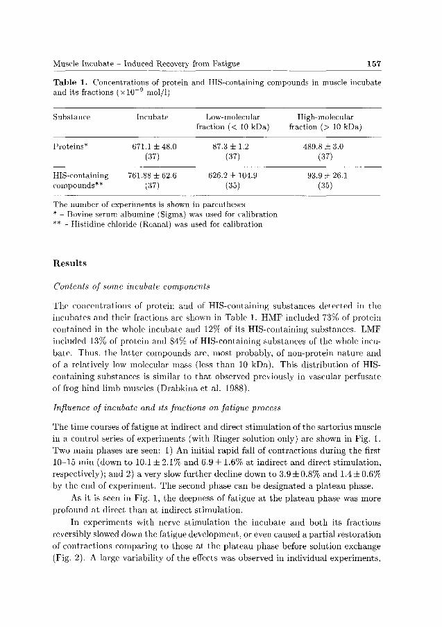
Table 1. Concentrations of protein and its fractions (xlO~9 mol/1)
Substance
Proteins*
HIS-containing compounds**
Incubate
671.1 ±48.0 (37)
761.88 ±62.6 (37)
and HIS-containing compounds in muscle incubate
Low-molecular fraction (< 10 kDa)
87.3 ±1 .2 (37)
626.2 ± 104.9 (35)
High-molecular fraction (> 10 kDa)
489.8 ±3 .0 (37)
93.9 ±26.1 (35)
The number of experiments is shown in parentheses * - Bovine serum albumíne (Sigma) was used for calibration ** - Histidine chloride (Reanal) was used for calibration
R e s u l t s
Contents of some incubate components
The concentrations of protein and of HIS-containing substances detected in the
incubates and their fractions are shown in Table 1. HMF included 73% of protein
contained in the whole incubate and 12% of its HIS-containing substances. LMF
included 13% of protein and 84% of HIS-containing substances of the whole incu
bate. Thus, the latter compounds are, most probably, of non-protein nature and
of a relatively low molecular mass (less than 10 kDa). This distribution of HIS-
containing substances is similar to tha t observed previously in vascular perfusate
of frog hind limb muscles (Drabkina et al. 1988).
Influence of incubate and its fractions on fatigue process
The time courses of fatigue at indirect and direct stimulation of the sartorius muscle
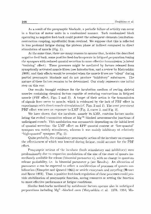
in a control series of experiments (with Ringer solution only) are shown in Fig. 1.
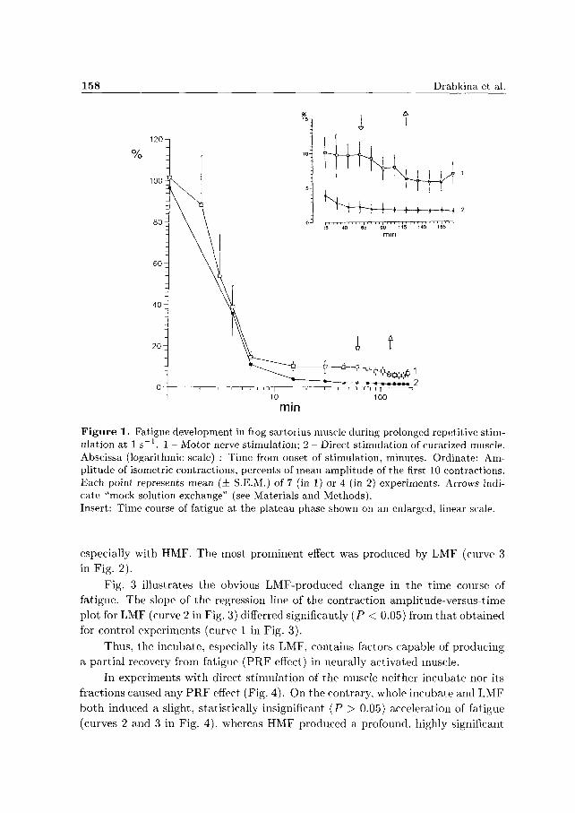
Two main phases are seen: 1) An initial rapid fall of contractions during the first
10-15 min (down to 10.1 ± 2 .1% and 6.9 ± 1.6% at indirect and direct stimulation,
respectively); and 2) a very slow further decline down to 3.9 ± 0.8% and 1.4 ± 0.6%
by the end of experiment. The second phase can be designated a plateau phase.
As it is seen in Fig. 1, the deepness of fatigue at the plateau phase was more
profound at direct than at indirect stimulation.
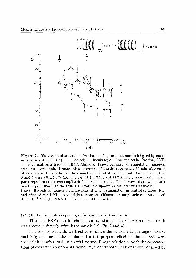
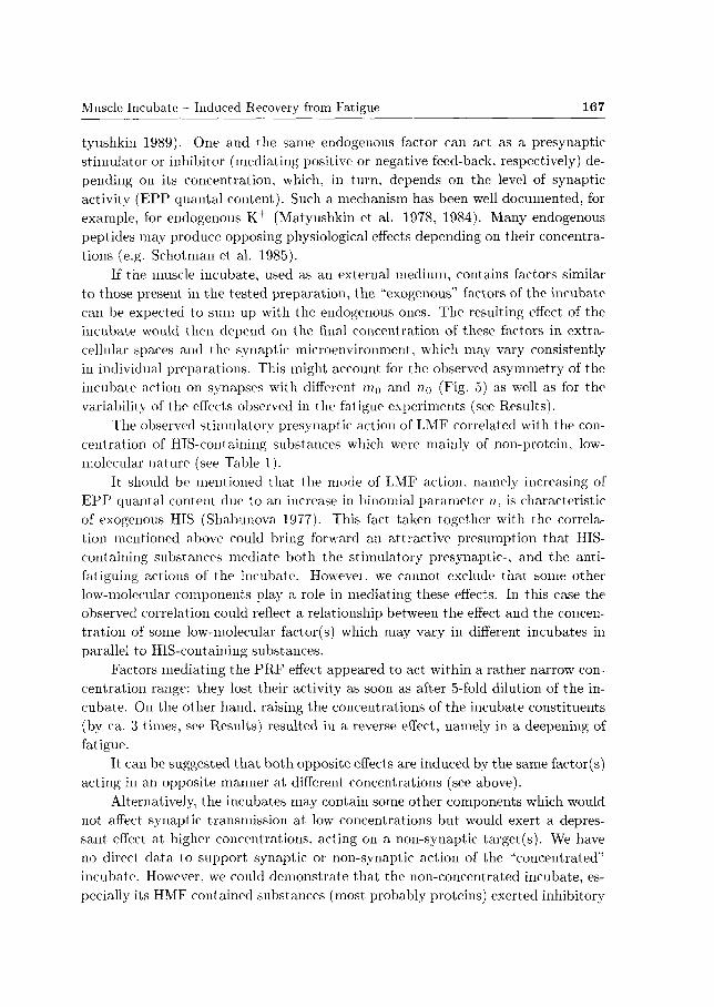
In experiments with nerve stimulation the incubate and both its fractions
reversibly slowed down the fatigue development, or even caused a partial restoration
of contractions comparing to those at the plateau phase before solution exchange
(Fig. 2). A large variability of the effects was observed in individual experiments,
158 Drabkina et al.
i í
O - | 1 i i—i i—i i i | i 1 1—i—i—r"rr -)—
1 10 100
min
Figure 1. Fatigue development in hog sartoiius muscle during prolonged repetitive stimulation at 1 s - 1 . 1 - Motor nerve stimulation; 2 - Direct stimulation of curarized muscle. Abscissa (logarithmic scale) : Time from onset of stimulation, minutes. Ordinate: Amplitude of isometric contractions, percents of mean amplitude of the first 10 contractions. Each point represents mean (± S.E.M.) of 7 (in 1) or 4 (in 2) experiments. Arrows indicate "mock solution exchange" (see Materials and Methods). Insert: Time course of fatigue at the plateau phase shown on an enlarged, linear scale.
especially with HMF. The most prominent effect was produced by LMF (curve 3 in Fig. 2).
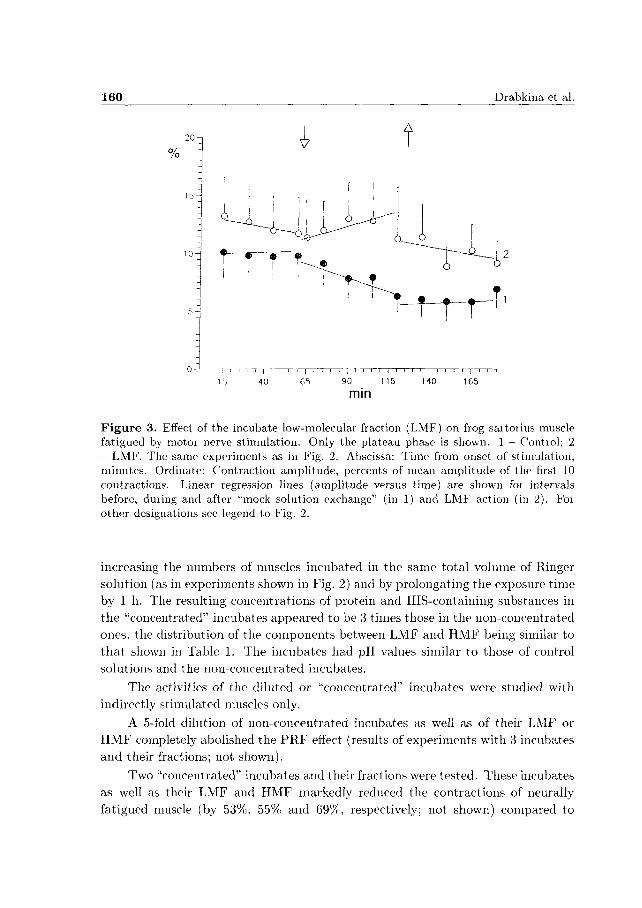
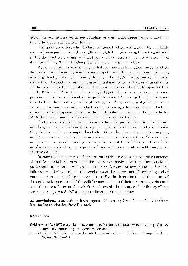
Fig. 3 illustrates the obvious LMF-produced change in the t ime course of fatigue. The slope of the regression line of the contraction amplitude-versus-time plot for LMF (curve 2 in Fig. 3) differred significantly (P < 0.05) from that obtained for control experiments (curve 1 in Fig. 3).
Thus, the incubate, especially its LMF, contains factors capable of producing
a part ial recovery from fatigue (PRF effect) in neurally activated muscle.
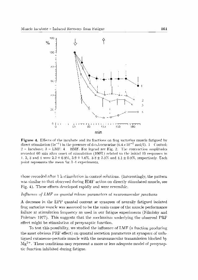
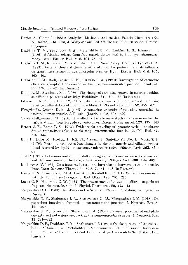
In experiments with direct stimulation of the muscle neither incubate nor its
fractions caused any P R F effect (Fig. 4). On the contrary, whole incubate and LMF
bo th induced a slight, statistically insignificant (P > 0.05) acceleration of fatigue
(curves 2 and 3 in Fig. 4), whereas HMF produced a profound, highly significant
Muscle Incubate - Induced Recovery from Fatigue 159
1 0 0 -
60-
9 8x10~3 N I 19 6x10'J N
4
3
90 - r ' i I i
180 210
mm
Figure 2. Effects of incubate and its fractions on frog sartorius muscle fatigued by motor neive stimulation (1 s _ 1 ) . 1 - Control; 2 - Incubate; 3 - Low-molecular fraction, LMF; 4 - High-molecular fraction, HMF. Abscissa: Time from onset of stimulation, minutes. Ordinate: Amplitude of contractions, percents of amplitude recorded 60 min after onset of stimulation. (The values of these amplitudes related to the initial 10 responses in 1, 2, 3 and 4 were 9.8 ± 1.8%, 13.4 ± 3.6%, 11.7 ± 3.1% and 11.2 ± 2.4%, respectively). Each point represents the mean amplitude for 7-8 experiments. The downward arrow indicates onset of perfusion with the tested solution, the upward arrow indicates wash-out. Insert: Records of isometric contractions after 1 h stimulation in control solution (left) and after 45 min LMF action (right). Note the difference in amplitude calibration: left 9.8 x 10"3 N; right 19.6 x 10"3 N. Time calibration 5 s.
(P < 0.01) reversible deepening of fatigue (curve 4 in Fig. 4) .
Thus, the P R F effect is related to a function of motor nerve endings since it
was absent in directly st imulated muscle (cf. Fig. 2 and 4).
In a few experiments we tried to estimate the concentration range of active
anti-fatigue factors of the incubate. For this purpose, effects of the incubate were
studied either after its dilution with normal Ringer solution or with the concentra
tions of extracted components raised. "Concentrated" incubates were obtained by
160 Drabkina et al.
O J r T r T T - p - i — i — r - i — j — r - T - r - T I i i i i | i i i i | i i i I | I i n
15 40 65 90 115 140 165
min
F i g u r e 3. Effect of the incubate low-molecular fraction (LMF) on frog saitoiius muscle fatigued by motoi nerve stimulation. Only the plateau phase is shown. 1 - Contiol; 2 - LMF. The same experiments as in Fig. 2. Abscissa: Time from onset of stimulation, minutes. Ordinate: Contraction amplitude, percents of mean amplitude of the first 10 contractions. Linear regression lines (amplitude versus time) are shown foi intervals before, duiing and after "mock solution exchange" (in 1) and LMF action (in 2). Foi other designations see legend to Fig. 2.
increasing the numbers of muscles incubated in the same total volume of Ringer
solution (as in experiments shown in Fig. 2) and by prolongating the exposure t ime
by 1 h. The resulting concentrations of protein and HIS-containing substances in
t h e "concentrated" incubates appeared to be 3 times those in the non-concentrated
ones, the distribution of the components between LMF and HMF being similar to
t ha t shown in Table 1. The incubates had pH values similar to those of control
solutions and the non-concentrated incubates.
The activities of the diluted or "concentrated" incubates were studied with
indirectly stimulated muscles only.
A 5-fold dilution of non-concentrated incubates as well as of their LMF or
H M F completely abolished the P R F effect (results of experiments with 3 incubates
and their fractions; not shown).
Two "concentrated" incubates and their fractions were tested. These incubates
as well as their LMF and H M F markedly reduced the contractions of neurally
fatigued muscle (by 53%, 55%) and 69%, respectively; not shown) compared to
Muscle Incubate - Induced Recovery from Fatigue 161
120
%
100
80
60
40-
20 -
1 H
0 - j i i r i i [ i r -i ~T i — y T — i i r~I [ ~ r r - i i i — | — i — i — i — i i — | — i — i — i
0 00 90 120 150 180
min
Figure 4. Effects of the incubate and its fractions on frog sartorius muscle fatigued by direct stimulation (ls^1) in the presence of d-tubocurariiie (6.4x 10~G mol/1). 1 - Control; 2 - Incubate; 3 - LMF; 4 - HMF. For legend see Fig. 2. The contraction amplitudes recorded 60 min after onset of stimulation (100%i) related to the initial 10 responses in 1, 2, 3 and 4 were 2.2 ± 0.9%, 3.9 ± 1.6%, 3.8 ± 2.5% and 4.1 ± 0.9%, respectively. Each point represents the mean for 3-4 experiments.
those recorded after 1 h stimulation in control solutions. (Interestingly, the pat tern was similar to that observed during HMF action on directly stimulated muscle, see Fig. 4). These effects developed rapidly and were reversible.
Influence of LMF on quantal release parameters at neuromuscular junctions
A decrease in the E P P quantal content at synapses of neurally fatigued isolated frog sartorius muscle was assumed to be the main cause of the muscle performance failure at stimulation frequency as used in our fatigue experiments (Nikolsky and Poletaev 1977). This suggests tha t the mechanism underlying the observed PRF effect might be stimulation of presynaptic function.
To test this possibility, we studied the influence of LMF (a fraction producing the most obvious PRF effect) on quantal secretion parameters at synapses of unfa-tigued cutaneous-pectoris muscle with the neuromuscular transmission blocked by Mg~+ . These conditions may represent a more or less adequate model of presynaptic function inhibited during fatigue.
i í
162 Drabkina et al.
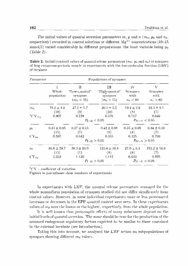
The initial values of quantal secretion parameters m, p and n (mo, po a n d no*
respectively) recorded in control solutions at different Mg~+ concentrations (10-15
mmol/1) varied considerably in different preparations, the least variable being po
(Table 2).
Table 2. Initial (control) values of quantal release parameters (mo, Po and no) at synapses of frog cutaneous-pectoris muscle in experiments with the low-molecular fraction (LMF) of incubate
Parameter Populations of synapses
I II Whole "Low-quantal"
population synapses (mo < 15)
III IV V "High-quant al" Synapses Synapses
synapses with with (mo > 15) n() < 60 n„ > 60
mo 19.2 ±4 .0 27.1 ±1 .7 30.4 ± 5.5 (19) (9) (10)
T V „ 1 0 0.907 0.729 0.576 P I - I I < 0.05
10.4 ±2.6 33.1 ±8 .1 (8) (7)
0.717 0.646 Piv-v < 0.05
p„ 0.35 ±0.05 0.27 ±0.15 0.41 ± 0.08 0.35 ± 0.05 0.34 ±0.10 (15) (7) (8) (8) (7)
CV„„ 0.597 0.642 0.555 0.425 0.799 Pi-u > 0.05 Piv-v > 0.05
n0 85.0 ±29.7 38.3 ± 16.9 125.8 ± 50.8 27.0 ± 6.1 151.2 ± 54.6 (15) (7) (8) (8) (7)
C'V„0 1.353 1.130 1.142 0.643 0.955 P i -n > 0.05 P i \ - V < 0 . 0 5
*CV - coefficient of variation Figures in parentheses show numbers of experiments
In experiments with LMF, the quantal release parameters averaged for the
whole nonuniform population of synapses studied did not differ significantly from
control values. However, in some individual experiments more or less pronounced
increases or decreases in the E P P quantal content were seen. In these experiments
values of mo were the lowest or the highest, respectively, from the whole population.
It is well known tha t presynaptic effects of many substances depend on the
initial levels of quantal secretion. The same should be t rue for the production of the
assumed endogenous regulatory factors expected to be similar to those contained
in the external incubate (see Introduction).
Taking this into account, we analyzed the LMF action on subpopulations of
synapses showing different mo values.
Muscle Incubate - Induced Recovery from Fatigue 163
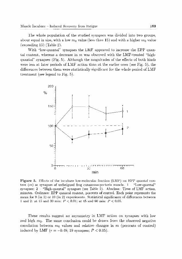
The whole population of the studied synapses was divided into two groups,
about equal in size, with a low ITIQ value (less than 15) and with a higher mg value
(exceeding 15) (Table 2).
Wi th "low-quantal" synapses the LMF appeared to increase the E P P quan
tal content, whereas a decrease in m was observed with the LMF-treated "high-
quantal" synapses (Fig. 5). Although the magnitudes of the effects of both kinds
were less at later periods of LMF action than at the earlier ones (see Fig. 5), the
differences between them were statistically significant for the whole period of LMF
treatment (see legend to Fig. 5).
200
150
100
mm
Figure 5. Effects of the incubate low-moleculai fraction (LMF) on EPP quantal content (m) at synapses of unfatigued frog cutaneous-pectoris muscle. 1 - "Low-quantal" synapses; 2 - "High-quantal" synapses (see Table 2). Abscissa: Time of LMF action, minutes. Ordinate: EPP quantal content, percents of control. Each point represents the mean for 9 (in 1) or 10 (in 2) experiments. Statistical significance of differences between 1 and 2: at 15 and 30 min: P < 0.01; at 45 and 60 min: P < 0.05.
These results suggest an asymmetry in LMF action on synapses with low
and high mo. The same conclusion could be drawn from the observed negative
correlation between mo values and relative changes in m (percents of control)
induced by LMF (r = - 0 . 4 8 ; 19 synapses; P < 0.05).
164 Drabkina et al.
In addition, LMF seemed to change the binomial parameter n, increasing it
for "low-quantal" synapses and slightly decreasing it for "high-quantal" synapses.
For a more detailed analysis of the LMF action on parameter n, we divided the
whole population of synapses into two groups according to UQ values (the groups
with different mo and no values were partially overlapping but did not coincide:
see Table 2).
With synapses with a relatively low n0 LMF increased n up to 152.0 ± 19.9%
(P < 0.05) whereas with synapses with a higher no it caused a slight decrease in
n (down to 86.1 ± 19.0%. P > 0.05). A negative correlation was observed between
?ro values and relative changes in n produced by LMF (r = —0.57: 15 synapses;
P < 0.05).
The asymmetry of presynaptic action could be convincingly confirmed in two
experiments where we had an opportunity to examine the influence of the same
LMF on two synapses with considerably different quantal release parameters. The
values of mo and "o were 2.8 and 8.5, respectively, for one synapse; and 16.0
and 66.0, respectively, for the other one. In the first synapse LMF increased the
E P P quantal content and parameter n by 146% and 110%-, respectively, while in
the second one it decreased the E P P quantal content by 30%i without substantial
change in n.
In some experiments, namely in those using synapses with low mo values,
the E P P quantal content was not altered significantly by the LMF action, but it
increased dramatically (by 100-200%>) in response to wash-out of LMF. Such a kind
of presynaptic after-effect has been described previously for exogenous carnosine
(Drabkina et al. 1990).
For the whole nonuniform population of synapses, the LMF-induced relative
changes in the E P P quantal content (both increases and decreases) were due to
respective changes in binomial parameter n: the coefficient of correlation was +0.59
(15 synapses; P < 0.05).
In experiments with LMF the relative changes in E P P quantal content ap
peared the larger, the higher the concentration of HIS-containing substances in this
fraction. For the whole population of synapses studied, the coefficient of correlation
was +0.50 (19 synapses; P < 0.05). An even higher correlation between relative
changes in m and the concentration of HIS-containing compounds was obtained
for the group of synapses with low no values (r = +0.87; 7 synapses; P < 0.05).
It was exactly in these synapses tha t LMF produced significant increase in n (see
above).
No correlation was found between changes in m and protein concentration.
No statistically significant change in binomial parameter p was observed, when
analyzing LMF action either on the whole population of synapses or on their above
described subpopulations. We were unable to separate any representative subpop-
ulations of synapses with different po values because of the relatively low variability
Muscle Incubate - Induced Recovery from Fatigue 165
of this parameter in our experiments (see Table 2).
In experiments with LMF no substantial changes were observed in muscle fibre
resting potential or in amplitude or t ime course of M E P P . This suggests that LMF
does not significantly modify electrogenesis in the muscle cell membrane or the
characteristics of the acetylcholine-receptive postsynaptic membrane.
D i s c u s s i o n
Fatigue of isolated skeletal muscle or neuromuscular preparation is a very compli
cated process which remains not completely understood. Different cellular mecha
nisms can be involved in the reversible reduction of muscle performance in fatigued
preparation depending on the stimulation pat tern or the mode of muscle activation
(Nikolsky and Poletaev 1977; Westerblad et al. 1991; Westerblad and Allen 1992;
Edman and Lou 1992).
In a comprehensive study of fatigue mechanisms in isolated, directly stimu
lated muscle fibres. Edman and Lou (1992) have shown that the force reduction
during an initial period of fatiguing stimulation at I s - 1 is caused by an impairment
on the level of contractile elements. This functional disorder (termed "true myofib
rillar fatigue"), is generally assumed to be a consequence of metabolic shifts in the
myoplasm during repetitive contractions (Renaud and Mainwood 1985; Westerblad
et al. 1991; Nagesser et al. 1992).
More prolonged stimulation at the above frequency causes a failure of the
contractile apparatus activation due to a failure of action potential propagation
along transverse tubules (Westerblad et al. 1991; Edman and Lou 1992).
It should be noted tha t such a blockade of excitation-contraction coupling
prevents the myofibrillar apparatus from further exhaustion (see Edman and Lou
1992). Probably, this is manifested in the slow phase of contraction decline (pla
teau) seen in our experiments with directly stimulated muscles (Fig. 1, curve 2).
In isolated frog sartorius muscle fatigued by nerve stimulation at 1 s _ 1 , a
block develops firstly at the presynaptic level (Nikolsky and Poletaev 1977). It
occurs due to a decrease in E P P quantal content below a threshold for muscle
action potential generation. However, in this case the presynaptic block is not a
result of "fatigue" of the presynaptic function per se; rather, it is a consequence
of inhibitory antidromic actions of metabolites released from contracting muscle
fibres and accumulated in extracellular clefts (Nikolsky and Poletaev 1977).
Although direct evidence is lacking, one can suggest the involvement of me
tabolites such as K + , H + , lactate, adenosine, etc. These substances are expected
to be released from muscle fibres, and are known to inhibit the t ransmit ter release
(Lucier and Mainwood 1972; Hnik et al. 1976; Matyushkin et al. 1978, 1984;
Silinsky 1980; Gaudri-Tallarmain 1986; Matyushkin 1989; Smith 1991).
166 Drabkina et al.
As a result of the presynaptic blockade, a periodic failure of activity can occur
in a fraction of motor units in a randomized manner. Such randomized block
operat ing as negative feed-back could protect the subsequent elements (excitation-
contraction coupling, myofibrills) from overload. We suppose tha t this is reflected
in less profound fatigue during the plateau phase at indirect compared to direct
st imulation of muscle (Fig. 1).
At the same time, there are many reasons to assume tha t , besides the described
negative feed-back, some positive feed-backs operate in fatigued preparat ion tuning
the synapses with reduced quantal secretion to more effective transmission (a latent
"training" effect). These processes might be mediated by factors released from
synaptically activated muscle fibres (see Introduction, and a review by Matyushkin
1989), and their effects would be revealed when the muscle fibres are "silent" during
par t ia l presynaptic blockade and do not produce "inhibitory" substances. The
na ture of these factors remains to be determined. Our study represents one initial
s tep on this way.
Our results brought evidence for the incubation medium of resting skeletal
muscles containing chemical factors capable of restoring contractions in fatigued
muscle (PRF effect, Figs. 2 and 3). A target of their action is the transmission
of signals from nerve to muscle, which is evidenced by the lack of P R F effect in
experiments with direct muscle stimulation (cf. Figs. 2 and 4). The most prominent
P R F effect was seen on exposure to LMF (Fig. 2, curve 3, and Fig. 3).
We have shown tha t the incubate, namely its LMF, contains factors modu
lating the evoked t ransmit ter release at Mg2 +-blocked neuromuscular junctions of
unfatigued muscle. This modulation was asymmetric depending on the initial level
of quantal secretion: the LMF effect on E P P quantal content at "low-quantal"
synapses was mainly stimulatory, whereas it was mainly inhibitory at relatively
"high-quantal" synapses (Fig. 5).
Quite probably, the stimulatory presynaptic action of the incubate on synapses
the effectiveness of which was lowered during fatigue, could account for the P R F
effect.
Presynaptic actions of the incubate (both stimulatory and inhibitory) were
predominantly due to respective modulat ion of the size of the store of quanta im
mediately available for release (binomial parameter n), with no change in quantum
release probability, i.e. in binomial parameter p (see Results). An alteration of
parameter n can be expected to reflect a modification of processes of quanta mo
bilization (Elmqvist and Quastel 1965) or vesicle exocytosis and recycling (Heuser
and Reese 1973). Thus, a positive feed-back regulation of these processes could pro
vide stabilization of presynaptic function, saving resources or setting the function
to more effective performance at fatigue conditions.
Similar feed-backs mediated by antidromic factors operate also in unfatigued
preparat ions including Mg2 +-blocked ones (Matyushkin et al. 1978, 1984; Ma-
Muscle Incubate - Induced Recovery from Fatigue 167
tyushkin 1989). One and the same endogenous factor can act as a presynaptic
stimulator or inhibitor (mediating positive or negative feed-back, respectively) de
pending on its concentration, which, in turn, depends on the level of synaptic
activity (EPP quantal content). Such a mechanism has been well documented, for
example, for endogenous K + (Matyushkin et al. 1978, 1984). Many endogenous
peptides may produce opposing physiological effects depending on their concentra
tions (e.g. Schotman et al. 1985).
If the muscle incubate, used as an external medium, contains factors similar
to those present in the tested preparation, the "exogenous" factors of the incubate
can be expected to sum up with the endogenous ones. The resulting effect of the
incubate would then depend on the final concentration of these factors in extra
cellular spaces and the synaptic microenvironment, which may vary consistently
in individual preparations. This might account for the observed asymmetry of the
incubate action on synapses with different mo and r?o (Fig. 5) as well as for the
variability of the effects observed in the fatigue experiments (see Results).
The observed stimulatory presynaptic action of LMF correlated with the con
centration of HIS-containing substances which were mainly of non-protein, low-
molecular nature (see Table 1).
It should be mentioned that the mode of LMF action, namely increasing of
E P P quantal content due to an increase in binomial parameter n, is characteristic
of exogenous HIS (Shabunova 1977). This fact taken together with the correla
tion mentioned above could bring forward an attractive presumption tha t HIS-
containing substances mediate both the stimulatory presynaptic-, and the anti-
fatiguing actions of the incubate. Howevei, we cannot exclude that some other
low-molecular components play a role in mediating these effects. In this case the
observed correlation could reflect a relationship between the effect and the concen
trat ion of some low-molecular factor(s) which may vary in different incubates in
parallel to HIS-containing substances.
Factors mediating the P R F effect appeared to act within a rather narrow con
centration range: they lost their activity as soon as after 5-fold dilution of the in
cubate. On the other hand, raising the concentrations of the incubate constituents
(by ca. 3 times, see Results) resulted in a reverse effect, namely in a deepening of
fatigue.
It can be suggested tha t both opposite effects are induced by the same factor(s)
acting in an opposite manner at different concentrations (see above).
Alternatively, the incubates may contain some other components which would
not affect synaptic transmission at low concentrations but would exert a depres
sant effect at higher concentrations, acting on a non-synaptic target(s) . We have
no direct da ta to support synaptic or non-synaptic action of the "concentrated"
incubate. However, we could demonstrate that the non-concentrated incubate, es
pecially its HMF contained substances (most probably proteins) exerted inhibitory
168 Drabkina et al.
action on excitation-contraction coupling or contractile apparatus of muscle fa
tigued by direct stimulation (Fig. 4).
The question arises, why the last mentioned action was lacking (or markedly
reduced) in experiments with neurally stimulated muscles, even those treated with
HMF, the fraction causing profound contraction decrease in muscles stimulated
directly (cf. Fig. 2 and 4). One plausible explanation is as follows.
As noted above, in experiments with direct muscle stimulation the contraction
decline at the plateau phase was mainly due to excitation-contraction uncoupling
in a large fraction of muscle fibres (Edman and Lou 1992). In the remaining fibres,
still active, the safety factor of action potential generation in T-tubular membranes
can be expected to be reduced due to K + accumulation in the tubular spaces (Hnik
et al. 1976; Juel 1986; Renaud and Light 1992). It can be suggested that some
proteins of the external incubate (especially when HMF is used) might be come
adsorbed on the mouths or walls of T-tubules. As a result, a slight increase in
external resistance can occur, which would be enough for complete blockade of
action potential propagation from surface to tubular membrane, if the safety factor
of the last membrane was lowered to just suprathreshold levels.
On the contrary, in the case of neurally fatigued preparation the muscle fibres
in a large part of motor units are kept unfatigued (with intact electrical proper
ties) due to part ial presynaptic blockade. Thus, the above described uncoupling
mechanism can be expected to become inoperative in this situation. Whatever the
mechanism, the same reasoning seems to be true if the inhibitory action of the
incubate on muscle elements requires a fatigue-induced alteration in the properties
of these elements.
In conclusion, the results of the present study have shown a complex influence
of muscle metabolites, present in the incubation medium of a resting muscle on
presynaptic function as well as on muscular elements of motor units. Such an
influence could play a role in the regulation of the motor units functioning and of
muscle performance in fatiguing conditions. For the determination of the nature of
the active substances and of the cellular mechanisms of their actions, experimental
conditions are to be created in which the observed stimulatory and inhibitory effects
are reliably separated. Efforts in this direction are under way.
Acknowledgements. This woik was supported in pait by Grant No. 94-04-12118a from Russian Foundation for Basic Research.
References
Boldyrev A. A. (1977): Biochemical Aspects of Excitation-Contraction Coupling. Moscow Univeisity Publishing, Moscow (in Russian)
Crush K. G. (1970): Carnosine and related substances in animal tissues. Comp. Biochem. Physiol. 34, 3—30
Muscle Incubate - Induced Recovery from Fatigue 1 6 9
Darbre A., Clamp J. (1986): Analytical Methods. In: Practical Protein Chemistry (Ed. A. Darbre), 243—363, J. Wiley & Sons Ltd. Chichester- N.-Y.-Brisbane- Toronto-Singapore
Drabkina T. M., Shabunova I. A., Matyushkin D. P., Gankina E. S., Efimova I. I. (1986): /i-Alanine release from frog muscle determined by thin-layer chromatography. Byull. Eksper. Biol. Med. 101, 38—41
Drabkina T. M., Kubasov I. V., Matyushkin D. P., Romanovsky D. Yu., Yurkyanets E. A. (1988): Some biochemical characteristics of muscular perfusate and its influence on transmitter release in neuromuscular synapse. Byull. Eksper. Biol. Med. 105, 460—462
Drabkina T. M., Radzjukevich V. K., Shtanko S. A. (1990): Investigation of carnosine effect on synaptic transmission in the frog neuromuscular junction. Fiziol. Zh. SSSR 76, Í8—25 (in Russian)
Dupin A. M., Stvolinsky S. L. (1986): The change of carnosine content in muscles working at different patterns of activity. Biokhimiya 51, 160—163 (in Russian)
Edman K. A. P.. Lou F. (1992): Myofibrillar fatigue versus failure of activation during repetitive stimulation of frog muscle fibres. J. Physiol. (London) 457, 655—673
Elmqvist D., Quastel D. M. J. (1965): A quantitative study of end-plate potentials in isolated human muscle. J. Physiol. (London) 178, 505 -529
Gaudri-Tallarmam I. M. (1986): The effect of lactate on acetycholine release evoked by various stimuli from Torpedo synaptosomes. Európ. J. Pharmacol. 129, 135- 143
Heuser J. E., Reese T. S. (1973): Evidence for recycling of synaptic vesicle membrane dming transmitter release at the frog neuromuscular junction. J. Cell. Biol. 57, 315—344
Hrn'k P., Holas M., Krekule L, Kríž N.. Mejsnar J.. Smieško V , Ujec E., Vyskočil F. (1976): Work-induced potassium changes in skeletal muscle and effluent venous blood assessed by liquid ion-exchanger microelectrodes. Pfliigers Arch. 362, 85— 94
Juel C. (1986): Potassium and sodium shifts during in vitro isometric muscle contraction and the time course of the ion-gradient recovery. Pfliigers Arch. 406, 458—463
Kibjakov A. V. (1935): On a humoral factor in the interrelation between nerve and muscle. Proc. Tatar Institute Theor. Clin. Med. 2, 141—148 (in Russian)
Lowry O. N.. Roscnbrough M. J., Fair A. L., Randall R. J. (1951): Protein measurement with the Folin phenol reagent. J. Biol. Chem. 193, 265—275
Lucier G. E., Mainwood G. W. (1972): The measurement of potassium efflux in superfused frog sartorius muscle. Can. J. Physiol. Pharmacol. 50, 123—131
Matyushkin D. P. (1989): Feed-Backs in the Synapse. "Nauka" Publishing, Leningrad (in Russian)
Matyushkin D. P., Shabunova I. A., Sharovarova G. M., Vinogradova I. M. (1978): On potassium functional feedback in neuromuscular junction. J. Neurosci. Res. 3, 441—450
Matyushkin D. P., Krivoi I. I., Shabunova I. A. (1984): Reversal potential of end-plate currents and potassium feedback at the neuromuscular synapse. J. Neurosci. Res. 11, 281—292
Matyushkin D. P., Drabkina T. M., Shabunova I. I. (1986): On the question of the contribution of some muscle metabolites to antidromic regulation of transmitter release from motor nerve terminal. Vestník Leningradskogo Universiteta Ser. 3, 76—84 (in Russian)
170 Drabkina et al.
Matyushkin D. P., Krivoi I. L, Drabkina T. M. (1993): The positive action of potassium ions outflow on the neuromuscular transmission. XXXII Congress of the Intern. Union of Physiol. Sciences. August lst-6th. Glasgow. Abstracts. 244.64/P
Miyamoto M. D. (1975): Binomial analysis of quantal transmitter release at glycerol-treated frog neuromuscular junctions. J. Physiol. (London) 250, 121—142
Nagesser A. S., van der Laarse W. J., Elzinga G. (1992): Metabolic changes with fatigue in different types of single muscle fibres of Xenopus laevis. J. Physiol. (London) 448, 511—523
Nikolsky E. E., Poletaev G. I. (1977): Study of mechanism of blockade of transmission from nerve to skeletal muscle at low-frequency neuronal stimulation. Neirofiziologiya 9, 78—85 (in Russian)
Ovadi J., Libor S., Elodi P. (1967): Spectrophotometric determination of histidine in proteins with diethylpyrocarbonate. Acta Biochim. Biophys. Hung. 2, 455—458
Renaud J. M., Mainwood G. W. (1985): The effects of pH on the kinetics of fatigue and recovery in frog sartorius muscle. Can. J. Physiol. Pharmacol. 63, 1435— 1443
Renaud J. M., Light P. (1992): Effects of K+ on the twitch and tetanic contraction in the sartorius muscle of the frog, Rana pipiens. Implication for fatigue m vitro. Can. J. Physiol. Pharmacol. 70, 1236 — 1246
Schotman P.. Schrama L. H., Edwards Ph. M. (1985): Peptidergic system. In: Handbook of Neurochemistry (Ed. A. Lojtha) 8, pp. 243—279, Plenum Press, N.-Y.-London
Severin S. E., Vulfson P. L. (1963): The role of carnosine in neurotrophic relations. In: The Effect of Use and Disuse of Neuromuscular Function. (Eds. Gut man E. and Hnik P.) pp. 53—62. Elsevier, Amsterdam
Shabunova I. A. (1977): On the presynaptic action of histidine at neuromuscular junction. Fiziol. Zh. SSSR 63, 964—969 (in Russian)
Shabunova I. A. (1979): The histidine release from the frog skeletal muscles at rest and in rhythmic activity. Fiziol. Zh. SSSR 65, 1371—1373 (in Russian)
Silinsky E. M. (1980): Evidence for specific adenosine receptors at cholinergic nerve endings. Brit. J. Pharmacol. 71, 191—194
Smith D. O. (1991): Sources of adenosine released during neuromuscular transmission in the rat. J. Physiol. (London) 432, 343—354
Westerblad H , Allen D. G. (1992): Changes of intracellular pH due to repetitive stimulation of single fibres from mouse skeletal muscle. J. Physiol. (London) 449, 49—71
Westerblad H., Lee J. A., Lannergren J., Allen D. G. (1991): Cellular mechanisms of fatigue in skeletal muscle. Amer. J. Physiol. 261, C195 -209
Williams B., Wilson K. (1975): Methods of Chromatography. In: Principles and Technique of Practical Biochemistry (Ed. B. Williams and K. Wilson) pp. 123—134, Edward Arnold, London
Final version accepted April 6, 1995