Embed Size (px)
Citation preview
Original Paper Veterinarni Medicina, 55, 2010 (6): 264–274
264
The influence of orally administered short chain fatty acids on intestinal histopathological changes and intensity of Trichinella spiralis infection in mice
D. Mista, J. Piekarska, M. Houszka, W. Zawadzki, M. Gorczykowski
Faculty of Veterinary Medicine, Wroclaw University of Environmental and Life Sciences, Wroclaw, Poland
ABSTRACT: The influence of short chain fatty acids (SCFA) on histopathological changes in the small intestine and the intensity of invasion of T. spiralis in mice were investigated in this study. The animals were infected with doses of 500 and 250 T. spiralis larvae per mouse. A SCFA solution containing acetic, propionic and butyric acid (30 : 15 : 20mM) was administered orally to the mice starting from the 5th day before infection to the 20th day after infection (day). Fragments of the jejunum collected during dissection on the 7th and 10th day were used to prepare specimens to assess the histopathological changes. In the infected animals, the intestinal trichinellae were counted on the 7th and 10th day, while on the 42nd day the muscle larvae number were determined. The strongest host reaction in the intestine was observed on the 7th day at a dose of T. spiralis 500 larvae, and on the 10th day at a dose of 250 larvae. Numerous inflammatory infiltrations, strong shortening of the intestinal villi, extension of the intestinal crypts, and the lowest ratio of the villi length to the intestinal crypts depth were observed. The ratio was 1.3 ± 0.3 on the 7th day at a dose of 500 larvae, and on the 10th day, at dose of 250 larvae the ratio reached 1.5 ± 0.5. Both values differed significantly from the control group: 3.3 ± 0.5 (P < 0.01). Administration of SCFA to the animals infected with T. spiralis caused remission of local histopathological changes resulting from the presence of the parasite in the small intestine after the mentioned periods. This manifested as limited villi shortening and reduced deepening of intestinal crypts. At the higher infectious dose, in animals receiving the acid solution, on the 7th day the intestinal villi were considerably longer (356 µm ± 35) than in the group infected with T. spiralis but not treated with the acids (279 µm ± 57; P < 0.01). At a lower dose of parasites, on the 10th day these values were 339 µm ± 88 and 306 µm ± 47 respectively and the observed differences were not statistically significant. The solution of SCFA also caused a decrease in the numbers of mature parasites in the intestine and the muscle larvae at a dose of 500 larvae/mouse. In animals receiving the SCFA, 24 050 ± 10 415 larvae were observed in muscles, while in the infected mice, which did not receive the acids, 32 875 ± 16 762 larvae were detected (P < 0.05). An increase in the intensity of infection accelerated the rate of host reaction to the presence of T. spiralis in the intestines (self-cure). To summarize, the administered solution of short chain fatty acids alleviated the forma-tion of histopathological changes in the intestine in response to the parasite’s presence, and lowered the intensity of T. spiralis invasion after infection with a higher dose of larvae.
Keywords: trichinellosis; SCFA; intestinal morphology; mouse
Short chain fatty acids (SCFA) are the most im-portant end products of the fermentation con-ducted by microorganisms in the gastrointestinal tract. They result from the decomposition of car-bohydrates originating mainly from plant cells: cellulose, pectin, starch, and other dextrans and soluble carbohydrates, and also from the bacte-
rial decomposition of proteins. The concentration of the end products of bacterial fermentation in the caecum, especially ammonia and SCFA, re-flects the activity of the intestinal microflora, which conditions the normal processes occur-ring in this section of the gastrointestinal tract (Bergman, 1990).

Veterinarni Medicina, 55, 2010 (6): 264–274 Original Paper
265
In the gastrointestinal tract, the most numerously produced SCFA are: acetate, propionate and butyrate, which after absorption are used as energy sources for primary metabolism, animal growth, and lipogenesis. SCFA affect the intestinal mucosa in various manners. They increase intercellular pH, and modulate sodium absorption and chloride excretion. They stimulate the release of mucins, and increase the blood flow in the mucosa (Engelhardt et al., 1998; Sellin, 1999). Inhibition of the growth of pathogenic bacteria in the large intestine, including E. coli (Byrne and Dankert, 1979), as well as anti-inflammatory properties has been described for SCFA (Rotstein, 1993; Cavaglieri et al., 2003). The joint interaction of three acids: acetic, propionic, and butyric, with the intestinal epithelium stimulates normal intestinal crypt cells to proliferate, while butyric acid alone inhibits the growth of the colon cancer cell lines by induction of apoptosis (Chiou et al., 1994; Engelhardt et al., 1998). The main influence of SCFA on inflammatory proc-esses lies in their effect on lymphocyte and cytokine production. Butyrate production inhibits the prolifera-tion of lymphocytes and IL-2 and IFN-acetate while propionate partially prevents this inhibitory effect of butyrate. Also, both acids increase IFN-γ and IL-10 production (Cavaglieri et al., 2003).
Such properties of SCFA can modulate the im-munological processes during T. spiralis intestinal infection. In the mature stadium, the nematodes are present in the epithelial layer of the host small intes-tine. Structural, cellular, and physiological changes in the intestine resulting from their presence are the reason for the immunological response mani-fested as acute inflammation (Garside et al., 2000; Khan and Collins, 2004; Dehlawi et al., 2006).
In the intestinal phase of trichinellosis, the “self-cure” phenomenon plays an important role. It oc-curs as a result of a rapid expulsion of the parasites from the gastrointestinal tract. The early phase of the intestinal phase of trichinellosis is character-ized by accumulation of inflammatory cells (mainly mast cells), eosinophils, Th1 and Th2 lymphocytes responses, production of IgE antibodies, and re-lease of mediators and cytokines (Grencis et al., 1991; Finkelman et al., 1997; Piekarska, 2004). It is known that faster expulsion of the mature forms of T. spiralis from the intestines can be connected with the production of IL-2, IL-3, and IFN– IL-4. IL-5 contributes to slower removal of trichinel-lae from the intestine (Kelly et al., 1991; Mink et al., 1994). An increase in chemokine expression in the mouse intestinal epithelium enhances the
recruitment of neutrophil granulocytes and lym-phocytes to the intestine (Kostro et al., 2000). Since SCFA alter chemokine expression in enterocytes (Sanderson, 2004), they can also play a role in the anti-parasite response.
In the intestinal phase of T. spiralis, a decrease in SCFA production and change in their relative levels has been described (Mista et al., 2005). The influence of parasitic infection on the fermenta-tion pattern and SCFA levels has been also con-firmed in ascariasis in sheeps (Varadyova et al., 2001). The effect of fermentation products on the intestinal phase of T. spiralis could be an example of the interaction between the saprophytic intestinal microflora and the parasite.
The aim of the study was to show the intestinal changes resulting from the inflammatory proc-esses caused by the parasites, and to determine the effect of SCFA administration on these proc-esses, by observing the histopathological changes, counting the number of mature parasites in the intestinal trichinellosis, and the number of larvae in muscles.
MATERIAL AND METHODS
The material for the research consisted of 117 CFW inbred mice at ages of around three months, weigh-ing around 20 g. 96 mice were infected per os with T. spiralis T1(ISS1820) larvae. 48 mice were in-fected with a dose of 500 larvae/mouse, while the remaining 48 were administered a dose of 250 lar-vae/mouse. There were six experimental groups:
C – healthy mice (7)S – healthy mice receiving SCFA (14)T 500 – mice infected with T. spiralis at dose of
500 larvae (24)TS 500 – mice infected with T. spiralis at dose of
500 larvae, and receiving SCFA (24)T 250 – mice infected with T. spiralis at dose of
250 larvae (24)TS 250 – mice infected with T. spiralis at dose of
250 larvae, and receiving SCFA (24)During the whole time of the experiment, all ani-
mals were maintained under identical conditions. SCFA solution was administered with drinking water from the 5th day before infection to the 20th day of infection. The molar concentrations of the following acids: acetic, propionic, and butyric, in the dosed solutions were 30, 15, and 20mM, re-spectively.
Original Paper Veterinarni Medicina, 55, 2010 (6): 264–274
266
On the 7th and 10th day of infection, after slaugh-tering seven animals in each group, tissue samples from their jejunum were collected. After fixation in a neutralized 8% formaldehyde water solution, four thick paraffin sections were stained with hematoxy-lin and eosin (H & E). Cellular infiltration intensity, the shape and size of villi and intestinal crypts, as well as the presence of goblet cells were assessed. The analysis of the intestinal villi and crypts was conducted using the Photomicroscope-Axiophot, and the computer program MultiScan Base V 8.08. Parasitological studies of the intestines were per-formed to determine the invasion intensity on the 7th and 10th days after infection. On the 42nd day, 10 animals from each group were slaughtered in or-der to establish the number of muscle larvae using the digestion method (Kozar and Kozar, 1972).
The results were analysed using one-way ANOVA and STATISTICA 8.0 software. The significance of the differences was confirmed by the multiple comparison Tukey’s test for P < 0.05 and P < 0.01. Obtained results are presented in Tables 1–3 as the mean values and standard deviations.
RESULTS AND DISCUSSION
In the parasitological studies conducted in this experiment, the observed differences in the number of parasites related only to the animals infected with the dose of 500 larvae/mouse. On the 7th day, a lower number of parasites in the small intestine of the infected mice which received SCFA (TS 500) was observed in comparison with the infected mice which did not receive the acid solution (T 500). However, the differences were not confirmed sta-tistically (Table 1). In above-mentioned groups, on the 10th day only single adult parasites were ob-served in the intestine. This observation points to the “self-cure” phenomena. Significant differences were confirmed in the number of muscle larvae on the 42nd day. In the animals of the TS 500 group,
the number of larvae was smaller in comparison to the T 500 group (Table 2).
Based on results mentioned above, it can be sup-posed that administration of the SCFA resulted in decreased numbers of newborn T. spiralis larvae in the intestines of mice infected with the higher dose of T. spiralis. The reason for this might have been direct interaction of SCFA with the parasite or (which seems to be more likely) interaction of the SCFA with the mucosa and the intestinal environ-ment. This could have resulted in changes which prevented the females from giving birth to larvae or the larvae from surviving. Changes in local micro-flora composition caused by the SCFA administra-tion may have importance because previous studies showed a significant role for intestinal microflora in determining the invasive capacity of T. spiralis in mice (Przyjalkowski et al., 1969, 1983).
At a dose of 250 larvae/mouse no differences in the intensification of T. spiralis intestinal infec-tion were observed among the groups. However, in comparison to the groups in which mice received the higher dose of T. spiralis, significantly lower numbers of parasites in the intestines was observed on the 7th day (Table 2). On the 10th day, at this dose, the number of T. spiralis in the intestine decreased only slightly. Thus, the “self-cure” phenomena oc-curred later than in those mice which received the 500 larvae dose. The number of muscle larvae on the 42nd day was similar in both infected groups
Table 1. The number of adult trichinellae in the jejunum on the 7th and 10th days after infection
Group T 500 TS 500 T 250 TS 250
7th day 121.6 ± 38.3 79.8 ± 30.3 33.1 ± 10.6 29.6 ± 39.4
10th day 3.4 ± 2.8 3.7 ± 2.6 20.0 ± 16.8 16.8 ± 20.6
Results are expressed as the mean ± SD of 7 animalsT = T. spiralis-infected mice, TS = SCFA-administered mice and infected with T. spiralis
Table 2. The number of muscle larvae per mouse on the 42nd day after infection
Group T TS
500 larvae/mouse 32 875 ± 16 762a 24 050 ± 10 415b
250 larvae/mouse 18 542 ± 17 967 17 000 ± 13 875
Results are expressed as the mean ± SD of 10 animalsT = T. spiralis-infected mice, TS = SCFA-administered mice and infected with T. spiralisabsignificant differences at P < 0.05
Veterinarni Medicina, 55, 2010 (6): 264–274 Original Paper
267
(T 250 and TS 250). These results suggest that the magnitude of the host’s reaction to the presence of parasites in the intestines increases together with the increase in invasion intensity. The influence of SCFA on intestinal trichinellosis is stronger with the higher number of parasites.
An effect of SCFA administration on parasite invasions in the intestine was also described by Petkevicius et al. (2004). Infusion of SCFA and lac-tic acid into the caecum had a strong anti-parasitic effect in the case of Oesophagostomum dentatum, manifesting in a decreased number of eggs, mature forms, and a lowered fertility of females. Similar effects were obtained during intestinal parasitoses in pigs after administering fodder containing highly fermentable carbohydrates (i.a. inulin), which de-compose in the large intestine resulting in high levels of SCFA. Decreased numbers and fertil-ity of Oesophagostomum dentatum and Trichuris suis have been observed in the large intestine (Petkevicius et al., 2003, 2007). Lower numbers of parasites in the intestines of animals fed with highly fermentable carbohydrates in comparison to animals receiving fodder supplemented with resist-ant carbohydrates was also observed by Thomsen et al. (2005, 2006). Considering the influence of carbohydrates contained in the fodder on the sur-vival of intestinal nematodes, it seems that the type of dietary carbohydrates plays a crucial role. Fermentable carbohydrates cause an increase in the
intensity of bacterial fermentation in the intestines. This leads to the elevated production of SCFA, the inhibiting influence of which on the survival and breeding of the nematodes, has been demonstrated (Petkevicius et al., 2004).
The results of the present study also confirm the inhibitory effect of the products of bacterial fer-mentation in intestines on the survival and fertility of nematodes. The diminished numbers of parasites after oral administration of SCFA allows us to spec-ulate that parasitoses of the gastrointestinal tract are influenced by the activity of saprophytic bac-teria present in the intestines, as well as a number of factors, which influence local microflora (e.g. feeding of the animals), especially the carbohydrate profile of the feed. A change of the environment in the intestines, which takes place along with the ac-cumulation of large amounts of organic acids, may play a significant role in this model (Petkevicius et al., 2004; Petkevicius, 2007).
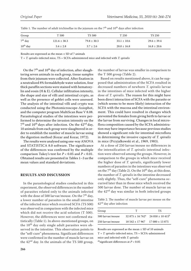
The influence of SCFA on histopathological changes in the jejunum was also observed in this study. In control animals, long intestinal villi with regular, finger-like shape, and oligocellular mesen-chymal stroma, as well as shallow intestinal crypts were observed. Goblet cells were sparse within the villi, and moderately numerous in the intestinal glands (Figure 1). Administration of the SCFA so-lution to healthy animals caused sparse inflamma-tory infiltrations in the stroma of the intestinal villi
Table 3. The lengths of intestinal villi and crypts in experimental trichinellosis in mice (µm)
Group C S T 500 TS 500 T 250 TS 250
7th day
Villi 465 ± 13A 456 ± 36A 279 ± 57B 356 ± 35C 316 ± 31BC 326 ± 46BC
Crypts 143 ± 27A 135 ± 22Aa 230 ± 46Bc 208 ± 22B 188 ± 43bc 177 ± 23ab
v/c 3.3 ± 0.5A 3.5 ± 0.7A 1.3 ± 0.3B 1.8 ± 0.4B 1.8 ± 0.5B 1.9 ± 0.4B
10th day
Villi 465 ± 13Aa 462 ± 55A 385 ± 49b 388 ± 45b 306 ± 47B 339 ± 88B
Crypts 143 ± 27Aab 143 ± 25Aa 192 ± 18B 186 ± 9c 214 ± 40B 180 ± 25bc
v/c 3.3 ± 0.5A 3.4 ± 0.7A 2.1 ± 0.3B 2.1 ± 0.1B 1.5 ± 0.5B 1.9 ± 0.6B
Results are expressed as the mean ± SD of 7 animalsC = the control group, S = the group of animals that received SCFA solution, T = T. spiralis infected mice, TS = SCFA-administered mice and infected with T. spiralis500, 250 = the number of larvae/mousev/c = the villus to crypt height ratioabcsignificant differences at P < 0.05ABCsignificant differences at P < 0.01
Original Paper Veterinarni Medicina, 55, 2010 (6): 264–274
268
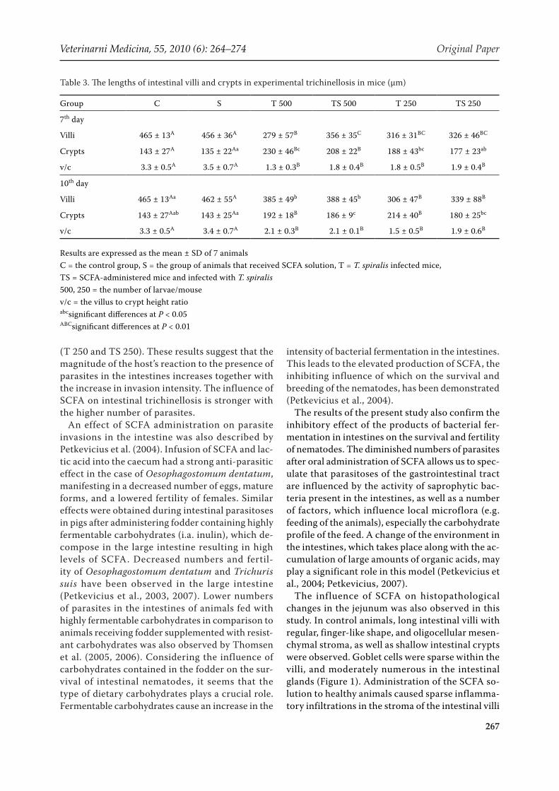
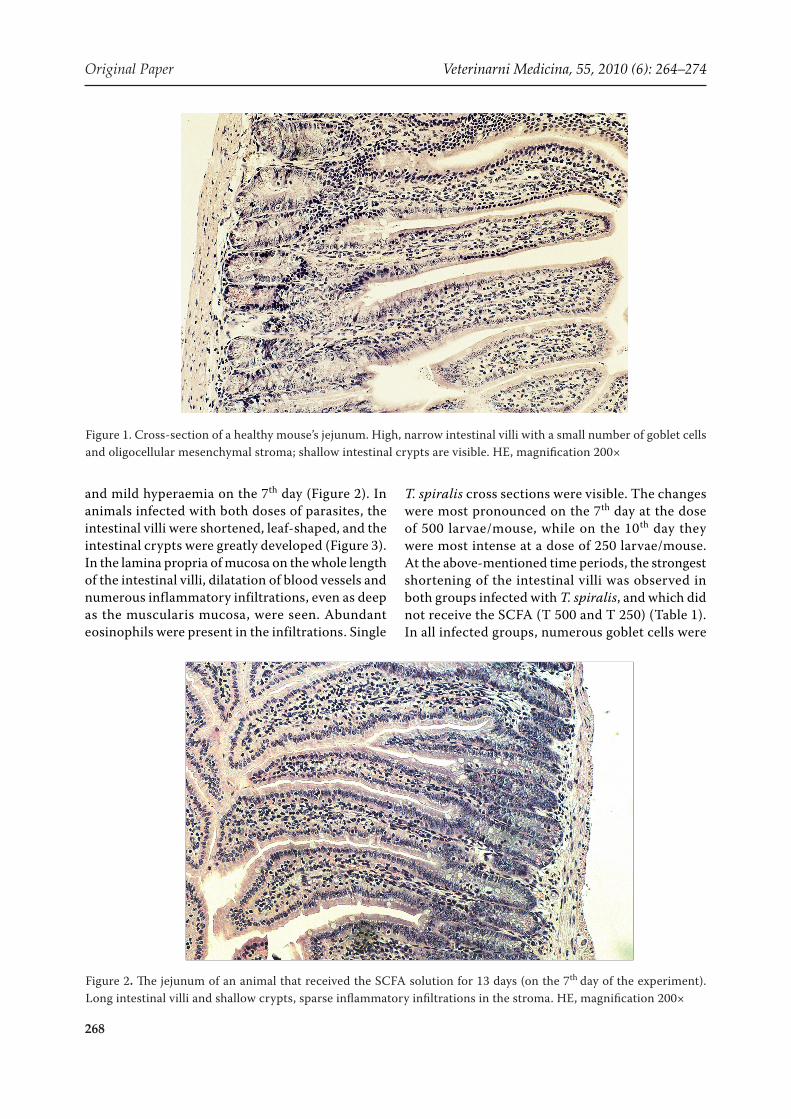
and mild hyperaemia on the 7th day (Figure 2). In animals infected with both doses of parasites, the intestinal villi were shortened, leaf-shaped, and the intestinal crypts were greatly developed (Figure 3). In the lamina propria of mucosa on the whole length of the intestinal villi, dilatation of blood vessels and numerous inflammatory infiltrations, even as deep as the muscularis mucosa, were seen. Abundant eosinophils were present in the infiltrations. Single
T. spiralis cross sections were visible. The changes were most pronounced on the 7th day at the dose of 500 larvae/mouse, while on the 10th day they were most intense at a dose of 250 larvae/mouse. At the above-mentioned time periods, the strongest shortening of the intestinal villi was observed in both groups infected with T. spiralis, and which did not receive the SCFA (T 500 and T 250) (Table 1). In all infected groups, numerous goblet cells were
Figure 1. Cross-section of a healthy mouse’s jejunum. High, narrow intestinal villi with a small number of goblet cells and oligocellular mesenchymal stroma; shallow intestinal crypts are visible. HE, magnification 200×
Figure 2. The jejunum of an animal that received the SCFA solution for 13 days (on the 7th day of the experiment). Long intestinal villi and shallow crypts, sparse inflammatory infiltrations in the stroma. HE, magnification 200×
Veterinarni Medicina, 55, 2010 (6): 264–274 Original Paper
269
observed. On the 7th day at the higher dose of the parasite, these cells were most abundant in the mid-dle and lower regions of the intestinal villi and in the crypts (Figure 3).
Increases in the number of goblet cells have been described as phenomena which accompany the invasion of multiple nematodes species, includ-ing T. spiralis (Ishikawa et al., 1997; Khan et al., 2001; Sagar et al., 2004). During the invasion of Nippostrongylus brasiliensis qualitative changes in mucus composition have also been observed in these cells (Koninkx et al., 1988; Ishikawa et al., 1993). Th2 cells play the main role in the devel-opment of goblet cells during intestinal trichinel-losis (Ishikawa et al., 1997). The greater number and increased activity of mucus-producing cells suggests that nematode invasions remain under the immunological control of the organism (Khan et al., 1995; Khan, 2008). Beside the mucus, the intestinal goblet cells also produce TFF3 (trefoil factor), which helps to prevent disturbances in the mucosa and improves regeneration (Dehlawi et al., 2006). Moreover, according to researches, in the intestines of Specific Pathogen Free (SPF) animals, goblet cells are less numerous than in conventional animals. This indicates the participation of sapro-phytic bacteria in mucus production. Increases in SCFA concentrations resulting from the activity of
these bacteria could be correlated with increases in mucus production (Theodoropoulos et al., 2005). In this study, no such dependence has been con-firmed. Indeed, the addition of dietary fibre is the main stimulus for increases in mucus production in the intestine; however, it is still believed that the produced short chain fatty acids can participate in the process (Montagne et al., 2003).
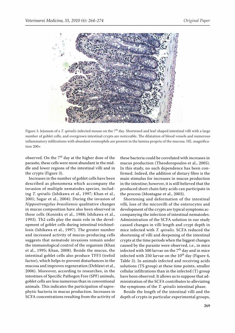
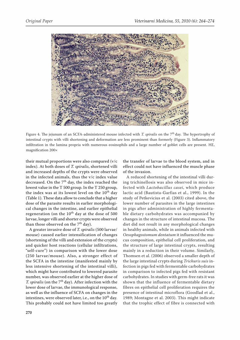
Shortening and deformation of the intestinal villi, loss of the microvilli of the enterocytes and development of the crypts are typical symptoms ac-companying the infection of intestinal nematodes. Administration of the SCFA solution in our study caused changes in villi length and crypt depth in mice infected with T. spiralis. SCFA reduced the shortening of villi and deepening of the intestinal crypts at the time periods when the biggest changes caused by the parasite were observed, i.e., in mice infected with 500 larvae on the 7th day and in mice infected with 250 larvae on the 10th day (Figure 4, Table 3). In animals infected and receiving acids solutions (TS group) at these time points, smaller cellular infiltrations than in the infected (T) group have been observed. It allows us to suppose that ad-ministration of the SCFA contributes to alleviating the symptoms of the T. spiralis intestinal phase.
Beside the length of the intestinal villi and the depth of crypts in particular experimental groups,
Figure 3. Jejunum of a T. spiralis-infected mouse on the 7th day. Shortened and leaf-shaped intestinal villi with a large number of goblet cells, and overgrown intestinal crypts are noticeable. The dilatation of blood vessels and numerous inflammatory infiltrations with abundant eosinophils are present in the lamina propria of the mucosa. HE, magnifica-tion 200×
Original Paper Veterinarni Medicina, 55, 2010 (6): 264–274
270
their mutual proportions were also compared (v/c index). At both doses of T. spiralis, shortened villi and increased depths of the crypts were observed in the infected animals, thus the v/c index value decreased. On the 7th day, the index reached the lowest value in the T 500 group. In the T 250 group, the index was at its lowest level on the 10th day (Table 1). These data allow to conclude that a higher dose of the parasite results in earlier morphologi-cal changes in the intestine, and earlier epithelial regeneration (on the 10th day at the dose of 500 larvae, longer villi and shorter crypts were observed than those observed on the 7th day).
A greater invasive dose of T. spiralis (500 larvae/mouse) caused earlier intensification of changes (shortening of the villi and extension of the crypts) and quicker host reactions (cellular infiltrations, “self-cure”) in comparison with the lower dose (250 larvae/mouse). Also, a stronger effect of the SCFA in the intestine (manifested mainly by less intensive shortening of the intestinal villi), which might have contributed to lowered parasite number, was observed earlier at the higher dose of T. spiralis (on the 7th day). After infection with the lower dose of larvae, the immunological response, as well as the influence of SCFA on changes in the intestines, were observed later, i.e., on the 10th day. This probably could not have limited too greatly
the transfer of larvae to the blood system, and in effect could not have influenced the muscle phase of the invasion.
A reduced shortening of the intestinal villi dur-ing trichinellosis was also observed in mice in-fected with Lactobacillus casei, which produce lactic acid (Bautista-Garfias et al., 1999). In the study of Petkevicius et al. (2003) cited above, the lower number of parasites in the large intestines in pigs after administration of highly fermenta-ble dietary carbohydrates was accompanied by changes in the structure of intestinal mucosa. The diet did not result in any morphological changes in healthy animals, while in animals infected with Oesophagostomum dentatum it influenced the mu-cus composition, epithelial cell proliferation, and the structure of large intestinal crypts, resulting mainly in a reduction in their volume. Similarly, Thomsen et al. (2006) observed a smaller depth of the large intestinal crypts during Trichuris suis in-fection in pigs fed with fermentable carbohydrates in comparison to infected pigs fed with resistant carbohydrates. In studies with germ-free rats it was shown that the influence of fermentable dietary fibres on epithelial cell proliferation requires the presence of intestinal microflora (Goodlad et al., 1989; Montagne et al. 2003). This might indicate that the trophic effect of fibre is connected with
Figure 4. The jejunum of an SCFA-administered mouse infected with T. spiralis on the 7th day. The hypertrophy of intestinal crypts with villi shortening and deformation are less prominent than formerly (Figure 3). Inflammatory infiltration in the lamina propria with numerous eosinophils and a large number of goblet cells are present. HE, magnification 200×
Veterinarni Medicina, 55, 2010 (6): 264–274 Original Paper
271
the activity of its bacterial decomposition products, mainly the SCFA. In contrast to earlier articles, where a stimulating effect of SCFA on a epithelial cell proliferation was described (Sakata, 1987), in the above-cited studies of Thomsen et al. (2006), no influence of SCFA on mitotic counts in the in-testinal crypts were observed. This result is repro-duced by the present study, where no differences in intestinal villi length between mice from groups C and S have been confirmed. A conclusion can be drawn that the influence of SCFA on the intestinal epithelium manifests itself mainly during patho-logical conditions.
Similarly, according to the studies of Ramos et al. (1997, 1999), orally administered SCFA reduced the inflammation of mucosa in the small and large in-testines in mice. It resulted in faster regeneration of the epithelium, a reduction in inflammatory infil-tration cells, and elongation of the intestinal villi in comparison to the animals that did not receive the acid solution. The impact of SCFA was independ-ent of endogenous production of short-chain fatty acids. In healthy animals, no influence of SCFA on the intestinal mucosa has been observed. The trophic effect of SCFA on damaged mucosa sug-gests that their administration might be beneficial not only during parasitic invasions, but also during such diseases as ulcerative intestine inflammation or colon cancer (Kim, 1998).
Short-chain fatty acids produced during micro-biological fermentation also act in the large intes-tine. Studies have demonstrated that gastric and rectal instillation of SCFA stimulated epithelial cell proliferation in rat jejunum and ileum. The increase in proliferation was significantly higher with rectal instillation than gastric instillation (Ichikawa et al., 2002). Previous studies have also demonstrated the effect of SCFA on distal sections of the intestine (Sakata, 1987). A trophic effect in rodent small intestines was also observed with in-travenous infusion of SCFA (Koruda et al., 1990). SCFA are rapidly absorbed by intestinal mucous membranes and their effects on distal sections of the alimentary tract may be explained by media-tion of the autonomic nervous and cardiovascu-lar system (Reilly et al., 1993; Frankel et al., 1994; Ichikawa et al., 2002). The effect of SCFA on in-testinal mucous membranes may proceed through stimulation of the production of regulatory pep-tides of the alimentary tract. It was observed that the trophic effect of colonically-infused SCFA on the mucous membrane of rat jejunum proceeded
partially through the intestinal gastrin (Reilly et al., 1995). Reilly et al. (1995) suggested that an au-tonomic nerve impulse generated under the influ-ence of colonic instillation of SCFA is directed to the central nervous system where the response is released in the form of a nerve or hormone signal. Enteroendocrine cells start producing gastrin un-der the influence of this signal. Numerous studies have demonstrated a stimulating effect of SCFA and dietary fibre on the secretion of glucagon-like peptide 2 (GLP-2) produced by L cells in the ileum and colon. The trophic effect of GLP-2 relies on increases in proliferation in crypt cells and apop-tosis suppression in intestinal mucosa. The effect of GLP-2 infusion on the depth of the crypts as well as the hight of the intestinal villi in the ileum and jejunum were demonstrated in parenterally fed pigs (Burrin et al., 2000). This mechanism plays a role in the adaptation of intestines for the absorp-tion of solid food after weaning as well as during adaptation of the organism after resection of the intestine (Burrin et al., 2003). SCFA can also act on the proliferation of enterocytes through modula-tion of blood flow or through direct action on genes regulating cell proliferation (Blottiere et al., 2003). The impact of SCFA on GLP-2 production and ex-pression of genes responsible for proliferation of enterocytes in the small intestine was observed in rats (Tappenden and McBurney, 1998; Tappenden et al., 1998), whereas a protective effect of SCFA on damaged mucosa of the small intestine was shown in mice (Ramos et al., 1999). The effect of GLP-2 on increases in proliferation and decreases in apopto-sis in the epithelium of the small intestines of mice was also observed (Tsai et al., 1997). Reimer and McBurney (1996) and Reimer et al. (1997) showed the impact of dietary fibre on the increase in GLP-2 secretion in rodents. Based on the mechanisms of SCFA action in the intestine, the effect of SCFA on the morphology of the small intestine can oc-cur with oral instillation and may also result from increased microbiological fermentation in the large intestine.
In summary, during intensive T. spiralis infec-tion (about 500 larvae per mouse), an orally admin-istered solution of short chain fatty acids caused decreases in numbers of mature nematodes in the intestines and larvae in muscles. The effect was not visible with a lower infectious dose (about 250 lar-vae). More intensive infection accelerated the rate of the host’s reaction to the presence of adult para-sites in the intestines – the so-called “self-cure ef-
Original Paper Veterinarni Medicina, 55, 2010 (6): 264–274
272
fect”. Moreover, higher invasive doses resulted in the greatest shortening of the intestinal villi and crypt development on the 7th day. The lower dose caused this change on the 10th day. Administration of SCFA to the animals infected with T. spiralis al-leviated the local histopathological changes caused by the parasite in the small intestine, and mani-fested mainly in less-intensive shortening of the villi and deepening of the intestinal crypts. Such results support a role for short chain fatty acids in influencing the course of trichinellosis in mice and should be followed by further investigation in order to clarify these mechanisms.
Acknowledgement
The study was approved by the II Local Ethics Committee in Wroclaw (No 52/2007; 30/2008).
REfERENCES
Bautista-Garfias CR, Ixta O, Orduna M, Martinez F, Aguilar B, Cortes A (1999): Enhancement of resistance in mice treated with Lactobacillus casei: Effect on Trichinella spiralis infection. Veterinary Parasitology 80, 251–260.
Bergman EN (1990): Energy contributions of volatile fatty acids from the gastrointestinal tract in various species. Physiological Reviews 70, 567–590.
Blottiere HM, Beucher B, Galmiche JP, Cherbut C (2003): Molecular analysis of the effect of short-chain fatty acids on intestinal cell proliferation. Proceedings of the Nutrition Society 62, 101–106.
Burrin DG, Stoll B, Jiang R, Petersen Y, Elnif J, Budding-ton RK, Schmidt M, Holst JJ, Hartmann B, Sangild PT (2000): GLP-2 stimulates intestinal growth in prema-ture TPN-fed pigs by suppressing proteolysis and app-tosisAmerican Journal of Physiology – Gastrointestinal and Liver Physiology 279, G1249–G1256.
Burrin DG, Stoll B, Guan X (2003): Glukagon-like pep-tyd 2 function in domestic animals. Domestic Animal Endocrinology 24, 103–122.
Byrne BM, Dankert J (1979): Volatile fatty acids and aero-bic flora in the gastrointestinal tract of mice under vari-ous conditions. Infection and Immunity 3, 559–563.
Cavaglieri CR, Nishiyama A, Fernandes LC, Curi R, Miles EA, Calder PC (2003): Differential effects of short-chain fatty acids on proliferation and production of pro- and anti-inflammatory cytokines by cultured lymphocytes. Life Sciences 73, 1683–1690.
Chiou PWS, Yu B, Lin CH (1994): Effect of different components of dietary fibre on the intestinal morphol-ogy of domestic rabbits. Comparative Biochemistry and Physiology 108A, 629–638.
Dehlawi MS, Mahida YR, Hughes K, Wakelin D (2006): Effects of Trichinella spiralis infection on intestinal pathology in mice lacking interleukin-4 (IL-4) or in-testinal trefoil factor (ITF/TFF3). Parasitology Inter-national 55, 207–211.
Engelhardt W, Bartels J, Kirchberger S, Meyer zu Dut-tingdorf HD, Busche R (1998): Role of short-chain fatty acids in the hindgut. Veterinary Quarterly 20, 52–59.
Finkelman FD, Shea-Donohue T, Goldhill J, Sullivan CA, Morris SC, Madden KB, Gause WC, Urban JF (1997): Cytokine regulation of host defense against parasitic gastrointestinal nematodes: lessons from studies with rodent models. Annual Review of Immunology 15, 505–533.
Frankel WL, Zhang W, Singh A, Klurfeld DM, Don S, Sakata T, Modlin I., Rombeau JL (1994): Stimulation of the autonomic nervous system mediates short-chain fatty acid induced jejunotrophism. Surgical Forum 43, 24–26.
Garside P, Kennedy MW, Wakelin D, Lawrence CE (2000): Immunopathology of intestinal helminth infec-tion. Parasite Immunology 22, 605–612.
Goodlad RA, Ratcliffe B, Fordham JP, Wright NA (1989): Does dietary fibre stimulate intestinal epithelial cell proliferation in germ free rats? Gut 30, 820–825.
Grencis RK, Hultner L, Else KJ (1991): Host protective immunity to Trichinella spiralis in mice: activation of Th cell subsets and lymphokine secretion in mice ex-pressing different response phenotypes. Immunology 74, 329–332.
Ichikawa H, Shineha R, Satomi S, Sakata T (2002): Gas-tric or rectal instillation of short-chain fatty acids stimulates epithelial cell proliferation of small and large intestine in rats. Digestive Diseases and Sciences 47, 1141–1146.
Ishikawa N, Horii Y, Nawa Y (1993): Immunemediat-edalteration of the terminal sugars of goblet cell mu-cins in the small intestine of Nippostrongylus brasiliensis-infected rats. Immunology 78, 303–307.
Ishikawa N, Wakelin D, Mahida YR (1997): Role of T helper 2 cells in intestinal goblet cell hyperplasia in mice infected with Trichinella spiralis. Gastroenterol-ogy 113, 542–549.
Kelly EAB, Enderson SC, Hauda KM, Wassom DL (1991): IFN-gamma and IL-5 producing cells compartmental-ize to different lymphoid organs in Trichinella spiralis-infected mice. Journal of Immunology 147, 306–311.

Veterinarni Medicina, 55, 2010 (6): 264–274 Original Paper
273
Khan WI (2008): Physiological changes in the gastroin-testinal tract and host protective immunity: learning from the mouse-Trichinella spiralis model. Parasitol-ogy 135, 671–682.
Khan W Collins SM (2004): Immune-mediated alteration in gut physiology and its role in host defence in nem-atode infection. Parasite Immunology 26, 319–326.
Khan WI, Abe T, Ishikawa N, Nawa Y, Yoshimura K (1995): Reduced amount of intestinal mucus by treat-ment with anti-CD4 antibody interferes with the spon-taneous cure of Nippostrongylus brasiliensis-infection in mice. Parasite Immunology 17, 485–491.
Khan WI, Blennerhasset PA, Ma C, Collins SM (2001): Stat6 dependent goblet cell hyperplasiaduring intestinal nematode infection. Parasite Immunology 23, 39–42.
Kim Y (1998): Short-chain fatty acids in ulcerative coli-tis. Nutrition Reviews 56, 17–24.
Koninkx JF, Mirck MH, Hendriks HG, Mouwen JM, van Dijk JE (1988): Nippostrongylus brasiliensis: histo-chemical changes in the composition of mucins in goblet cells during infection in rats. Experimental Parasitology 65, 84–90.
Koruda M, Rollanderi R, Bliss D, Hastings J, Rombeau JL, Settle RG (1990): Parenteral nutrition supple-mented with short-chain fatty acids: effect on the small bowel mucosa in normal rats. American Journal of Clinical Nutrition 51, 685–689.
Kostro K, Glinski Z, Wojcicka-Lorenowicz K, Krakowski L (2000): Immunological and immunopathological mechanisms of inflammation (in Polish). Medycyna Weterynaryjna 56, 479–485.
Kozar Z, Kozar M (1972): Diagnostics of Human Para-sitic Diseases (in Polish). PZWL, Warszawa. 87–90.
Mink CM, Van Esch WJE, Savelkoul HFJ, Van Loveren H, Bernardina WE, Ruitenberg EJ (1994): Role of in-terleukin-4 and interleukin-5 in the gut immune re-sponse to Trichinella spiralis mice. In: Campbell WC, Pozio E, Bruschi F (eds.): Trichinellosis. Istituto Su-periore di Sanita Press, Rome. 255–260.
Mista D, Piekarska J, Kaczmarek-Oliwa A, Zawadzki W, Steininger M, Kroliczewska B (2005): Effects of ex-perimental trichinellosis on the volatile fatty acid con-centration in mice intestines (in Polish). Medycyna Weterynaryjna 61, 1280–1283.
Montagne L, Pluske JR, Hampson DJ (2003): A review of interactions between dietary fibre and the intestinal mucosa, and their consequences on digestive health in young non-ruminant animals. Animal Feed Science and Technology 108, 95–117.
Petkevicius S (2007): The interaction between intestinal helminth infection and host nutrition. Review. Vet-erinarija ir Zootechnika 37, 53–59.
Petkevicius S, Bach Knudsen KE, Murrell KD (2003): Effects of Oesophagostomum dentatum and dietary carbohydrates on morphology of the large intestine of pigs. Veterinary Parasitology 116, 125–138.
Petkevicius S, Murrell KD, Bach Knudsen KE, Jørgensen H, Roepstorff A, Laue A, Wachmann H (2004): Effects of short-chain fatty acids and lactic acids on survival of Oesophagostomum dentatum in pigs. Veterinary Parasitology 122, 293–301.
Petkevicius S, Thomsen LE, Bach Knudsen KE, Murrell KD, Roepstorff A, Boes J (2007): The effect of inulin on new and on patent infections of Trichuris suis in growing pigs. Parasitology 134, 121–127.
Piekarska J (2004): Influence of the select immunotrophic compounds on mast cells, eosinophiles and macro-phages in the course of the experimental trichinellosis in mice (in Polish). Medycyna Weterynaryjna 60, 637–641.
Przyjalkowski W, Wescott RB (1969): Trichinella spira-lis: Establishment in gnotobiotic mice affected by Ba-cillus mesentericus, B. subtilis, and Pseudomonas aeruginosa. Experimental Parasitology 25, 8–12.
Przyjalkowski W, Cabaj W, Rykalo R (1983): Intestinal Trichinella spiralis and Trichinella pseudospiralis in germfree and conventional mice. Progress in Food and Nutrition Science 7, 117–126.
Ramos MG, Bambirra EA, Cara DC, Vieira EC, Alvarez-Leite JI (1997): Oral administration of short-chain fatty acids reduces the intestinal mucositis caused by treat-ment with Ara-C in mice fed commercial or elemental diets. Nutrition and Cancer 28, 212–217.
Ramos MG, Bambirra EA, Nicol, JR, Cara DC, Vieira EC, Alvarez-Leite JI (1999): Protection by short-chain fatty acids against 1-β-D-arabinofuranosylcytosine-induced intestinal lesions in germ free mice. Antimi-crobial Agents and Chemotherapy 43, 950–953.
Reilly K, Frankel W, Klurfeld D, Choi D, Rombeau JL (1993): The parasympathetic (PSNS) and sympathetic (SNS) nervous system mediate the systemic effects of short chain fatty acids (SCFA) on jejunal structure and function. Surgical Forum 44, 20–22.
Reilly K, Frankel W, Bain AM, Rombeau JL (1995): Co-lonic short chain fatty acids mediate jejunal growth by increasing gastrin. Gut 37, 81–86.
Reimer RA, McBurney MI (1996): Dietary fiber modu-lates intestinal proglucagon messenger ribonucleic acidand postprandial secretion of glucagon-like pep-tide 1 and insulin in rats. Endocrinology 137, 3948–3956.
Reimer RA, Thomson ABR, Rajotte RV, Basu TK, Oo-raikul B, McBurney MI (1997): A physiological level of rhubarb fiber increases proglucagon gene expres-
Original Paper Veterinarni Medicina, 55, 2010 (6): 264–274
274
Corresponding Author:
Dorota Mista, Wroclaw University of Environmental and Life Sciences, Faculty of Veterinary Medicine, Department of Biostructure and Animal Physiology, Norwida 31, 50-375 Wroclaw, PolandTel. +48 713 205 437, E-mail: [email protected]
sion and modulates intestinal glucose uptake in rats. Journal of Nutrition 127, 1923–1928.
Rotstein OD (1993): Interactions between leukocytes and anaerobic bacteria in polymicrobial surgical infec-tions. Clinical Infectious Diseases: an official publica-tion of the Infectious Diseases Society of America 16, 190–194.
Sagar M, Padol I, Khan WI, Bonin RP, Blennerhassett PA, Hunt RH (2004): Establishment of T-Helper-2 immune response based gerbil model of enteric infection. Scan-dinavian Journal of Gastroenterology 39, 668–673.
Sakata T (1987): Stimulatory effect of short chain fatty acids on epithelial cell proliferation in the rat intestine: a possible explanation for trophic effects of fermenta-ble fibre, gut microbes and luminal trophic factors. British Journal of Nutrition 58, 95–103.
Sanderson IR (2004): Short chain fatty acid regulation of signaling genes expressed by the intestinal epithe-lium. Journal of Nutrition 134, 2450–2454.
Sellin JH (1999): SCFAs: The enigma of weak electrolyte transport in the colon. News in physiological sciences: an international journal of physiology produced jointly by the International Union of Physiological Sciences and the American Physiological Society 14, 58–64.
Tappenden KA, McBurney MI (1998): Systemic short-chain fatty acids rapidly alter gastrointestinal struc-ture, function, and expression of early response genes. Digestive Diseases and Sciences 43, 1526–1536.
Tappenden KA, Drozdowski LA, Thomson AB, McBur-ney MI (1998): Short-chain fatty acid-supplemented
total parenteral nutrition alters intestinal structure, glucose transporter 2 (GLUT2) mRNA and protein, and proglucagon mRNA abundance in normal rats. American Journal of Clinical Nutrition 68, 118–125.
Theodoropoulos G, Hicks SJ, Corfield AP, Miller BG, Kapel CMO, Trivizaki M, Balaskas C, Petrakos G, Car-rington SD (2005): Trichinella spiralis: enteric mucin-related response to experimental infection in conventional and SPF pigs. Experimental Parasitology 109, 63–71.
Thomsen LE, Petkevicius S, Bach Knudsen KE, Roep-storff A (2005): The influence of dietary carbohydrates on experimental infection with Trichuris suis in pigs. Parasitology 131, 857–865.
Thomsen LE, Knudsen KE, Hedemann MS, Roepstorff A (2006): The effect of dietary carbohydrates and Trichuris suis infection on pig large intestine tissue structure, epithelial cell proliferation and mucin characteristics. Veterinary Parasitology 142, 112–122.
Tsai CH, Hill M, Asa SL, Brubaker PL, Drucker DJ (1997): Intestinal growth-promoting properties of glucagon-like peptide-2 in mice. American Journal of Physiology – Endocrinology and Metabolism 273, E77–E84.
Varadyova Z, Zelenaka I, Siroka P, Dubinsky P (2001): In vitro fermentation of celulosis amorphous and meadow hay in experimentally Ascaris suum-infected lambs. Small Ruminant Research 40, 155–164.
Received: 2010–04–06Accepted after corrections: 2010–06–25